gata-1: friends, brothers, and coworkers
TRANSCRIPT

537
Ann. N.Y. Acad. Sci. 1030: 537–554 (2004). © 2004 New York Academy of Sciences.doi: 10.1196/annals.1329.064
GATA-1: Friends, Brothers, and Coworkers
FRANCK MORCEAU, MICHAEL SCHNEKENBURGER, MARIO DICATO,AND MARC DIEDERICH
a
Laboratoire de Biologie Moléculaire et Cellulaire du Cancer, Hôpital Kirchberg, L-2540 Luxembourg, Luxembourg
A
BSTRACT
: GATA-1 is the founding member of the GATA family of transcrip-tion factors. GATA-1 and GATA family member GATA-2 are expressed inerythroid and megakaryocytic lineages, in which they play a crucial role in cellmaturation and differentiation. GATA-1 regulates the transcription of manyspecific and nonspecific erythroid genes by binding to DNA at the consensussequence WGATAR, which is recognized by all of the GATA family of tran-scription factors. However, it was identified in eosinophilic cells and also inSertoli cells in testis. Its activity depends on close cooperation with a functionalnetwork of cofactors, among them Friend of GATA, PU.1, and CBP/p300. TheGATA-1 protein structure has been well described and includes two zinc fin-gers that are directly involved in the interaction with DNA and other proteins
in vivo
. GATA-1 mutations in the zinc fingers can cause deregulation of re-quired interactions and lead to severe dysfunction in the hematopoietic system.
K
EYWORDS
: GATA-1; FOG; PU.1; CBP/p300; erythroid; megakaryocyte;differentiation
INTRODUCTION
The GATA-binding proteins form a group of structurally related transcription fac-tors that control gene expression and differentiation in a variety of cell types. GATA-1was the founding member of this family of transcription factors, which currently in-cludes six known members, GATA-1 to GATA-6. GATA-1 was first referred to aserythroid transcription factor 1 (Eryf 1), nuclear factor erythroid 1 (NF-E1), andglobin transcription factor (GF1) before getting its definite and consensus name in1990. GATA-1 gene transcription is regulated by numerous transcription factors, in-cluding GATA-1 itself. All proteins of the GATA transcription factor family containzinc finger motifs. In GATA-1, zinc fingers physically interact with the WGATARDNA sequence within target genes as well as with other proteins as cofactors.
GATA-1 represents a crucial transcription factor in erythroid and megakaryocyticdifferentiation processes, since it is particularly expressed in these lineages, in whichit regulates the transcription of many specific genes. However, GATA-1 is also in-volved in the regulation of unspecific hematopoietic genes. Positive or negative reg-ulation of GATA-1 protein by various cofactors, its capacity to regulate its own gene
a
Address for correspondence: Marc Diederich, Laboratoire de Biologie Moléculaire et Cellu-laire du Cancer, Hôpital Kirchberg, L-2540 Luxembourg, Luxembourg. Voice:
+
352-2468-4040;fax:
+
352-2468-4060.e-mail: [email protected]

538 ANNALS NEW YORK ACADEMY OF SCIENCES
transcription, the range of its target genes, and the diseases induced by GATA-1 dysfunc-tion make this complex nuclear protein a key transcription factor for hematopoiesis.
THE GATA-1 LEGEND
At the beginning, it was shown that the 3
′
region of the chicken adult
β
-globingene contained a DNA sequence that acted as a strong tissue-specific enhancer.
1,2
Invitro
DNase protection experiments revealed the presence of four protein-bindingdomains (I–IV) within this positive regulatory region.
3
Studying the interaction of anuclear protein only found in erythrocytes with region IV of the
β
-globin enhancer,Evans
et al.
4
identified an erythrocyte-specific DNA-binding factor, Eryf 1. Theyshowed that this nuclear protein recognized a regulatory sequence common to allchicken globin genes and that an Eryf 1-binding site was also present within the en-hancer of at least one human globin gene enhancer. Furthermore, proteins from hu-man erythroid cells were able to recognize the chicken sites. Finally, they suggestedthat the sequence WGATAR (where W
5
A or T and R
5
A or G) could representthe Eryf 1 “consensus” binding sequence. At the same time, Wall
et al.
5
showed thepresence of four regions within the minimal enhancer fragment of the human
β
-globin gene that bound an erythroid cell-specific nuclear factor. This factor, whichthey named
NF-E1, was found in erythroid cells at different developmental stages ofglobin gene expression. In addition to the intragenic enhancer and the promoter of the
β
-globin gene, the sequence recognized by NF-E1 was shown to be present in the pro-moter of other erythroid-specific genes. Performing footprinting, electromobility shift,and stable transfection assays, Plumb
et al.
6
proposed that a conserved GATAAGmotif could correspond to a
cis
element in the regulation of erythroid-specific genes.Indeed, this element was present in the promoter sequences of the mouse
α
- and
β
-globin genes as well as in the erythroid-specific promoter of the human porpho-bilinogen deaminase (PBGD)
7
and the erythropoietin receptor (R-Epo) genes.
8–10
The involvement of the GATAAG motif in the erythroid-specific stimulatory ef-fect on transcription was then strongly suggested. These observations were com-pleted by Martin
et al.
11
By studying the mutations involved in the hereditarypersistence of fetal hemoglobin disease, they demonstrated that two AGATA motifsin the
γ
-globin promoter could bind the erythroid-specific protein GF-1. Examiningspecific competition for GF-1 binding to the
γ
-promoter by sequences derived fromthe 3
′
β
-globin enhancers of chicken or human origin, they established that GF-1 aswell as the independently identified NF-E1 factor were counterparts of the chickentranscription factor Eryf 1. Moreover, they showed that GF-1 was a positive regula-tor of
γ
-gene expression.Together, these data provided evidence that the nuclear factors known as Eryf 1,
NF-E1, and GF-1 were very important in the regulation of globin and nonglobingenes specifically expressed along the erythroid differentiation pathway and thatthey were very close in their structure/function properties. A unique name was thenadopted in 1990 for the protein. Indeed, the name GATA-1 was agreed upon at theConference of Hemoglobin Switching (Warrenton, VA, USA, September 8–11,1990) by the investigators who had given the original names. However, during thelast 10 years, GATA-1 was found to be involved in the regulation of numerous genesthat are not necessarily erythroid-specific genes, and its expression was extended to
539MORCEAU
et al.
: GATA-1
other hematopoietic cells (T
ABLE
1). A few years later, it was found that GATA-1mRNA was also transcribed in Sertoli cells of the seminiferous tubules of prepuber-tal murine testis with a GATA-1 protein identical to the one expressed in erythroidcells.
12
It was also reported that GF-1/NF-E1 expression was shared by megacaryo-cytes and bone marrow-derived mast cells.
13,14
The authors concluded that thesetranscription factors should be expressed in committed multipotential cells and theirprogeny. According to these observations, Romeo
et al.
14
suggested a close associ-ation between the erythrocytic and megakaryocytic lineages, since they also showedthat NF-E2, another transcription factor involved in erythroid-specific gene regula-tion, was also expressed in megakaryocytes. More recently, Iwasaki
et al.
15
suggestedthat the regulation of GATA-1 is critical in maintaining multilineage homeostasis,since GATA-1 specifically induces megakaryocytic and erythrocytic commitmentwhile excluding other lineage development in stem and progenitor cells. Indeed,forced expression of ectopic GATA-1 led to both common lymphoid and granulo-cyte/monocyte progenitor reprogramming. These progenitors differentiate towardmegakaryocytic and erythroid lineages, whereas normal lymphoid or granulocyte/monocyte differentiation was inhibited. On the other hand, GATA-1 mRNA and pro-tein expression as well as binding activity
in vitro
were shown to be increased in thehuman CML K562 cell line induced to differentiate by the anthracycline aclacino-mycin or GTP. GATA-1 activation was correlated to
γ
-globin, PBGD, and R-Epogene overexpression and hemoglobin synthesis.
16–19
However, hemoglobinizationof K562 cells could be obtained as a result of an increase in specific mRNA half-life,rather than by the activation of GATA-1.
16
THE GATA FAMILY
A series of genes encoding zinc finger proteins that recognize the GATA motifwere cloned. All proteins of the GATA transcription factor family contain zinc fingermotifs. The GATA-2 gene was cloned by Lee
et al.
20
It is expressed in hematopoieticprogenitors, including early erythroid cells, mast cells, and megakaryocytes, andalso in nonhematopoietic embryonic stem cells. Labastie
et al.
21
cloned the humanGATA-3 gene and the 5
′
end of the mouse gene. This transcription factor is abun-dantly expressed in the T-lymphocyte lineage but also in brain. In addition to GATA-1,GATA-2, and GATA-3, all expressed in hematopoietic cells, another distinct subfam-ily of GATA transcription factors is formed by GATA-4, GATA-5, and GATA-6,since they are early and persistent markers of heart development in diverse vertebratespecies.
22–26
GATA-4 is expressed in gut epithelium and gonads, whereas GATA-6is expressed in pancreas, ovary, lung, and liver. GATA-6 is also detected in humanand rat vascular smooth muscle cell cultures.
THE GATA-1 GENE
The cDNA encoding the major DNA-binding protein (Eryf 1) was cloned fromboth murine
27
and chicken erythroid cells
28
before the identification of the humanGF-1 (hEryf 1) gene on the X chromosome (Xp21-11).
29,30
Hannon
et al.
31
isolateda genomic clone for the chicken GATA-1 and provided much information. Indeed,

540 ANNALS NEW YORK ACADEMY OF SCIENCES
TA
BLE
1.T
arge
t gen
es o
f GA
TA
-1 a
nd g
enes
reg
ulat
ed b
y G
AT
A-1
-exp
ress
ing
tissu
es
Ge
ne
Na
me
Ro
le o
f th
e G
ATA
-1 T
arg
et
Ge
ne
Pro
du
ctG
ATA
-1-T
arg
ete
dG
en
e T
issu
eR
efe
ren
ces
Ta
c2,
tach
ykin
in 2
En
cod
es
the
am
ida
ted
ne
uro
pe
ptid
e p
ro-n
eu
roki
nin
BE
ryth
roid
67
GS
TP
1,
glu
tath
ion
e
S
-tra
nsf
era
se P
1P
ha
se II
me
tab
oliz
ing
en
zym
e; c
ata
lyze
s th
e c
on
jug
atio
n o
f ele
ctro
ph
ilic
com
po
un
ds
to t
he
tri
pe
ptid
e g
luta
thio
ne
; p
lays
a r
ole
in c
ell
pro
tect
ion
Hu
ma
n,
ery
thro
id
89
α
-Sp
ect
rin
Hig
hly
exp
ress
ed
me
mb
ran
e p
rote
in c
ritic
al f
or
the
fle
xib
ility
an
dst
ab
ility
of
the
ery
thro
cyte
Hu
ma
n,
ery
thro
id
90
Ho
x B
2M
em
be
r o
f th
e v
ert
eb
rate
Ho
x g
en
e f
am
ily;
en
cod
es
for
spe
cific
d
eve
lop
me
nta
l-st
ag
e D
NA
-bin
din
g p
rote
ins
Hu
ma
n,
ery
thro
id
91
EK
LF
, e
ryth
roid
Kru
pp
el-
like
fa
cto
rE
ryth
roid
-sp
eci
fic t
ran
scri
ptio
n f
act
or
tha
t b
ind
s a
CA
CC
C m
otif
Hu
ma
n,
ery
thro
id
92
α
IIb
Co
mp
on
en
t o
f in
teg
rin
re
cep
tor
tha
t fu
nct
ion
s in
pla
tele
t a
gg
reg
atio
nH
um
an
, m
eg
aka
ryo
cyte
65,9
3
PF
4,
pla
tele
t fa
cto
r 4
Neu
tral
izat
ion
of h
epar
in-li
ke m
olec
ules
on
the
endo
thel
ial s
urfa
ce o
f blo
od
vess
els,
inhi
bitin
g lo
cal a
ntith
rom
bin
III a
ctiv
ity a
nd p
rom
otin
g co
agul
atio
nHu
ma
n,
me
ga
kary
ocy
te
94
GP
IIb
, g
lyco
pro
tein
IIb
Inte
gri
n c
lass
of
cell
ad
he
sio
n m
ole
cule
re
cep
tors
; e
arl
y a
nd
sp
eci
fic
ma
rke
r o
f th
e m
eg
aka
ryo
cytic
lin
ea
ge
Hu
ma
n,
me
ga
kary
ocy
te
95
FcR
,
β
-ch
ain
, F
c
«
RI
β
-ch
ain
Am
plif
ies
the
ma
st c
ell
resp
on
se;
ne
cess
ary
fo
r th
e c
ell
surf
ace
exp
ress
ion
of
Fc
«
RI
in m
ou
seM
ou
se,
ma
st c
ells
96
Gly
cop
rote
in,
gp
91
-ph
ox
Ele
ctro
n-t
ran
spo
rtin
g s
ub
un
it o
f th
e p
ha
go
cyte
NA
DP
H o
xid
ase
Hu
ma
n,
eo
sin
op
hil
97
Gra
nu
le (
MB
P)
ma
jor
ba
sic
pro
tein
En
ha
nce
s n
atu
ral k
ille
r ce
ll a
ctiv
ity;
kille
r T
-ce
ll a
ctiv
ity in
du
ced
by
the
mix
ed
lym
ph
ocy
te t
um
or
cell
rea
ctio
nH
um
an
, m
ou
se,
eo
sin
op
hil
98
FS
HR
, fol
licle
-stim
ulat
ing
ho
rmo
ne
re
cep
tor
Ess
en
tial f
or
no
rma
l re
pro
du
ctiv
e f
un
ctio
n in
ma
les
an
d f
em
ale
sT
est
icu
lar
(Se
rto
li ce
lls)
99
Inhi
bin/
activ
in
β
-B-s
ubun
itT
GF
-
β
su
pe
rfa
mily
me
mb
er;
ro
le in
re
pro
du
ctio
n a
nd
de
velo
pm
en
tM
ouse
, tes
ticul
ar (
Ser
toli
cells
)
100
Inh
ibin
α
-su
bu
nit
Gly
cop
rote
in h
orm
on
e t
ha
t re
gu
late
s p
ituita
ry F
SH
se
cre
tion
Mou
se, t
estic
ular
(S
erto
li ce
lls)
101
N
OT
E
:W
ell-
est
ab
lish
ed
GA
TA
-1-r
eg
ula
ted
ery
thro
id-s
pe
cific
ge
ne
s (g
lob
ins,
R-E
po
, P
BG
D,
etc
.) w
ere
vo
lun
tari
ly o
mitt
ed
.

541MORCEAU
et al.
: GATA-1
they established that this gene is a 6.5-kb DNA fragment split into six exons and con-taining multiple start sites for gene transcription. These sites are clustered in a smallregion (180–218 nucleotides) upstream of the translation start site. In hematopoieticand Sertoli cells, GATA-1 mRNA is transcribed from two different promoters or un-translated first exons. The erythroid or proximal promoter (IE), with respect to theAUG, is active in hematopoietic cells,
32,33
whereas the testis or distal promoter (IT),located 8 kb upstream of the proximal promoter, is active in Sertoli cells. No tran-scription from this promoter was detected in mouse erythroleukemia cells.
12
How-ever, GATA-1 mRNAs derived from the “testis” promoter were found to be presentat low levels at all stages of hematopoiesis in mice.
34
Furthermore, it was clearlyshown that the increase in GATA-1 expression during the differentiation of primaryerythroid cells was correlated with increased levels of the distal, but not the proxi-mal, GATA-1 mRNA.35 These data provided evidence that promoter IT was involvedin the initial activation of GATA-1 expression in early erythroid progenitors.
DNase I protection assays were performed on the nucleotide sequence upstreamof the GATA-1 protein-encoding region using nuclear extracts from erythroid orbrain cells. The results revealed numerous sites for specific binding of nuclear fac-tors. Seven protected sites that contain the binding sequence for the β-globin protein1 (BGP1) factor were found. BGP1 was first shown to bind a sequence of 16 deoxy-guanosine residues within the promoter region of the chicken adult β-globin gene.This specific protein was identified in chicken erythroid cells. It appears at the sametime or shortly before chromatin structure modifications. BGP1 is restricted to eryth-rocytes and is most abundant in definitive erythrocytes. Thus, its presence corre-sponds to the tissue- and stage-specific occupancy of the poly-G sequence in vivo.36,37
On the other hand, one of the seven BGP1-binding sites in the chicken GATA-1 geneoverlaps a consensus binding sequence for the c-myb transcription factor.31 It wasthen suggested that c-myb might be involved in regulating GATA-1 expression inerythropoietic stem cells, particularly at some earlier developmental stage. Indeed,high levels of c-myb expression were found predominantly in immature hematopoi-etic cells, where they decreased during differentiation.38,39 Furthermore, Melotti andCalabretta40 showed that undifferentiated ES cells transfected with intact c-myb ledto GATA-1 expression, among other gene products, whereas the gene was silent incells transfected with a c-myb mutant.
Another important specific DNA sequence, protected by brain but not erythroidextracts, was found and corresponded to a palindromic sequence recognized by sev-eral members of the steroid hormone-receptor superfamily. Interestingly, a cluster ofthree GATA-1 sites in this region of the chicken GATA-1 gene was shown. Thesesites were strongly protected by erythroid extracts and by purified GATA-1 protein.In contrast, nuclear extracts from brain cells, which do not express the GATA-1 gene,failed to protect these sites. The role of the GATA-1 binding sites was then assessedby performing cotransfection assays with GATA-1 cDNA expression vector andchloramphenicol acetyl transferase reporter gene. The reporter gene driven by aGATA-1 promoter region containing the upstream sequence GATA was powerfullystimulated in GATA-1-overexpressing day-10 erythroid cells. Together, these resultsindicated that GATA-1 might play a role in the regulation of its own expression.
Similar results were obtained in mice. Tsai et al.33 characterized the mouse GATA-1gene and cis-elements within its promoter. They demonstrated that promoter activityrequires double GATA consensus sequences at its 5′ end that bind a single GATA-1

542 ANNALS NEW YORK ACADEMY OF SCIENCES
molecule in an asymmetric way. According to their findings, they proposed thatGATA-1 gene expression was regulated by the GATA-1 protein itself. Furthermore,they showed the presence of two crucial proximal CACCC elements for the promoteractivity in erythroid cells at the 3′ end of the GATA-1 gene. On the other hand, hu-man and mouse GATA-1 gene promoter sequences were reported by Nicolis et al.32
They demonstrated homologies between the human and mouse proximal promoter andthe region containing the double GATA site involved in autoregulation. Zon andOrkin41 provided precise details concerning this homology and showed a closer align-ment of the two mammalian sequences. This finding led the authors to predict strongfunctional similarities between human and mouse GATA-1 regulatory regions.
GATA-1 PROTEIN STRUCTURE AND DNA INTERACTION
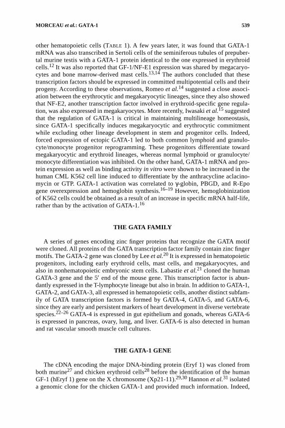
GF-1 and Eryf 1 proteins were described as very similar, particularly in theirbinding domains. However, the avian form of the erythroid-specific transcriptionfactor diverged from mammalian polypeptides at the level of the transactivator re-gion. Mouse GATA-1 protein was characterized as a 413-amino acid DNA-bindingprotein, and some similarities with the human protein have been revealed.27 It is con-stituted of two cysteine-rich motifs (Cys-x-x-Cys-x17-Cys-x-x-Cys)27,28 that arereminiscent of a zinc-finger structure and have been shown to be encoded by separateexons of 48 and 42 codons.31 Moreover, at the C terminus of each pair of zinc fin-gers, two highly conserved lysine-rich motifs have been described that are found inGATA-1 proteins from different species and in other members of the GATA family42
(FIG. 1).Trainor et al. demonstrated that erythroid regulatory factors in human, chicken,
and mouse evolved from a common precursor composed of two distinct kinds of re-peated domains.29 Indeed, the central third of the cDNA, containing the two “finger”motifs, is almost identical in the different organisms. In contrast, the N- and C-terminal thirds of the human protein are similar to those of mouse but are strikinglydifferent from the corresponding domains in chicken.
Omichinski et al.43 determined the three-dimensional solution structure of a com-plex between the DNA-binding domain of the chicken erythroid transcription factorGATA-1 and its cognate DNA site using multidimensional heteronuclear magneticresonance spectroscopy. The DNA-binding domain of GATA-1 contains a zinc atomlinked to four cysteines and a C-terminal tail. This core structure is composed of twoirregular antiparallel β sheets and α helixes that interact together with the majorgroove of the DNA. The C-terminal tail is an essential determinant of specific bind-ing, by wrapping around into the minor groove. The crucial role of the C-terminalfinger as necessary and sufficient in the binding activity of GATA-1 to the WGATARrecognition sequence was widely demonstrated.27,28,43–45 Particularly, a 66-aminoacid peptide containing the C-terminal finger domain was able to bind tightly andspecifically to the GATA target sequence.46 In addition, the binding properties ofGATA-1 were shown to be only weakly affected by mutation or deletion of the N-terminal finger.47 Even though the N-terminal finger does not bind DNA indepen-dently, it stabilizes GATA-1 interactions with some sites crucial for gene expression.Since GATA-1 binds as a monomer to such sites, it has been proposed that both theN- and C-terminal fingers are involved in DNA recognition.31,33,48,49 However, a
543MORCEAU et al.: GATA-1
critical role for the N-terminal finger has been identified in terminal erythroid differ-entiation.50 On the other hand, phosphorylation of transcription factors, a post-trans-lational modification, modulates their activity. GATA-1 phosphorylation increasesits binding affinity for a canonical binding site.51
Interestingly, Newton et al.52 demonstrated that the murine GATA-1 N-terminalfinger is capable of recognizing GATC motifs in vitro and that a naturally occurringmutation in GATA-1 (R216Q) abolishes this interaction. Moreover, they showedthat GATA-1 is capable of activating transcription from promoters exhibiting theGATC sequence, thus extending the range of genes that may be regulated by GATA-1.Furthermore, DNA-binding activity, which requires stoichiometric quantities ofZn2+, can be efficiently obtained with some other heavy metals, such as Fe2+.46 Byperforming circular permutation DNA-bending experiments with a variety ofGATA-binding sites, Ghirlando and Trainor53 showed that the GATA-1 proteinbends DNA by 24° in a site-independent manner and that the C-terminal finger issufficient to account for this bending. According to the finding that DNA is appar-ently unconstrained by the orientations of the two zinc fingers, the N- and C-termi-nal zinc fingers might adopt different geometries when bound to different doubleGATA sites.
FIGURE 1. The GATA-1 protein structure includes two zinc fingers and two conservedlysine-rich motifs. GATA-1 N-terminal and C-terminal zinc fingers are shaped by the linkof one zinc atom (Zn) to four cysteines (C). The lysine-rich N- and C-motifs (white bars) C-terminal to each finger are conserved among GATA-1, -2, and -3, and among species (human[h], mouse [m], and chicken [c]). (Based on Hung et al.42)
544 ANNALS NEW YORK ACADEMY OF SCIENCES
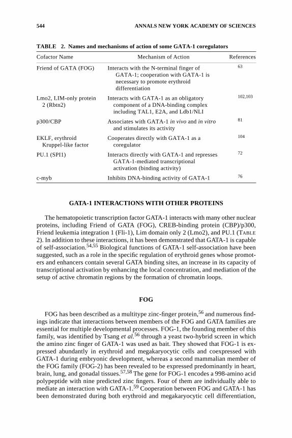
GATA-1 INTERACTIONS WITH OTHER PROTEINS
The hematopoietic transcription factor GATA-1 interacts with many other nuclearproteins, including Friend of GATA (FOG), CREB-binding protein (CBP)/p300,Friend leukemia integration 1 (Fli-1), Lim domain only 2 (Lmo2), and PU.1 (TABLE
2). In addition to these interactions, it has been demonstrated that GATA-1 is capableof self-association.54,55 Biological functions of GATA-1 self-association have beensuggested, such as a role in the specific regulation of erythroid genes whose promot-ers and enhancers contain several GATA binding sites, an increase in its capacity oftranscriptional activation by enhancing the local concentration, and mediation of thesetup of active chromatin regions by the formation of chromatin loops.
FOG
FOG has been described as a multitype zinc-finger protein,56 and numerous find-ings indicate that interactions between members of the FOG and GATA families areessential for multiple developmental processes. FOG-1, the founding member of thisfamily, was identified by Tsang et al.56 through a yeast two-hybrid screen in whichthe amino zinc finger of GATA-1 was used as bait. They showed that FOG-1 is ex-pressed abundantly in erythroid and megakaryocytic cells and coexpressed withGATA-1 during embryonic development, whereas a second mammalian member ofthe FOG family (FOG-2) has been revealed to be expressed predominantly in heart,brain, lung, and gonadal tissues.57,58 The gene for FOG-1 encodes a 998-amino acidpolypeptide with nine predicted zinc fingers. Four of them are individually able tomediate an interaction with GATA-1.59 Cooperation between FOG and GATA-1 hasbeen demonstrated during both erythroid and megakaryocytic cell differentiation,
TABLE 2. Names and mechanisms of action of some GATA-1 coregulators
Cofactor Name Mechanism of Action References
Friend of GATA (FOG) Interacts with the N-terminal finger ofGATA-1; cooperation with GATA-1 is necessary to promote erythroiddifferentiation
63
Lmo2, LIM-only protein 2 (Rbtn2)
Interacts with GATA-1 as an obligatory component of a DNA-binding complex including TAL1, E2A, and Ldb1/NLI
102,103
p300/CBP Associates with GATA-1 in vivo and in vitro and stimulates its activity
81
EKLF, erythroidKruppel-like factor
Cooperates directly with GATA-1 as a coregulator
104
PU.1 (SPI1) Interacts directly with GATA-1 and represses GATA-1-mediated transcriptional activation (binding activity)
72
c-myb Inhibits DNA-binding activity of GATA-1 76

545MORCEAU et al.: GATA-1
since they synergistically activated transcription from a hematopoietic-specific reg-ulatory region. Performing the yeast two-hybrid assay, Tsang et al.56 further speci-fied that FOG interacts with GATA-1 by binding the N-terminal finger of thetranscription factor, indicating that FOG acts as a cofactor of GATA-1. Yu et al.60
recently showed that a single amino acid substitution (Arg216Gln) in the N-terminalfinger of GATA-1, which was found in a previously reported family with X-linkedthrombocytopenia and β-thalassemia, affected neither the interaction with FOG-1nor the erythroid differentiation program. The mutated protein was slightly less ef-ficient than the wild-type counterpart. However, mutant GATA-1 harboring this sub-stitution destabilizes the binding of GATA-1 to complex DNA sites. By identifyingthis mutation in GATA-1, the authors suggested a critical in vivo requirement for theDNA-binding contribution of the N-terminal finger of GATA-1, as distinguishedfrom its involvement in the FOG-1 interaction.
Cantor et al.61 disfavored a model in which FOG-1 acts to bridge multiple GATA-binding DNA elements. Indeed, they demonstrated that interaction with GATA-1through a single GATA-binding zinc finger is sufficient for rescue erythroid andmegakaryocytic maturation. In addition, they proposed that FOG-1 may modulatethe outcome of a precursor cell to differentiate along the erythroid or megakaryo-cytic pathway, since they discovered that distinct domains of FOG-1 differentiallyinfluence erythroid versus megakaryocytic differentiation. However, mutation ofFOG (FOG−/−) was responsible for differential effects in erythroid and megakaryo-cytic cells. Erythroid maturation was indeed partially blocked, whereas megakaryocytesfailed to develop in the absence of FOG. Clearly, it was established that the interactionof FOG with GATA-1 is essential for the function of GATA-1 in erythroid differentia-tion. Mice lacking FOG died during midembryonic development from severe ane-mia.62 GATA-1 mutants that failed to interact with FOG but retained normal DNAbinding did not promote erythroid differentiation of GATA-1-deficient erythroidcells. However, differentiation was rescued by a compensatory FOG mutant that re-stored the interaction with GATA-1.63 A similar point mutation of GATA-1 in hu-mans leads to severe congenital dyserythropoietic anemia and thrombocytopenia.64
By contrast, FOG was shown to be capable of acting via a GATA-1-independentroute to stimulate α IIb gene expression in megakaryocytic cells, which suggestedadditional hematopoietic-restricted cofactors such as GATA-2.65 Recently, Lettinget al.66 demonstrated that FOG-1 uses distinct mechanisms when cooperating withGATA-1 during transcriptional activation and repression. Thus, GATA-1 DNA bind-ing at DNase I-hypersensitive site (HS) 3 of the β-globin locus control region isFOG-1-independent, whereas FOG-1 is required for GATA-1 occupancy at HS2.FOG-1 is not needed at other sites, including the FOG-1-independent GATA-1 targetgene EKLF. Similarly, at the GATA-2 gene, interaction with FOG-1 is not necessaryfor GATA-1 occupancy and is required for transcriptional inhibition and histonedeacetylation. On the other hand, Pal et al.67 proposed that FOG-1 is a prototype of“chromatin occupancy facilitators,” a new class of coregulators that confer coregu-lation in certain contexts by enhancing trans-acting factor binding to chromatin invivo. A role for FOG expression has been described in other hematopoietic lineages.Yamaguchi et al.68 found that FOG may act as a negative cofactor for the eosinophillineage, unlike its positive regulatory function for the erythroid and megakaryocytelineages. Zhou et al.69 showed that FOG might regulate GATA-3 activity in naive Tcells. Indeed, FOG repressed GATA-3-induced processes, including GATA-3 auto-

546 ANNALS NEW YORK ACADEMY OF SCIENCES
activation and GATA-3-dependent activation of the interleukin-5 promoter in T cells.In addition, they established that overexpression of FOG during primary activationof naive T cells inhibited T helper (Th) 2 development in CD4+ T cells. Thus, theyconcluded that FOG-1 may be one factor capable of regulating Th2 development.Ketola et al.70 indicated that FOG-2 had a role in early gonadal development andsexual differentiation with GATA-4 and FOG-1 at later fetal stages, whereas GATA-1executes its action postnatally. Recently, a role for the transforming acidic coiled-coil protein 3 was suggested in the regulation of the cellular distribution of FOG-1,since it was shown to affect its nuclear localization, thus altering the interactionbetween GATA-1 and FOG-1.71
PU.1
PU.1 (also known as Spi-1) is a hematopoietic-specific Ets family transcriptionfactor that is required for the development of some lymphoid and myeloid lineagesand that can block erythroid differentiation. It is suggested that deregulation of PU.1increases erythroblast proliferation, blocks erythroid differentiation, and contributesto erythroleukemia.
By studying the mechanism by which PU.1 inhibits erythroid differentiation,Rekhtman et al. found that PU.1 represses GATA-1-mediated transcriptional activa-tion via direct interaction with GATA-1.72 Both the DNA-binding and transactivationdomains of PU.1 are required for repression, and both domains are also needed toblock terminal differentiation in MEL cells. Similar mechanisms were previously re-ported by Yamada et al.,73 who suggested that reduction of the DNA-binding activityof GATA-1 might partly account for PU.1-mediated apoptosis in MEL cells. Ectopicexpression of PU.1 in Xenopus embryos was also shown to be sufficient to blockerythropoiesis during normal development. Introduction of exogenous GATA-1 inboth MEL cells and Xenopus embryos relieves the block to erythroid differentiationimposed by PU.1. Importantly, the stoichiometry of interacting PU.1 and GATA-1was suggested to be a determinant for processes of both normal differentiation andmalignant transformation.72 Moreover, the inhibition of hemin-induced erythroiddifferentiation in the induced overexpressing PU.1 K562 erythroleukemia cells wascorrelated with a drastic decrease in GATA-1 DNA binding.74 However, the lineage-specific transcription factors GATA-1 and PU.1 can physically interact to inhibiteach other’s functions (FIG. 2). Thus, Zhang et al.75 demonstrated that the N-terminalregion (TAD) of the PU.1 interacts with the conserved C-terminal zinc finger ofGATA-1 and GATA-2 (FIG. 2A). Furthermore, they described the role of the N-terminal70 amino acids of PU.1 and determined that they are essential to repress GATA-1function by blocking GATA-1 binding to DNA in vitro and in vivo.74 In addition,GATA-1 activities regulated by direct interaction with PU.1 and c-Myb may be cru-cial for Ras-mediated megakaryocytic differentiation, as suggested by Matsumura etal.76 Rekhtman et al.77 recently reported that PU.1 cooperates with the corepressorpRB to inhibit GATA-1 and block erythroid differentiation. Indeed, they suggestedthat inhibition of GATA-1 by PU.1 occurs by forming a PU.1/GATA-1 complex atGATA-1 target genes. By binding to these sites, the corepressor pRB blocks GATA-1transcriptional activity and thereby erythroid differentiation. On the other hand, it wasshown that GATA-1 was capable of inhibiting the activity of the PU.1 protein in a

547MORCEAU et al.: GATA-1
dose-dependent manner via a direct interaction between the PU.1 ETS domain andthe C-terminal finger region of GATA-1.78 Furthermore, GATA-1 inhibits PU.1 func-tion by displacing the PU.1 coactivator c-Jun75 (FIG. 2B). Regulation of GATA-1 ac-tivity is complex because of the network of interactions involved with activating andrepressing cofactors, which cooperate with other cellular factors as well. Hong etal.79 suggested that inhibition of normal cellular differentiation by aberrant expres-sion of PU.1 and possibly other members of the Ets family of oncoproteins is par-tially due to their specific and efficient inhibitory action on the CBP-mediatedacetylation of several nuclear proteins, including GATA-1. Another member of theEts family of transcription factors, Fli-1, has been shown to be an important regulatorduring megakaryocytic differentiation. Eisbacher et al.80 have suggested that Fli-1 andGATA-1 work together to activate the expression of genes associated with the terminaldifferentiation of megakaryocytes. By mapping the minimal domains required forthe interaction, they showed that the zinc fingers of GATA-1 interact with the Etsdomain of Fli-1.
p300/CBP
GATA-1 is associated in vivo with the acetyltransferase CBP/p300, since GATA-1and CBP were shown to coimmunoprecipitate from nuclear extracts of erythroid
FIGURE 2. GATA-1 and PU.1 physically interact to inhibit each other’s functionsthrough protein–protein interactions. (A) The N-terminal region of PU.1 blocks GATA-1function by binding to the GATA-1 C-terminal finger and inhibiting GATA-1 binding toDNA. (B) GATA-1 inhibits PU.1 transactivation of PU.1 target genes by blocking the bind-ing of the PU.1 coactivator c-Jun to the β3/β4 region of PU.1. (Based on Zhang et al.74)

548 ANNALS NEW YORK ACADEMY OF SCIENCES
cells. Interaction mapping allowed the location of contact sites to the zinc finger re-gion of GATA-1 and to the adenovirus oncoprotein E1A-binding region of CBP. Thisregion acetylates GATA-1 at two highly conserved lysine-rich motifs present at theC-terminal tails of both zinc fingers.42,81 It was shown that GATA-1 is acetylated invitro by p300 and that acetylation directly stimulates GATA-1-dependent transcrip-tion in vivo. A conformational change in GATA-1 has been suggested because acety-lation significantly increased the GATA-1 binding activity to DNA and altered themobility of GATA-1/DNA complexes.82 On the other hand, another aspect of the com-plexity of the GATA-1 regulation has been reported. Indeed, CBP acts also as a tran-scriptional adapter for c-myb transcription factor. Matsumura et al.76 showed thatGATA-1 interacts with c-myb, resulting in the inhibition of the DNA-binding activityof GATA-1. This was confirmed notably by Bartunek et al.,83 who provided evidenceof a direct molecular link between GATA-1 activity and c-myb proto-oncogene expres-sion during terminal red cell differentiation. Takahashi et al.84 showed that any possi-ble dimeric complexes involving c-Myb, GATA-1, or CBP could be formed and thatthe transcription factors c-Myb and GATA-1 mutually inhibit transcriptional activi-ties. They suggested that binding of GATA-1 or c-Myb to CBP might be importantfor hematopoietic differentiation and that CBP functions as a key molecule duringthe process. In addition, it was demonstrated that GATA-1 and its cofactor CBP areessential for the formation of an erythroid-specific acetylation pattern.85
CONCLUSION
Target genes of GATA-1 were originally identified in the erythroid lineage, andinvestigations rapidly demonstrated the crucial role of this transcription factor inblood cell differentiation processes. Indeed, expression of the GATA-1 gene was ex-tended to various tissues, particularly in blood but also in testis. In the same way, thegroup of genes regulated by GATA-1 was increased beyond erythroid genes. Thus,during the past 10 years, increased knowledge about GATA-1 activity has enforcedthe biological meaning of this nuclear factor, particularly in the hematopoietic sys-tem. In addition, competitive or cooperative effects with coexpressed transcriptionfactors of the GATA family (particularly GATA-2) and the GATA-1-dependent ac-tivity on numerous “coworkers” make this transcription factor very complex. Dereg-ulation of interactions in this network of cofactors can have severe physiologicalconsequences, and some hematological dysfunctions are attributed to GATA-1 genemutations. Indeed, a mutation (R216Q) identified in the DNA-binding site of GATA-1N-terminal zinc finger induces X-linked thrombocytopenia with thalassemia.86 Fur-thermore, the development of acute megakaryocytic leukemia in Down syndromenewborn infants has been correlated with the expression of GATA-1 with a defectiveN-terminal activation domain that contributes to the expansion of transient myelo-proliferative disorder blast cells.87 On the other hand, alterations of the FOG-1 bind-ing site are responsible for dyserythropoietic anemia with thrombocytopenia,88 andderegulation of the GATA-1 repressor PU.1 in erythroid precursors can cause eryth-roleukemias in mice.72 Understanding of GATA-1 regulation and mechanisms of ac-tion should be critical for improving the treatment of hematological diseases andleukemias as well as for developing alternative therapeutic approaches such as dif-ferentiation therapy.
549MORCEAU et al.: GATA-1
ACKNOWLEDGMENTS
Research at the laboratory of M. Diederich is supported by the Fondation de Re-cherche Cancer et Sang, the Recherches Scientifiques Luxembourg association,Télévie grants, and the Een Häerz fir kriibskrank Kanner association. M.S. was sup-ported by a fellowship from the Government of Luxembourg.
REFERENCES
1. HESSE, J.E., J.M. NICKOL, M.R. LIEBER, et al. 1986. Regulated gene expression in trans-fected primary chicken erythrocytes. Proc. Natl. Acad. Sci. USA 83: 4312–4316.
2. CHOI, O.R. & J.D. ENGEL. 1986. A 3′ enhancer is required for temporal and tissue-spe-cific transcriptional activation of the chicken adult β-globin gene. Nature 323: 731–734.
3. EMERSON, B.M., J.M. NICKOL, P.D. JACKSON, et al. 1987. Analysis of the tissue-specificenhancer at the 3′ end of the chicken adult β-globin gene. Proc. Natl. Acad. Sci. USA84: 4786–4790.
4. EVANS, T., M. REITMAN & G. FELSENFELD. 1988. An erythrocyte-specific DNA-bindingfactor recognizes a regulatory sequence common to all chicken globin genes. Proc.Natl. Acad. Sci. USA 85: 5976–5980.
5. WALL , L., E. DEBOER & F. GROSVELD. 1988. The human β-globin gene 3′ enhancercontains multiple binding sites for an erythroid-specific protein. Genes Dev. 2:1089–1100.
6. PLUMB, M., J. FRAMPTON, H. WAINWRIGHT, et al. 1989. GATAAG: a cis-control regionbinding an erythroid-specific nuclear factor with a role in globin and non-globin geneexpression. Nucleic Acids Res. 17: 73–92.
7. FRAMPTON, J., M. WALKER, M. PLUMB, et al. 1990. Synergy between the NF-E1 erythroid-specific transcription factor and the CACCC factor in the erythroid-specific promoter ofthe human porphobilinogen deaminase gene. Mol. Cell. Biol. 10: 3838–3842.
8. ZON, L.I., H. YOUSSOUFIAN, C. MATHER, et al. 1991. Activation of the erythropoietinreceptor promoter by transcription factor GATA-1. Proc. Natl. Acad. Sci. USA 88:10638–10641.
9. CHIBA, T., Y. IKAWA & K. TODOKORO. 1991. GATA-1 transactivates erythropoietinreceptor gene, and erythropoietin receptor-mediated signals enhance GATA-1 geneexpression. Nucleic Acids Res. 19: 3843–3848.
10. CHIN, K., N. ODA, K. SHEN, et al. 1995. Regulation of transcription of the humanerythropoietin receptor gene by proteins binding to GATA-1 and Sp1 motifs. NucleicAcids Res. 23: 3041–3049.
11. MARTIN, D.I., S.F. TSAI & S.H. ORKIN. 1989. Increased γ-globin expression in a non-deletion HPFH mediated by an erythroid-specific DNA-binding factor. Nature 338: 435–438.
12. ITO, E., T. TOKI, H. ISHIHARA, et al. 1993. Erythroid transcription factor GATA-1 isabundantly transcribed in mouse testis. Nature 362: 466–468.
13. MARTIN, D.I., L.I. ZON, G. MUTTER, et al. 1990. Expression of an erythroid transcrip-tion factor in megakaryocytic and mast cell lineages. Nature 344: 444–447.
14. ROMEO, P.H., M.H. PRANDINI , V. JOULIN, et al. 1990. Megakaryocytic and erythrocyticlineages share specific transcription factors. Nature 344: 447–449.
15. IWASAKI , H., S. MIZUNO, R.A. WELLS, et al. 2003. GATA-1 converts lymphoid andmyelomonocytic progenitors into the megakaryocyte/erythrocyte lineages. Immunity19: 451–462.
16. MORCEAU, F., B. CHENAIS, R. GILLET, et al. 1996. Transcriptional and posttranscrip-tional regulation of erythroid gene expression in anthracycline-induced differentia-tion of human erythroleukemic cells. Cell Growth Differ. 7: 1023–1029.
17. MORCEAU, F., A. ARIES, R. LAHLIL , et al. 1996. Evidence for distinct regulation pro-cesses in the aclacinomycin- and doxorubicin-mediated differentiation of humanerythroleukemic cells. Biochem. Pharmacol. 51: 839–845.

550 ANNALS NEW YORK ACADEMY OF SCIENCES
18. MORCEAU, F., C. DUPONT, V. PALISSOT, et al. 2000. GTP-mediated differentiation ofthe human K562 cell line: transient overexpression of GATA-1 and stabilization ofthe γ-globin mRNA. Leukemia 14: 1589–1597.
19. GILLET, R., H. BOBICHON & C. TRENTESAUX. 2002. Nuclear transcription factor GATA-1is activated during aclacinomycin-induced erythroid differentiation. Biol. Cell 94:267–273.
20. LEE, M.E., D.H. TEMIZER, J.A. CLIFFORD, et al. 1991. Cloning of the GATA-bindingprotein that regulates endothelin-1 gene expression in endothelial cells. J. Biol.Chem. 266: 16188–16192.
21. LABASTIE, M.C., D. BORIES, C. CHABRET, et al. 1994. Structure and expression of thehuman GATA3 gene. Genomics 21: 1–6.
22. ARCECI, R.J., A.A. KING, M.C. SIMON, et al. 1993. Mouse GATA-4: a retinoic acid-inducible GATA-binding transcription factor expressed in endodermally derived tis-sues and heart. Mol. Cell. Biol. 13: 2235–2246.
23. LAVERRIERE, A.C., C. MACNEILL, C. MUELLER, et al. 1994. GATA-4/5/6, a subfamilyof three transcription factors transcribed in developing heart and gut. J. Biol. Chem.269: 23177–23184.
24. JIANG, Y. & T. EVANS. 1996. The Xenopus GATA-4/5/6 genes are associated with car-diac specification and can regulate cardiac-specific transcription during embryogene-sis. Dev. Biol. 174: 258–270.
25. MORRISEY, E.E., H.S. IP, M.M. LU, et al. 1996. GATA-6: a zinc finger transcription fac-tor that is expressed in multiple cell lineages derived from lateral mesoderm. Dev.Biol. 177: 309–322.
26. MORRISEY, E.E., H.S. IP, Z. TANG, et al. 1997. GATA-4 activates transcription via twonovel domains that are conserved within the GATA-4/5/6 subfamily. J. Biol. Chem.272: 8515–8524.
27. TSAI, S.F., D.I. MARTIN, L.I. ZON, et al. 1989. Cloning of cDNA for the major DNA-binding protein of the erythroid lineage through expression in mammalian cells.Nature 339: 446–451.
28. EVANS, T. & G. FELSENFELD. 1989. The erythroid-specific transcription factor Eryf1: anew finger protein. Cell 58: 877–885.
29. TRAINOR, C.D., T. EVANS, G. FELSENFELD, et al. 1990. Structure and evolution of ahuman erythroid transcription factor. Nature 343: 92–96.
30. ZON, L.I., S.F. TSAI, S. BURGESS, et al. 1990. The major human erythroid DNA-bindingprotein (GF-1): primary sequence and localization of the gene to the X chromosome.Proc. Natl. Acad. Sci. USA 87: 668–672.
31. HANNON, R., T. EVANS, G. FELSENFELD, et al. 1991. Structure and promoter activity ofthe gene for the erythroid transcription factor GATA-1. Proc. Natl. Acad. Sci. USA88: 3004–3008.
32. NICOLIS, S., C. BERTINI, A. RONCHI, et al. 1991. An erythroid specific enhancerupstream to the gene encoding the cell-type specific transcription factor GATA-1.Nucleic Acids Res. 19: 5285–5291.
33. TSAI, S.F., E. STRAUSS & S.H. ORKIN. 1991. Functional analysis and in vivo footprint-ing implicate the erythroid transcription factor GATA-1 as a positive regulator of itsown promoter. Genes Dev. 5: 919–931.
34. MORONI, E., L. CAIRNS, S. OTTOLENGHI, et al. 1997. Expression in hematopoietic cellsof GATA-1 transcripts from the alternative “testis” promoter during development andcell differentiation. Biochem. Biophys. Res. Commun. 231: 299–304.
35. VANNUCCHI, A.M., S. LINARI , C.S. LIN, et al. 1999. Increased expression of the distal,but not of the proximal, Gata1 transcripts during differentiation of primary erythroidcells. J. Cell Physiol. 180: 390–401.
36. LEWIS, C.D., S.P. CLARK, G. FELSENFELD, et al. 1988. An erythrocyte-specific proteinthat binds to the poly(dG) region of the chicken β-globin gene promoter. Genes Dev.2: 863–873.
37. CLARK, S.P., C.D. LEWIS & G. FELSENFELD. 1990. Properties of BGP1, a poly(dG)-binding protein from chicken erythrocytes. Nucleic Acids Res. 18: 5119–5126.
38. GONDA, T.J. & D. METCALF. 1984. Expression of myb, myc and fos proto-oncogenesduring the differentiation of a murine myeloid leukaemia. Nature 310: 249–251.

551MORCEAU et al.: GATA-1
39. SHEINESS, D. & M. GARDINIER. 1984. Expression of a proto-oncogene (proto-myb) inhemopoietic tissues of mice. Mol. Cell. Biol. 4: 1206–1212.
40. MELOTTI, P. & B. CALABRETTA. 1996. Induction of hematopoietic commitment anderythromyeloid differentiation in embryonal stem cells constitutively expressingc-myb. Blood 87: 2221–2234.
41. ZON, L.I. & S.H. ORKIN. 1992. Sequence of the human GATA-1 promoter. NucleicAcids Res. 20: 1812.
42. HUNG, H.L., J. LAU, A.Y. KIM, et al. 1999. CREB-binding protein acetylates hemato-poietic transcription factor GATA-1 at functionally important sites. Mol. Cell. Biol.19: 3496–3505.
43. OMICHINSKI , J.G., G.M. CLORE, O. SCHAAD, et al. 1993. NMR structure of a specificDNA complex of Zn-containing DNA binding domain of GATA-1. Science 261:438–446.
44. YAMAMOTO, M., L.J. KO, M.W. LEONARD, et al. 1990. Activity and tissue-specificexpression of the transcription factor NF-E1 multigene family. Genes Dev. 4: 1650–1662.
45. YANG, H.Y. & T. EVANS. 1992. Distinct roles for the two cGATA-1 finger domains.Mol. Cell. Biol. 12: 4562–4570.
46. OMICHINSKI , J.G., C. TRAINOR, T. EVANS, et al. 1993. A small single-“finger” peptidefrom the erythroid transcription factor GATA-1 binds specifically to DNA as a zincor iron complex. Proc. Natl. Acad. Sci. USA 90: 1676–1680.
47. MARTIN, D.I. & S.H. ORKIN. 1990. Transcriptional activation and DNA binding by theerythroid factor GF-1/NF-E1/Eryf 1. Genes Dev. 4: 1886–1898.
48. EVANS, T. & G. FELSENFELD. 1991. Trans-activation of a globin promoter in nonery-throid cells. Mol. Cell. Biol. 11: 843–853.
49. TRAINOR, C.D., J.G. OMICHINSKI , T.L. VANDERGON, et al. 1996. A palindromic regula-tory site within vertebrate GATA-1 promoters requires both zinc fingers of theGATA-1 DNA-binding domain for high-affinity interaction. Mol. Cell. Biol. 16:2238–2247.
50. WEISS, M.J., C. YU & S.H. ORKIN. 1997. Erythroid-cell-specific properties of tran-scription factor GATA-1 revealed by phenotypic rescue of a gene-targeted cell line.Mol. Cell. Biol. 17: 1642–1651.
51. PARTINGTON, G.A. & R.K. PATIENT. 1999. Phosphorylation of GATA-1 increases itsDNA-binding affinity and is correlated with induction of human K562 erythroleu-kaemia cells. Nucleic Acids Res. 27: 1168–1175.
52. NEWTON, A., J. MACKAY & M. CROSSLEY. 2001. The N-terminal zinc finger of the ery-throid transcription factor GATA-1 binds GATC motifs in DNA. J. Biol. Chem. 276:35794–35801.
53. GHIRLANDO, R. & C.D. TRAINOR. 2000. GATA-1 bends DNA in a site-independentfashion. J. Biol. Chem. 275: 28152–28156.
54. CROSSLEY, M., M. MERIKA & S.H. ORKIN. 1995. Self-association of the erythroid tran-scription factor GATA-1 mediated by its zinc finger domains. Mol. Cell. Biol. 15:2448–2456.
55. MACKAY, J.P., K. KOWALSKI, A.H. FOX, et al. 1998. Involvement of the N-finger in theself-association of GATA-1. J. Biol. Chem. 273: 30560–30567.
56. TSANG, A.P., J.E. VISVADER, C.A. TURNER, et al. 1997. FOG, a multitype zinc fingerprotein, acts as a cofactor for transcription factor GATA-1 in erythroid and mega-karyocytic differentiation. Cell 90: 109–119.
57. SVENSSON, E.C., R.L. TUFTS, C.E. POLK, et al. 1999. Molecular cloning of FOG-2: amodulator of transcription factor GATA-4 in cardiomyocytes. Proc. Natl. Acad. Sci.USA 96: 956–961.
58. TEVOSIAN, S.G., A.E. DECONINCK, A.B. CANTOR, et al. 1999. FOG-2: a novel GATA-family cofactor related to multitype zinc-finger proteins Friend of GATA-1 and U-shaped. Proc. Natl. Acad. Sci. USA 96: 950–955.
59. FOX, A.H., C. LIEW, M. HOLMES, et al. 1999. Transcriptional cofactors of the FOGfamily interact with GATA proteins by means of multiple zinc fingers. EMBO J. 18:2812–2822.
60. YU, C., K.K. NIAKAN , M. MATSUSHITA, et al. 2002. X-linked thrombocytopenia with

552 ANNALS NEW YORK ACADEMY OF SCIENCES
thalassemia from a mutation in the amino finger of GATA-1 affecting DNA bindingrather than FOG-1 interaction. Blood 100: 2040–2045.
61. CANTOR, A.B., S.G. KATZ & S.H. ORKIN. 2002. Distinct domains of the GATA-1 cofac-tor FOG-1 differentially influence erythroid versus megakaryocytic maturation. Mol.Cell. Biol. 22: 4268–4279.
62. TSANG, A.P., Y. FUJIWARA, D.B. HOM, et al. 1998. Failure of megakaryopoiesis andarrested erythropoiesis in mice lacking the GATA-1 transcriptional cofactor FOG.Genes Dev. 12: 1176–1188.
63. CRISPINO, J.D., M.B. LODISH, J.P. MACKAY, et al. 1999. Use of altered specificitymutants to probe a specific protein-protein interaction in differentiation: the GATA-1:FOG complex. Mol. Cell 3: 219–228.
64. NICHOLS, K.E., J.D. CRISPINO, M. PONCZ, et al. 2000. Familial dyserythropoieticanaemia and thrombocytopaenia due to an inherited mutation in GATA1. Nat. Genet.24: 266–270.
65. GAINES, P., J.N. GEIGER, G. KNUDSEN, et al. 2000. GATA-1- and FOG-dependent acti-vation of megakaryocytic α IIB gene expression. J. Biol. Chem. 275: 34114–34121.
66. LETTING, D.L., Y.Y. CHEN, C. RAKOWSKI, et al. 2004. Context-dependent regulation ofGATA-1 by Friend of GATA-1. Proc. Natl. Acad. Sci. USA 101: 476–481.
67. PAL, S., A.B. CANTOR, K.D. JOHNSON, et al. 2004. Coregulator-dependent facilitationof chromatin occupancy by GATA-1. Proc. Natl. Acad. Sci. USA 101: 980–985.
68. YAMAGUCHI , Y., H. NISHIO, K. KISHI, et al. 1999. C/EBPβ and GATA-1 synergisticallyregulate activity of the eosinophil granule major basic protein promoter: implicationfor C/EBPβ activity in eosinophil gene expression. Blood 94: 1429–1439.
69. ZHOU, M., W. OUYANG, Q. GONG, et al. 2001. Friend of GATA-1 represses GATA-3-dependent activity in CD4+ T cells. J. Exp. Med. 194: 1461–1471.
70. KETOLA, I., M. ANTTONEN, T. VASKIVUO, et al. 2002. Developmental expression andspermatogenic stage specificity of transcription factors GATA-1 and GATA-4 andtheir cofactors FOG-1 and FOG-2 in the mouse testis. Eur. J. Endocrinol. 147: 397–406.
71. GARRIGA-CANUT, M. & S.H. ORKIN. 2004. Transforming acidic coiled-coil protein 3(TACC3) controls Friend of GATA-1 (FOG-1) subcellular localization and regulatesthe association between GATA-1 and FOG-1 during hematopoiesis. J. Biol. Chem.279: 23597–23605.
72. REKHTMAN, N., F. RADPARVAR, T. EVANS, et al. 1999. Direct interaction of hematopoi-etic transcription factors PU.1 and GATA-1: functional antagonism in erythroid cells.Genes Dev. 13: 1398–1411.
73. YAMADA , T., F. KIHARA-NEGISHI, H. YAMAMOTO, et al. 1998. Reduction of DNA bindingactivity of the GATA-1 transcription factor in the apoptotic process induced by overex-pression of PU.1 in murine erythroleukemia cells. Exp. Cell Res. 245: 186–194.
74. ZHANG, P., X. ZHANG, A. IWAMA , et al. 2000. PU.1 inhibits GATA-1 function and ery-throid differentiation by blocking GATA-1 DNA binding. Blood 96: 2641–2648.
75. ZHANG, P., G. BEHRE, J. PAN, et al. 1999. Negative cross-talk between hematopoieticregulators: GATA proteins repress PU.1. Proc. Natl. Acad. Sci. USA 96: 8705–8710.
76. MATSUMURA, I., A. KAWASAKI , H. TANAKA , et al. 2000. Biologic significance ofGATA-1 activities in Ras-mediated megakaryocytic differentiation of hematopoieticcell lines. Blood 96: 2440–2450.
77. REKHTMAN, N., K.S. CHOE, I. MATUSHANSKY, et al. 2003. PU.1 and pRB interact andcooperate to repress GATA-1 and block erythroid differentiation. Mol. Cell. Biol. 23:7460–7474.
78. NERLOV, C., E. QUERFURTH, H. KULESSA, et al. 2000. GATA-1 interacts with the mye-loid PU.1 transcription factor and represses PU.1-dependent transcription. Blood 95:2543–2551.
79. HONG, W., A.Y. KIM, S. KY, et al. 2002. Inhibition of CBP-mediated protein acetyla-tion by the Ets family oncoprotein PU.1. Mol. Cell. Biol. 22: 3729–3743.
80. EISBACHER, M., M.L. HOLMES, A. NEWTON, et al. 2003. Protein-protein interactionbetween Fli-1 and GATA-1 mediates synergistic expression of megakaryocyte-specific genes through cooperative DNA binding. Mol. Cell. Biol. 23: 3427–3441.
553MORCEAU et al.: GATA-1
81. BLOBEL, G.A., T. NAKAJIMA , R. ECKNER, et al. 1998. CREB-binding protein cooper-ates with transcription factor GATA-1 and is required for erythroid differentiation.Proc. Natl. Acad. Sci. USA 95: 2061–2066.
82. BOYES, J., P. BYFIELD, Y. NAKATANI , et al. 1998. Regulation of activity of the tran-scription factor GATA-1 by acetylation. Nature 396: 594–598.
83. BARTUNEK, P., J. KRALOVA, G. BLENDINGER, et al. 2003. GATA-1 and c-myb crosstalkduring red blood cell differentiation through GATA-1 binding sites in the c-myb pro-moter. Oncogene 22: 1927–1935.
84. TAKAHASHI , T., N. SUWABE, P. DAI , et al. 2000. Inhibitory interaction of c-Myb andGATA-1 via transcriptional co-activator CBP. Oncogene 19: 134–140.
85. LETTING, D.L., C. RAKOWSKI, M.J. WEISS, et al. 2003. Formation of a tissue-specifichistone acetylation pattern by the hematopoietic transcription factor GATA-1. Mol.Cell. Biol. 23: 1334–1340.
86. BALDUINI , C.L., A. PECCI, G. LOFFREDO, et al. 2004. Effects of the R216Q mutation ofGATA-1 on erythropoiesis and megakaryocytopoiesis. Thromb. Haemostasis 91:129–140.
87. XU, G., M. NAGANO, R. KANEZAKI , et al. 2003. Frequent mutations in the GATA-1gene in the transient myeloproliferative disorder of Down syndrome. Blood 102:2960–2968.
88. MEHAFFEY, M.G., A.L. NEWTON, M.J. GANDHI , et al. 2001. X-linked thrombocytope-nia caused by a novel mutation of GATA-1. Blood 98: 2681–2688.
89. SCHNEKENBURGER, M., F. MORCEAU, A. DUVOIX, et al. 2003. Expression of glu-tathione S-transferase P1-1 in differentiating K562: role of GATA-1. Biochem. Bio-phys. Res. Commun. 311: 815–821.
90. BOULANGER, L., D.E. SABATINO, E.Y. WONG, et al. 2002. Erythroid expression of thehuman α-spectrin gene promoter is mediated by GATA-1- and NF-E2-binding pro-teins. J. Biol. Chem. 277: 41563–41570.
91. VIEILLE-GROSJEAN, I. & P. HUBER. 1995. Transcription factor GATA-1 regulateshuman HOXB2 gene expression in erythroid cells. J. Biol. Chem. 270: 4544–4550.
92. CROSSLEY, M., A.P. TSANG, J.J. BIEKER, et al. 1994. Regulation of the erythroid Krup-pel-like factor (EKLF) gene promoter by the erythroid transcription factor GATA-1.J. Biol. Chem. 269: 15440–15444.
93. WANG, X., J.D. CRISPINO, D.L. LETTING, et al. 2002. Control of megakaryocyte-specific gene expression by GATA-1 and FOG-1: role of Ets transcription factors.EMBO J. 21: 5225–5234.
94. MINAMI , T., K. TACHIBANA , T. IMANISHI , et al. 1998. Both Ets-1 and GATA-1 areessential for positive regulation of platelet factor 4 gene expression. Eur. J. Biochem.258: 879–889.
95. MARTIN, F., M.H. PRANDINI , D. THEVENON, et al. 1993. The transcription factor GATA-1 regulates the promoter activity of the platelet glycoprotein IIb gene. J. Biol. Chem.268: 21606–21612.
96. MAEDA, K., C. NISHIYAMA , T. TOKURA, et al. 2003. Regulation of cell type-specificmouse Fc ϵ RI β-chain gene expression by GATA-1 via four GATA motifs inthe promoter. J. Immunol. 170: 334–340.
97. YANG, D., S. SUZUKI , L.J. HAO, et al. 2000. Eosinophil-specific regulation ofgp91(phox) gene expression by transcription factors GATA-1 and GATA-2. J. Biol.Chem. 275: 9425–9432.
98. YAMAGUCHI , Y., S.J. ACKERMAN, N. MINEGISHI, et al. 1998. Mechanisms of transcrip-tion in eosinophils: GATA-1, but not GATA-2, transactivates the promoter of theeosinophil granule major basic protein gene. Blood 91: 3447–3458.
99. KIM, J.S. & M.D. GRISWOLD. 2001. E2F and GATA-1 are required for the Sertoli cell-specific promoter activity of the follicle-stimulating hormone receptor gene. J.Androl. 22: 629–639.
100. FENG, Z.M., A.Z. WU, Z. ZHANG, et al. 2000. GATA-1 and GATA-4 transactivateinhibin/activin β-B-subunit gene transcription in testicular cells. Mol. Endocrinol.14: 1820–1835.
101. FENG, Z.M., A.Z. WU & C.L. CHEN. 1998. Testicular GATA-1 factor up-regulates the

554 ANNALS NEW YORK ACADEMY OF SCIENCES
promoter activity of rat inhibin α-subunit gene in MA-10 Leydig tumor cells. Mol.Endocrinol. 12: 378–390.
102. OSADA, H., G.G. GRUTZ, H. AXELSON, et al. 1997. LIM-only protein Lmo2 forms aprotein complex with erythroid transcription factor GATA-1. Leukemia 11(Suppl.3): 307–312.
103. WADMAN , I.A., H. OSADA, G.G. GRUTZ, et al. 1997. The LIM-only protein Lmo2 is abridging molecule assembling an erythroid, DNA-binding complex which includesthe TAL1, E47, GATA-1 and Ldb1/NLI proteins. EMBO J. 16: 3145–3157.
104. GREGORY, R.C., D.J. TAXMAN , D. SESHASAYEE, et al. 1996. Functional interaction ofGATA1 with erythroid Kruppel-like factor and Sp1 at defined erythroid promoters.Blood 87: 1793–1801.