gastrointestinal stromal tumor early detection, … stromal tumor early detection, diagnosis, and...
TRANSCRIPT
Gastrointestinal Stromal Tumor EarlyDetection, Diagnosis, and Staging Detection and Diagnosis
Catching cancer early often allows for more treatment options. Some early cancers mayhave signs and symptoms that can be noticed, but that is not always the case.
Can Gastrointestinal Stromal Tumors Be Found Early?●
Signs and Symptoms of Gastrointestinal Stromal Tumors●
Tests for Gastrointestinal Stromal Tumors●
Stages and Outlook (Prognosis)
After a cancer diagnosis, staging provides important information about the extent ofcancer in the body and anticipated response to treatment.
Gastrointestinal Stromal Tumor Stages●
Survival Rates for Gastrointestinal Stromal Tumors●
Questions to Ask About Gastrointestinal Stromal Tumors
Here are some questions you can ask your cancer care team to help you betterunderstand your cancer diagnosis and treatment options.
Questions to Ask Your Doctor About Gastrointestinal Stromal Tumors●
Can Gastrointestinal Stromal Tumors BeFound Early?

Screening is testing for diseases like cancer in people who do not have any symptoms.Screening tests can find some types of cancer early, when treatment is most likely to beeffective. But at this time, no effective screening tests have been found forgastrointestinal stromal tumors (GISTs), so routine testing of people without anysymptoms is not recommended.
Many GISTs are found because of symptoms a person is having, but some GISTs maybe found early by chance. Sometimes they are seen on an exam for another problem,like during colonoscopy to look for colon cancer. Rarely, a GIST may be seen when animaging test, like a computed tomography (CT) scan or barium study, is done foranother reason. Some GISTs may also be found incidentally (unexpectedly) duringabdominal surgery for another problem.
References●
Casali PG, Dei Tos AP, Gronchi A. Chapter 55: Gastrointestinal Stromal Tumor. In:DeVita VT, Lawrence TS, Rosenberg SA, eds. DeVita, Hellman, and Rosenberg’sCancer: Principles and Practice of Oncology. 10th ed. Philadelphia, Pa: LippincottWilliams & Wilkins; 2015.
National Cancer Institute. Physician Data Query (PDQ). Gastrointestinal StromalTumors Treatment. 2017. Accessed at www.cancer.gov/types/soft-tissue-sarcoma/hp/gist-treatment-pdq on April 17, 2017.
Last Medical Review: May 17, 2017 Last Revised: May 17, 2017
American Cancer Society medical information is copyrighted material. For reprintrequests, please see our Content Usage Policy.
Signs and Symptoms of GastrointestinalStromal Tumors Most gastrointestinal stromal tumors (GISTs) occur in the stomach or small intestine.These tumors often grow into the empty space inside the gastrointestinal (GI) tract, sothey might not cause symptoms right away unless they are in a certain location or reacha certain size.
Small tumors might not cause any symptoms and may be found accidentally when the

doctor is looking for some other problem. These small tumors often grow slowly.
Symptoms related to blood loss
GISTs tend to be fragile tumors that can bleed easily. In fact, they are often foundbecause they cause bleeding into the GI tract. Signs and symptoms of this bleedingdepend on how fast it occurs and where the tumor is located.
Brisk bleeding into the esophagus or stomach can cause the person to throw upblood. When the blood is thrown up it may be partially digested, so it might look likecoffee grounds.
●
Brisk bleeding into the stomach or small intestine can make bowel movements(stools) black and tarry.
●
Brisk bleeding into the large intestine is likely to turn the stool red with visibleblood.
●
If the bleeding is slow, it often doesn’t cause the person to throw up blood or have achange in their stool. Over time, though, slow bleeding can lead to a low red bloodcell count (anemia), and make a person feel tired and weak.
●
Bleeding from the GI tract can be very serious. If you have any of these signs orsymptoms, see a doctor right away.
Other possible symptoms of GISTs
Other symptoms of GISTs can include:
Abdominal (belly) pain●
A mass or swelling in the abdomen●
Nausea, vomiting●
Feeling full after eating only a small amount of food●
Loss of appetite●
Weight loss●
Problems swallowing (for tumors in the esophagus)●
Sometimes the tumor grows large enough to block the passage of food through thestomach or intestine. This is called an obstruction, and it can cause severe abdominalpain and vomiting. Because GISTs are often fragile, they can sometimes rupture, whichcan lead to a hole (perforation) in the wall of the GI tract. This can also result in severeabdominal pain. Emergency surgery might be needed in these situations.

Although many of the possible symptoms of GISTs (like belly pain and nausea) can becaused by things other than cancer, if you have these symptoms, especially if they lastfor more than a few days, it's important to see a doctor.
References●
Casali PG, Dei Tos AP, Gronchi A. Chapter 55: Gastrointestinal Stromal Tumor. In:DeVita VT, Lawrence TS, Rosenberg SA, eds. DeVita, Hellman, and Rosenberg’sCancer: Principles and Practice of Oncology. 10th ed. Philadelphia, Pa: LippincottWilliams & Wilkins; 2015.
National Cancer Institute. Physician Data Query (PDQ). Gastrointestinal StromalTumors Treatment. 2017. Accessed at www.cancer.gov/types/soft-tissue-sarcoma/hp/gist-treatment-pdq on April 17, 2017.
Last Medical Review: May 17, 2017 Last Revised: May 17, 2017
American Cancer Society medical information is copyrighted material. For reprintrequests, please see our Content Usage Policy.
Tests for Gastrointestinal StromalTumors Gastrointestinal stromal tumors (GISTs) are often found because a person is havingsigns or symptoms. Others are found during exams or tests for other problems. Butthese symptoms or initial tests aren’t usually enough to know for sure if a person has aGIST or another type of gastrointestinal (GI) tumor. If a GI tumor is suspected, you willneed further tests to confirm the diagnosis.
Medical history and physical exam
The doctor will ask you questions about your medical history, including your symptoms,possible risk factors, family history, and other medical conditions.
Your doctor will give you a thorough physical exam to get more information about thepossible signs of a GI tumor, like a mass in the abdomen, or other health problems.

If there is a reason to suspect that you may have a GIST (or other type of GI tumor), thedoctor will use imaging tests or endoscopy exams to help find out if it is cancer orsomething else. If it is a GIST, further tests will be done to help determine the stage(extent) of the cancer.
Imaging tests
Imaging tests use x-rays, magnetic fields, or radioactive substances to create picturesof the inside of the body. Imaging tests may be done for a number of reasons, including:
To help find out if a suspicious area might be cancer●
To learn how far cancer has spread●
To help determine if treatment has been effective●
To look for signs that the cancer has come back●
Most people who have or might have a GI tumor will have one or more of these tests.
Barium x-rays
Barium x-rays are not used as much today as in the past. In many cases they are beingreplaced by endoscopy – where the doctor actually looks into your colon or stomachwith a narrow fiber-optic scope (see below).
For these types of x-rays, a chalky liquid containing barium is used to coat the innerlining of the esophagus, stomach, and intestines. This makes abnormal areas of thelining easier to see on x-ray. These tests are sometimes used to diagnose GI tumors,but they can miss some small intestine tumors.
You will probably have to fast starting the night before the test. If the colon is beingexamined, you might need to take laxatives and/or enemas to clean out the bowel thenight before or the morning of the exam.
Barium swallow: This is often the first test done if someone is having a problemswallowing. For this test, you drink a liquid containing barium to coat the inner lining ofthe esophagus. A series of x-rays is then taken over the next few minutes.
Upper GI series: This test is similar to the barium swallow, except that x-rays are takenafter the barium has time to coat the stomach and the first part of the small intestine. Tolook for problems in the rest of the small intestine, more x-rays can be taken over thenext few hours as the barium passes through. This is called a small bowel followthrough.

Enteroclysis: This test is another way to look at the small intestine. A thin tube ispassed through your mouth or nose, down your esophagus, and through your stomachinto the start of the small intestine. Barium is sent through the tube, along with asubstance that creates more air in the intestines, causing them to expand. Then x-raysare taken of the intestines. This test can give better images of the small intestine than asmall bowel follow through, but it is also more uncomfortable.
Barium enema: This test (also known as a lower GI series) is used to look at the innersurface of the large intestine. For this test, the barium solution is given through a small,flexible tube inserted in the anus while you are lying on the x-ray table. When the colonis about half full of barium, you roll over so the barium spreads throughout the colon.For a regular barium enema, x-rays are then taken. After the barium is put in the colon,air may be blown in to help push the barium toward the wall of the colon and better coatthe inner surface. Then x-rays are taken. This is called an air-contrast barium enema ordouble-contrast barium enema.
Computed tomography (CT) scan
A CT scan uses x-rays to make detailed, cross-sectional images of your body. Unlike aregular x-ray, a CT scan creates detailed images of the soft tissues in the body.
CT scans can be useful in patients who have (or might have) GISTs to find the locationand size of a tumor, as well as to see if it has spread into the abdomen or the liver.
In some cases, CT scans can also be used to guide a biopsy needle precisely into asuspected cancer. However, this can be risky if the tumor might be a GIST (because ofthe risk of bleeding and a possible increased risk of tumor spread), so these types ofbiopsies are usually done only if the result might affect the decision on treatment. (Seethe biopsy information below.)
Magnetic resonance imaging (MRI) scan
Like CT scans, MRI scans show detailed images of soft tissues in the body. But MRIscans use radio waves and strong magnets instead of x-rays.
MRI scans can sometimes be useful in people with GISTs to help find the extent of thecancer in the abdomen, but usually CT scans are enough. MRIs can also be used tolook for cancer that might havecome back (recurred) or spread (metastasized) to distantorgans, particularly in the brain or spine.
Positron emission tomography (PET) scan
For a PET scan, you are injected with a slightly radioactive form of sugar, which collectsmainly in cancer cells. A special camera is then used to create a picture of areas ofradioactivity in the body. The picture is not detailed like a CT or MRI scan, but a PETscan can look for possible areas of cancer spread in all areas of the body at once.
Some newer machines can do both a PET and CT scan at the same time (PET/CTscan). This lets the doctor see areas that “light up” on the PET scan in more detail.
PET scans can be useful for looking at GISTs, especially if the results of CT or MRIscans aren’t clear. This test can also be used to look for possible areas of cancerspread to help determine if surgery is an option.
PET scans can also be helpful in finding out if a drug treatment is working, as they canoften give an answer quicker than CT or MRI scans. The scan is usually obtained about4 weeks after starting the medicine. If the drug is working, the tumor will stop taking upthe radioactive sugar. If the tumor still takes up the sugar, your doctor may decide tochange your drug treatment.
Endoscopy
For these tests, the doctor puts a flexible lighted tube (endoscope) with a tiny videocamera on the end into the body to see the inner lining of the gastrointestinal (GI) tract.If abnormal areas are found, small pieces can be biopsied (removed) through theendoscope. The biopsy samples can be looked at under the microscope to find out ifthey contain cancer and if so, what kind of cancer it is.
GISTs are often below the surface (mucosa) of the inner lining of the GI tract. This canmake them harder to see with endoscopy than more common GI tract tumors, whichtypically start in the mucosa. The doctor may see only a bulge under the normallysmooth surface if a GIST is present. GISTs that are below the mucosa are also harderto biopsy through the endoscope. This is one reason that many GISTs are notdiagnosed before surgery.
If the tumor breaks through the inner lining of the GI tract and is easy to see onendoscopy, there is a greater chance that the GIST is cancerous (malignant).
Upper endoscopy
For this procedure,an endoscope is passed through the mouth and down the throat tolook at the inner lining of the esophagus, stomach, and first part of the small intestine.

Biopsy samples may be taken from any abnormal areas.
Upper endoscopy can be done in a hospital, in an outpatient surgery center, or in adoctor’s office. You are typically given medicine through an intravenous (IV) line tomake you sleepy before the exam. The exam itself usually takes 10 to 20 minutes, but itmight take longer if a tumor is seen or if biopsy samples are taken. If medicine is givento make you sleepy, you will need someone you know to drive you home (not just a cabor rideshare service).
This test is also known as an EGD (short for esophagogastroduodenoscopy).
Colonoscopy (lower endoscopy)
For this test, a type of endoscope known as a colonoscope is inserted through the anusand up into the colon. This lets the doctor look at the inner lining of the rectum andcolon and to take biopsy samples from any abnormal areas.
To get a good look at the inside of the colon, it must be cleaned out before the test.Your doctor will give you specific instructions. You might need to follow a special diet fora day or more before the test. You will also likely have to drink a large amount of a liquidlaxative the evening before, which means you will spend a lot of time in the bathroom.
A colonoscopy can be done in a hospital, in an outpatient surgery center, or in adoctor’s office. You will be given intravenous (IV) medicine to make you feel relaxed andsleepy during the procedure. The exam typically takes 15 to 30 minutes, but it can takelonger if a tumor is seen and/or a biopsy taken. Because medicine is given to make yousleepy, you will need someone you know to drive you home (not just a cab or rideshareservice).
Capsule endoscopy
Unfortunately, neither upper endoscopy nor colonoscopy can reach all areas of thesmall intestine. Capsule endoscopy is one way to look at the small intestine.
This procedure does not actually use an endoscope. Instead, you swallow a capsule(about the size of a large vitamin pill) that contains a light source and a very smallcamera. Like any other pill, the capsule goes through the stomach and into the smallintestine. As it travels through the intestine (usually over about 8 hours), it takesthousands of pictures. These images are transmitted electronically to a device wornaround your waist. The pictures can then be downloaded onto a computer, where thedoctor can view them as a video. The capsule passes out of the body during a normal
bowel movement and is discarded.
This test requires no sedation – you can just continue normal daily activities as thecapsule travels through the GI tract. This technique is fairly new, and the best ways touse it are still being studied. One disadvantage is that any abnormal areas seen can’tbe biopsied during the test.
Double balloon enteroscopy (endoscopy)
This is another way to look at the small intestine. The small intestine is too long and hastoo many curves to be examined well with regular endoscopy. But this method getsaround these problems by using a special endoscope that is made of 2 tubes, oneinside the other.
You are given intravenous (IV) medicine to help you relax, or even general anesthesia(so that you are asleep). The endoscope is then inserted either through the mouth orthe anus, depending on if there is a specific part of the small intestine to be examined.
Once inside the small intestine, the inner tube, which has the camera on the end, isadvanced forward about a foot as the doctor looks at the lining of the intestine. Then aballoon on the end of the endoscope is inflated to anchor it. The outer tube is thenpushed forward to near the end of the inner tube and is anchored in place with a secondballoon. The first balloon is deflated and the endoscope is advanced again. Thisprocess is repeated over and over, letting the doctor see the intestine a foot at a time.The test can take hours to complete.
This test may be done along with capsule endoscopy. The main advantage of this testover capsule endoscopy is that the doctor can take a biopsy if something abnormal isseen. Like other forms of endoscopy, because you are given medicine to make yousleepy for the procedure, someone you know will need to drive you home (not just a cabor rideshare service).
Endoscopic ultrasound (EUS)
This is a type of imaging test that uses an endoscope. Ultrasound uses sound waves totake pictures of parts of the body. For most ultrasound exams, a wand-like probe (calleda transducer) is placed on the skin. The probe gives off sound waves and detects thepattern of echoes that come back.
For an EUS, the ultrasound probe is on the tip of an endoscope. This allows the probeto be placed very close to (or on top of) a tumor in the wall of the GI tract. Like a regular

ultrasound, the probe gives off sound waves and then detects the echoes that bounceback. A computer then translates the echoes into an image of the area being looked at.
EUS can be used to find the precise location of the GIST and to determine its size. It isuseful in finding out how deeply a tumor has grown into the wall of the GI tract. The testcan also help show if the tumor has spread to nearby lymph nodes or has startedgrowing into other tissues nearby. In some cases it may be used to help guide a biopsy(see below).
You are typically given medicine before this procedure to make you sleepy. Because ofthis, you need to have someone you know drive you home (not just a cab or rideshareservice).
Biopsy
Even if something abnormal is seen on an imaging test such as a barium x-ray or CTscan, these tests often cannot tell if the abnormal area is a GIST, some other type oftumor (benign or cancerous), or some other condition (like an infection). The only way toknow what it is for sure is to remove cells from the area. This procedure is called abiopsy. The cells are then sent to a lab, where a doctor called a pathologist looks atthem under a microscope and might do other tests on them.
Not everyone who has a tumor that might be a GIST needs a biopsy before treatment. Ifthe doctor suspects a tumor may be a GIST, biopsies are usually done only if they willhelp determine treatment options. GISTs are often fragile tumors that tend to breakapart and bleed easily. Any biopsy must be done very carefully, because of the risk thatthe biopsy might cause bleeding or possibly increase the risk of cancer spreading.
There are several ways to biopsy a GI tract tumor.
Endoscopic biopsy
Biopsy samples can be obtained through an endoscope. When a tumor is found, thedoctor can insert biopsy forceps (pincers or tongs) through the tube to take a smallsample of the tumor.
Even though the sample will be very small, doctors can often make an accuratediagnosis. However, with GISTs, sometimes the biopsy forceps can’t go deep enough toreach the tumor because it's underneath the inner lining of the stomach or intestine.
Bleeding from a GIST after a biopsy is rare, but it can be a serious problem. If thisoccurs, doctors can sometimes inject drugs into the tumor through an endoscope toconstrict blood vessels and stop the bleeding.
Needle biopsy
Sometimes, a biopsy is done using a thin, hollow needle to remove pieces of the area.The most common way to do this is during an endoscopic ultrasound (described above).The doctor uses the ultrasound image to guide a needle on the tip of the endoscope intothe tumor.
Less often, the doctor may place a needle through the skin and into the tumor whileguided by an imaging test such as a CT scan.
Surgical biopsy
If a sample can’t be obtained from an endoscopic or needle biopsy, or if the result of abiopsy wouldn’t affect treatment options, your doctor might recommend waiting untilsurgery to remove the tumor to get a sample of it.
If the surgery is done through a large cut (incision) in the abdomen, it is called alaparotomy. Sometimes the tumor can be sampled (or small tumors can be removed)using a thin, lighted tube called a laparoscope, which lets the surgeon see inside thebelly through a small incision. The surgeon can then sample (or remove) the tumorusing long, thin surgical tools that are passed through other small incisions in theabdomen. This is known as laparoscopic or keyhole surgery.
Lab tests of biopsy samples
Once tumor samples are obtained, a pathologist looks at them under a microscope. Thepathologist might be able to tell that a tumor is most likely a GIST just by looking at thecells. But sometimes further lab tests might be needed to be sure.
Immunohistochemistry: For this lab test, a part of the sample is treated with man-made antibodies that will attach only to a certain protein. The antibodies cause colorchanges if the protein is present, which can be seen under a microscope.
Some of the proteins most often tested for if GIST is suspected are KIT (also known asCD117) and DOG1. Most GIST cells have these proteins, but cells of most other typesof cancer do not, so tests for these proteins can help tell whether a GI tumor is a GIST

or not. Other proteins, such as PDGFRA, might be tested for as well.
Molecular genetic testing: If the doctor is still unsure if the tumor is a GIST, testingmight be done to look for mutations in the KIT or PDGFRA genes themselves, as mostGIST cells have mutations in one or the other. Less often, tests might be done to lookfor changes in other genes, such as SDH.
Mitotic rate: If a GIST is diagnosed, the doctor will also look at the cancer cells in thesample to see how many of them are actively dividing into new cells. This is known asthe mitotic rate. A low mitotic rate means the cancer cells are growing and dividingslowly, while a high rate means they are growing quickly. The mitotic rate is animportant part of the stage of the cancer. (See Gastrointestinal Stromal Tumor Stages.)
Blood tests
Your doctor may order some blood tests if he or she thinks you may have a GIST.
There are no blood tests that can tell for sure if a person has a GIST. But blood testscan sometimes point to a possible tumor (or to its spread). For example, a completeblood count (CBC) can tell if you have a low red blood cell count (that is, if you areanemic). Some people with GIST may become anemic because of bleeding from thetumor. Abnormal liver function tests may mean that the GIST has spread to your liver.
Blood tests are also done to check your overall health before you have surgery or whileyou get other treatments such as targeted therapy.
References●
Casali PG, Dei Tos AP, Gronchi A. Chapter 55: Gastrointestinal Stromal Tumor. In:DeVita VT, Lawrence TS, Rosenberg SA, eds. DeVita, Hellman, and Rosenberg’sCancer: Principles and Practice of Oncology. 10th ed. Philadelphia, Pa: LippincottWilliams & Wilkins; 2015.
National Cancer Institute. Physician Data Query (PDQ). Gastrointestinal StromalTumors Treatment. 2017. Accessed at www.cancer.gov/types/soft-tissue-sarcoma/hp/gist-treatment-pdq on April 17, 2017.
National Comprehensive Cancer Network (NCCN). NCCN Clinical Practice Guidelinesin Oncology: Soft Tissue Sarcoma. V.2.2017. Accessed atwww.nccn.org/professionals/physician_gls/pdf/sarcoma.pdf on April 17, 2017.
Last Medical Review: May 17, 2017 Last Revised: May 17, 2017

American Cancer Society medical information is copyrighted material. For reprintrequests, please see our Content Usage Policy.
Gastrointestinal Stromal Tumor Stages After someone is diagnosed with cancer, doctors will try to figure out if it has spread,and if so, how far. This process is called staging. The stage of a cancer describes howmuch cancer is in the body. It helps determine how serious the cancer is and how besttotreat it. Doctors also use a cancer's stage when talking about survival statistics.
The stages for gastrointestinal stromal tumors (GIST) range from stages I (1) through IV(4). As a rule, the lower the number, the less the cancer has spread. A higher number,such as stage IV, means cancer has spread more. And within a stage, an earlier lettermeans a lower stage. Although each person’s cancer experience is unique, cancerswith similar stages tend to have a similar outlook and are often treated in much thesame way.
How is the stage determined?
The staging system most often used for GIST tumors is the American Joint Committeeon Cancer (AJCC) TNM system, which is based on 4 key pieces of information:
The extent of the tumor (T): How large is the cancer?●
The spread to nearby lymph nodes (N): Has the cancer spread to nearby lymphnodes?
●
The spread (metastasis) to distant sites (M): Has the cancer spread to distantorgans such as the liver?
●
The mitotic rate is a lab test measurement of how fast the cancer cells are growingand dividing. It is described as either low or high. A low mitotic rate predicts a betteroutcome.
●
Numbers or letters after T, N, and M provide more details about each of these factors.Higher numbers mean the cancer is more advanced. Once a person’s T, N, and Mcategories have been determined, this information is combined in a process calledstage grouping to assign an overall stage. The stage grouping for GIST tumorsdepends on where the tumor starts:
The stomach or the omentum (The omentum is an apron-like layer of fatty tissue●
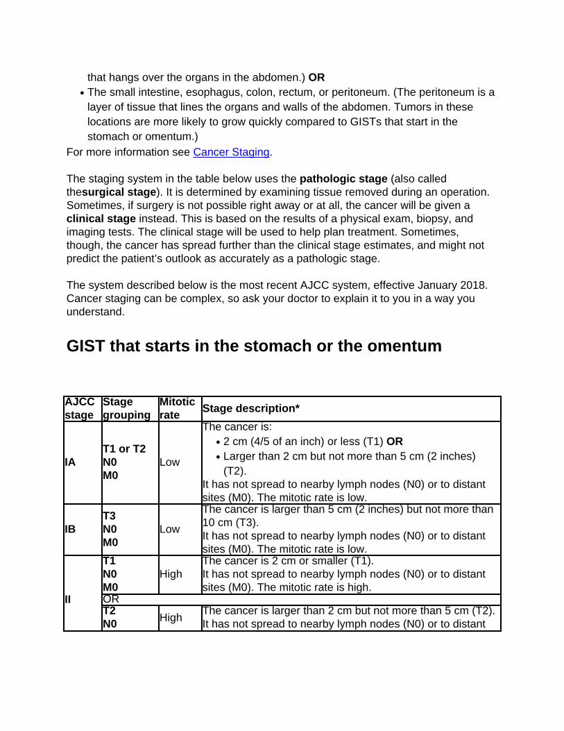
that hangs over the organs in the abdomen.) ORThe small intestine, esophagus, colon, rectum, or peritoneum. (The peritoneum is alayer of tissue that lines the organs and walls of the abdomen. Tumors in theselocations are more likely to grow quickly compared to GISTs that start in thestomach or omentum.)
●
For more information see Cancer Staging.
The staging system in the table below uses the pathologic stage (also calledthesurgical stage). It is determined by examining tissue removed during an operation.Sometimes, if surgery is not possible right away or at all, the cancer will be given aclinical stage instead. This is based on the results of a physical exam, biopsy, andimaging tests. The clinical stage will be used to help plan treatment. Sometimes,though, the cancer has spread further than the clinical stage estimates, and might notpredict the patient’s outlook as accurately as a pathologic stage.
The system described below is the most recent AJCC system, effective January 2018.Cancer staging can be complex, so ask your doctor to explain it to you in a way youunderstand.
GIST that starts in the stomach or the omentum
AJCCstage
Stagegrouping
Mitoticrate
Stage description*
IAT1 or T2N0M0
Low
The cancer is:2 cm (4/5 of an inch) or less (T1) OR●
Larger than 2 cm but not more than 5 cm (2 inches)(T2).
●
It has not spread to nearby lymph nodes (N0) or to distantsites (M0). The mitotic rate is low.
IBT3N0M0
Low
The cancer is larger than 5 cm (2 inches) but not more than10 cm (T3).It has not spread to nearby lymph nodes (N0) or to distantsites (M0). The mitotic rate is low.
II
T1N0M0
HighThe cancer is 2 cm or smaller (T1).It has not spread to nearby lymph nodes (N0) or to distantsites (M0). The mitotic rate is high.
ORT2N0 High
The cancer is larger than 2 cm but not more than 5 cm (T2).It has not spread to nearby lymph nodes (N0) or to distant

M0 sites (M0). The mitotic rate is high.ORT4N0M0
LowThe cancer is larger than 10 cm (T4). It has not spread tonearby lymph nodes (N0) or to distant sites (M0). The mitoticrate is low.
IIIA
T3N0M0
High
The cancer is larger than 5 cm (2 inches) but not more than10 cm(T3).It has not spread to nearby lymph nodes (N0) or to distantsites (M0). The mitotic rate is high.
IIIBT4N0M0
HighThe cancer is larger than 10 cm (T4).It has not spread to nearby lymph nodes (N0) or to distantsites (M0). The mitotic rate is high.
IV
Any TN1M0
Any rate
The cancer is any size (Any T) AND it has spread to nearbylymph nodes (N1).It has not spread to distant sites (M0). The cancer can haveany mitotic rate.
OR
Any TAny NM1
Any rate
The cancer is any size (Any T) AND it might or might nothave spread to nearby lymph nodes (Any N).It has spread to distant sites such as the liver(M1). Thecancer can have any mitotic rate.
*The following additional categories are not listed in the table above:
TX: Main tumor cannot be assessed due to lack of information.●
T0: No evidence of a primary tumor.●
NX: Regional lymph nodes cannot be assessed due to lack of information.●
GIST of the small intestine, esophagus, colon, rectum,or peritoneum
AJCCstage
Stagegrouping
Mitoticrate
Stage description*
IT1 or T2N0M0
Low
The cancer is:2 cm (4/5 of an inch) or less (T1) OR●
Larger than 2 cm but not more than 5 cm (2 inches)(T2).
●
It has not spread to nearby lymph nodes (N0) or to distantsites (M0). The mitotic rate is low.

II
T3N0M0
Low
The cancer is larger than 5 cm (2 inches) but not more than10 cm(T3).It has not spread to nearby lymph nodes (N0) or to distantsites (M0). The mitotic rate is low.
IIIA
T1N0M0
HighThe cancer is 2 cm or smaller (T1).It has not spread to nearby lymph nodes (N0) or to distantsites (M0). The mitotic rate is high.
ORT4N0M0
LowThe cancer is larger than 10 cm (T4).It has not spread to nearby lymph nodes (N0) or to distantsites (M0). The mitotic rate is low.
IIIB
T2N0M0
HighThe cancer is larger than 2 cm but not more than 5 cm (T2).It has not spread to nearby lymph nodes (N0) or to distantsites (M0). The mitotic rate is high.
OR
T3N0M0
High
The cancer is larger than 5 cm (2 inches) but not more than10cm (T3).It has not spread to nearby lymph nodes (N0) or to distantsites (M0). The mitotic rate is high.
ORT4N0M0
HighThe cancer is larger than 10 cm (T4).It has not spread to nearby lymph nodes (N0) or to distantsites (M0). The mitotic rate is high.
IV
Any TN1M0
Any rate
The cancer is any size (Any T) AND it has spread to nearbylymph nodes (N1).It has not spread to distant sites (M0). The cancer can haveany mitotic rate.
OR
Any TAny NM1
Any rate
The cancer is any size (Any T) AND it might or might nothave spread to nearby lymph nodes (Any N).It has spread to distant sites such as the liver(M1). Thecancer can have any mitotic rate.
*The following additional categories are not listed in the table above:
TX: Main tumor cannot be assessed due to lack of information.●
T0: No evidence of a primary tumor.●
NX: Regional lymph nodes cannot be assessed due to lack of information.●
Resectable versus unresectable tumors
The AJCC staging system gives a detailed summary of how far a GIST has spread. But

for treatment purposes, doctors are often more concerned about whether the tumor canbe removed (resected) completely with surgery.
Whether or not a tumor is resectable depends on its size and location, if it has spread toother parts of the body, and if a person is healthy enough for surgery:
Tumors that can clearly be removed without causing major health problems aredefined as resectable.
●
Tumors that can’t be removed completely (because they have spread or for otherreasons) are described as unresectable.
●
In some cases, doctors may describe a tumor as marginally resectable orborderline resectable if it’s not clear if it can be removed completely.
●
If a tumor is considered unresectable or marginally resectable when it is first found,treatments such as targeted therapy may be used first to try to shrink the tumor enoughto make it resectable.
References●
American Joint Committee on Cancer. Gastrointestinal Stromal Tumor. In: AJCCCancer Staging Manual. 8th ed. New York, NY: Springer; 2017:523.
Casali PG, Dei Tos AP, Gronchi A. Chapter 55: Gastrointestinal Stromal Tumor. In:DeVita VT, Lawrence TS, Rosenberg SA, eds. DeVita, Hellman, and Rosenberg’sCancer: Principles and Practice of Oncology. 10th ed. Philadelphia, Pa: LippincottWilliams & Wilkins; 2015.
National Cancer Institute. Physician Data Query (PDQ). Gastrointestinal StromalTumors Treatment. 2017. Accessed at www.cancer.gov/types/soft-tissue-sarcoma/hp/gist-treatment-pdq on April 17, 2017.
National Comprehensive Cancer Network (NCCN). NCCN Clinical Practice Guidelinesin Oncology: Soft Tissue Sarcoma. V.2.2017. Accessed atwww.nccn.org/professionals/physician_gls/pdf/sarcoma.pdf on April 17, 2017.
Last Medical Review: December 20, 2017 Last Revised: December 20, 2017
American Cancer Society medical information is copyrighted material. For reprintrequests, please see our Content Usage Policy.

Survival Rates for GastrointestinalStromal Tumors Survival rates tell you what portion of people with the same type and stage of cancerare still alive a certain amount of time (usually 5 years) after they were diagnosed. Theycan’t tell you how long you will live, but they may help give you a better understandingabout how likely it is that your treatment will be successful. Some people will want toknow the survival rates for their cancer, and some people won’t. If you don’t want toknow, you don’t have to.
What is a 5-year survival rate?
Statistics on the outlook for a certain type and stage of cancer are often given as 5-yearsurvival rates. The 5-year survival rate is the percentage of people who live at least 5years after being diagnosed with cancer. For example, a 5-year survival rate of 70%means that an estimated 70 out of 100 people who have that cancer are still alive 5years after being diagnosed. Keep in mind, however, that many of these people livemuch longer than 5 years after diagnosis.
Relative survival rates are a more accurate way to estimate the effect of cancer onsurvival. These rates compare people with cancer to people in the overall population.For example, if the 5-year relative survival rate for a specific stage of gastrointestinalstromal tumor (GIST) is 80%, it would mean that people who have that stage of cancerare, on average, about 80% as likely as people who don’t have that cancer to live for atleast 5 years after being diagnosed.
But remember, the 5-year relative survival rates are estimates – your outlook can varybased on a number of factors specific to you.
Cancer survival rates don’t tell the whole story
Survival rates are often based on previous outcomes of large numbers of people whohad the disease, but they can’t predict what will happen in any particular person’s case.There are a number of limitations to remember:
The numbers below are among the most current available. But to get 5-yearsurvival rates, doctors have to look at people who were treated at least 5 years ago.
●

As treatments are improving over time, people who are now being diagnosed withGISTs may have a better outlook than these statistics show.These statistics are based on the stage of the cancer when it was first diagnosed.They do not apply to cancers that later come back or spread, for example.
●
The outlook for people with GISTs varies by the stage (extent) of the cancer – ingeneral, the survival rates are higher for people with earlier stage cancers. Butmany other factors can affect a person’s outlook, such as age and overall health,where the cancer is in the body, and how well the cancer responds to treatment.The outlook for each person is specific to their circumstances.
●
Your doctor can tell you how these numbers may apply to you, as he or she is familiarwith your particular situation.
Survival rates for GISTs
It is very hard to get accurate numbers on survival rates for GISTs. Part of this isbecause these tumors are not common. In the past, they were often classified as othertypes of cancers, which made the numbers available for study even smaller. Treatmenthas also changed dramatically in recent years now that newer, targeted therapy drugsare being used. The survival rates below are based on people treated many years ago,largely before these newer treatments were used, so people being treated for GISTstoday are likely to have a better outlook.
Based on people diagnosed between 2003 and 2009 the overall relative 5-year survivalrate of people diagnosed with a malignant GIST was estimated to be about 76%.
If the tumor was still just in the organ where it started, the 5-year relative survivalwas 91%.
●
If it had grown into nearby structures (or spread to nearby lymph nodes) when itwas first diagnosed, the 5-year relative survival was around 74%.
●
If it had spread to distant parts of the body when it was first diagnosed, the 5-yearrelative survival was 48%.
●
Remember, these survival rates are only estimates – they can’t predict what will happento any individual person. We understand that these statistics can be confusing and maylead you to have more questions. Talk to your doctor to better understand your specificsituation.
Last Medical Review: May 17, 2017 Last Revised: May 17, 2017

American Cancer Society medical information is copyrighted material. For reprintrequests, please see our Content Usage Policy.
Questions to Ask Your Doctor AboutGastrointestinal Stromal Tumors It’s important to have honest, open discussions with your cancer care team. Ask anyquestion, no matter how small it might seem. Some questions to consider:
When you’re told you have a gastrointestinal stromal tumor (GIST)
How sure are you that my tumor is a GIST?●
Where is my tumor located? How big is it?●
How likely is this tumor to grow or spread quickly?●
Has my tumor spread beyond where it started?●
What is the stage of my cancer, and what does that mean?●
Will I need any other testsbefore we can decide on treatment?●
Will I need to see any other doctors?●
If I'm concerned about costs and insurance coverage for my diagnosis andtreatment, who can help me?
●
When deciding on a treatment plan
How much experience do you have treating these tumors?●
What are my treatment options?●
What do you recommend? Why?●
What’s the goal of the treatment?●
Should I get a second opinion? How do I do that? Can you recommend someone?●
What are the chances my cancer can be cured?●
How quickly do we need to decide on treatment?●
What should I do to be ready for treatment?●
How long will treatment last? What will it be like? Where will it be done?●
What risks or side effects I should expect? How long are they likely to last?●
Will treatment affect my daily activities?●
How likely is it that the cancer will come back after treatment? Is there anything Ican do to lower this risk?
●

During treatment
How will we know if the treatment is working?●
Is there anything I can do to help manage side effects?●
What symptoms or side effects should I tell you about right away?●
How can I reach you on nights, holidays, or weekends?●
Do I need to change what I eat during treatment?●
Are there any limits on what I can do? ●
Should I exercise? What should I do, and how often?●
Can you suggest a mental health professional I can see if I start to feeloverwhelmed, depressed, or distressed?
●
After treatment
Are there any limits on what I can do?●
What symptoms should I watch for?●
What kind of exercise should I do now?●
What type of follow-up will I need after treatment?●
How often will I need to have follow-up exams and tests?●
How will we know if the cancer has come back? What should I watch for?●
What will my options be if the cancer comes back?●
Along with these sample questions, be sure to write down some of your own. Forinstance, you might want more information about second opinions or about clinical trialsfor which you may qualify.
Keep in mind that doctors aren’t the only ones who can give you information. Otherhealth care professionals, such as nurses and social workers, can answer some of yourquestions. To find more about speaking with your health care team, see The Doctor-Patient Relationship.
Last Medical Review: May 17, 2017 Last Revised: May 17, 2017
American Cancer Society medical information is copyrighted material. For reprintrequests, please see our Content Usage Policy.

2016 Copyright American Cancer Society