gamatogenesis - bhattadev university
TRANSCRIPT
GAMATOGENESIS
DR. ALAKESH BARMAN
ASST. PROFESSOR
DEPT. OF ZOOLOGY; BHATTADEV UNIVERSITY

WHAT IS THE BASIC DIFFERENCES BETWEEN
SPERMATOGENESIS AND OOGENESIS

One of the fundamental differences concerns the timing of meiosis onset.

SO HOW TIMING IS DIFEERENT ??
In females, meiosis begins in theembryonic gonads BUT in males, meiosisis not initiated until puberty. HOW ITPOSSIBLE ???

This critical difference in timing is due toRetinoic Acid (RA) produced by the mesonephric kidneys.
This RA stimulates the germ cells to undergo anew round of DNA replication and initiate meiosis(Baltus et al. 2006; Bowles et al. 2006; Lin etal. 2008).
In males, however, the embryonic testes secretethe RA-degrading enzyme Cyp26bl. This prevents RA frompromoting meiosis. Later, Nanos2 will be expressedin the male germ cells, and this will alsoprevent meiosis and ensure that the cells followthe pathway to become sperm (Koubova et al. 2006;Suzuki and Saga 2008).


SPERMATOGENESIS
Once mammalian PGCs arrive at the genitalridge of a male embryo, they are calledgonocytes and become incorporated into thesex cords (Culty 2009). They remain thereuntil maturity, at which time the sex cordshollow out to form the seminiferous tubules.The epithelium of the tubules differentiatesinto the Sertoli cells that will nourish andprotect the developing sperm cells.
The gonocytes differentiate into a population ofstem cells that have recently been named theundifferentiated type A spermatogonia (Yoshidaet al. 2007). These cells can reestablishspermatogenesis when transferred into micewhose sperm production was eliminated bytoxic chemicals. They appear to reside in stemcell niches created by the junction of Sertolicells, interstitial (testosterone-producing) cells,and blood vessels.
The decision to proliferate or differentiate mayinvolve interactions between to pathways –Wnt and BMP pathways.
Wnt signalling appears to promote proliferation ofstem cells, and the spermatogonia appear to havereceptors for both Wnts and BMPs (Golestaneh et al.2009). The initiation of spermatogenesis duringpuberty is probably regulated by the synthesis ofBMPs by the spermatogenic germ cells, thespermatogonia.
HOW BMPS INITIATES SPERMATOGENESIS ??

When BMP8b reaches a critical concentration, thegerm cells begin to differentiate.The differentiating cells produce high levels of BMP8b,which can then further stimulate their differentiation.Mice lacking BMP8b do not initiate spermatogenesis atpuberty (Zhao et al. 1996).The spermatogenic germ cells are bound to the Sertolicells by N-cadherin molecules on the surfaces of bothcell types, and by galactosyltransferase molecules onthe spermatogenic cells that bind a carbohydratereceptor on the Sertoli cells (Newton et al. 1993; Prattet al. 1993).SpermatogenesiS occurs in the recesses between theSertoli cells.

FORMING THE HAPLOID SPERMATID
The undifferentiated type A1 spermatogonia
(sometimes called the dense type A
spermatogonia) are found adjacent to the outer
basal lamina of the sex cords.
They are stem cells, and upon reaching
maturity are thought to divide to make another
type A1 spermatogonium as well as a
second,the type A2 spermatogonia.
The A2 spermatogonia divide to produce type
A3 spermatogonia, which then beget type A4
spermatogonia..
The A4 spermatogonia are thought to
differentiate into the first committed stem cell type,
the intermediate spermatogonia.
Intermediate spermatogonia are committed to
becoming spermatozoa, and they divide mitotically
once to form type B spermatogonia (see Figure
16.27).
These cells are the precursors of the
spermatocytes and are the last cells of the line that
undergo mitosis. They divide once to generate the
primary spermatocytes—the cells that enter
meiosis
The transition between spermatogonia andspermatocytes appears to be mediated by theopposing influences of two factors- glial cell line-derived neurotrophic factor (GDNF) and stem cellfactor (SCF), both of which are secreted by theSertoli cells.
GDNF levels determine whether the dividingspermatogonia remain spermatogonia or enter thepathway to become spermatocytes. Low levels ofGDNF favor the differentiation of thespermatogonia, whereas high levels favor self-renewal of the stem cells (Meng et al. 2000)
SCF promotes the transition to spermatogenesis(Rossi et al. 2000).
Both GDNF and SCF are upregulated by follicle-stimulating hormone (FSH).
These two factors may serve as a link between theSertoli cells and the endocrine system, and theyprovide a mechanism for FSH to instruct the testes toproduce more sperm (Tadokoro et al. 2002).
Looking at Figure 16.28, we find that during thespermatogonial divisions, cytokinesis is notcomplete. Rather, the cells form a syncytium inwhich each cell communicates with the others viacytoplasmic bridges .
The successive divisions produce clones ofinterconnected cells, and because ions andmolecules readily pass through these cytoplasmicbridges, each cohort matures synchronously.
During this time, the spermatocyte nucleus oftentranscribes genes whose products will be used laterto form the axoneme and acrosome

Each primary spermatocyte undergoes the firstmeiotic division to yield a pair of secondaryspermatocytes, which complete the second division ofmeiosis. The haploid cells thus formed are calledspermatids, and they are still connected to oneanother through their cytoplasmic bridges.
The spermatids that are connected in this mannerhave haploid nuclei but are functionally diploid, sincea gene product made in one cell can readily diffuseinto the cytoplasm of its neighbors (Braun et al. 1989).
During the divisions from type A1 spermatogoniato spermatids, the cells move farther and fartheraway from the basal lamina of the seminiferoustubule and closer to its lumen (see Figure 16.27; Siuand Cheng 2004).
Thus, each type of cell can be found in a particularlayer of the tubule.
The spermatids are located at the border of thelumen, and here they lose their cytoplasmicconnections and differentiate into spermatozoa. Inhumans, the progression from spermatogonial stemcell to mature spermatozoa takes 65 days (Dym1994).

OOGENESIS
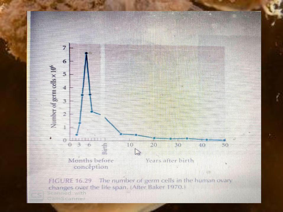
In the human embryo, the thousand of oogonia
divide rapidly from the second to the seventh month
of gestation to form roughly 7 million germ cells.
After the seventh month of embryonic
development, however, the number of germ cells
drops precipitously.
Most oogonia die during this period, while the
remaining oogonia enter the first meiotic division
(Pinkerton et al. 1961).
These latter cells, called primary oocytes, progress
through the first meiotic prophase until the diplotene
stage, at which point they are maintained until the
female matures.
With the onset of puberty, groups of oocytes
periodically resume meiosis.
Thus, in the human female, the first part of
meiosis begins in the embryo, and the signal to
resume meiosis is not given until roughly 12 years
later.
In fact, some oocytes are maintained in meiotic
prophase for nearly 50 years. Of the millions of
primary oocytes present at her birth, only about
400 mature during a woman's lifetime.
OOGENIC MEIOSIS
Oogenic meiosis differs from spermatogenic
meiosis in its placement of the metaphase plate.
When the primary oocyte divides, its nucleus, called
the germinal vesicle, breaks down, and the
metaphase spindle migrates to the periphery of the
cell. At telophase, one of the two daughter cells
contains hardly any cytoplasm, whereas the other
cell retains nearly the entire volume of cellular
constituents (Figure 16.30). The smaller cell is
called the first polar body, and the larger cell is
referred to as the secondary oocyte.
During the second division of meiosis, a similar
unequal cytokinesis takes place. Most of the
cytoplasm is retained by the mature egg (the
ovum), and a second polar body receives little
more than a haploid nucleus. (The first polar
body usually does not divide.) Thus, oogenic
meiosis conserves the volume of oocyte
cytoplasm in a single cell rather than splitting it
equally among four progeny.
MATURATION OF THE MAMMALIAN OOCYTE
Ovulation in mammals follows one of two
patterns, depending on the species. One type of
ovulation is stimulated by the act of copulation.
Physical stimulation of the cervix triggers the
release of gonadotropins from the pituitary. These
gonadotropins signal the egg to resume meiosis and
initiate the events that will expel it from the ovary.
This mechanism ensures that most copulations will
result in fertilized ova, and animals that utilize this
method of ovulation—such as rabbits and minks—
have a reputation for procreative success.

Most mammals, however, have a periodic
ovulation pattern, in which the female ovulates
only at specific times of the year. This ovulatory
period is called estrus (or its English equivalent,
heat).
In these animals, environmental cues (most
notably the amount and type of light during the
day) stimulate the hypothalamus to release
gonadotropinreleasing hormone (GRH). GRH
stimulates the pituitary to release the
gonadotropins—follicle-stimulating hormone
(FSH) and luteinizing hormone (LH)—that cause
the ovarian follicle cells to proliferate and secrete
estrogen.

Estrogen enters certain neurons and evokes the
pattern of mating behavior characteristic of the
species.
The gonadotropins also stimulate follicular
growth and initiate ovulation. Thus, mating
behavior and ovulation occur close together.
Humans have a variation on the theme of
periodic ovulation. Although human females have
cyclical ovulation (averaging about once every
29.5 days) and no definitive yearly estrus, most of
human reproductive physiology is shared with
other primates. The characteristic primate
periodicity in maturing and releasing ova is called
the men strual cycle
The menstrual cycle represents the integration of
three very different cycles:
1. The ovarian cycle, the function of which is to
mature and release an oocyte.
2. The uterine cycle, the function of which is to
provide the appropriate environment for the
developing blastocyst.
3. The cervical cycle, the function of which is to
allow sperm to enter the female reproductive
tract only at the appropriate time. These three
functions are integrated through the
hormones of the pituitary, hypothalamus, and
ovary.

The majority of the oocytes in the adult huma n
ovary are maintained in the diplotene stage of the
first meiotic prophase, often referred to as the
dictyate state.
Each oocyte is enveloped by a primordial follicle
consisting of a single layer of epithelial granulosa
cells and a less organized layer of mesenchymal
thecal cells (Figure 16.31).
Periodically, a group of primordial follicles enters
a stage of follicular growth. During this time, the
oocyte undergoes a 500-fold increase in volume
(corresponding to an increase in oocyte diameter
from 10 um in a primordial follicle to 80 um in a fully
developed follicle).

Books used as References to prepare the presentation:
1. DEVELOPMENTAL BIOLOGY: 9TH ED. BY GILBERT