gallstone disease rufi
TRANSCRIPT

Gallstone Disease
Definitions
• Cholelithiasis = presence of gallstones
• Acute calculous cholecystitis = occlusion of the cystic duct by gallstone leading to gallbladder inflammation
• Chronic calculous cholecystitis = recurrent episodes of cystic duct obstruction leading to scarring and a nonfunctional gallbladder
• Chronic acalculous cholecystitis = symptoms of biliary colic, no gallstones, and an abnormal gallbladder ejection fraction
• Acute cholangitis = bacterial infection of the biliary ducts
• Choledocholithiasis = CBD stones
• Mirizzi syndrome = when gallstones lodged in either the cystic duct or the Hartmann pouch of the gallbladder, externally compressed the common hepatic duct (CHD), causing symptoms of obstructive jaundice

Gallstone Disease
Types of Gallstones
•Pure cholesterol (70%)-radioluscent•Pigmented (30%)-radiopaque
• PURE 20%-Black stones (contain Ca bilirubinate, a/w cirrhosis and hemolysis)
• MIXED 10%-Brown stones (a/w biliary tract infection)

Gallstone Disease
Risk Factors
Calculous• “Female, Fat, Forty, Fertile”• Oral contraceptives• Obesity• Fatty diet• Hemolytic states• Cirrhosis• Bile duct stasis (biliary stricture, congenital
cysts, pancreatitis, sclerosing cholangitis)• IBD• Vagotomy• Hyperlipidemia
Acalculous• Rapid weight loss
(gastric bypass pts.)• DM• Prolonged fasting• TPN• Ileal resection• Old age• Severe illness• Burns ,trauma
Gallstone Disease
Gallstone Complications• Gallstone ileus, gallstone pancreatitis
• Acute cholecystitis: 10-20% of pts. w/ symptomatic gallstones• GB gangrene • GB perforation• GB empyema (pus in the GB)• Emphysematous cholecystitis (a/w GB vascular compromise,
stones, impaired immune system, infection w/gas-forming organisms - clostridium, E. coli, Klebsiella)
• Cholecystoenteric fistula
• Choledocholithiasis: 8-15% of pts. w/ symptomatic gallstones• Cirrhosis• Cholangitis• Pancreatitis

Gallstone Disease
RUQ DDx
• Gallbladder: cholecystitis, Choledocholithiasis, cholangitis
• Duodenal ulcer
• Hepatitis
• Appendicitis (atypical presentation)
• Pancreatitis

Gallstone Disease
Strasberg S. N Engl J Med 2008;358:2804-2811
Diagnostic Criteria for Acute Cholecystitis, According to Tokyo Guidelines

Gallstone Disease
CLUE
• Provocation/Timing: meals (50%), night time• Quality: constant• Radiation: RUQ to the R scapula (Boas’ sign)• Severity: “severe”

Gallstone DiseaseComplication History Examination Blood tests
Biliary Colic - Intermittent RUQ/epigastric pain (minutes/hours) into back or right shoulder- N&V
-Tender RUQ-No peritonism-Murphy’s –-Apyrexial, HR and BP (N)
-WCC (N) CRP (N)- LFT (N)
Acute Cholecystitis -Constant RUQ pain into back or right shoulder-N&V-Feverish
-Tender RUQ-Periotnism RUQ (guarding/rebound)-Murphy’s +-Pyrexia, HR (↑)
-WCC and CRP (↑)-LFT (N or mildly (↑)
Empyema -Constant RUQ pain into back or right shoulder-N&V-Feverish
-Tender RUQ -Peritonism RUQ-Murphy’s +-Pyrexia, HR (↑), BP (↔ or ↓)-More septic than acute cholecystitis
-WCC and CRP (↑)-LFT (N or mildly (↑)
Obstructive Jaundice -Yellow discolouration-Pale stool, dark urine-painless or assocaited with mild RUQ pain
-Jaundiced-Non-tender or minimally tender RUQ-No peritonism-Murphy’s –-Apyrexial, HR and BP (N)
-WCC and CRP (N)-LFT: obstructive pattern bili (↑), ALP (↑), GGT (↑), ALT/AST (↔)-INR (↔ or ↑)
Ascending Cholangitis Charcot”s triad-RUQ pain (constant)-Jaundice -Rigors
-Jaundiced-Tender RUQ -Peritonism RUQ-Spiking high pyrexia (38-39)-HR (↑), BP (↔ or ↓)-Can develop septic shock
-WCC and CRP (↑)-LFT : obstructive pattern bili (↑), ALP (↑), GGT (↑), ALT/AST (↔)-INR (↔ or ↑)
Acute Pancreatitis -Severe upper abdominal pain (constant) into back-Profuse vomiting
-Tender upper abdomen-Upper abdominal or generalised peritonism-Usually apyrexial, HR (↑), BP (↔ or ↓)
-WCC and CRP (↑)-LFT: (N) if passed stone or obstructive pattern ifstone still in CBD-Amylase (↑)-INR/APTT (N) or (↑) if DIC
Gallstone Ileus - 4 cardinal features of SBO -distended tympanic abdomen-hyperactive/tinkling bowel sounds

Gallstone Disease
Labs
• Order: BMP, amylase/lipase, LFTs, CBC, coags
• Acute cholecystitis: increased WBC, increased alk phos, slight increase in amylase and T bili

Gallstone Disease
Imaging
• KUB - only 15% of gallstones are radiopaque
• U/S - gallstone identification false(-) rate is 5-15%. It identifies bile duct dilatation w/ 80% accuracy.
Look for• thickened GB wall (>3.5mm)-independent predictor of acute cholecystitis .(ppv 95%)• pericholecystic fluid, distended GB, •Murphy’s sign-sensitivity as high as 88%

Gallstone Disease
Strasberg S. N Engl J Med 2008;358:2804-2811
Ultrasonographic Images of Three Gallbladders

Gallstone Disease
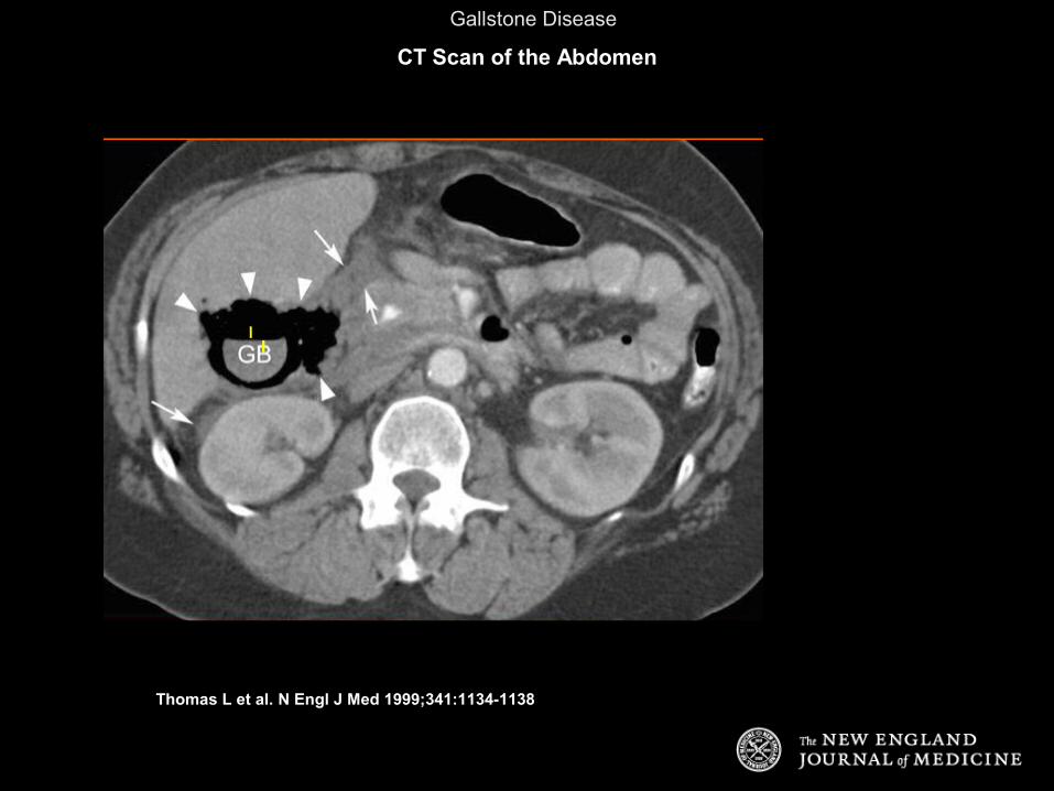
• CT scan - used to diagnose complications
• MRI - can detect gallstones and common duct stones
• ERCP - to look for CBD stones
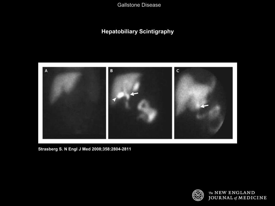
• HIDA scan - radionuclide IV, extracted from blood, excreted into bile
Gallstone Disease
Thomas L et al. N Engl J Med 1999;341:1134-1138
CT Scan of the Abdomen
Gallstone Disease
Strasberg S. N Engl J Med 2008;358:2804-2811
Hepatobiliary Scintigraphy

Gallstone Disease
Management
• biliary colic: oral or parenteral opiate analgesics and NSAIDS( ketorolac).
• complicated: analgesia, antiemetics , cessation of oral intake, volume and electrolyte replacement, antibiotics(cefotaxime or ceftriaxone, 1 gram IV) and metronidazole; or a fluoroquinolone and metronidazole)
• ERCP is undertaken for patients with common bile duct stones or dilated common bile ducts

Gallstone Disease
Case 1
• HPI: 46y F p/w 4hr h/o nausea and RUQ pain radiating to the R scapula. Symptoms began 1 hr. after a fatty meal. Pt. currently has no pain. No prior episodes.
• PMHx/PSHx None
• PE: RUQ minimally TTP, (-)Murphy’s
• Labs: WBC 8, LFT normal

Gallstone Disease
Case 1
What is the diagnosis?
→→
►

Gallstone Disease
Case 1: Continued
• Dx: symptomatic cholethiasis• Plan: NPO, IVF, cholecystectomy

Gallstone Disease
Case 2
• 46yF p/w h/o >24hr of RUQ pain radiating to the R scapula, started after fatty meal, a/w nausea, vomiting, fever
• Exam: Febrile, RUQ +, (+)Murphy’s sign
• Labs: WBC 13, Mild ↑LFT

Gallstone Disease
Case 2: Continued
◄

Gallstone Disease
Case 2: Continued
• → denotes the GB wall thickening
• ► denotes the fluid around the GB
• GB also appears distended
• ?DIAGNOSIS
→
►

Gallstone Disease
Case 2: Continued
• Dx: acute calculous cholecystitis
• Persistent cystic duct obstruction leads to GB distension, wall inflammation & edema
• Risk of: empyema, gangrene, rupture
• Treatment: • NPO• IVF• ABX:
• Common organisms: E coli, Bacteroides fragilis, Klebsiella, Enterococcus, and Pseudomonas
Sanford guide:2006/11• Piperacillin/tazobactam (Zosyn), ampicillin/sulbactam
(Unasyn), or meropenem• Cholecystectomy

Gallstone Disease
Case 3• 87y M critically ill, on long-term TPN c/o RUQ
pain
• PE: febrile, RUQ +
• U/S: GB wall thickening, pericholecystic fluid, no gallstones
• What is the diagnosis?

Gallstone Disease
Case 3: Continued
• Dx: acute acalculous cholecystitis
• Caused by gallbladder stasis from lack of enteral stimulation by cholecystokinin
• Risk of: gangrene, empyema, perforation due to ischemia
• TX: rapid progression of acute acalculous cholecystitis to gangrene & perforation, early recognition and intervention are required.

Gallstone Disease
Case 4
• 46y F p/w RUQ pain, jaundice, acholic stools, dark tea-colored urine, w/o fever
• PMHx: Cholelithiasis• Exam: unremarkable• WBC 8, T.Bili 8, AST/ALT NL, Hep B/C neg• U/S: gallstones, CBD stone, dilated CBD > 1cm
• What is the diagnosis?

Gallstone Disease
Case 4: Continued
• DX: Choledocholithiasis
• Similar presentation as Cholelithiasis, except with the addition of jaundice
• DDx: Cholelithiasis, hepatitis, cholangitis, CA, choledochal cyst, bile duct stricture, UC, pancreatitis
• Plan: • Endoscopic retrograde cholangiopancreatography
(ERCP) w/ stone extraction and sphincterotomy• Interval cholecystectomy after recovery from ERCP

Gallstone Disease
Case 5
• 46y F p/w 4hr h/o nausea and RUQ pain radiating to the R scapula. Symptoms began 1 hr. after a fatty meal. Pt. currently has no pain. Has had multiple similar episodes.
• PMHx/PSHx None
• PE: RUQ minimally TTP, (-)Murphy’s
• Labs: WBC 6, LFT normal
• Studies: RUQ U/S w/Cholelithiasis without GB wall thickening or pericholecystic fluid
• Diagnosis: ?

Gallstone Disease
Case 5: Continued
• Dx: chronic calculous cholecystitis• Recurrent inflammatory process due to
recurrent cystic duct obstruction leading to scarring/wall thickening
• Treatment: cholecystectomy

Gallstone Disease
Case 6
• 46y F p/w persistent epigastric & back pain• PMHx: symptomatic gallstones• SHx: no ETOH• PE: Tender epigastrum• Labs: Amylase 2000, ALT 150• U/S: gallstones
• What is the diagnosis?• What is the plan?

Gallstone Disease
Case 6: Continued
• Dx: gallstone pancreatitis • 35% of acute pancreatitis secondary to stones• Pathophysiology: reflux of bile into pancreatic duct and/or
obstruction of ampulla by stone• ALT >150 (3-fold elevation) has 95% PPV for diagnosing
gallstone pancreatitis• Treatment:
• ABC, resuscitate, NPO/IVF, pain medication• ERCP once pancreatitis resolves • Cholecystectomy before d/c

Gallstone Disease
Take Home Points
• Start with ABCs• Cholelithiasis = “Female, Fat, Forty, Fertile”• Stone formation based on the relative concentration of
cholesterol, bile salts, and phospholipid • Cholecystitis PE = Murphy’s sign• RUQ evaluation: U/S, HIDA, CT, MRI, ERCP• Acalculous cholecystitis a/w TPN, ICU setting• Cholangitis = Charcot’s triad, Reynold’s pentad

Gallstone Disease
•THANKS