gabab receptors at glutamatergic synapses in the … · receptor subunits, gaba b1 and gaba b2. in...
TRANSCRIPT
GR
CLa
pb
Bc
td
H
AvdtimrvtvpgGwpwisptasaeultgTupmsat
1
Q2
*EAtGgV
Neuroscience 136 (2005) 1083–1095
0d
ABAB RECEPTORS AT GLUTAMATERGIC SYNAPSES IN THE
AT STRIATUMnsonaGo
KV
Tntfmpas1tptdauJwtGbs2C
aiawt1sppRepeta
. J. LACEY,a2 J. BOYES,a,b2 O. GERLACH,a,c
. CHEN,a,d1 P. J. MAGILLa AND J. P. BOLAMa*
Medical Research Council Anatomical Neuropharmacology Unit, De-artment of Pharmacology, University of Oxford, Oxford OX1 3TH, UK
School of Life and Health Sciences, University of Aston, Birmingham4 7ET, UK
Department of Psychiatry and Neuropsychology, University of Maas-richt, Maastricht, The Netherlands
Department of Physiology, The Chinese University of Hong Kong,ong Kong, China
bstract—Although multiple effects of GABAB receptor acti-ation on synaptic transmission in the striatum have beenescribed, the precise locations of the receptors mediatinghese effects have not been determined. To address thisssue, we carried out pre-embedding immunogold electron
icroscopy in the rat using antibodies against the GABAB
eceptor subunits, GABAB1 and GABAB2. In addition, to in-estigate the relationship between GABAB receptors and glu-amatergic striatal afferents, we used antibodies against theesicular glutamate transporters, vesicular glutamate trans-orter 1 and vesicular glutamate transporter 2, as markers forlutamatergic terminals. Immunolabeling for GABAB1 andABAB2 was widely and similarly distributed in the striatum,ith immunogold particles localized at both presynaptic andostsynaptic sites. The most commonly labeled structuresere dendritic shafts and spines, as well as terminals form-
ng asymmetric and symmetric synapses. In postsynaptictructures, the majority of labeling associated with thelasma membrane was localized at extrasynaptic sites, al-hough immunogold particles were also found at the postsyn-ptic specialization of some symmetric, putative GABAergicynapses. Labeling in axon terminals was located within, ort the edge of, the presynaptic active zone, as well as atxtrasynaptic sites. Double labeling for GABAB receptor sub-nits and vesicular glutamate transporters revealed that
abeling for both GABAB1 and GABAB2 was localized on glu-amatergic axon terminals that expressed either vesicularlutamate transporter 1 or vesicular glutamate transporter 2.he patterns of innervation of striatal neurons by the vesic-lar glutamate transporter 1- and vesicular glutamate trans-orter 2-positive terminals suggest that they are selectivearkers of corticostriatal and thalamostriatal afferents, re-
pectively. These results thus provide evidence that presyn-ptic GABAB heteroreceptors are in a position to modulatehe two major excitatory inputs to striatal spiny projection
Present address: Department of Physiology, Qingdao University,ingdao 266021, China.These authors contributed equally to this manuscript.
Corresponding author. Tel: �44-1865-271869; fax: �44-1865-271647.-mail address: [email protected] (J. P. Bolam).bbreviations: ABC, avidin–biotin–peroxidase complex; EPSP, exci-
atory postsynaptic potential; GABAB1 and GABAB2, subunits of theABAB receptor; IPSP, inhibitory postsynaptic potential; NGS, normal
doat serum; PB, phosphate buffer; PBS, phosphate-buffered saline;GLUT, vesicular glutamate transporter.
306-4522/05$30.00�0.00 © 2005 Published by Elsevier Ltd on behalf of IBRO.oi:10.1016/j.neuroscience.2005.07.013
1083
eurons arising in the cortex and thalamus. In addition, pre-ynaptic GABAB autoreceptors are present on the terminalsf spiny projection neurons and/or striatal GABAergic inter-eurons. Furthermore, the data indicate that GABA may alsoffect the excitability of striatal neurons via postsynapticABAB receptors. © 2005 Published by Elsevier Ltd on behalff IBRO.
ey words: basal ganglia, vesicular glutamate transporters,GLUT1, VGLUT2, corticostriatal, thalamostriatal.
he inhibitory amino acid GABA plays a critical role in theeuronal networks of the basal ganglia. Two receptorypes mediate the actions of GABA in the basal ganglia:ast-acting ionotropic GABAA receptors and slower-actingetabotropic GABAB receptors. Both postsynaptic andresynaptic GABAB receptors exert their influence on syn-ptic transmission through G-protein coupling and down-tream intracellular effector systems (Kaupmann et al.,997). Activation of postsynaptic GABAB receptors leads
o increases in potassium conductance and hence, hyper-olarization, and activation of presynaptic GABAB recep-ors leads to an inhibition of transmitter release through aecrease in calcium influx (Bowery et al., 2002; Calver etl., 2002). GABAB receptors are heterodimers of two sub-nits, GABAB1 and GABAB2 (Kaupmann et al., 1997, 1998;ones et al., 1998; White et al., 1998; Kuner et al., 1999)hich are both necessary and sufficient for normal func-
ioning in vivo (Prosser et al., 2001; Schuler et al., 2001;assmann et al., 2004). The GABAB1 subunit is responsi-le for binding GABA whereas GABAB2 is necessary forurface trafficking and G-protein activation (Galvez et al.,000; Jones et al., 2000; Margeta-Mitrovic et al., 2000;alver et al., 2001; Robbins et al., 2001).
The striatum is the major division of the basal gangliand the majority of its neurons are GABAergic. These
nclude the spiny projection neurons, which account forbout 95% of the total population of striatal neurons andhich form the major output of the striatum, and at least
hree classes of GABAergic interneurons (see Kawaguchi,993; Tepper et al., 2004 for reviews). In addition, thetriatum receives GABAergic afferents from the globusallidus (Kita and Kitai, 1994; Bevan et al., 1998) andossibly the substantia nigra (van der Kooy et al., 1981;odríguez and González-Hernández, 1999). Although theffects of GABA in the striatum are mediated principally byostsynaptic GABAA receptors (Tepper et al., 2004), sev-ral effects of GABAB receptor activation on synapticransmission in the striatum have been described. Thus,pplication of the GABA agonist, baclofen, causes a re-
Buction in cortically evoked excitatory postsynaptic poten-
tnebtGeis1gt
srGM2HitCotstmGusntgcsuarzpa2adGGf
A
TrtpdmdSv
sp(c
A
Acb(Gab2pVuVta
I
TspTciiPww
iGttSbrfipS
io1egbtbtpasbv
Vpvta1o
C. J. Lacey et al. / Neuroscience 136 (2005) 1083–10951084
ials (EPSPs) in striatal neurons, including spiny projectioneurons, in vitro (Calabresi et al., 1990, 1991; Nisenbaumt al., 1992, 1993). In support of this effect being mediatedy presynaptic GABAB receptors, lesions of the corticos-
riatal pathway are associated with a marked reduction inABAB binding sites in the striatum (Moratalla and Bow-ry, 1991). In addition, GABAB activation also reduces
nhibitory postsynaptic potentials (IPSPs) following intra-triatal stimulation (Calabresi et al., 1991; Seabrook et al.,991; Nisenbaum et al., 1992, 1993). These findings sug-est that GABAB receptors are present on both glutama-
ergic and GABAergic inputs to striatal neurons.The presence of functional GABAB receptors in the
triatum is supported by immunocytochemical studies inats and monkeys demonstrating that both GABAB1 andABAB2 are expressed by striatal neurons (Margeta-itrovic et al., 1999; Yung et al., 1999; Charara et al.,000, 2004; Charles et al., 2001; Waldvogel et al., 2004).owever, this is inconsistent with in situ hybridization stud-
es showing that GABAB2 mRNA is virtually absent fromhe striatum (Durkin et al., 1999; Martin et al., 1999, 2004;lark et al., 2000). In view of this apparent mismatch, onef the main objectives of the present study was to charac-erize the precise localization of both GABAB receptorubunits at the electron microscopic level in the rat stria-um, using the pre-embedding immunogold labelingethod. In monkeys, immunoperoxidase labeling forABAB receptors has previously been detected in a pop-lation of terminals forming asymmetric, putative excitatoryynapses, which is characteristic of corticostriatal termi-als (Charara et al., 2000, 2004). However, in addition to
he input from the cortex, the striatum receives excitatory,lutamatergic afferents from the intralaminar thalamic nu-lei (see Smith et al., 2004). Glutamatergic terminals in thetriatum can be characterized by their expression of vesic-lar glutamate transporter (VGLUT) subtypes; VGLUT1nd VGLUT2 are localized in terminals that form asymmet-ic synapses in the striatum (Bellocchio et al., 1998; Her-og et al., 2001; Fujiyama et al., 2004) and have beenroposed to be selectively associated with corticostriatalnd thalamostriatal afferents, respectively (Fremeau et al.,001; Varoqui et al., 2002; Bacci et al., 2004; Fujiyama etl., 2004). Thus, a further objective of the study was toetermine the nature of the afferent terminals that expressABAB receptors by combining immunogold labeling forABAB receptor subunits with immunoperoxidase labeling
or VGLUTs.
EXPERIMENTAL PROCEDURES
nimals and tissue preparation
he experiments were carried out on 12 male Sprague–Dawleyats (180–300 g; Charles River, Margate, Kent, UK). Environmen-al conditions for housing of the rats and all procedures that wereerformed were in accordance with the Animals (Scientific Proce-ures) Act of 1986 (UK). Every effort was made to use the mini-um number of animals and to minimize suffering. They wereeeply anesthetized with sodium pentobarbitone (200 mg/kg; i.p.;agatal; Rhône Mérieux, Tallaght, Dublin, Ireland) and perfused
ia the ascending aorta with 50–100 ml of phosphate-buffered waline (PBS; 0.01 M phosphate, pH 7.4) followed by 300 ml of 3%araformaldehyde and 0.1% glutaraldehyde in phosphate bufferPB; 0.1 M, pH 7.4). Coronal sections (65–70 �m) of striatum wereut on a vibrating microtome and collected in PBS.
ntibodies
ll antibodies used in these studies were obtained from commer-ial sources. For GABAB immunocytochemistry, polyclonal anti-odies raised in guinea-pig against the C termini of GABAB1
AB1531; Chemicon, Chandlers Ford, Hampshire, UK) andABAB2 (AB5394; Chemicon) were used. The specificity of bothntibodies has been established by immunohistochemistry onrain sections from GABAB-subunit knockout mice (Prosser et al.,001; Gassmann et al., 2004). For VGLUT immunocytochemistry,olyclonal antibodies raised in rabbit against VGLUT1 andGLUT2 (both Synaptic Systems, Göttingen, Germany) weresed. In addition, a guinea-pig polyclonal antibody againstGLUT1 (AB5905; Chemicon) was also used. The specificity of
he VGLUT antibodies has been established by others (Todd etl., 2003; Montana et al., 2004).
mmunolabeling
o enhance the tissue penetration of the immunoreagents, theections were equilibrated in a cryoprotectant solution (0.05 M PB,H 7.4, containing 25% sucrose and 10% glycerol) for at least 3 h.he sections were then freeze-thawed by freezing in isopentaneooled in liquid nitrogen, followed by liquid nitrogen, and thawingn PBS. The sections were then washed several times in PBS andncubated in 10% normal goat serum (NGS; Vector Laboratories,eterborough, UK) in PBS for 2 h. All further incubation stepsere carried out in PBS containing 2% NGS and sections wereashed three to four times between steps.
For immunogold labeling of GABAB receptors, sections werencubated with anti-GABAB1 (diluted 1:1000–1:2000) or anti-ABAB2 (diluted 1:2000–1:4000) for 48–72 h at 4 °C. The sec-
ions were then incubated in goat anti-guinea-pig IgG conjugatedo 1.4 nm gold particles (diluted 1:100; Nanoprobes, Yaphank,tony Brook, NY, USA), sometimes supplemented with 0.5%ovine serum albumin (Sigma, Gillingham, Dorset, UK), for 2 h atoom temperature or overnight at 4 °C. The sections were post-xed in 0.1% glutaraldehyde (in PBS) for 10 min, and then the goldarticles were enhanced by silver intensification using the HQilver kit (Nanoprobes).
For immunoperoxidase labeling of VGLUTs, sections werencubated with guinea-pig or rabbit anti-VGLUT1 (diluted 1:50,000r 1:2000–1:4000 respectively) or rabbit anti-VGLUT2 (diluted:2000) for 48–72 h at 4 °C. The sections were then incubated inither biotin-conjugated goat anti-guinea-pig IgG or biotin-conju-ated goat anti-rabbit IgG (diluted 1:200; Vector) for 2 h, followedy avidin–biotin–peroxidase complex (ABC; 1:100 in PBS; Vec-or) for 1.5–2 h, at room temperature. After equilibrating in Trisuffer (0.05 M, pH 7.6), the peroxidase was revealed by incuba-ion in 0.025% diaminobenzidine (Sigma) in Tris buffer in theresence of 0.01% H2O2. The peroxidase reaction was stoppedfter 6–8 min by several washes in Tris buffer. In two animals,ections were incubated in a cocktail of the rabbit primary anti-odies against VGLUT1 and VGLUT2 (1:2000 dilution) and re-ealed by the peroxidase method.
For double immunolabeling of GABAB receptors andGLUTs, the immunogold labeling and silver intensification wereerformed before the immunoperoxidase labeling in order to pre-ent the non-specific deposition of silver on the peroxidase reac-ion product. The sections were incubated in a mixture of primaryntibodies: guinea-pig anti-GABAB1 or anti-GABAB2 (diluted:1000 and 1:2000, respectively) plus either rabbit anti-VGLUT1r anti-VGLUT2 (diluted 1:2000) for 48 h at 4 °C. The sections
ere then incubated in gold-conjugated anti-guinea-pig IgG (di-
l(sgrd
I
A0fNfgbqgs4M4Vi(trw
C
Tsmbiwfdtsr
P
A1ipsetwotwlt(ugU3s
A
Tf
fttmioaGtcacrtdwow
pprmrnitbwdomatcsn
I
Ir(wdvlp
St
Ibcttoabs
wp
C. J. Lacey et al. / Neuroscience 136 (2005) 1083–1095 1085
uted 1:100; Nanoprobes) and biotin-conjugated anti-rabbit IgGdiluted 1:200; Vector), overnight at 4 °C. GABAB-immunoreactiveites were then revealed by silver intensification of the immuno-old labeling. Subsequently, VGLUT-immunoreactive sites wereevealed by incubation in ABC and a peroxidase reaction, asescribed above.
mmunofluorescence
fter washing in PBS, sections were incubated in 10% NGS and.2% Triton X-100 (Sigma) in PBS for 2 h at room temperature. Allurther incubation steps were carried out in PBS containing 2%GS and 0.2% Triton X-100 and sections were washed three to
our times between steps. The sections were incubated in theuinea-pig anti-VGLUT1 (diluted 1:100,000–1:200,000) and rab-it anti-VGLUT2 (diluted 1:2000–1:5000) for 48 h at 4 °C. Subse-uently, the sections were incubated in a mixture of Cy3-conju-ated donkey anti-guinea-pig (for VGLUT1; diluted 1:200; Jack-on Immunoresearch, West Grove, PA, USA) and Alexa Fluor88-conjugated donkey anti-rabbit (for VGLUT2; diluted 1:1000;olecular Probes, Eugene, OR, USA), overnight in the dark at°C. The sections were then rinsed in PBS and mounted inectashield (Vector) under coverslips. The sections were exam-
ned using a Zeiss LSM 510/Axiovert 100 M confocal microscopeCarl Zeiss, Welwyn Garden City, Hertfordshire, UK). Three sec-ions, ranging from rostral to caudal striatum, from each of threeats were examined and images captured using Zeiss LSM soft-are.
ontrol experiments
he specificity of the GABAB and VGLUT antibodies was as-essed by incubating sections of striatum through the whole im-unolabeling protocols but with the omission of the primary anti-odies. Under these conditions, there was a complete lack of
mmunolabeling for the respective agents. Sections of cerebellumere included as a positive control for the GABA receptor. To test
or possible cross-reactivity of the secondary antibodies in theouble immunolabeling experiments, sections were incubatedhrough the whole double immunolabeling protocol but with omis-ion of one of the primary antibodies. No evidence of cross-eactivity was evident under these conditions.
rocessing for electron microscopy
ll sections were washed several times in PB and post-fixed with% osmium tetroxide (in PB; Oxkem, Oxford, UK) for 7 min (for
mmunogold- and double-labeled sections) or 25 min (for immuno-eroxidase-labeled sections). After several washes in PB, theections were dehydrated through a graded series of dilutions ofthanol, with 1% uranyl acetate (TAAB, Berkshire, UK) added tohe 70% ethanol solution. Following absolute ethanol, the sectionsere treated with propylene oxide (Sigma) and placed in resinvernight (Durcupan, ACM; Fluka, Dorset, UK). The sections werehen mounted on glass slides, a coverslip applied and the slidesere placed in an oven at 60 °C for 48 h. After examination in the
ight microscope, selected regions of striatum were cut out fromhe slides and glued onto resin blocks. Serial ultrathin sectionsapproximately 70 nm) were cut on a Riechert-Jung Ultracut Eltramicrotome (Leica, Nussloch, Germany) and collected on sin-le-slot copper grids coated with pioloform (Agar Scientific, Essex,K). Ultrathin sections were then contrasted with lead citrate for–4 min and examined in a Philips CM 10 or 100 electron micro-cope.
nalysis of material
he analysis of the subcellular distribution of immunogold labeling
or GABAB1 and GABAB2 was performed on six blocks of tissue mrom dorsal striatum (two from each of three rats). Areas of stria-um close to the surface of the tissue were selected at random inhe electron microscope and a series of 18 adjacent electronicrographs were taken parallel to, and at least 5 �m from, the
nterface between ‘empty resin’ and the tissue at a magnificationf �15,500, giving a total sampled area of tissue of 1074 �m2 pernimal. To quantify the overall distribution of immunolabeling forABAB1 and GABAB2 within presynaptic and postsynaptic struc-
ures, every immunogold particle in each photomicrograph wasounted and categorized as being either within the cytoplasm orssociated with the plasma membrane, i.e. those gold particles inontact with the membrane. In addition, to assess the spatialelationship between postsynaptic GABAB receptors and neuro-ransmitter release sites, membrane-associated gold particles inendritic shafts and spines were classified as synaptic, if theyere located within the postsynaptic specialization at asymmetricr symmetric synapses, or extrasynaptic, if they were associatedith the non-synaptic parts of the membrane.
The analysis of VGLUT expression in the dorsal striatum waserformed on random serial electron micrographs taken at ap-roximately the same distance from the interface between ‘emptyesin’ and the tissue and in systematic scans in the electronicroscope (five rats for VGLUT1, seven rats for VGLUT2 and two
ats for double immunolabeling). In the micrographs, each termi-al forming an asymmetric synapse was categorized as being
mmunolabeled or unlabeled. In both the micrographs and sys-ematic scans, the structures postsynaptic to the immunolabeledoutons, i.e. dendritic shaft or spine, were characterized. Spinesere identified on the basis of their size, the absence of mitochon-ria and/or the presence of spine apparatus. In addition, the sizesf VGLUT1 and VGLUT2-labeled synaptic boutons were deter-ined from the micrographs by measuring the cross-sectionalrea of the boutons, the diameter of the boutons and the length ofhe synaptic specialization, using the public-domain image-pro-essing program, ImageJ. Data were represented as means andtandard deviations and comparisons made using the Mann-Whit-ey U test with P�0.05 considered significant.
mage capture and manipulation
mages from the electron microscope were recorded on film orecorded digitally using a Gatan multiscan CCD digital cameraGatan, Oxfordshire, UK). Qualitative and quantitative analysesere carried out on the digital images, on high quality prints or onigital images created from the prints or negative. Images wereiewed and manipulated (contrast, brightness and application ofettering, arrows etc) using Adobe Photoshop on a Macintosh com-uter.
RESULTS
ubcellular localization of GABAB1 and GABAB2 inhe striatum
mmunolabeling for GABAB1 and GABAB2 was identifiedy the presence of the electron dense immunogold parti-les. Labeled structures generally contained multiple par-icles and it is on this labeling that our qualitative descrip-ion of the distribution is based. However, structures werebserved that possessed only one immunogold particlend, although we cannot be sure that they do not representackground labeling, the profile of structures labeled wasimilar to those labeled by multiple particles.
The immunolabeling for GABAB1 and GABAB2 wasidely distributed in the striatum, being detected in bothresynaptic and postsynaptic structures (Fig. 1). The im-
unogold particles were thus present in unmyelinated ax-
FoiPbeiidiibaisptsd
C. J. Lacey et al. / Neuroscience 136 (2005) 1083–10951086
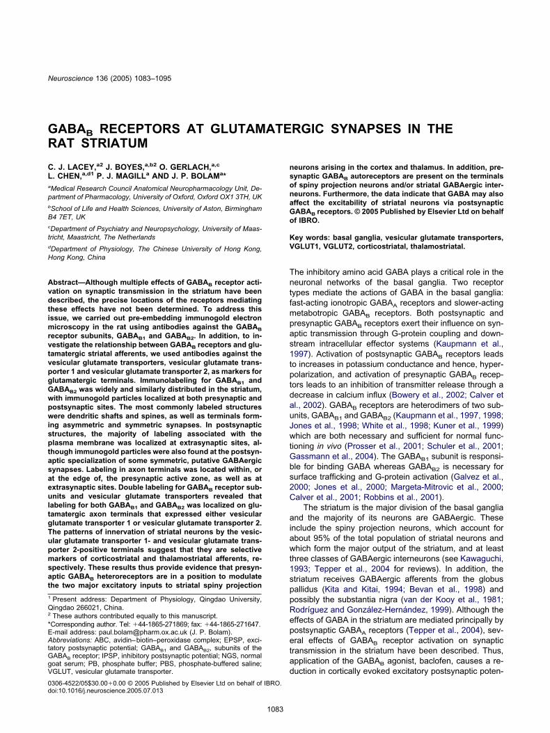
ig. 1. Localization of immunogold labeling for GABAB1 (GB1; A–C) and GABAB2 (GB2; D–F) in the striatum. (A) Immunolabeling for GABAB1 (arrows)n the presynaptic membrane of a bouton (b1) forming an asymmetric synapse with a dendritic spine (s). GABAB1 immunolabeling (double arrowhead)
s also associated with the postsynaptic density at a second asymmetric synapse between an unlabeled bouton (b2) and a dendritic shaft (d). (B)resynaptic labeling for GABAB1 in boutons (b1–b3) forming asymmetric synapses with spines (s). Immunogold particles (some of which are indicatedy arrows) are located at synaptic (b1 and b2) and extrasynaptic sites (b1–3) on the membrane of the boutons. The spine postsynaptic to bouton b1,merges from a dendrite (d) that is also immunolabeled for GABAB1. Similarly, the spine postsynaptic to bouton b2, is also immunolabeled; the
mmunogold particles are associated with the spine apparatus (double arrowhead). The spine postsynaptic to bouton b3 receives symmetric synapticnput from a second bouton (b4) that is labeled on the presynaptic membrane (arrow). (C) A bouton (b) forms symmetrical synaptic contact with aendrite that is immunolabeled for GABAB1. The immunogold particles are associated with the postsynaptic membrane (double arrowhead) and
ntracellular sites. (D) Immunolabeling for GABAB2 at the presynaptic specialization (arrows) of a bouton (b1) forming an asymmetric synapse with anmmunonegative spine (s). Another GABAB2-positive bouton (b2) forms an asymmetric synapse with a GABAB2-positive spine (s). An immunonegativeouton (b3) forms an asymmetric synapse with a GABAB2-positive spine (s). The GABAB2-labeled bouton, b4, is in symmetrical synaptic contact withdendrite. Note that some of the immunogold labeling in this bouton is associated with the presynaptic membrane specialization. (E) A GABAB2-
mmunolabeled bouton (b) forming asymmetric synapses with two spines (s). The immunogold particles (arrows) are associated with the presynapticpecialization at each synapse and the postsynaptic spines also express GABAB2-immunolabeling. (F) Immunogold labeling for GABAB2 (some goldarticles indicated by arrows) in boutons (b1–b5) forming asymmetric synapses with spines or a dendrite. The immunogold labeling is associated withhe presynaptic specialization (b4), extrasynaptic sites on the membrane (b2, b3) and intracellular sites (b1, b2, b5). Several of the postsynaptictructures are also immunolabeled including the spine contacted by b5. In this case immunogold labeling is closely associated with the postsynaptic
ensity (double arrowhead). Scale bars�0.25 �m.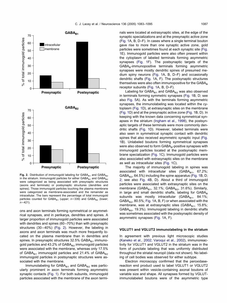
orlwsacsgwois
usp
ns(gp1tsGsddtr
iast(kaadas1wibaa
aG2pmisGmGwa
V
I(tfti
rwv
Fiw(swipn
C. J. Lacey et al. / Neuroscience 136 (2005) 1083–1095 1087
ns and axon terminals forming symmetrical or asymmet-ical synapses, and in perikarya, dendrites and spines. Aarger proportion of immunogold particles were associatedith dendrites and spines (60–70%) than with presynaptictructures (30–40%) (Fig. 2). However, the labeling inxons and axon terminals was much more frequently lo-ated on the plasma membrane than in dendrites andpines. In presynaptic structures 32.5% GABAB1 immuno-old particles and 43.2% of GABAB2 immunogold particlesere associated with the membrane, whereas only 17.5%f GABAB1 immunogold particles and 21.4% of GABAB2
mmunogold particles in postsynaptic structures were as-ociated with the membrane.
Immunolabeling for GABAB1 and GABAB2 was partic-larly prominent in axon terminals forming asymmetricynaptic contacts (Fig. 1). For both subunits, immunogold
ig. 2. Distribution of immunogold labeling for GABAB1 and GABAB2
n the striatum. Immunogold particles for either GABAB1 and GABAB2
ere categorized as being associated with presynaptic structuresaxons and terminals) or postsynaptic structures (dendrites andpines). Those immunogold particles touching the plasma membraneere categorized as membrane-associated and the remainder as
ntracellular. The bars represent the percentage of total immunogoldarticles counted for GABAB1 (upper; n�339) and GABAB2 (lower;�421).
articles associated with the membrane of the axon termi- i
als were located at extrasynaptic sites, at the edge of theynaptic specializations and at the presynaptic active zoneFig. 1A, B, D–F). In cases where a single terminal boutonave rise to more than one synaptic active zone, goldarticles were sometimes found at each synaptic site (Fig.E). Immunogold particles were also often present withinhe cytoplasm of labeled terminals forming asymmetricynapses (Fig. 1F). The postsynaptic targets of theABAB-immunopositive terminals forming asymmetric
ynapses were mostly dendritic spines of presumed me-ium spiny neurons (Fig. 1A, B, D–F) and occasionallyendritic shafts (Fig. 1A, F). The postsynaptic structureshemselves were also often immunopositive for the GABAB
eceptor subunits (Fig. 1A, B, D–F).Labeling for GABAB1 and GABAB2 was also observed
n terminals forming symmetric synapses (Fig. 1B, D; seelso Fig. 5A). As with the terminals forming asymmetricynapses, the immunolabeling was located within the cy-oplasm (Fig. 1D), at extrasynaptic sites on the membraneFig. 1D) and at the presynaptic active zone (Fig. 1B, D). Ineeping with the known data concerning symmetrical syn-pses in the striatum (Ingham et al., 1998), the postsyn-ptic targets of these terminals were more commonly den-ritic shafts (Fig. 1D). However, labeled terminals werelso seen in symmetrical synaptic contact with dendriticpines that also received asymmetric synaptic input (Fig.B). Unlabeled boutons forming symmetrical synapsesere also observed to form GABAB-positive synapses with
mmunogold particles located at the postsynaptic mem-rane specialization (Fig. 1C). Immunogold particles werelso associated with extrasynaptic sites on the membranes well as intracellular sites (Fig. 1C).
The majority of immunogold labeling in spines wasssociated with intracellular sites (GABAB1, 67.2%;ABAB2, 64.5%) including the spine apparatus (Fig. 1B, D;; see also Fig. 4B, D). About a third of immunogoldarticles were associated with extrasynaptic sites on theembrane (GABAB1, 32.1%; GABAB2, 31.6%). Similarly,
n large and small dendritic shafts, labeling for GABAB
ubunits was mostly intracellular (GABAB1, 84.1%;ABAB2, 80.5%; Fig. 1A, B, F) or when associated with theembrane, was at extrasynaptic sites (GABAB1, 15.8%;ABAB2, 19.3%). Immunogold labeling in dendritic shaftsas sometimes associated with the postsynaptic density ofsymmetric synapses (Fig. 1A, F).
GLUT1 and VGLUT2 immunolabeling in the striatum
n agreement with previous light microscopic studiesKaneko et al., 2002; Varoqui et al., 2002), immunoreac-ivity for VGLUT1 and VGLUT2 in the striatum was in theorm of punctate labeling that was uniformly distributedhroughout the striatal neuropil (data not shown). No label-ng of cell bodies was observed for either subtype.
Electron microscopy confirmed that the peroxidase-eaction end product used to label VGLUT1 or VGLUT2as present within vesicle-containing axonal boutons ofariable size and shape. All synapses formed by VGLUT-
mmunolabeled boutons were of the asymmetric type
(os3diaiabwacadt
0toahV(wlw(Vm
lmtti(Vt
GV
Itt(saanld
FVmsform synapses with more than one spine. (B) VGLUT2-positive bou-
tamdV
C. J. Lacey et al. / Neuroscience 136 (2005) 1083–10951088
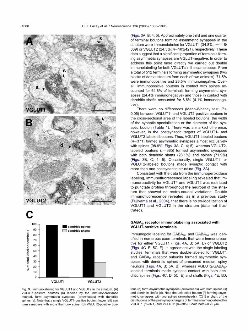
Figs. 3A, B; 4; 5). Approximately one third and one quarterf terminal boutons forming asymmetric synapses in thetriatum were immunolabeled for VGLUT1 (34.8%; n�118/39) or VGLUT2 (24.5%; n�103/421), respectively. Theseata suggest that a significant proportion of terminals form-
ng asymmetric synapses are VGLUT-negative. In order toddress this point more directly we carried out double
mmunolabeling for both VGLUTs in the same tissue. Fromtotal of 512 terminals forming asymmetric synapses (twolocks of dorsal striatum from each of two animals), 71.5%ere immunopositive and 28.5% immunonegative. Over-ll, immunopositive boutons in contact with spines ac-ounted for 64.8% of terminals forming asymmetric syn-pses (24.4% immunonegative) and those in contact withendritic shafts accounted for 6.6% (4.1% immunonega-ive).
There were no differences (Mann-Whitney test, P�.05) between VGLUT1- and VGLUT2-positive boutons inhe cross-sectional area of the labeled boutons, the widthf the synaptic specialization or the diameter of the syn-ptic bouton (Table 1). There was a marked difference,owever, in the postsynaptic targets of VGLUT1- andGLUT2-labeled boutons. Thus, VGLUT1-labeled boutons
n�371) formed asymmetric synapses almost exclusivelyith spines (98.9%; Figs. 3A, C; 4; 5), whereas VGLUT2-
abeled boutons (n�385) formed asymmetric synapsesith both dendritic shafts (28.1%) and spines (71.9%)
Figs. 3B, C; 4; 5). Occasionally, single VGLUT1- orGLUT2-labeled boutons made synaptic contact withore than one postsynaptic structure (Fig. 3A).
Consistent with the data from the immunoperoxidaseabeling, immunofluorescence labeling revealed that im-
unoreactivity for VGLUT1 and VGLUT2 was restrictedo punctate profiles throughout the neuropil of the stria-um that showed no rostro-caudal variations. Doublemmunofluorescence revealed, as in a previous studyFujiyama et al., 2004), that there is no co-localization ofGLUT1 and VGLUT2 in the striatum (data not illus-
rated).
ABAB receptor immunolabeling associated withGLUT-positive terminals
mmunogold labeling for GABAB1 and GABAB2 was iden-ified in numerous axon terminals that were immunoreac-ive for either VGLUT1 (Figs. 4A, B; 5A, B) or VGLUT2Figs. 4C–E; 5C–F). In agreement with the single labelingtudies, terminals that were double-labeled for VGLUT1nd GABAB receptor subunits formed asymmetric syn-pses with dendritic spines of presumed medium spinyeurons (Figs. 4A, B; 5A, B), whereas VGLUT2/GABAB-
abeled terminals made synaptic contact with both den-ritic spines (Figs. 4C, D; 5C, E) and shafts (Figs. 4E; 5D,
ons (b) form asymmetric synapses (arrowheads) with both spines (s)nd dendritic shafts (d). Note the unlabeled bouton (*) forming asym-etric synapses with two spines (arrowheads). (C) Bar chart of the
istributions of the postsynaptic targets of terminals immunolabeled forig. 3. Immunolabeling for VGLUT1 and VGLUT2 in the striatum. (A)GLUT1-positive boutons (b) labeled by the immunoperoxidaseethod, form asymmetric synapses (arrowheads) with dendritic
pines (s). Note that a single VGLUT1-positive bouton (lower left) can
GLUT1 (n�371) and VGLUT2 (n�385). Scale bars�0.25 �m.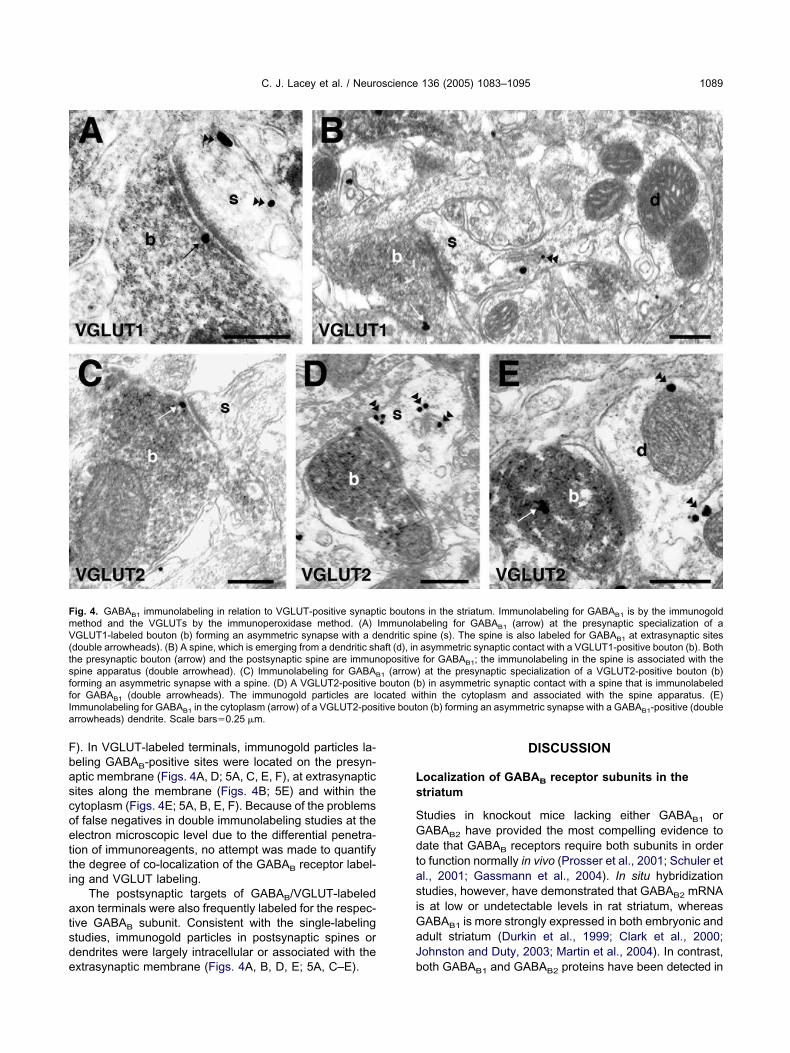
Fbascoetti
atsde
Ls
SGdtasiGaJ
FmV(tsffI itive bouta
C. J. Lacey et al. / Neuroscience 136 (2005) 1083–1095 1089
). In VGLUT-labeled terminals, immunogold particles la-eling GABAB-positive sites were located on the presyn-ptic membrane (Figs. 4A, D; 5A, C, E, F), at extrasynapticites along the membrane (Figs. 4B; 5E) and within theytoplasm (Figs. 4E; 5A, B, E, F). Because of the problemsf false negatives in double immunolabeling studies at thelectron microscopic level due to the differential penetra-ion of immunoreagents, no attempt was made to quantifyhe degree of co-localization of the GABAB receptor label-ng and VGLUT labeling.
The postsynaptic targets of GABAB/VGLUT-labeledxon terminals were also frequently labeled for the respec-ive GABAB subunit. Consistent with the single-labelingtudies, immunogold particles in postsynaptic spines orendrites were largely intracellular or associated with the
ig. 4. GABAB1 immunolabeling in relation to VGLUT-positive synaptethod and the VGLUTs by the immunoperoxidase method. (A)GLUT1-labeled bouton (b) forming an asymmetric synapse with a d
double arrowheads). (B) A spine, which is emerging from a dendritic shhe presynaptic bouton (arrow) and the postsynaptic spine are immunpine apparatus (double arrowhead). (C) Immunolabeling for GABAB
orming an asymmetric synapse with a spine. (D) A VGLUT2-positiveor GABAB1 (double arrowheads). The immunogold particles are lommunolabeling for GABAB1 in the cytoplasm (arrow) of a VGLUT2-posrrowheads) dendrite. Scale bars�0.25 �m.
xtrasynaptic membrane (Figs. 4A, B, D, E; 5A, C–E). b
DISCUSSION
ocalization of GABAB receptor subunits in thetriatum
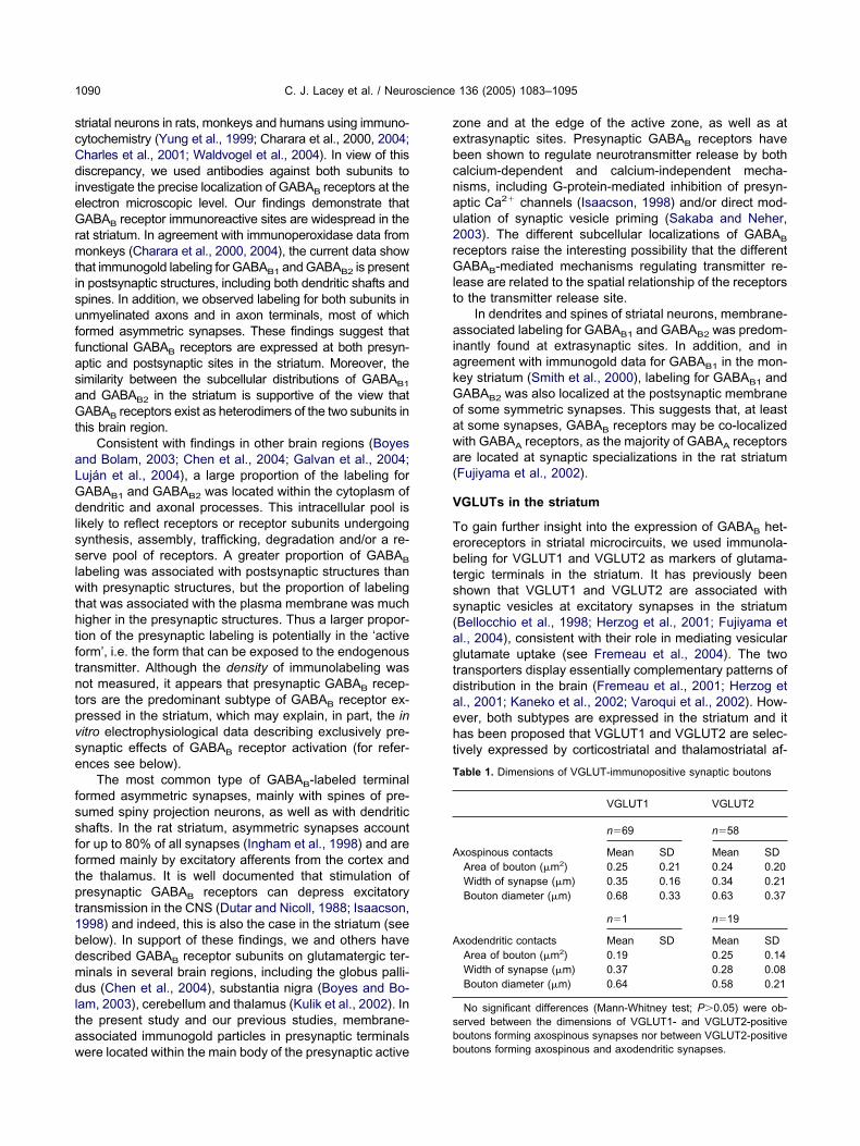
tudies in knockout mice lacking either GABAB1 orABAB2 have provided the most compelling evidence toate that GABAB receptors require both subunits in ordero function normally in vivo (Prosser et al., 2001; Schuler etl., 2001; Gassmann et al., 2004). In situ hybridizationtudies, however, have demonstrated that GABAB2 mRNAs at low or undetectable levels in rat striatum, whereas
ABAB1 is more strongly expressed in both embryonic anddult striatum (Durkin et al., 1999; Clark et al., 2000;ohnston and Duty, 2003; Martin et al., 2004). In contrast,
s in the striatum. Immunolabeling for GABAB1 is by the immunogoldbeling for GABAB1 (arrow) at the presynaptic specialization of a
pine (s). The spine is also labeled for GABAB1 at extrasynaptic sitesasymmetric synaptic contact with a VGLUT1-positive bouton (b). Bothfor GABAB1; the immunolabeling in the spine is associated with the
) at the presynaptic specialization of a VGLUT2-positive bouton (b)b) in asymmetric synaptic contact with a spine that is immunolabeledithin the cytoplasm and associated with the spine apparatus. (E)on (b) forming an asymmetric synapse with a GABAB1-positive (double
ic boutonImmunolaendritic saft (d), inopositive
1 (arrowbouton (cated w
oth GABAB1 and GABAB2 proteins have been detected in
scCdieGrmtisuffasaGt
aLGdlsslwthtftntpvse
fssfftpt1bdmdltaw
zebcnau2rGlt
aiakGoawa(
V
Tebtss(agtdaeht
T
A
A
sb
C. J. Lacey et al. / Neuroscience 136 (2005) 1083–10951090
triatal neurons in rats, monkeys and humans using immuno-ytochemistry (Yung et al., 1999; Charara et al., 2000, 2004;harles et al., 2001; Waldvogel et al., 2004). In view of thisiscrepancy, we used antibodies against both subunits to
nvestigate the precise localization of GABAB receptors at thelectron microscopic level. Our findings demonstrate thatABAB receptor immunoreactive sites are widespread in the
at striatum. In agreement with immunoperoxidase data fromonkeys (Charara et al., 2000, 2004), the current data show
hat immunogold labeling for GABAB1 and GABAB2 is presentn postsynaptic structures, including both dendritic shafts andpines. In addition, we observed labeling for both subunits innmyelinated axons and in axon terminals, most of whichormed asymmetric synapses. These findings suggest thatunctional GABAB receptors are expressed at both presyn-ptic and postsynaptic sites in the striatum. Moreover, theimilarity between the subcellular distributions of GABAB1
nd GABAB2 in the striatum is supportive of the view thatABAB receptors exist as heterodimers of the two subunits in
his brain region.Consistent with findings in other brain regions (Boyes
nd Bolam, 2003; Chen et al., 2004; Galvan et al., 2004;uján et al., 2004), a large proportion of the labeling forABAB1 and GABAB2 was located within the cytoplasm ofendritic and axonal processes. This intracellular pool is
ikely to reflect receptors or receptor subunits undergoingynthesis, assembly, trafficking, degradation and/or a re-erve pool of receptors. A greater proportion of GABAB
abeling was associated with postsynaptic structures thanith presynaptic structures, but the proportion of labeling
hat was associated with the plasma membrane was muchigher in the presynaptic structures. Thus a larger propor-ion of the presynaptic labeling is potentially in the ‘activeorm’, i.e. the form that can be exposed to the endogenousransmitter. Although the density of immunolabeling wasot measured, it appears that presynaptic GABAB recep-
ors are the predominant subtype of GABAB receptor ex-ressed in the striatum, which may explain, in part, the initro electrophysiological data describing exclusively pre-ynaptic effects of GABAB receptor activation (for refer-nces see below).
The most common type of GABAB-labeled terminalormed asymmetric synapses, mainly with spines of pre-umed spiny projection neurons, as well as with dendritichafts. In the rat striatum, asymmetric synapses accountor up to 80% of all synapses (Ingham et al., 1998) and areormed mainly by excitatory afferents from the cortex andhe thalamus. It is well documented that stimulation ofresynaptic GABAB receptors can depress excitatory
ransmission in the CNS (Dutar and Nicoll, 1988; Isaacson,998) and indeed, this is also the case in the striatum (seeelow). In support of these findings, we and others haveescribed GABAB receptor subunits on glutamatergic ter-inals in several brain regions, including the globus palli-us (Chen et al., 2004), substantia nigra (Boyes and Bo-
am, 2003), cerebellum and thalamus (Kulik et al., 2002). Inhe present study and our previous studies, membrane-ssociated immunogold particles in presynaptic terminals
ere located within the main body of the presynaptic active bone and at the edge of the active zone, as well as atxtrasynaptic sites. Presynaptic GABAB receptors haveeen shown to regulate neurotransmitter release by bothalcium-dependent and calcium-independent mecha-isms, including G-protein-mediated inhibition of presyn-ptic Ca2� channels (Isaacson, 1998) and/or direct mod-lation of synaptic vesicle priming (Sakaba and Neher,003). The different subcellular localizations of GABAB
eceptors raise the interesting possibility that the differentABAB-mediated mechanisms regulating transmitter re-
ease are related to the spatial relationship of the receptorso the transmitter release site.
In dendrites and spines of striatal neurons, membrane-ssociated labeling for GABAB1 and GABAB2 was predom-
nantly found at extrasynaptic sites. In addition, and ingreement with immunogold data for GABAB1 in the mon-ey striatum (Smith et al., 2000), labeling for GABAB1 andABAB2 was also localized at the postsynaptic membranef some symmetric synapses. This suggests that, at leastt some synapses, GABAB receptors may be co-localizedith GABAA receptors, as the majority of GABAA receptorsre located at synaptic specializations in the rat striatumFujiyama et al., 2002).
GLUTs in the striatum
o gain further insight into the expression of GABAB het-roreceptors in striatal microcircuits, we used immunola-eling for VGLUT1 and VGLUT2 as markers of glutama-ergic terminals in the striatum. It has previously beenhown that VGLUT1 and VGLUT2 are associated withynaptic vesicles at excitatory synapses in the striatumBellocchio et al., 1998; Herzog et al., 2001; Fujiyama etl., 2004), consistent with their role in mediating vesicularlutamate uptake (see Fremeau et al., 2004). The tworansporters display essentially complementary patterns ofistribution in the brain (Fremeau et al., 2001; Herzog etl., 2001; Kaneko et al., 2002; Varoqui et al., 2002). How-ver, both subtypes are expressed in the striatum and itas been proposed that VGLUT1 and VGLUT2 are selec-ively expressed by corticostriatal and thalamostriatal af-
able 1. Dimensions of VGLUT-immunopositive synaptic boutons
VGLUT1 VGLUT2
n�69 n�58
xospinous contacts Mean SD Mean SDArea of bouton (�m2) 0.25 0.21 0.24 0.20Width of synapse (�m) 0.35 0.16 0.34 0.21Bouton diameter (�m) 0.68 0.33 0.63 0.37
n�1 n�19
xodendritic contacts Mean SD Mean SDArea of bouton (�m2) 0.19 0.25 0.14Width of synapse (�m) 0.37 0.28 0.08Bouton diameter (�m) 0.64 0.58 0.21
No significant differences (Mann-Whitney test; P�0.05) were ob-erved between the dimensions of VGLUT1- and VGLUT2-positiveoutons forming axospinous synapses nor between VGLUT2-positive
outons forming axospinous and axodendritic synapses.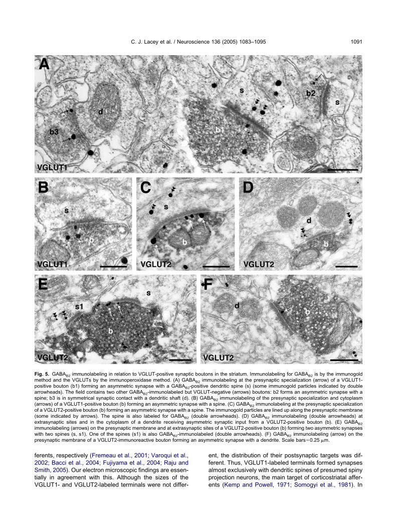
f2StV
efap
Fmpas(o(eiwp an asym
C. J. Lacey et al. / Neuroscience 136 (2005) 1083–1095 1091
erents, respectively (Fremeau et al., 2001; Varoqui et al.,002; Bacci et al., 2004; Fujiyama et al., 2004; Raju andmith, 2005). Our electron microscopic findings are essen-
ially in agreement with this. Although the sizes of the
ig. 5. GABAB2 immunolabeling in relation to VGLUT-positive synaptethod and the VGLUTs by the immunoperoxidase method. (A) GAositive bouton (b1) forming an asymmetric synapse with a GABAB2
rrowheads). The field contains two other GABAB2-immunolabeled bupine; b3 is in symmetrical synaptic contact with a dendritic shaft (d).arrows) of a VGLUT1-positive bouton (b) forming an asymmetric synaf a VGLUT2-positive bouton (b) forming an asymmetric synapse with asome indicated by arrows). The spine is also labeled for GABAB2
xtrasynaptic sites and in the cytoplasm of a dendrite receiving ammunolabeling (arrows) on the presynaptic membrane and at extrasynith two spines (s, s1). One of the spines (s1) is also GABAB2-immuresynaptic membrane of a VGLUT2-immunoreactive bouton forming
GLUT1- and VGLUT2-labeled terminals were not differ- e
nt, the distribution of their postsynaptic targets was dif-erent. Thus, VGLUT1-labeled terminals formed synapseslmost exclusively with dendritic spines of presumed spinyrojection neurons, the main target of corticostriatal affer-
s in the striatum. Immunolabeling for GABAB2 is by the immunogoldunolabeling at the presynaptic specialization (arrow) of a VGLUT1-dendritic spine (s) (some immunogold particles indicated by double-negative (arrows) boutons: b2 forms an asymmetric synapse with aAB2 immunolabeling of the presynaptic specialization and cytoplasma spine. (C) GABAB2 immunolabeling at the presynaptic specializationhe immunogold particles are lined up along the presynaptic membranearrowheads). (D) GABAB2 immunolabeling (double arrowheads) atc synaptic input from a VGLUT2-positive bouton (b). (E) GABAB2
s of a VGLUT2-positive bouton (b) forming two asymmetric synapsesd (double arrowheads). (F) GABAB2 immunolabeling (arrow) on themetric synapse with a dendrite. Scale bars�0.25 �m.
ic boutonBAB2 imm-positivet VGLUT(B) GAB
pse withspine. T(double
symmetriaptic sitenolabele
nts (Kemp and Powell, 1971; Somogyi et al., 1981). In
cbcsnB2diaTefomd
sVftesmpdniheeihaTcV
G
Dtt(ctrtpbsairGwfssg
1mmgrs
GtmtstblatameGatiehi
dbosbmw2bgmaaptfmg
Pt
SGb1ponte
C. J. Lacey et al. / Neuroscience 136 (2005) 1083–10951092
ontrast, VGLUT2-labeled terminals formed synapses withoth dendritic shafts (28%) and spines (72%), which isharacteristic of the more complex pattern of innervation oftriatal neurons by afferents from the intralaminar thalamicuclei (Dubé et al., 1988; Xu et al., 1991; Lapper andolam, 1992; Rudkin and Sadikot, 1999; Ichinohe et al.,001; Smith et al., 2004). Furthermore, by the use ofouble immunofluorescence, we confirmed the recent find-
ng of Fujiyama et al. (2004) that VGLUT1 and VGLUT2re not co-localized in individual boutons in the striatum.aken together, these data are further support for a differ-ntial expression of VGLUTs by cortical and thalamic af-erents to the striatum. Although the functional significancef this is unclear, the expression of the two transportersay be linked with the probability of glutamate release atifferent synapses (Fremeau et al., 2001).
Approximately 70% of terminals forming asymmetricynapses in the striatum were immunoreactive for theGLUTs. Although we cannot exclude the possibility of
alse-negatives, these data suggest that up to 30% oferminals forming asymmetric synapses do not expressither VGLUT1 or VGLUT2. This implies either that aignificant proportion of synaptic boutons forming asym-etric synapses are not glutamatergic or that there is aopulation of glutamatergic afferents to the striatum thatoes not express VGLUT1 or 2. One possible source ofon-glutamatergic terminals forming asymmetric synapses
s the serotonergic projection from the dorsal raphé (Sog-omonian et al., 1989; Descarries et al., 1992). It is inter-sting to note that VGLUT3, a third VGLUT subtype that isxpressed in populations of non-glutamatergic neurons,
ncluding neurons of the raphé nuclei (Gras et al., 2002),as been found in a small proportion of terminals formingsymmetric synapses in striatum (Fujiyama et al., 2004).he possibility that sub-populations of thalamostriatal ororticostriatal neurons do not express any of the knownGLUTs remains to be established.
ABAB receptors at glutamatergic synapses
ouble immunolabeling demonstrated that GABAB recep-ors are expressed by VGLUT1-positive axon terminals inhe striatum, which are likely to be derived from the cortexsee above). In support of this conclusion, lesions of theorticostriatal pathway are associated with a decrease inhe density of GABAB binding sites in the striatum (Mo-atalla and Bowery, 1991). Furthermore, as indicated inhe introduction, in corticostriatal slices, baclofen de-resses the EPSPs in striatal neurons that are evokedy stimulation of cortical afferents or by intrastriataltimulation (Calabresi et al., 1990, 1991; Seabrook etl., 1990; Nisenbaum et al., 1992, 1993), an effect that
s absent in mice with mutations that render the GABAB
eceptor inactive (Thuault et al., 2004). Labeling forABAB receptors was also expressed by terminals thatere labeled for VGLUT2, which are likely to be derived
rom the thalamus (see above). This is consistent with initu hybridization studies showing expression of GABAB
ubunit mRNA in the intralaminar thalamic nuclei that
ive rise to the thalamostriatal projections (Durkin et al., r999; Lu et al., 1999). Our data thus predict that gluta-ate release at thalamostriatal synapses is subject toodulation by presynaptic GABAB receptors. Taken to-ether, the findings suggest that presynaptic GABAB
eceptors regulate both of the major excitatory inputs totriatal neurons.
One of the key questions that arises concerningABAB receptor expression by glutamatergic terminals is
he source of the GABA that activates these receptors. Theost plausible explanation is that these receptors are ac-
ivated by extrasynaptic GABA that diffuses out of theynaptic cleft at neighboring GABAergic synapses. Cross-alk between GABAergic and glutamatergic synapses haseen demonstrated in both the hippocampus and cerebel-
um (Isaacson et al., 1993; Vogt and Nicoll, 1999; Mitchellnd Silver, 2000). Calabresi et al. (1990) demonstrated
hat increasing GABA levels in the striatum in vitro causesdecrease in spontaneous and evoked EPSPs that isimicked by baclofen, indicating that striatal GABAB het-roreceptors are sensitive to extracellular GABA. Thus,ABA released from spiny projection neurons and/or stri-tal interneurons may activate presynaptic GABAB recep-ors located on cortical and thalamic terminals to simplynhibit transmission or may possibly regulate release tonable sustained transmission at higher frequencies, asas been observed in the avian auditory system (Brenow-
tz et al., 1998).We also observed labeling for GABAB1 and GABAB2 in
endritic spines that were postsynaptic to terminals la-eled for either VGLUT1 or VGLUT2. Although the majorityf this was intracellular, labeling was also localized extra-ynaptically on spine membranes. Interestingly, in severalrain regions, postsynaptic GABAB receptors appear to beore closely associated with glutamatergic synapses thanith GABAergic synapses (Kulik et al., 2002; Galvan et al.,004; Luján et al., 2004). There is evidence in the cere-ellum for an interaction between GABAB receptors andlutamate receptors (Hirono et al., 2001), however, thisight not be the case in the striatum, as baclofen does notlter the responses of striatal neurons to exogenouslypplied glutamate (Calabresi et al., 1991). An intriguingossibility, recently proposed by Tabata et al. (2004), ishat GABAB receptors at glutamatergic synapses mightunction independently of GABA through calcium-mediatedechanisms, to increase the sensitivity of metabotropiclutamate receptors to glutamate.
resynaptic GABAB receptors on other classes oferminals in the striatum
everal studies have reported that baclofen reducesABAA-mediated IPSPs in striatal neurons in vitro (Cala-resi et al., 1991; Seabrook et al., 1991; Nisenbaum et al.,992, 1993). In support of the idea that this is an effect ofresynaptic GABAB receptors on GABAergic terminals, webserved GABAB1 and GABAB2 immunolabeling of termi-als forming symmetric axodendritic synapses with theypical appearance of striatal GABAergic terminals (Bolamt al., 1985). Thus, in addition to regulating glutamate
elease through presynaptic GABAB heteroreceptors in the
soTireprRtalii(ataie2oc1qft
Tssatttcp
AsKsf
A
B
B
B
B
B
B
B
C
C
C
C
C
C
C
C
C
D
D
D
D
F
F
F
C. J. Lacey et al. / Neuroscience 136 (2005) 1083–1095 1093
triatum, GABA may also exert a feedback control over itswn release through presynaptic GABAB autoreceptors.here are several sources of GABA in the striatum, includ-
ng the recurrent axon collaterals of spiny projection neu-ons, the axons of GABAergic striatal interneurons andxtrinsic sources, and it has been proposed that differentopulations of GABAergic synapses may be differentiallyegulated by GABAB autoreceptors (Seabrook et al., 1991;adnikow et al., 1997). It is clearly important to establish
he identity of those terminals forming symmetrical syn-pses and expressing GABAB receptors because the se-
ective expression of GABAB receptors will have significantmplications for our understanding of GABA transmissionn the striatum. Based on the ultrastructural features alonei.e. synapses on the necks of spines that also receivesymmetric synaptic input), it is likely that at least some ofhe GABAB-labeled terminals forming symmetric synapsesre derived from dopaminergic nigrostriatal neurons. This
s supported by the observation that GABAB receptors arexpressed by nigrostriatal neurons (Boyes and Bolam,003), although the issue of presynaptic GABAB receptorsn dopaminergic terminals in the striatum is somewhatontroversial (Arias-Montaño et al., 1991; Santiago et al.,993; Westerink et al., 1994; Smolders et al., 1995). Thisuestion awaits the application of double immunolabelingor a marker of dopaminergic terminals and GABAB recep-ors.
CONCLUSIONS
he findings of the present study provide an anatomicalubstrate for the presynaptic effects of GABAB receptortimulation at glutamatergic cortical and thalamic syn-pses in the striatum thus underpinning the close interac-ions between the major inhibitory and excitatory neuro-ransmitter systems in this nucleus. They also demonstratehat stimulation of synaptic and extrasynaptic GABAB re-eptors is likely to affect GABAergic transmission at bothresynaptic and postsynaptic sites.
cknowledgments—The work was funded by the Medical Re-earch Council UK and the Research Grants Council of Hongong. C.J.L. is in receipt of a Medical Research Council student-hip. We thank Caroline Francis, Ben Micklem, and Liz Normanor their expert technical assistance.
REFERENCES
rias-Montaño JA, Martinez-Fong D, Aceves J (1991) �-Aminobutyricacid (GABAB) receptor-mediated inhibition of tyrosine hydroxylaseactivity in the striatum of rat. Neuropharmacology 30:1047–1051.
acci JJ, Kachidian P, Kerkerian-Le Goff L, Salin P (2004) Intralaminarthalamic nuclei lesions: widespread impact on dopamine denerva-tion-mediated cellular defects in the rat basal ganglia. J Neuro-pathol Exp Neurol 63:20–31.
ellocchio EE, Hu H, Pohorille A, Chan J, Pickel VM, Edwards RH(1998) The localization of the brain-specific inorganic phosphatetransporter suggests a specific presynaptic role in glutamatergictransmission. J Neurosci 18:8648–8659.
evan MD, Booth PAC, Eaton SA, Bolam JP (1998) Selective inner-vation of neostriatal interneurons by a subclass of neuron in the
globus pallidus of the rat. J Neurosci 18:9438–9452.olam JP, Powell JF, Wu J-Y, Smith AD (1985) Glutamate decarbox-ylase-immunoreactive structures in the rat neostriatum. A corre-lated light and electron microscopic study including a combinationof Golgi-impregnation with immunocytochemistry. J Comp Neurol237:1–20.
owery NG, Bettler B, Froestl W, Gallagher JP, Marshall F, Raiteri M,Bonner TI, Enna SJ (2002) International Union of Pharmacology.XXXIII. Mammalian �-aminobutyric acidB receptors: structure andfunction. Pharmacol Rev 54:247–264.
oyes J, Bolam JP (2003) The subcellular localization of GABAB
receptor subunits in the rat substantia nigra. Eur J Neurosci18:3279–3293.
renowitz S, David J, Trussell L (1998) Enhancement of synapticefficacy by presynaptic GABAB receptors. Neuron 20:135–141.
alabresi P, Mercuri NB, De Murtas M, Bernardi G (1990) Endoge-nous GABA mediates presynaptic inhibition of spontaneous andevoked excitatory synaptic potentials in the rat neostriatum. Neu-rosci Lett 118:99–102.
alabresi P, Mercuri NB, De Murtas M, Bernardi G (1991) Involvementof GABA systems in feedback regulation of glutamate-and GABA-mediated synaptic potentials in rat neostriatum. J Physiol 440:581–599.
alver AR, Robbins MJ, Cosio C, Rice SQ, Babbs AJ, Hirst WD,Boyfield I, Wood MD, Russell RB, Price GW, Couve A, Moss SJ,Pangalos MN (2001) The C-terminal domains of the GABAB re-ceptor subunits mediate intracellular trafficking but are not requiredfor receptor signaling. J Neurosci 21:1203–1210.
alver AR, Davies CH, Pangalos M (2002) GABAB receptors: Frommonogamy to promiscuity. Neurosignals 11:299–314.
harara A, Heilman TC, Levey AI, Smith Y (2000) Pre- and postsyn-aptic localization of GABAB receptors in the basal ganglia in mon-keys. Neuroscience 95:127–140.
harara A, Galvan A, Kuwajima M, Hall RA, Smith Y (2004) Anelectron microscope immunocytochemical study of GABAB R2 re-ceptors in the monkey basal ganglia: A comparative analysis withGABAB R1 receptor distribution. J Comp Neurol 476:65–79.
harles KJ, Evans ML, Robbins MJ, Calver AR, Leslie RA, PangalosMN (2001) Comparative immunohistochemical localisation ofGABAB1a, GABAB1b and GABAB2 subunits in rat brain, spinal cordand dorsal root ganglion. Neuroscience 106:447–467.
hen L, Boyes J, Yung WH, Bolam JP (2004) Subcellular localizationof GABAB receptor subunits in rat globus pallidus. J Comp Neurol474:340–352.
lark JA, Mezey E, Lam AS, Bonner TI (2000) Distribution of theGABAB receptor subunit gb2 in rat CNS. Brain Res 860:41–52.
escarries L, Soghomonian J-J, Garcia S, Doucet G, Bruno JP (1992)Ultrastructural analysis of the serotonin hyperinnervation in adultrat neostriatum following neonatal dopamine denervation with6-hydroxydopamine. Brain Res 569:1–13.
ubé L, Smith AD, Bolam JP (1988) Identification of synaptic terminalsof thalamic or cortical origin in contact with distinct medium-sizespiny neurons in the rat neostriatum. J Comp Neurol 267:455–471.
urkin MM, Gunwaldsen CA, Borowsky B, Jones KA, Branchek TA(1999) An in situ hybridization study of the distribution of theGABAB2 protein mRNA in the rat CNS. Brain Res Mol Brain Res71:185–200.
utar P, Nicoll RA (1988) A physiological role for GABAB receptors inthe central nervous system. Nature 332:156–158.
remeau RT Jr, Troyer MD, Pahner I, Nygaard GO, Tran CH, ReimerRJ, Bellocchio EE, Fortin D, Storm-Mathisen J, Edwards RH(2001) The expression of vesicular glutamate transporters definestwo classes of excitatory synapse. Neuron 31:247–260.
remeau RT Jr, Voglmaier S, Seal RP, Edwards RH (2004) VGLUTsdefine subsets of excitatory neurons and suggest novel roles forglutamate. Trends Neurosci 27:98–103.
ujiyama F, Fritschy J-M, Stephenson FA, Bolam JP (2002) Synapticlocalization of GABA receptor subunits in the striatum of the rat.
AJ Comp Neurol 416:158–172.
F
G
G
G
G
H
H
I
I
I
I
J
J
J
K
K
K
K
K
K
K
K
L
L
L
M
M
M
M
M
M
M
N
N
P
R
R
R
C. J. Lacey et al. / Neuroscience 136 (2005) 1083–10951094
ujiyama F, Kuramoto E, Okamoto K, Hioki H, Furuta T, Zhou L,Nomura S, Kaneko T (2004) Presynaptic localization of an AMPA-type glutamate receptor in corticostriatal and thalamostriatal axonterminals. Eur J Neurosci 20:3322–3330.
alvan A, Charara A, Pare JF, Levey AI, Smith Y (2004) Differentialsubcellular and subsynaptic distribution of GABAA and GABAB
receptors in the monkey subthalamic nucleus. Neuroscience127:709–721.
alvez T, Prezeau L, Milioti G, Franek M, Joly C, Froestl W, Bettler B,Bertrand HO, Blahos J, Pin JP (2000) Mapping the agonist-bindingsite of GABAB type 1 subunit sheds light on the activation processof GABAB receptors. J Biol Chem 275:41166–41174.
assmann M, Shaban H, Vigot R, Sansig G, Haller C, Barbieri S,Humeau Y, Schuler V, Müller M, Kinzel B, Klebs K, Schmutz M,Froestl W, Heid J, Kelly PH, Gentry C, Jaton AL, Van der Putten H,Mombereau C, Lecourtier L, Mosbacher J, Cryan JF, Fritschy JM,Lüthi A, Kaupmann K, Bettler B (2004) Redistribution of GABAB(1)
protein and atypical GABAB responses in GABAB(2)-deficient mice.J Neurosci 24:6086–6097.
ras C, Herzog E, Bellenchi GC, Bernard V, Ravassard P, Pohl M,Gasnier B, Giros B, El Mestikawy S (2002) A third vesicular glu-tamate transporter expressed by cholinergic and serotoninergicneurons. J Neurosci 22:5442–5451.
erzog E, Bellenchi GC, Gras C, Bernard V, Ravassard P, Bedet C,Gasnier B, Giros B, El Mestikawy S (2001) The existence of asecond vesicular glutamate transporter specifies subpopulations ofglutamatergic neurons. J Neurosci 21:RC181.
irono M, Yoshioka T, Konishi S (2001) GABAB receptor activationenhances mGluR-mediated responses at cerebellar excitatorysynapses. Nat Neurosci 4:1207–1216.
chinohe N, Iwatsuki H, Shoumura K (2001) Intrastriatal targets ofprojection fibers from the central lateral nucleus of the rat thala-mus. Neurosci Lett 302:105–108.
ngham CA, Hood SH, Taggart P, Arbuthnott GW (1998) Plasticity ofsynapses in the rat neostriatum after unilateral lesion of the nigro-striatal dopaminergic pathway. J Neurosci 18:4732–4743.
saacson JS (1998) GABAB receptor-mediated modulation of presyn-aptic currents and excitatory transmission at a fast central syn-apse. J Neurophysiol 80:1571–1576.
saacson JS, Solis JM, Nicoll RA (1993) Local and diffuse synapticactions of GABA in the hippocampus. Neuron 10:165–175.
ohnston T, Duty S (2003) Changes in GABAB receptor mRNA ex-pression in the rodent basal ganglia and thalamus following lesionof the nigrostriatal pathway. Neuroscience 120:1027–1035.
ones KA, Borowsky B, Tamm JA, Craig DA, Durkin MM, Dai M, YaoWJ, Johnson M, Gunwaldsen C, Huang LY, Tang C, Shen Q,Salon JA, Morse K, Laz T, Smith KE, Nagarathnam D, Noble SA,Branchek TA, Gerald C (1998) GABAB receptors function as aheteromeric assembly of the subunits GABABR1 and GABABR2.Nature 396:674–679.
ones KA, Tamm JA, Craig DA, Yao W, Panico R (2000) Signaltransduction by GABAB receptor heterodimers. Neuropsychophar-macology 23:S41–S49.
aneko T, Fujiyama F, Hioki H (2002) Immunohistochemical localiza-tion of candidates for vesicular glutamate transporters in the ratbrain. J Comp Neurol 444:39–62.
aupmann K, Huggel K, Heid J, Flor PJ, Bischoff S, Mickel SJ,McMaster G, Angst C, Bittiger H, Froestl W, Bettler B (1997)Expression cloning of GABAB receptors uncovers similarity tometabotropic glutamate receptors. Nature 386:239–246.
aupmann K, Malitschek B, Schuler V, Heid J, Froestl W, Beck P,Mosbacher J, Bischoff S, Kulik A, Shigemoto R, Karschin A, BettlerB (1998) GABAB-receptor subtypes assemble into functional het-eromeric complexes. Nature 396:683–687.
awaguchi Y (1993) Physiological, morphological, and histochemicalcharacterization of three classes of interneurons in rat neostriatum.
J Neurosci 13:4908–4923.emp JM, Powell TPS (1971) The site of termination of afferent fibresin the caudate nucleus. Phil Trans R Soc Lond B 262:413–427.
ita H, Kitai ST (1994) The morphology of globus pallidus projectionneurons in the rat: an intracellular staining study. Brain Res636:308–319.
ulik A, Nakadate K, Nylri G, Notomi T, Malitschek B, Bettler B,Shigemoto R (2002) Distinct localization of GABAB receptors rel-ative to synaptic sites in the rat cerebellum and ventrobasal thal-amus. Eur J Neurosci 15:291–307.
uner R, Köhr G, Grünewald S, Eisenhardt G, Bach A, Kornau HC(1999) Role of heteromer formation in GABAB receptor function.Science 283:74–77.
apper SR, Bolam JP (1992) Input from the frontal cortex and theparafascicular nucleus to cholinergic interneurons in the dorsalstriatum of the rat. Neuroscience 51:533–545.
u XY, Ghasemzadeh MB, Kalivas PW (1999) Regional distributionand cellular localization of �-aminobutyric acid subtype 1 receptormRNA in the rat brain. J Comp Neurol 407:166–182.
uján R, Shigemoto R, Kulik A, Juiz JM (2004) Localization of theGABAB receptor 1a/b subunit relative to glutamatergic synapses inthe dorsal cochlear nucleus of the rat. J Comp Neurol 475:36–46.
argeta-Mitrovic M, Mitrovic I, Riley RC, Jan LY, Basbaum AI (1999)Immunohistochemical localization of GABAB receptors in the ratcentral nervous system. J Comp Neurol 405:299–321.
argeta-Mitrovic M, Jan YN, Jan LY (2000) A trafficking checkpointcontrols GABAB receptor heterodimerization. Neuron 27:97–106.
artin SC, Russek SJ, Farb DH (1999) Molecular identification of thehuman GABABR2: cell surface expression and coupling to adenylylcyclase in the absence of GABABR1. Mol Cell Neurosci 13:180–191.
artin SC, Steiger JL, Gravielle MC, Lyons HR, Russek SJ, Farb DH(2004) Differential expression of �-aminobutyric acid type B recep-tor subunit mRNAs in the developing nervous system and receptorcoupling to adenylyl cyclase in embryonic neurons. J Comp Neurol473:16–29.
itchell SJ, Silver RA (2000) Glutamate spillover suppresses inhibitionby activating presynaptic mGluRs. Nature 404:498–502.
ontana V, Ni Y, Sunjara V, Hua X, Parpura V (2004) Vesicularglutamate transporter-dependent glutamate release from astro-cytes. J Neurosci 24:2633–2642.
oratalla R, Bowery NG (1991) Chronic lesion of corticostriatal fibersreduces GABAB but not GABAA binding in rat caudate putamen: anautoradiographic study. Neurochem Res 16:309–315.
isenbaum ES, Berger TW, Grace AA (1992) Presynaptic modulationby GABAB receptors of glutamatergic excitation and GABAergicinhibition of neostriatal neurons. J Neurophysiol 67:477–481.
isenbaum ES, Berger TW, Grace AA (1993) Depression of glutama-tergic and GABAergic synaptic responses in striatal spiny neuronsby stimulation of presynaptic GABAB receptors. Synapse 14:221–242.
rosser HM, Gill CH, Hirst WD, Grau E, Robbins M, Calver A, SoffinEM, Farmer CE, Lanneau C, Gray J, Schenck E, Warmerdam BS,Clapham C, Reavill C, Rogers DC, Stean T, Upton N, HumphreysK, Randall A, Geppert M, Davies CH, Pangalos MN (2001) Epilep-togenesis and enhanced prepulse inhibition in GABAB1-deficientmice. Mol Cell Neurosci 17:1059–1070.
adnikow G, Rohrbacher J, Misgeld U (1997) Heterogeneity in use-dependent depression of inhibitory postsynaptic potentials in therat neostriatum in vitro. J Neurophysiol 77:427–434.
aju DV, Smith Y (2005) Differential localization of vesicular glutamatetransporters 1 and 2 in the rat striatum. In: The basal ganglia VIII(Bolam JP, Ingham CA, Magill PJ, eds), pp 601–610. New York:Springer Science and Business Media, in press.
obbins MJ, Calver AR, Filippov AK, Hirst WD, Russell RB, Wood MD,Nasir S, Couve A, Brown DA, Moss SJ, Pangalos MN (2001)GABA is essential for G-protein coupling of the GABA receptor
B2 Bheterodimer. J Neurosci 21:8043–8052.
R
R
S
S
S
S
S
S
S
S
S
S
T
T
T
T
v
V
V
W
W
W
X
Y
C. J. Lacey et al. / Neuroscience 136 (2005) 1083–1095 1095
odríguez M, González-Hernández T (1999) Electrophysiological andmorphological evidence for a GABAergic nigrostriatal pathway.J Neurosci 19:4682–4694.
udkin TM, Sadikot AF (1999) Thalamic input to parvalbumin-immuno-reactive GABAergic interneurons: organization in normal striatumand effect of neonatal decortication. Neuroscience 88:1165–1175.
akaba T, Neher E (2003) Direct modulation of synaptic vesicle prim-ing by GABAB receptor activation at a glutamatergic synapse.Nature 424:775–778.
antiago M, Machado A, Cano J (1993) In vivo release of dopaminefrom rat striatum, substantia nigra and prefrontal cortex: differentialmodulation by baclofen. Br J Pharmacol 109:814–818.
chuler V, Lüscher C, Blanchet C, Klix N, Sansig G, Klebs K, SchmutzM, Heid J, Gentry C, Urban L, Fox A, Spooren W, Jaton AL,Vigouret J, Pozza M, Kelly PH, Mosbacher J, Froestl W, Käslin E,Korn R, Bischoff S, Kaupmann K, van der Putten H, Bettler B(2001) Epilepsy, hyperalgesia, impaired memory, and loss of pre-and postsynaptic GABAB responses in mice lacking GABAB1. Neu-ron 31:47–58.
eabrook GR, Howson W, Lacey MG (1990) Electrophysiologicalcharacterization of potent agonists and antagonists at pre- andpostsynaptic GABAB receptors on neurones in rat brain slices. Br JPharmacol 101:949–957.
eabrook GR, Howson W, Lacey MG (1991) Subpopulations ofGABA-mediated synaptic potentials in slices of rat dorsal striatumare differentially modulated by presynaptic GABAB receptors. BrainRes 562:332–334.
mith Y, Charara A, Hanson JE, Paquet M, Levey AI (2000) GABAB
and group I metabotropic glutamate receptors in the striatopallidalcomplex in primates. J Anat 196:555–576.
mith Y, Raju DV, Pare JF, Sidibé M (2004) The thalamostriatalsystem: a highly specific network of the basal ganglia circuitry.Trends Neurosci 27:520–527.
molders I, De Klippel N, Sarre S, Ebinger G, Michotte Y (1995) TonicGABA-ergic modulation of striatal dopamine release studied by invivo microdialysis in the freely moving rat. Eur J Pharmacol284:83–91.
oghomonian J-J, Descarries L, Watkins KC (1989) Serotonin inner-vation in adult rat neostriatum. II. Ultrastructural features: a radio-autographic and immunocytochemical study. Brain Res 481:67–86.
omogyi P, Bolam JP, Smith AD (1981) Monosynaptic cortical inputand local axon collaterals of identified striatonigral neurons. A lightand electron microscopic study using the Golgi-peroxidase trans-
port-degeneration procedure. J Comp Neurol 195:567–584.abata T, Araishi K, Hashimoto K, Hashimotodani Y, van der Putten H,Bettler B, Kano M (2004) Ca2� activity at GABAB receptors con-stitutively promotes metabotropic glutamate signaling in the ab-sence of GABA. Proc Natl Acad Sci U S A 101:16952–16957.
epper JM, Koós T, Wilson CJ (2004) GABAergic microcircuits in theneostriatum. Trends Neurosci 27:662–669.
huault SJ, Brown JT, Sheardown SA, Jourdain S, Fairfax B, SpencerJP, Restituito S, Nation JH, Topps S, Medhurst AD, Randall AD,Couve A, Moss SJ, Collingridge GL, Pangalos MN, Davies CH,Calver AR (2004) The GABAB2 subunit is critical for the traffickingand function of native GABAB receptors. Biochem Pharmacol68:1655–1666.
odd AJ, Hughes DI, Polgár E, Nagy GG, Mackie M, Ottersen OP,Maxwell DJ (2003) The expression of vesicular glutamate trans-porters VGLUT1 and VGLUT2 in neurochemically defined axonalpopulations in the rat spinal cord with emphasis on the dorsal horn.Eur J Neurosci 17:13–27.
an der Kooy D, Coscina DV, Hattori T (1981) Is there a non-dopam-inergic nigrostriatal pathway? Neuroscience 6:345–357.
aroqui H, Schäfer MK, Zhu H, Weihe E, Erickson JD (2002) Identi-fication of the differentiation-associated Na�/PI transporter as anovel vesicular glutamate transporter expressed in a distinct set ofglutamatergic synapses. J Neurosci 22:142–155.
ogt KE, Nicoll RA (1999) Glutamate and �-aminobutyric acid mediatea heterosynaptic depression at mossy fiber synapses in the hippo-campus. Proc Natl Acad Sci U S A 96:1118–1122.
aldvogel HJ, Billinton A, White JH, Emson PC, Faull RL (2004)Comparative cellular distribution of GABAA and GABAB recep-tors in the human basal ganglia: Immunohistochemical colocal-ization of the �1 subunit of the GABAA receptor, and the GAB-ABR1 and GABABR2 receptor subunits. J Comp Neurol470:339 –356.
esterink BHC, de Boer P, Santiago M, De Vries JB (1994) Donerve terminals and cell bodies of nigrostriatal dopaminergicneurons of the rat contain similar receptors? Neurosci Lett167:109 –112.
hite JH, Wise A, Main MJ, Green A, Fraser NJ, Disney GH, BarnesAA, Emson P, Foord SM, Marshall FH (1998) Heterodimerization isrequired for the formation of a functional GABAB receptor. Nature396:679–682.
u ZC, Wilson CJ, Emson PC (1991) Restoration of thalamostriatalprojections in rat neostriatal grafts: an electron microscopic anal-ysis. J Comp Neurol 303:22–34.
ung KK, Ng TK, Wong CK (1999) Subpopulations of neurons in therat neostriatum display GABA R1 receptor immunoreactivity. Brain
BRes 830:345–352.
(Accepted 1 July 2005)(Available online 14 October 2005)