gaba binding to an insect gaba receptor: a molecular dynamics and mutagenesis study
TRANSCRIPT
Biophysical Journal Volume 103 November 2012 2071–2081 2071
GABA Binding to an Insect GABA Receptor: A Molecular Dynamics andMutagenesis Study
Jamie A. Ashby,† Ian V. McGonigle,† Kerry L. Price,† Netta Cohen,‡ Federico Comitani,‡ Dennis A. Dougherty,§
Carla Molteni,‡ and Sarah C. R. Lummis†*†Department of Biochemistry, Cambridge, United Kingdom; ‡Department of Physics, King’s College London, Strand, London, United Kingdom;and §Division of Chemistry and Chemical Engineering, California Institute of Technology, Pasadena, California
ABSTRACT RDL receptors are GABA-activated inhibitory Cys-loop receptors found throughout the insect CNS. They area key target for insecticides. Here, we characterize the GABA binding site in RDL receptors using computational and electro-physiological techniques. A homology model of the extracellular domain of RDL was generated and GABA docked into thebinding site. Molecular dynamics simulations predicted critical GABA binding interactions with aromatic residues F206, Y254,and Y109 and hydrophilic residues E204, S176, R111, R166, S176, and T251. These residues were mutated, expressed inXenopus oocytes, and their functions assessed using electrophysiology. The data support the binding mechanism providedby the simulations, which predict that GABA forms many interactions with binding site residues, the most significant of whichare cation-p interactions with F206 and Y254, H-bonds with E204, S205, R111, S176, T251, and ionic interactions with R111and E204. These findings clarify the roles of a range of residues in binding GABA in the RDL receptor, and also show that molec-ular dynamics simulations are a useful tool to identify specific interactions in Cys-loop receptors.
INTRODUCTION
Cys-loop receptors are pentameric ligand-gated ion chan-nels that are involved in fast synaptic neurotransmission inthe central and peripheral nervous systems. Members ofthe vertebrate Cys-loop receptor family include nACh,GABAA, glycine, and 5-HT3 receptors (1). Such receptorsalso exist in invertebrates; one of the best studied is theRDL (resistant to dieldrin) receptor, which is a GABA-gatedchloride channel present in many insects, and is a majortarget site for insecticides. Understanding the specific aminoacids important for agonist binding is thus of benefit forthe development of RDL-targeting insecticides, facilitatingthe rational design of novel and more specific insecticides,in addition to clarifying similarities and differencesbetween the mechanisms by which GABA binds in differentGABA-gated Cys loop receptors.
The agonist binding site of Cys-loop receptors is located inthe extracellular region of the receptor at the interfacebetween adjacent subunits. It is composed of six discontin-uous loops (A–F). The ligand binds between loops A, B,and C on the principal subunit; and loops D, E, and F onthe complementary subunit (Fig. 1). Many residues involved
Submitted March 30, 2012, and accepted for publication October 11, 2012.
*Correspondence: [email protected]
This is an Open Access article distributed under the terms of the Creative
Commons-Attribution Noncommercial License (http://creativecommons.
org/licenses/by-nc/2.0/), which permits unrestricted noncommercial use,
distribution, and reproduction in any medium, provided the original work
is properly cited.
Abbreviations used: nACh, nicotinic acetylcholine; AChBP, acetylcholine
binding protein; GABA, gamma-aminobutyric acid; MD, molecular
dynamics; RDL, resistant to dieldrin; RMSD, root mean-square displace-
ment; RMSF, root mean-square fluctuation.
Editor: Cynthia Czajkowski.
� 2012 by the Biophysical Society
0006-3495/12/11/2071/11 $2.00
in GABA binding in vertebrate GABAA and GABAr(also known as GABAC) receptors have been identifiedwithin these extracellular binding-site loops, and roles foraromatic, hydroxyl-containing, and charged residues havebeen determined (2). Although there is a certain degree ofsimilarity in GABA-binding site residues across differentreceptors, there are differences in ligand orientation andconformation. In GABAA receptors for example GABAprefers to bind in a partially folded conformation, whereasinGABACandRDL receptors it appears to prefer an extendedconformation (3–5). Specific binding-site interactions arealso not always conserved: in GABAA receptors a loop Aresidue forms a cation-p interaction with GABA, whereasin GABAC receptors it is a loop B residue, and in RDL recep-tors two residues (in loops B and C) contribute (6–8).
Computer-aided modeling of ligand-receptor interactionshas proved helpful in understanding the nature of neuro-transmitter binding to their receptor binding sites. In partic-ular, MD simulations of structures derived from x-raycrystallography and homology modeling have allowed aquantitative description of ligand binding. Many of theearlier studies used models based on AChBP, a proteinhomologous to the extracellular domain (ECD) of nAChreceptors, and which was the first such structure published(9). Since then many AChBP structures have been solved,and, although these proteins do not possess ligand-activatedpore regions, a range of studies, including the structuraldetermination of the nACh receptor a1 subunit and the func-tionality of a modified AChBP linked to a transmembranedomain (TMD), suggest that they provide a good represen-tation of the ECD of Cys-loop receptors (10,11). Thecomplete structures of two related bacterial ligand-gatedchannels, Erwinia chrysanthemi (ELIC) and Gloeobacter
http://dx.doi.org/10.1016/j.bpj.2012.10.016
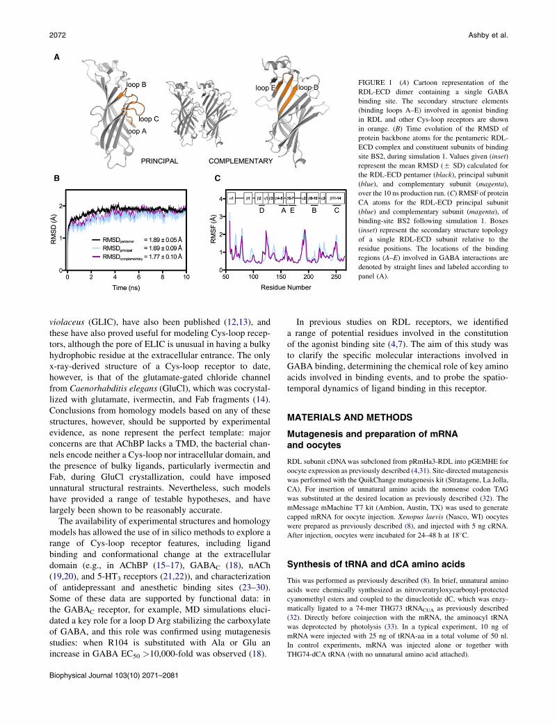
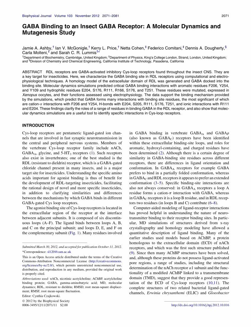
FIGURE 1 (A) Cartoon representation of the
RDL-ECD dimer containing a single GABA
binding site. The secondary structure elements
(binding loops A–E) involved in agonist binding
in RDL and other Cys-loop receptors are shown
in orange. (B) Time evolution of the RMSD of
protein backbone atoms for the pentameric RDL-
ECD complex and constituent subunits of binding
site BS2, during simulation 1. Values given (inset)
represent the mean RMSD (5 SD) calculated for
the RDL-ECD pentamer (black), principal subunit
(blue), and complementary subunit (magenta),
over the 10 ns production run. (C) RMSF of protein
CA atoms for the RDL-ECD principal subunit
(blue) and complementary subunit (magenta), of
binding-site BS2 following simulation 1. Boxes
(inset) represent the secondary structure topology
of a single RDL-ECD subunit relative to the
residue positions. The locations of the binding
regions (A–E) involved in GABA interactions are
denoted by straight lines and labeled according to
panel (A).
2072 Ashby et al.
violaceus (GLIC), have also been published (12,13), andthese have also proved useful for modeling Cys-loop recep-tors, although the pore of ELIC is unusual in having a bulkyhydrophobic residue at the extracellular entrance. The onlyx-ray-derived structure of a Cys-loop receptor to date,however, is that of the glutamate-gated chloride channelfrom Caenorhabditis elegans (GluCl), which was cocrystal-lized with glutamate, ivermectin, and Fab fragments (14).Conclusions from homology models based on any of thesestructures, however, should be supported by experimentalevidence, as none represent the perfect template: majorconcerns are that AChBP lacks a TMD, the bacterial chan-nels encode neither a Cys-loop nor intracellular domain, andthe presence of bulky ligands, particularly ivermectin andFab, during GluCl crystallization, could have imposedunnatural structural restraints. Nevertheless, such modelshave provided a range of testable hypotheses, and havelargely been shown to be reasonably accurate.
The availability of experimental structures and homologymodels has allowed the use of in silico methods to explore arange of Cys-loop receptor features, including ligandbinding and conformational change at the extracellulardomain (e.g., in AChBP (15–17), GABAC (18), nACh(19,20), and 5-HT3 receptors (21,22)), and characterizationof antidepressant and anesthetic binding sites (23–30).Some of these data are supported by functional data: inthe GABAC receptor, for example, MD simulations eluci-dated a key role for a loop D Arg stabilizing the carboxylateof GABA, and this role was confirmed using mutagenesisstudies: when R104 is substituted with Ala or Glu anincrease in GABA EC50 >10,000-fold was observed (18).
Biophysical Journal 103(10) 2071–2081
In previous studies on RDL receptors, we identifieda range of potential residues involved in the constitutionof the agonist binding site (4,7). The aim of this study wasto clarify the specific molecular interactions involved inGABA binding, determining the chemical role of key aminoacids involved in binding events, and to probe the spatio-temporal dynamics of ligand binding in this receptor.
MATERIALS AND METHODS
Mutagenesis and preparation of mRNAand oocytes
RDL subunit cDNAwas subcloned from pRmHa3-RDL into pGEMHE for
oocyte expression as previously described (4,31). Site-directed mutagenesis
was performed with the QuikChange mutagenesis kit (Stratagene, La Jolla,
CA). For insertion of unnatural amino acids the nonsense codon TAG
was substituted at the desired location as previously described (32). The
mMessage mMachine T7 kit (Ambion, Austin, TX) was used to generate
capped mRNA for oocyte injection. Xenopus laevis (Nasco, WI) oocytes
were prepared as previously described (8), and injected with 5 ng cRNA.
After injection, oocytes were incubated for 24–48 h at 18�C.
Synthesis of tRNA and dCA amino acids
This was performed as previously described (8). In brief, unnatural amino
acids were chemically synthesized as nitroveratryloxycarbonyl-protected
cyanomethyl esters and coupled to the dinucleotide dC, which was enzy-
matically ligated to a 74-mer THG73 tRNACUA as previously described
(32). Directly before coinjection with the mRNA, the aminoacyl tRNA
was deprotected by photolysis (33). In a typical experiment, 10 ng of
mRNA were injected with 25 ng of tRNA-aa in a total volume of 50 nl.
In control experiments, mRNA was injected alone or together with
THG74-dCA tRNA (with no unnatural amino acid attached).
Binding of GABA at RDL Receptors 2073
Electrophysiology
Using a two-electrode voltage-clamp, Xenopus oocytes were clamped at
�60 mV using an OC-725 amplifier (Warner Instruments, CT), Digidata
1322A, and the Strathclyde Electrophysiology Software Package (Depart-
ment of Physiology and Pharmacology, University of Strathclyde, UK) or
using the OpusXpress voltage-clamp system (Molecular Devices, Union
City, CA). Currents were filtered at a frequency of 1 kHz. Microelectrodes
were fabricated from borosilicate glass (GC120TF-10, Harvard Apparatus,
Edenbridge, Kent, UK) using a one stage horizontal pull (P-87, Sutter
Instrument, California) and filled with 3 M KCl. Pipette resistances ranged
from 1.0 to 2.0 MU. Oocytes were perfused with ND96 (96 mM NaCl,
2 mM KCl, 1 mM MgCl2, 1.8 mM CaCl2, 5 mM HEPES, pH 7.4). Drug
application was via a simple gravity fed system or via the computer-
controlled perfusion system of the OpusXpress.
Analysis and curve fitting was performed using Prism (GraphPad
Software, San Diego, CA). Concentration-response data for each oocyte
was normalized to the maximum current for that oocyte. The mean 5
SE for a series of oocytes were plotted against agonist concentration and
iteratively fitted to the following equation:
IC ¼ Imin þ Imax � Imin
1þ 10nHðlog EC50�log CÞ;
where C is the concentration of ligand present; IC is the current in the pres-
ence of ligand concentration C; Imin is the current when C ¼ 0; Imax is the
current whenC¼N, EC50 is the concentration ofC, which evokes a current
equal to (Imax þ Imin)/2; and nH is the Hill coefficient. Significance was
calculated using a one-way ANOVA.
Modeling
The crystal structure of C. elegans glutamate-gated chloride channel
(GluCl; PDB ID: 3RIF (14)) was chosen as a template for building the
GABA RDL extracellular domain model. A sequence alignment of GluCl
(residues 1–212 [PDB numbering]) and GABA RDL (NCBI accession:
NM_168321.1, residues 57–266) was produced with FUGUE (34), and
used to model all five subunits simultaneously with MODELER 9v10
(35). The resulting 30 models were protonated with MolProbity (36) and
ranked according to their energetic favorability (ANOLEA (37); QMEAN
(38)) and stereo-chemical quality (PROCHECK (39); RAMPAGE (40)).
Interatomic clashes were removed from the selected model (RDL-ECD)
by 1500 steps of steepest descent minimization followed by 1000 steps of
conjugate gradient minimization in a solvated, neutralized simulation box
with positional restraints applied to the CA atoms.
Docking
The zwitterionic form of GABA was docked into the RDL-ECD binding
site, which was defined as being within a 10 A radius of F206, using
GOLD v4.0 (CCDC, Cambridge, UK). In all 20 docking poses, the
amine nitrogen of GABA was positioned consistent with the formation of
a cation-p interaction with F206 and Y254, thus satisfying previous exper-
imental criteria (7). Ten genetic algorithm runs were performed on each
docking exercise using default parameters. The docking pose with the high-
est GoldScore fitness function was chosen for MD simulations. The struc-
tures were visualized using PyMOL v 1.3 (Schrodinger).
MD simulations
These were performed using the AMBER 2003 force field (41) and the
GROMACS 4.5.4 suite of software (42). The charges of protein ionizable
groups were found to be in the standard protonation state at neutral pH, as
calculated using the Karlsberg webserver (43). The GABA ESP partial
charges were calculated with the Density Functional Theory as imple-
mented in the CPMD code, as previously described (18), and averaged
over eight geometry configurations; the charges of equivalent atoms
were also averaged. The protein-ligand complex was solvated with
TIP3P water molecules in a periodically repeating truncated octahedral
box. The net charge of the system was brought to neutrality and physio-
logical ionic strength (0.15 M) by the addition of dissociated NaCl,
resulting in a system comprising 38,611 water molecules, 121 Naþ ions,
and 126 Cl� ions. An integration time step of 2 fs was used and all bonds
were constrained using the LINCS algorithm (44). Long-range electro-
statics was evaluated with the particle mesh Ewald method (45) and van
der Waals forces were treated with a cutoff of 14 A. Following 5000
steps of steepest descent energy minimization, the system was equilibrated
with 100 ps of position-restrained MD under NVT conditions, followed by
100 ps under NPT conditions. Temperature and pressure were kept
constant (T ¼ 300 K, tt ¼ 0.1 ps; P ¼ 1 bar, tp ¼ 2 ps) by coupling to
a modified Berendsen thermostat based on stochastic velocity rescaling
(46,47) and Parrinello-Rahman barostat (48), respectively. A 10 ns
data production run was performed in the NPT ensemble with positional
restraints imposed on the last three C-terminal CA atoms of each subunit
(1000 kJ mol�1 nm�2), to mimic the presence of the TMD. The protein
RMSD reached a plateau around 2 ns, and thus, ligand binding statistics
were performed on the last 8 ns of the simulation. Hydrogen bonds
were defined as having an acceptor-hydrogen distance %3.5 A and an
acceptor-donor-hydrogen angle %30�. Groups of opposite charge <6 A
apart were defined as ionic interactions. Cation-p interactions were iden-
tified as previously described (18): a distance cutoff (<6 A) was applied
between the GABA amine nitrogen and the center of mass of the
phenyl ring, and an angle cutoff (<45�) was applied between the normal
to the phenyl ring and the vector pointing from the ring center of mass
to the GABA nitrogen. Cluster analysis was performed on each indepen-
dent ligand trajectory (n ¼ 20) using the gromos algorithm (49)
(RMSD cutoff <1 A), as implemented in the GROMACS program
g_cluster: distance-RMSD (dRMSD) and atomic distance calculations
were performed on each cluster member, data from equivalent clusters
were pooled and mean (5SD) values were derived from each pool. The
protein atom names used throughout correspond to the AMBER atom
nomenclature and GABA atom naming is found in Fig. 2 A. Unless
otherwise stated, statistics describing ligand motions and intermolecular
interactions refer to the mean (5SEM) value, as averaged over all 20
independent binding sites.
RESULTS
Homology modeling and ligand docking
The crystal structure of the invertebrate Cys-loop receptorGluCl is the closest structural homolog of RDL currentlyavailable in the Protein Data Bank and these proteins have38.1% extracellular domain sequence identity. This valueis above the accepted 30% threshold for model reliabilityand is considerably higher than those for other relatedcrystal structures (GLIC, 19.5%; ELIC, 21.0%; AChBPs,17–20%). Because protein structure is more highly con-served at the tertiary level than at the primary level, weused an alignment generated by FUGUE (34), a programthat performs profile analysis based on known sequence-structure compatibilities. Refinement of our model byenergy minimization resulted in 99.7% of residues fallingwithin the favored/allowed region of the Ramachandranplot. The number of rotamer deviations and bond angle
Biophysical Journal 103(10) 2071–2081
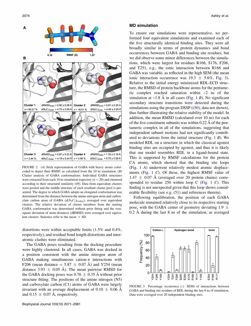
FIGURE 2 (A) Stick representation of GABA with heavy atoms color-
coded to depict their RMSF, as calculated from the 10 ns simulation. (B)
Cluster analysis of GABA conformations. Individual GABA structures
were extracted from each 10 ns simulation trajectory (n¼ 20) and clustered
according to their structural similarity (49). Data from equivalent clusters
were pooled and the middle structure of each resultant cluster pool is pre-
sented. The degree to which GABA adopts an elongated conformation was
determined from the distance between the amino nitrogen atom and carbox-
ylate carbon atom of GABA (d(N5C1)GABA), averaged over equivalent
clusters. The relative deviation of cluster members from the starting
GABA conformation was determined without prior fitting and the root-
square deviation of atom distances (dRMSD) were averaged over equiva-
lent clusters. Statistics refer to the mean 5 SD.
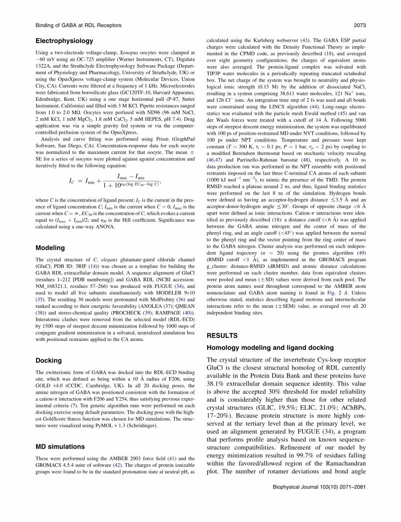
FIGURE 3 Percentage occurrence (5 SEM) of interactions between
GABA and binding site residues of RDL during the last 8 ns of simulation.
Data were averaged over 20 independent binding sites.
2074 Ashby et al.
distortions were within acceptable limits (1.5% and 0.4%,respectively), and residual bond length distortions and inter-atomic clashes were eliminated.
The GABA poses resulting from the docking procedurewere highly clustered. In all cases, GABA was docked ina position consistent with the amine nitrogen atom ofGABA making simultaneous cation-p interactions withF206 (mean distance ¼ 5.87 5 0.07 A) and Y254 (meandistance 3.93 5 0.05 A). The mean pairwise RMSD forthe GABA docking poses was 0.76 5 0.35 A without priorstructure fitting. The positions of the amine nitrogen (N5)and carboxylate carbon (C1) atoms of GABA were largelyinvariant with an average displacement of 0.10 5 0.06 Aand 0.15 5 0.07 A, respectively.
Biophysical Journal 103(10) 2071–2081
MD simulation
To ensure our simulations were representative, we per-formed four equivalent simulations and examined each ofthe five structurally identical binding sites. They were allbroadly similar in terms of protein dynamics and bondoccurrences between GABA and binding site residues, butwe did observe some minor differences between the simula-tions, which were largest for residues R166, S176, F206,and T251, e.g., the ionic interaction between R166 andGABAwas variable, as reflected in the high SEM (the meanionic interaction occurrence was 19.3 5 5.6%, Fig. 3).Relative to the initial energy minimized RDL-ECD struc-ture, the RMSD of protein backbone atoms for the pentame-ric complex reached saturation within ~2 ns of thesimulation at ~1.8 A in all cases (Fig. 1 B). No significantsecondary structure transitions were detected during thesimulations using the program DSSP ((50); data not shown),thus further illustrating the relative stability of the model. Inaddition, the mean RMSD (calculated over 10 ns) for eachof the five constituent subunits was within 0.22 A of the pen-tameric complex in all of the simulations, suggesting thatindependent subunit motions had not significantly contrib-uted to deviations from the initial structure (Fig. 1 B). Wemodeled RDL on a structure in which the classical agonistbinding sites are occupied by agonist, and thus it is likelythat our model resembles RDL in a ligand-bound state.This is supported by RMSF calculations for the proteinCA atoms, which showed that the binding site loops(Fig. 1 A) underwent relatively modest atomic displace-ments (Fig. 1 C). Of these, the highest RMSF value of1.47 5 0.07 A (averaged over 20 protein chains) corre-sponded to residue 250 within loop C (Fig. 1 C). Thisfinding is not unexpected given that this loop shows consid-erable flexibility (see e.g. (51) and references therein).
Following equilibration, the position of each GABAmolecule remained relatively close to its respective startingpose, with the GABA center of geometry deviating 1.9 50.2 A during the last 8 ns of the simulation, as averaged
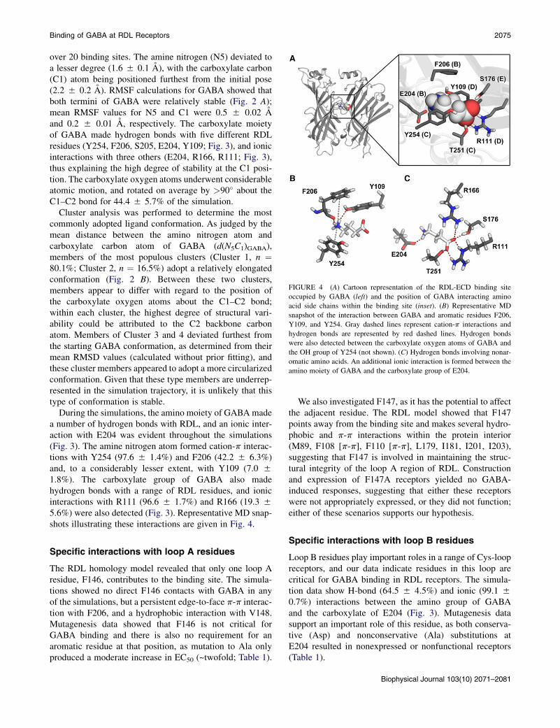
FIGURE 4 (A) Cartoon representation of the RDL-ECD binding site
occupied by GABA (left) and the position of GABA interacting amino
acid side chains within the binding site (inset). (B) Representative MD
snapshot of the interaction between GABA and aromatic residues F206,
Y109, and Y254. Gray dashed lines represent cation-p interactions and
hydrogen bonds are represented by red dashed lines. Hydrogen bonds
were also detected between the carboxylate oxygen atoms of GABA and
the OH group of Y254 (not shown). (C) Hydrogen bonds involving nonar-
omatic amino acids. An additional ionic interaction is formed between the
amino moiety of GABA and the carboxylate group of E204.
Binding of GABA at RDL Receptors 2075
over 20 binding sites. The amine nitrogen (N5) deviated toa lesser degree (1.6 5 0.1 A), with the carboxylate carbon(C1) atom being positioned furthest from the initial pose(2.2 5 0.2 A). RMSF calculations for GABA showed thatboth termini of GABA were relatively stable (Fig. 2 A);mean RMSF values for N5 and C1 were 0.5 5 0.02 Aand 0.2 5 0.01 A, respectively. The carboxylate moietyof GABA made hydrogen bonds with five different RDLresidues (Y254, F206, S205, E204, Y109; Fig. 3), and ionicinteractions with three others (E204, R166, R111; Fig. 3),thus explaining the high degree of stability at the C1 posi-tion. The carboxylate oxygen atoms underwent considerableatomic motion, and rotated on average by >90� about theC1–C2 bond for 44.4 5 5.7% of the simulation.
Cluster analysis was performed to determine the mostcommonly adopted ligand conformation. As judged by themean distance between the amino nitrogen atom andcarboxylate carbon atom of GABA (d(N5C1)GABA),members of the most populous clusters (Cluster 1, n ¼80.1%; Cluster 2, n ¼ 16.5%) adopt a relatively elongatedconformation (Fig. 2 B). Between these two clusters,members appear to differ with regard to the position ofthe carboxylate oxygen atoms about the C1–C2 bond;within each cluster, the highest degree of structural vari-ability could be attributed to the C2 backbone carbonatom. Members of Cluster 3 and 4 deviated furthest fromthe starting GABA conformation, as determined from theirmean RMSD values (calculated without prior fitting), andthese cluster members appeared to adopt a more circularizedconformation. Given that these type members are underrep-resented in the simulation trajectory, it is unlikely that thistype of conformation is stable.
During the simulations, the amino moiety of GABAmadea number of hydrogen bonds with RDL, and an ionic inter-action with E204 was evident throughout the simulations(Fig. 3). The amine nitrogen atom formed cation-p interac-tions with Y254 (97.6 5 1.4%) and F206 (42.2 5 6.3%)and, to a considerably lesser extent, with Y109 (7.0 51.8%). The carboxylate group of GABA also madehydrogen bonds with a range of RDL residues, and ionicinteractions with R111 (96.6 5 1.7%) and R166 (19.3 55.6%) were also detected (Fig. 3). Representative MD snap-shots illustrating these interactions are given in Fig. 4.
Specific interactions with loop A residues
The RDL homology model revealed that only one loop Aresidue, F146, contributes to the binding site. The simula-tions showed no direct F146 contacts with GABA in anyof the simulations, but a persistent edge-to-face p-p interac-tion with F206, and a hydrophobic interaction with V148.Mutagenesis data showed that F146 is not critical forGABA binding and there is also no requirement for anaromatic residue at that position, as mutation to Ala onlyproduced a moderate increase in EC50 (~twofold; Table 1).
We also investigated F147, as it has the potential to affectthe adjacent residue. The RDL model showed that F147points away from the binding site and makes several hydro-phobic and p-p interactions within the protein interior(M89, F108 [p-p], F110 [p-p], L179, I181, I201, I203),suggesting that F147 is involved in maintaining the struc-tural integrity of the loop A region of RDL. Constructionand expression of F147A receptors yielded no GABA-induced responses, suggesting that either these receptorswere not appropriately expressed, or they did not function;either of these scenarios supports our hypothesis.
Specific interactions with loop B residues
Loop B residues play important roles in a range of Cys-loopreceptors, and our data indicate residues in this loop arecritical for GABA binding in RDL receptors. The simula-tion data show H-bond (64.5 5 4.5%) and ionic (99.1 50.7%) interactions between the amino group of GABAand the carboxylate of E204 (Fig. 3). Mutagenesis datasupport an important role of this residue, as both conserva-tive (Asp) and nonconservative (Ala) substitutions atE204 resulted in nonexpressed or nonfunctional receptors(Table 1).
Biophysical Journal 103(10) 2071–2081
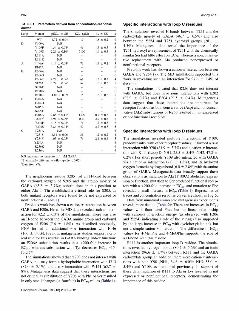
TABLE 1 Parameters derived from concentration-response
curves
Loop Mutant pEC50 5 SE EC50 (mM) nH 5 SE n
WT 4.72 5 0.04 19 1.8 5 0.2 9
D Y109A N/R – – 6
Y109F 4.34 5 0.04* 46 1.7 5 0.3 5
Y109S 2.29 5 0.10* 5100 1.9 5 0.3 5
R111A N/R – – 6
R111K N/R – – 6
A F146Ay 4.14 5 0.04* 73 1.7 5 0.2 3
F147A N/R – – 6
E R166A N/R – – 6
R166G N/R – – 6
R166K 4.22 5 0.06* 61 1.3 5 0.2 4
S176A 3.27 5 0.06* 540 1.6 5 0.3 5
S176T N/R – – 6
R178A N/R – – 6
R178K 4.82 5 0.09 15 1.3 5 0.3 5
B E204A N/R – – 6
E204D N/R – – 6
S205A N/R – – 6
S205T N/R – – 6
F206A 2.88 5 0.11* 1300 0.7 5 0.3 4
F206Yy 6.94 5 0.09* 0.11 2.2 5 0.3 6
Y208F 4.15 5 0.03* 71 1.7 5 0.3 6
Y208S 3.84 5 0.04* 47 2.2 5 0.3 5
C T251A N/R – - 6
T251S 4.51 5 0.06 31 1.2 5 0.2 4
Y254Fy 4.05 5 0.05* 76 2.1 5 0.4 5
Y254Ay N/R – – 6
R256K N/R – – 6
R256A N/R – – 6
N/R indicates no response to 1 mM GABA.
*Statistically different to wild-type (p < 0.05).yData from (7).
2076 Ashby et al.
The neighboring residue S205 had an H-bond betweenthe carbonyl oxygen of S205 and the amino moiety ofGABA (65.8 5 3.7%); substitutions in this position toeither Ala or Thr established a critical role for S205, asboth mutant receptors were found to be not expressed ornonfunctional (Table 1).
Previous work has shown a cation-p interaction betweenGABA and F206. Here, the MD data revealed such an inter-action for 42.2 5 6.3% of the simulations. There was alsoan H-bond between the GABA amino group and carbonyloxygen of F206 (7.6 5 1.8%). As described previously,F206 formed an additional p-p interaction with F146(100 5 0.0%). Previous mutagenesis studies support a crit-ical role for this residue in GABA binding and/or function:an F206A substitution results in a >200-fold increase inEC50, whereas substitution with Tyr decreases EC50 ~15-fold (7).
The simulations showed that Y208 does not interact withGABA, but may form a hydrophobic interaction with I213(87.0 5 5.1%), and a p-p interaction with W141 (65.7 58%). Mutagenesis data suggest that these interactions arenot critical as substitution of Y208 with Phe or Ser resultedin only small changes (< fourfold) in EC50 values (Table 1).
Biophysical Journal 103(10) 2071–2081
Specific interactions with loop C residues
The simulations revealed H-bonds between T251 and thecarboxylate moiety of GABA (46.7 5 6.5%) and alsobetween the Y254 and T251 hydroxyl groups (28.1 54.3%). Mutagenesis data reveal the importance of theT251 hydroxyl as replacement of T251 with the chemicallysimilar Ser had little effect on EC50, whereas a nonconserva-tive replacement with Ala produced nonexpressed ornonfunctional receptors.
Previous work has shown a cation-p interaction betweenGABA and Y254 (7). The MD simulations supported thiswork in revealing such an interaction for 97.6 5 1.4% ofthe time.
The simulations indicated that R256 does not interactwith GABA, but does have ionic interactions with E202(98.9 5 0.7%) and E204 (99.5 5 0.4%). Mutagenesisdata suggest that these interactions are important forreceptor function as both conservative (Asp) and nonconser-vative (Ala) substitutions of R256 resulted in nonexpressedor nonfunctional receptors.
Specific interactions with loop D residues
The simulations revealed multiple interactions of Y109,predominantly with other receptor residues: it formed a p-pinteraction with Y90 (81.9 5 3.7%) and a cation-p interac-tion with R111 (Loop D; NH1, 25.5 5 5.4%; NH2, 47.8 56.2%). For short periods Y109 also interacted with GABAvia a cation-p interaction (7.0 5 1.8%), and its hydroxyloxygen formed a hydrogenbond (6.05 2.8%)with the aminogroup of GABA. Mutagenesis data broadly support theseobservations as mutation to Ala (Y109A) abolished expres-sion or function, mutation to Ser produced functional recep-tors with a >200-fold increase in EC50, and mutation to Pherevealed a small increase in EC50 (Table 1). Representativetraces and concentration response curves are shown in Fig. 5.
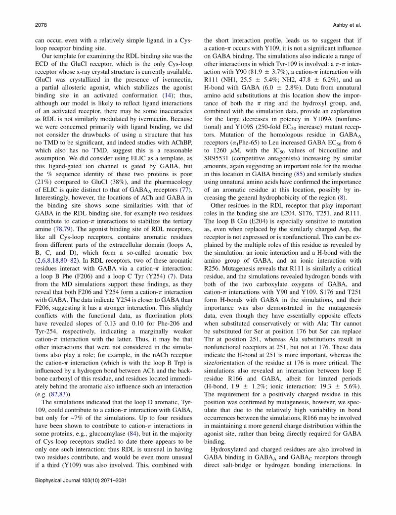
Data from unnatural amino acid mutagenesis experimentsreveals more details (Table 2): There are increases in EC50
values with fluorinated Phes but no linear relationshipwith cation-p interaction energy (as observed with F206and Y254) indicating a role of the p ring (also supportedby the large increase in EC50 with cyclohexylalanine), butnot a simple cation-p interaction. The difference in EC50
values for 4-Me Phe and 4-MeOPhe supports the role ofa H-bond with this residue.
R111 is another important loop D residue. The simula-tions revealed hydrogen bonds (80.2 5 5.6%) and an ionicinteraction (96.6 5 1.7%) between R111 and the GABAcarboxylate group. In addition, there were cation-p interac-tions with both Y90 (NH1, 34.6 5 6.0%; NH2 35.0 55.4%) and Y109, as mentioned previously. In support ofthese data, mutation of R111 to Ala or Lys resulted in notexpressed or nonfunctional receptors, demonstrating theimportance of this residue.
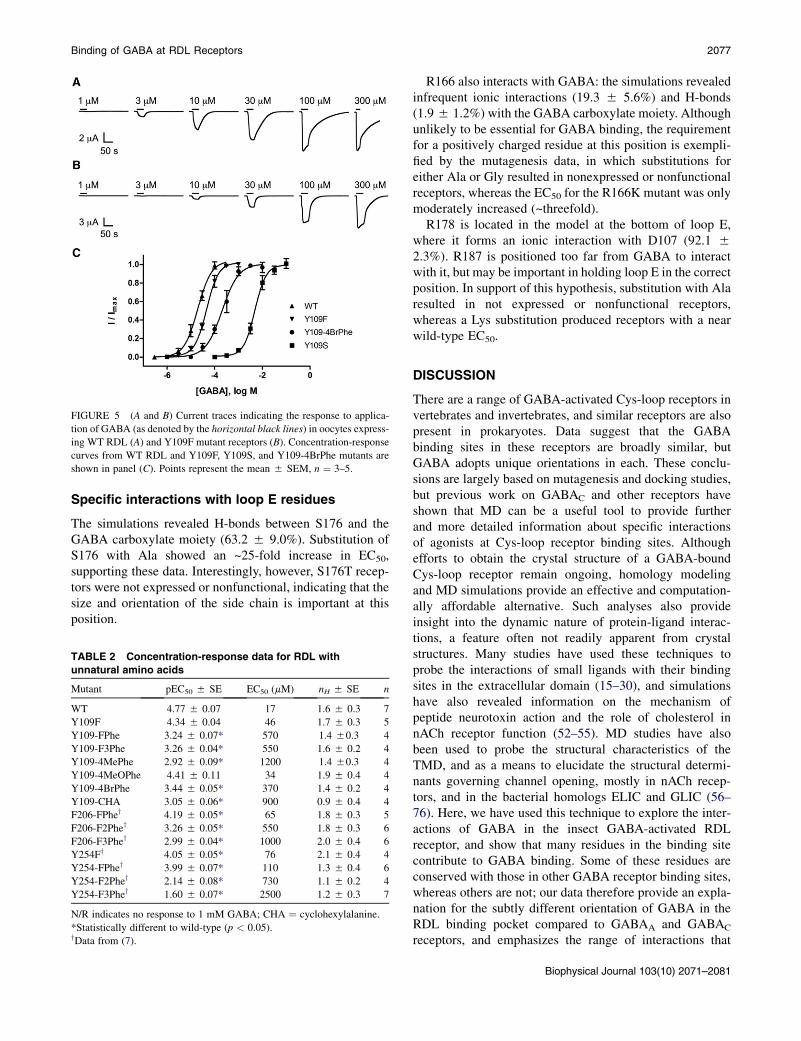
FIGURE 5 (A and B) Current traces indicating the response to applica-
tion of GABA (as denoted by the horizontal black lines) in oocytes express-
ing WT RDL (A) and Y109F mutant receptors (B). Concentration-response
curves from WT RDL and Y109F, Y109S, and Y109-4BrPhe mutants are
shown in panel (C). Points represent the mean 5 SEM, n ¼ 3–5.
Binding of GABA at RDL Receptors 2077
Specific interactions with loop E residues
The simulations revealed H-bonds between S176 and theGABA carboxylate moiety (63.2 5 9.0%). Substitution ofS176 with Ala showed an ~25-fold increase in EC50,supporting these data. Interestingly, however, S176T recep-tors were not expressed or nonfunctional, indicating that thesize and orientation of the side chain is important at thisposition.
TABLE 2 Concentration-response data for RDL with
unnatural amino acids
Mutant pEC50 5 SE EC50 (mM) nH 5 SE n
WT 4.77 5 0.07 17 1.6 5 0.3 7
Y109F 4.34 5 0.04 46 1.7 5 0.3 5
Y109-FPhe 3.24 5 0.07* 570 1.4 50.3 4
Y109-F3Phe 3.26 5 0.04* 550 1.6 5 0.2 4
Y109-4MePhe 2.92 5 0.09* 1200 1.4 50.3 4
Y109-4MeOPhe 4.41 5 0.11 34 1.9 5 0.4 4
Y109-4BrPhe 3.44 5 0.05* 370 1.4 5 0.2 4
Y109-CHA 3.05 5 0.06* 900 0.9 5 0.4 4
F206-FPhey 4.19 5 0.05* 65 1.8 5 0.3 5
F206-F2Phey 3.26 5 0.05* 550 1.8 5 0.3 6
F206-F3Phey 2.99 5 0.04* 1000 2.0 5 0.4 6
Y254Fy 4.05 5 0.05* 76 2.1 5 0.4 4
Y254-FPhey 3.99 5 0.07* 110 1.3 5 0.4 6
Y254-F2Phey 2.14 5 0.08* 730 1.1 5 0.2 4
Y254-F3Phey 1.60 5 0.07* 2500 1.2 5 0.3 7
N/R indicates no response to 1 mM GABA; CHA ¼ cyclohexylalanine.
*Statistically different to wild-type (p < 0.05).yData from (7).
R166 also interacts with GABA: the simulations revealedinfrequent ionic interactions (19.3 5 5.6%) and H-bonds(1.95 1.2%) with the GABA carboxylate moiety. Althoughunlikely to be essential for GABA binding, the requirementfor a positively charged residue at this position is exempli-fied by the mutagenesis data, in which substitutions foreither Ala or Gly resulted in nonexpressed or nonfunctionalreceptors, whereas the EC50 for the R166K mutant was onlymoderately increased (~threefold).
R178 is located in the model at the bottom of loop E,where it forms an ionic interaction with D107 (92.1 52.3%). R187 is positioned too far from GABA to interactwith it, but may be important in holding loop E in the correctposition. In support of this hypothesis, substitution with Alaresulted in not expressed or nonfunctional receptors,whereas a Lys substitution produced receptors with a nearwild-type EC50.
DISCUSSION
There are a range of GABA-activated Cys-loop receptors invertebrates and invertebrates, and similar receptors are alsopresent in prokaryotes. Data suggest that the GABAbinding sites in these receptors are broadly similar, butGABA adopts unique orientations in each. These conclu-sions are largely based on mutagenesis and docking studies,but previous work on GABAC and other receptors haveshown that MD can be a useful tool to provide furtherand more detailed information about specific interactionsof agonists at Cys-loop receptor binding sites. Althoughefforts to obtain the crystal structure of a GABA-boundCys-loop receptor remain ongoing, homology modelingand MD simulations provide an effective and computation-ally affordable alternative. Such analyses also provideinsight into the dynamic nature of protein-ligand interac-tions, a feature often not readily apparent from crystalstructures. Many studies have used these techniques toprobe the interactions of small ligands with their bindingsites in the extracellular domain (15–30), and simulationshave also revealed information on the mechanism ofpeptide neurotoxin action and the role of cholesterol innACh receptor function (52–55). MD studies have alsobeen used to probe the structural characteristics of theTMD, and as a means to elucidate the structural determi-nants governing channel opening, mostly in nACh recep-tors, and in the bacterial homologs ELIC and GLIC (56–76). Here, we have used this technique to explore the inter-actions of GABA in the insect GABA-activated RDLreceptor, and show that many residues in the binding sitecontribute to GABA binding. Some of these residues areconserved with those in other GABA receptor binding sites,whereas others are not; our data therefore provide an expla-nation for the subtly different orientation of GABA in theRDL binding pocket compared to GABAA and GABAC
receptors, and emphasizes the range of interactions that
Biophysical Journal 103(10) 2071–2081
2078 Ashby et al.
can occur, even with a relatively simple ligand, in a Cys-loop receptor binding site.
Our template for examining the RDL binding site was theECD of the GluCl receptor, which is the only Cys-loopreceptor whose x-ray crystal structure is currently available.GluCl was crystallized in the presence of ivermectin,a partial allosteric agonist, which stabilizes the agonistbinding site in an activated conformation (14); thus,although our model is likely to reflect ligand interactionsof an activated receptor, there may be some inaccuraciesas RDL is not similarly modulated by ivermectin. Becausewe were concerned primarily with ligand binding, we didnot consider the drawbacks of using a structure that hasno TMD to be significant, and indeed studies with AChBP,which also has no TMD, suggest this is a reasonableassumption. We did consider using ELIC as a template, asthis ligand-gated ion channel is gated by GABA, butthe % sequence identity of these two proteins is poor(21%) compared to GluCl (38%), and the pharmacologyof ELIC is quite distinct to that of GABAA receptors (77).Interestingly, however, the locations of ACh and GABA inthe binding site shows some similarities with that ofGABA in the RDL binding site, for example two residuescontribute to cation-p interactions to stabilize the tertiaryamine (78,79). The agonist binding site of RDL receptors,like all Cys-loop receptors, contains aromatic residuesfrom different parts of the extracellular domain (loops A,B, C, and D), which form a so-called aromatic box(2,6,8,18,80–82). In RDL receptors, two of these aromaticresidues interact with GABA via a cation-p interaction:a loop B Phe (F206) and a loop C Tyr (Y254) (7). Datafrom the MD simulations support these findings, as theyreveal that both F206 and Y254 form a cation-p interactionwith GABA. The data indicate Y254 is closer to GABA thanF206, suggesting it has a stronger interaction. This slightlyconflicts with the functional data, as fluorination plotshave revealed slopes of 0.13 and 0.10 for Phe-206 andTyr-254, respectively, indicating a marginally weakercation-p interaction with the latter. Thus, it may be thatother interactions that were not considered in the simula-tions also play a role; for example, in the nACh receptorthe cation-p interaction (which is with the loop B Trp) isinfluenced by a hydrogen bond between ACh and the back-bone carbonyl of this residue, and residues located immedi-ately behind the aromatic also influence such an interaction(e.g. (82,83)).
The simulations indicated that the loop D aromatic, Tyr-109, could contribute to a cation-p interaction with GABA,but only for ~7% of the simulations. Up to four residueshave been shown to contribute to cation-p interactions insome proteins, e.g., glucoamylase (84), but in the majorityof Cys-loop receptors studied to date there appears to beonly one such interaction; thus RDL is unusual in havingtwo residues contribute, and would be even more unusualif a third (Y109) was also involved. This, combined with
Biophysical Journal 103(10) 2071–2081
the short interaction profile, leads us to suggest that ifa cation-p occurs with Y109, it is not a significant influenceon GABA binding. The simulations also indicate a range ofother interactions in which Tyr-109 is involved: a p-p inter-action with Y90 (81.9 5 3.7%), a cation-p interaction withR111 (NH1, 25.5 5 5.4%; NH2, 47.8 5 6.2%), and anH-bond with GABA (6.0 5 2.8%). Data from unnaturalamino acid substitutions at this location show the impor-tance of both the p ring and the hydroxyl group, and,combined with the simulation data, provide an explanationfor the large decreases in potency in Y109A (nonfunc-tional) and Y109S (250-fold EC50 increase) mutant recep-tors. Mutation of the homologous residue in GABAA
receptors (a1Phe-65) to Leu increased GABA EC50 from 6to 1260 mM, with the IC50 values of bicuculline andSR95531 (competitive antagonists) increasing by similaramounts, again suggesting an important role for the residuein this location in GABA binding (85) and similarly studiesusing unnatural amino acids have confirmed the importanceof an aromatic residue at this location, possibly by in-creasing the general hydrophobicity of the region (8).
Other residues in the RDL receptor that play importantroles in the binding site are E204, S176, T251, and R111.The loop B Glu (E204) is especially sensitive to mutationas, even when replaced by the similarly charged Asp, thereceptor is not expressed or is nonfunctional. This can be ex-plained by the multiple roles of this residue as revealed bythe simulation: an ionic interaction and a H-bond with theamino group of GABA, and an ionic interaction withR256. Mutagenesis reveals that R111 is similarly a criticalresidue, and the simulations revealed hydrogen bonds withboth of the two carboxylate oxygens of GABA, andcation-p interactions with Y90 and Y109. S176 and T251form H-bonds with GABA in the simulations, and theirimportance was also demonstrated in the mutagenesisdata, even though they have essentially opposite effectswhen substituted conservatively or with Ala: Thr cannotbe substituted for Ser at position 176 but Ser can replaceThr at position 251, whereas Ala substitutions result innonfunctional receptors at 251, but not at 176. These dataindicate the H-bond at 251 is more important, whereas thesize/orientation of the residue at 176 is more critical. Thesimulations also revealed an interaction between loop Eresidue R166 and GABA, albeit for limited periods(H-bond, 1.9 5 1.2%; ionic interaction: 19.3 5 5.6%).The requirement for a positively charged residue in thisposition was confirmed by mutagenesis, however, we spec-ulate that due to the relatively high variability in bondoccurrences between the simulations, R166 may be involvedin maintaining a more general charge distribution within theagonist site, rather than being directly required for GABAbinding.
Hydroxylated and charged residues are also involved inGABA binding in GABAA and GABAC receptors throughdirect salt-bridge or hydrogen bonding interactions. In
Binding of GABA at RDL Receptors 2079
GABAA receptors the hydroxylated residues a1S68,b2T160, b2T202, b2S204, and b2S209, and the chargedresidues a1R120, a1D183, a1R66, and b2R207 are involvedin ligand binding (86–90). A smaller repertoire of residues isinvolved in GABA binding in the related GABAC receptor:R104, F138, R158, and T244 (6,91–94). There are alsodifferences in ligand orientation and conformation: InGABAA receptors GABA likely binds in a partially foldedconformation, in GABAC receptors and in the RDL receptorGABA has been described as binding in an extended orelongated conformation (3–5). The current data supportthis and reveal specific interactions that may occur duringthe binding process.
CONCLUSIONS
GABA binds to GABA-activated Cys-loop receptors ina variety of different orientations. Here, we use MD simula-tions, combined with mutagenesis and functional data, toexplore the different interactions with binding-site residuesin the GABA-activated RDL receptor. The data reveala range of interactions, predominantly with aromatic,charged, and polar residues and suggest that interactionswith R111, E204, F206, Y254, and R256 are the mostcritical.
We acknowledge financial support from TheWellcome Trust (WT 81925 to
SCRL; SCRL is a Wellcome Trust Senior Research Fellow in Basic
Biomedical Science), the European Union 7th Framework Program No.
HEALTH-F2-2007-202088 ("NeuroCypres" project) to SCRL; the Medical
Research Council (a studentship to I.McG.), the U. S. National Institutes of
Health (NS34407 to D.A.D.), and the EPSRC (EP/F037457/1 ‘‘Support for
the UK Car-Parrinello Consortium’’) to CM.
REFERENCES
1. Thompson, A. J., H. A. Lester, and S. C. R. Lummis. 2010. Thestructural basis of function in Cys-loop receptors. Q. Rev. Biophys.43:449–499.
2. Lummis, S. C. R. 2009. Locating GABA in GABA receptor bindingsites. Biochem. Soc. Trans. 37:1343–1346.
3. Jones, M. V., Y. Sahara, ., G. L. Westbrook. 1998. Defining affinitywith the GABAA receptor. J. Neurosci. 18:8590–8604.
4. McGonigle, I., and S. C. R. Lummis. 2010. Molecular characterizationof agonists that bind to an insect GABA receptor. Biochemistry.49:2897–2902.
5. Woodward, R. M., L. Polenzani, and R. Miledi. 1993. Characterizationof bicuculline/baclofen-insensitive (r-like) g-aminobutyric acid recep-tors expressed in Xenopus oocytes. II. Pharmacology of g-aminobuty-ric acidA and g-aminobutyric acidB receptor agonists and antagonists.Mol. Pharmacol. 43:609–625.
6. Lummis, S. C. R., D. L. Beene,., D. A. Dougherty. 2005. A cation-pbinding interaction with a tyrosine in the binding site of the GABAC
receptor. Chem. Biol. 12:993–997.
7. Lummis, S. C. R., I. McGonigle, ., D. A. Dougherty. 2011. Twoamino acid residues contribute to a cation-p binding interaction inthe binding site of an insect GABA receptor. J. Neurosci. 31:12371–12376.
8. Padgett, C. L., A. P. Hanek, ., S. C. Lummis. 2007. Unnatural aminoacid mutagenesis of the GABA(A) receptor binding site residues
reveals a novel cation-p interaction between GABA and b 2Tyr97.J. Neurosci. 27:886–892.
9. Brejc, K., W. J. van Dijk,., T. K. Sixma. 2001. Crystal structure of anACh-binding protein reveals the ligand-binding domain of nicotinicreceptors. Nature. 411:269–276.
10. Grutter, T., L. Prado de Carvalho,., J. P. Changeux. 2005. A chimeraencoding the fusion of an acetylcholine-binding protein to an ionchannel is stabilized in a state close to the desensitized form ofligand-gated ion channels. C. R. Biol. 328:223–234.
11. Dellisanti, C. D., Y. Yao, ., L. Chen. 2007. Crystal structure of theextracellular domain of nAChR alpha1 bound to alpha-bungarotoxinat 1.94 A resolution. Nat. Neurosci. 10:953–962.
12. Hilf, R. J. C., and R. Dutzler. 2008. X-ray structure of a prokaryoticpentameric ligand-gated ion channel. Nature. 452:375–379.
13. Bocquet, N., H. Nury, ., P. J. Corringer. 2009. X-ray structure of apentameric ligand-gated ion channel in an apparently open conforma-tion. Nature. 457:111–114.
14. Hibbs, R. E., and E. Gouaux. 2011. Principles of activation and perme-ation in an anion-selective Cys-loop receptor. Nature. 474:54–60.
15. Geitmann, M., K. Retra,., U. H. Danielson. 2010. Interaction kineticand structural dynamic analysis of ligand binding to acetylcholine-binding protein. Biochemistry. 49:8143–8154.
16. Gao, F., N. Bren, ., S. M. Sine. 2005. Agonist-mediated conforma-tional changes in acetylcholine-binding protein revealed by simulationand intrinsic tryptophan fluorescence. J. Biol. Chem. 280:8443–8451.
17. Stober, S. T., and C. F. Abrams. 2012. Enhanced meta-analysis ofacetylcholine binding protein structures reveals conformational signa-tures of agonism in nicotinic receptors. Protein Sci. 21:307–317.
18. Melis, C., S. C. R. Lummis, and C. Molteni. 2008. Molecular dynamicssimulations of GABA binding to the GABAC receptor: the role ofArg-104. Biophys. J. 95:4115–4123.
19. Perez, E. G., B. K. Cassels, and G. Zapata-Torres. 2009. Molecularmodeling of the alpha9alpha10 nicotinic acetylcholine receptorsubtype. Bioorg. Med. Chem. Lett. 19:251–254.
20. Henchman, R. H., H. L. Wang, ., J. A. McCammon. 2005. Ligand-induced conformational change in the alpha7 nicotinic receptor ligandbinding domain. Biophys. J. 88:2564–2576.
21. Melis, C., P. L. Chau, ., C. Molteni. 2006. Exploring the binding ofserotonin to the 5-HT3 receptor by density functional theory. J. Phys.Chem. B. 110:26313–26319.
22. Thompson, A. J., and S. C. R. Lummis. 2006. 5-HT3 receptors. Curr.Pharm. Des. 12:3615–3630.
23. Sanghvi, M., A. K. Hamouda, ., H. R. Arias. 2008. Identifying thebinding site(s) for antidepressants on the Torpedo nicotinic acetylcho-line receptor: [3H]2-azidoimipramine photolabeling and moleculardynamics studies. Biochim. Biophys. Acta. 1778:2690–2699.
24. Nury, H., C. Van Renterghem, ., P. J. Corringer. 2011. X-ray struc-tures of general anaesthetics bound to a pentameric ligand-gated ionchannel. Nature. 469:428–431.
25. Brannigan, G., D. N. LeBard, ., M. L. Klein. 2010. Multiple bindingsites for the general anesthetic isoflurane identified in the nicotinicacetylcholine receptor transmembrane domain. Proc. Natl. Acad. Sci.USA. 107:14122–14127.
26. Chen, Q. A., M. H. Cheng, ., P. Tang. 2010. Anesthetic binding ina pentameric ligand-gated ion channel: GLIC. Biophys. J. 99:1801–1809.
27. Willenbring, D., Y. Xu, and P. Tang. 2010. The role of structured waterin mediating general anesthetic action on alpha4beta2 nAChR. Phys.Chem. Chem. Phys. 12:10263–10269.
28. Mowrey, D., E. J. Haddadian, ., P. Tang. 2010. Unresponsivecorrelated motion in alpha7 nAChR to halothane binding explains itsfunctional insensitivity to volatile anesthetics. J. Phys. Chem. B.114:7649–7655.
29. Liu, L. T., E. J. Haddadian, ., P. Tang. 2010. Higher susceptibility tohalothane modulation in open- than in closed-channel alpha4beta2
Biophysical Journal 103(10) 2071–2081
2080 Ashby et al.
nAChR revealed by molecular dynamics simulations. J. Phys. Chem. B.114:626–632.
30. Liu, L. T., D. Willenbring, ., P. Tang. 2009. General anestheticbinding to neuronal alpha4beta2 nicotinic acetylcholine receptor andits effects on global dynamics. J. Phys. Chem. B. 113:12581–12589.
31. Millar, N. S., S. D. Buckingham, and D. B. Sattelle. 1994. Stableexpression of a functional homo-oligomeric Drosophila GABAreceptor in a Drosophila cell line. Proc. Biol. Sci. 258:307–314.
32. Nowak, M. W., J. P. Gallivan, ., H. A. Lester. 1998. In vivo incorpo-ration of unnatural amino acids into ion channels in Xenopus oocyteexpression system. Methods Enzymol. 293:504–529.
33. Kearney, P. C., H. Zhang,., H. A. Lester. 1996. Determinants of nico-tinic receptor gating in natural and unnatural side chain structures at theM2 90 position. Neuron. 17:1221–1229.
34. Shi, J., T. L. Blundell, and K. Mizuguchi. 2001. FUGUE: sequence-structure homology recognition using environment-specific substitu-tion tables and structure-dependent gap penalties. J. Mol. Biol.310:243–257.
35. Sali, A., and T. L. Blundell. 1993. Comparative protein modelling bysatisfaction of spatial restraints. J. Mol. Biol. 234:779–815.
36. Chen, V. B., W. B. Arendall, 3rd,., D. C. Richardson. 2010. MolPro-bity: all-atom structure validation for macromolecular crystallography.Acta Crystallogr. D Biol. Crystallogr. 66:12–21.
37. Melo, F., and E. Feytmans. 1998. Assessing protein structures witha non-local atomic interaction energy. J. Mol. Biol. 277:1141–1152.
38. Benkert, P., T. Schwede, and S. C. Tosatto. 2009. QMEANclust: esti-mation of protein model quality by combining a composite scoringfunction with structural density information. BMC Struct. Biol. 9:35.
39. Laskowski, R. A., M. W. Macarthur, ., J. M. Thornton. 1993.Procheck - a program to check the stereochemical quality of proteinstructures. J. Appl. Cryst. 26:283–291.
40. Lovell, S. C., I. W. Davis, ., D. C. Richardson. 2003. Structurevalidation by Calpha geometry: 4,j and Cbeta deviation. Proteins.50:437–450.
41. Ponder, J. W., and D. A. Case. 2003. Force fields for protein simula-tions. Adv. Protein Chem. 66:27–85.
42. Hess, B., C. Kutzner, ., E. Lindahl. 2008. GROMACS 4: algorithmsfor highly efficient, load-balanced, and scalable molecular simulation.J. Chem. Theory Comput. 4:435–447.
43. Kieseritzky, G., and E. W. Knapp. 2008. Optimizing pKa computationin proteins with pH adapted conformations. Proteins. 71:1335–1348.
44. Hess, B., H. Bekker, ., J. G. E. M. Fraaije. 1997. LINCS: a linearconstraint solver for molecular simulations. J. Comput. Chem.18:1463–1472.
45. Cerutti, D. S., R. E. Duke, ., T. P. Lybrand. 2009. Staggered MeshEwald: an extension of the Smooth Particle-Mesh Ewald methodadding great versatility. J. Chem. Theory Comput. 5:2322.
46. Bussi, G., D. Donadio, and M. Parrinello. 2007. Canonical samplingthrough velocity rescaling. J. Chem. Phys. 126:014101.
47. Bussi, G., T. Zykova-Timan, and M. Parrinello. 2009. Isothermal-isobaric molecular dynamics using stochastic velocity rescaling.J. Chem. Phys. 130:074101.
48. Parrinello, M., and A. Rahman. 1981. Polymorphic transitions insingle-crystals: a new molecular-dynamics method. J. Appl. Phys.52:7182–7190.
49. Daura, X., K. Gademann,., A. E. Mark. 1999. Peptide folding: whensimulation meets experiment. Angew. Chem. Int. Ed. 38:236–240.
50. Kabsch, W., and C. Sander. 1983. Dictionary of protein secondarystructure: pattern recognition of hydrogen-bonded and geometricalfeatures. Biopolymers. 22:2577–2637.
51. Sander, T., A. T. Bruun, and T. Balle. 2010. Docking to flexiblenicotinic acetylcholine receptors: a validation study using the acetyl-choline binding protein. J. Mol. Graph. Model. 29:415–424.
Biophysical Journal 103(10) 2071–2081
52. Yu, R., Q. Kaas, and D. J. Craik. 2012. Delineation of the unbindingpathway of a-conotoxin ImI from the a7 nicotinic acetylcholinereceptor. J. Phys. Chem. B. 116:6097–6105.
53. Yi, M., H. Tjong, and H. X. Zhou. 2008. Spontaneous conformationalchange and toxin binding in alpha7 acetylcholine receptor: insight intochannel activation and inhibition. Proc. Natl. Acad. Sci. USA.105:8280–8285.
54. Yu, R., D. J. Craik, and Q. Kaas. 2011. Blockade of neuronala7-nAChR by a-conotoxin ImI explained by computational scanningand energy calculations. PLOS Comput. Biol. 7:e1002011.
55. Brannigan, G., J. Henin, ., M. L. Klein. 2008. Embedded cholesterolin the nicotinic acetylcholine receptor. Proc. Natl. Acad. Sci. USA.105:14418–14423.
56. Hung, A., K. Tai, and M. S. Sansom. 2005. Molecular dynamics simu-lation of the M2 helices within the nicotinic acetylcholine receptortransmembrane domain: structure and collective motions. Biophys. J.88:3321–3333.
57. Saladino, A. C., Y. Xu, and P. Tang. 2005. Homology modeling andmolecular dynamics simulations of transmembrane domain structureof human neuronal nicotinic acetylcholine receptor. Biophys. J.88:1009–1017.
58. Xu, Y., F. J. Barrantes,., H. Jiang. 2005. Conformational dynamics ofthe nicotinic acetylcholine receptor channel: a 35-ns moleculardynamics simulation study. J. Am. Chem. Soc. 127:1291–1299.
59. Corry, B. 2004. Theoretical conformation of the closed and openstates of the acetylcholine receptor channel. Biochim. Biophys. Acta.1663:2–5.
60. Corry, B. 2006. An energy-efficient gating mechanism in the acetyl-choline receptor channel suggested by molecular and Browniandynamics. Biophys. J. 90:799–810.
61. Saiz, L., and M. L. Klein. 2005. The transmembrane domain of theacetylcholine receptor: insights from simulations on synthetic peptidemodels. Biophys. J. 88:959–970.
62. Zhu, F., and G. Hummer. 2009. Gating transition of pentameric ligand-gated ion channels. Biophys. J. 97:2456–2463.
63. Taly, A., M. Delarue, ., J. P. Changeux. 2005. Normal mode analysissuggests a quaternary twist model for the nicotinic receptor gatingmechanism. Biophys. J. 88:3954–3965.
64. Law, R. J., R. H. Henchman, and J. A. McCammon. 2005. A gatingmechanism proposed from a simulation of a human alpha7 nicotinicacetylcholine receptor. Proc. Natl. Acad. Sci. USA. 102:6813–6818.
65. Cheng, X., I. Ivanov, ., J. A. McCammon. 2007. Nanosecond-time-scale conformational dynamics of the human alpha7 nicotinic acetyl-choline receptor. Biophys. J. 93:2622–2634.
66. Cheng, X., B. Lu, ., J. A. McCammon. 2006. Channel openingmotion of alpha7 nicotinic acetylcholine receptor as suggested bynormal mode analysis. J. Mol. Biol. 355:310–324.
67. Cheng, X., H. Wang, ., J. A. McCammon. 2006. Targeted moleculardynamics study of C-loop closure and channel gating in nicotinicreceptors. PLOS Comput. Biol. 2:e134.
68. Liu, X., Y. Xu, ., F. J. Barrantes. 2008. Mechanics of channel gatingof the nicotinic acetylcholine receptor. PLOS Comput. Biol. 4:e19.
69. Haddadian, E. J., M. H. Cheng, ., P. Tang. 2008. In silico models forthe human alpha4beta2 nicotinic acetylcholine receptor. J. Phys. Chem.B. 112:13981–13990.
70. Law, R. J., and F. C. Lightstone. 2009. Modeling neuronal nicotinic andGABA receptors: important interface salt-links and protein dynamics.Biophys. J. 97:1586–1594.
71. Melis, C., G. Bussi, ., C. Molteni. 2009. Trans-cis switching mecha-nisms in proline analogues and their relevance for the gating of the5-HT3 receptor. J. Phys. Chem. B. 113:12148–12153.
72. Cheng, X., I. Ivanov,., J. A. McCammon. 2009. Molecular-dynamicssimulations of ELIC-a prokaryotic homologue of the nicotinic acetyl-choline receptor. Biophys. J. 96:4502–4513.
Binding of GABA at RDL Receptors 2081
73. Zhu, F., and G. Hummer. 2010. Pore opening and closing of a pentame-ric ligand-gated ion channel. Proc. Natl. Acad. Sci. USA. 107:19814–19819.
74. Nury, H., F. Poitevin, ., M. Baaden. 2010. One-microsecond molec-ular dynamics simulation of channel gating in a nicotinic receptorhomologue. Proc. Natl. Acad. Sci. USA. 107:6275–6280.
75. Wang, H. L., X. Cheng, and S. M. Sine. 2012. Intramembrane protonbinding site linked to activation of bacterial pentameric ion channel.J. Biol. Chem. 287:6482–6489.
76. Prevost, M. S., L. Sauguet, ., P. J. Corringer. 2012. A locally closedconformation of a bacterial pentameric proton-gated ion channel.Nat. Struct. Mol. Biol. 19:642–649.
77. Thompson, A. J., M. Alqazzaz, ., S. C. Lummis. 2012. Thepharmacological profile of ELIC, a prokaryotic GABA-gated receptor.Neuropharmacology. 63:761–767.
78. Pan, J., Q. Chen,., P. Tang. 2012. Structure of the pentameric ligand-gated ion channel ELIC cocrystallized with its competitive antagonistacetylcholine. Nat Commun. 3:714.
79. Spurny, R., J. Ramerstorfer, ., C. Ulens. 2012. Pentameric ligand-gated ion channel ELIC is activated by GABA and modulated bybenzodiazepines. Proc. Natl. Acad. Sci. USA. In press.
80. Bartos, M., J. Corradi, and C. Bouzat. 2009. Structural basis of activa-tion of cys-loop receptors: the extracellular-transmembrane interface asa coupling region. Mol. Neurobiol. 40:236–252.
81. Pless, S. A., K. S. Millen,., D. A. Dougherty. 2008. A cation-p inter-action in the binding site of the glycine receptor is mediated bya phenylalanine residue. J. Neurosci. 28:10937–10942.
82. Xiu, X., N. L. Puskar, ., D. A. Dougherty. 2009. Nicotine bindingto brain receptors requires a strong cation-p interaction. Nature.458:534–537.
83. Cashin, A. L., M. M. Torrice, ., D. A. Dougherty. 2007. Chemical-scale studies on the role of a conserved aspartate in preorganizingthe agonist binding site of the nicotinic acetylcholine receptor.Biochemistry. 46:630–639.
84. Aleshin, A. E., B. Stoffer, ., R. B. Honzatko. 1996. Crystallographiccomplexes of glucoamylase with maltooligosaccharide analogs: rela-tionship of stereochemical distortions at the nonreducing end to thecatalytic mechanism. Biochemistry. 35:8319–8328.
85. Sigel, E., R. Baur, ., P. Malherbe. 1992. Point mutations affectingantagonist affinity and agonist dependent gating of GABAA receptorchannels. EMBO J. 11:2017–2023.
86. Boileau, A. J., A. R. Evers, ., C. Czajkowski. 1999. Mapping theagonist binding site of the GABAA receptor: evidence for a beta-strand.J. Neurosci. 19:4847–4854.
87. Amin, J., and D. S. Weiss. 1993. GABAA receptor needs two homolo-gous domains of the b-subunit for activation by GABA but not bypentobarbital. Nature. 366:565–569.
88. Newell, J. G., and C. Czajkowski. 2003. The GABAA receptor a 1subunit Pro174-Asp191 segment is involved in GABA binding andchannel gating. J. Biol. Chem. 278:13166–13172.
89. Westh-Hansen, S. E., M. R. Witt, ., M. Nielsen. 1999. Arginineresidue 120 of the human GABAA receptor a 1, subunit is essentialfor GABA binding and chloride ion current gating. Neuroreport.10:2417–2421.
90. Wagner, D. A., C. Czajkowski, and M. V. Jones. 2004. An arginineinvolved in GABA binding and unbinding but not gating of theGABA(A) receptor. J. Neurosci. 24:2733–2741.
91. Harrison, N. J., and S. C. R. Lummis. 2006. Locating the carboxylategroup of GABA in the homomeric rho GABA(A) receptor ligand-binding pocket. J. Biol. Chem. 281:24455–24461.
92. Harrison, N. J., and S. C. R. Lummis. 2006. Molecular modeling ofthe GABA(C) receptor ligand-binding domain. J. Mol. Model. 12:317–324.
93. Zhang, J., F. Xue, and Y. Chang. 2009. Agonist- and antagonist-induced conformational changes of loop F and their contributions tothe r1 GABA receptor function. J. Physiol. 587:139–153.
94. Amin, J., and D. S. Weiss. 1994. Homomeric rho 1 GABA channels:activation properties and domains. Receptors Channels. 2:227–236.
Biophysical Journal 103(10) 2071–2081