gaba-acs chem bio
TRANSCRIPT
Mechanism of Inactivation of GABA Aminotransferase by (E)- and(Z)‑(1S,3S)‑3-Amino-4-fluoromethylenyl-1-cyclopentanoic AcidHyunbeom Lee,#,† Hoang V. Le,#,† Rui Wu,§ Emma Doud,‡ Ruslan Sanishvili,∥ John F. Kellie,‡
Phillip D. Compton,‡ Boobalan Pachaiyappan,† Dali Liu,§ Neil L. Kelleher,‡ and Richard B. Silverman*,†
†Departments of Chemistry and Molecular Biosciences, Chemistry of Life Processes Institute, and the Center for MolecularInnovation and Drug Discovery, Northwestern University, Evanston, Illinois 60208, United States§Department of Chemistry and Biochemistry, Loyola University Chicago, Chicago, Illinois 60660, United States‡Departments of Chemistry and Molecular Biosciences, and the Proteomics Center of Excellence, Northwestern University, Evanston,Illinois 60208, United States∥X-ray Science Division, Advanced Photon Source, Argonne National Laboratory, Lemont, Illinois 60439, United States
*S Supporting Information
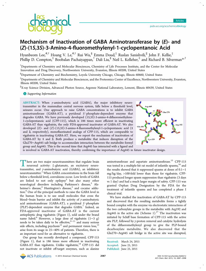
ABSTRACT: When γ-aminobutyric acid (GABA), the major inhibitory neuro-transmitter in the mammalian central nervous system, falls below a threshold level,seizures occur. One approach to raise GABA concentrations is to inhibit GABAaminotransferase (GABA-AT), a pyridoxal 5′-phosphate-dependent enzyme thatdegrades GABA. We have previously developed (1S,3S)-3-amino-4-difluoromethylene-1-cyclopentanoic acid (CPP-115), which is 186 times more efficient in inactivatingGABA-AT than vigabatrin, the only FDA-approved inactivator of GABA-AT. We alsodeveloped (E)- and (Z)-(1S,3S)-3-amino-4-fluoromethylenyl-1-cyclopentanoic acid (1and 2, respectively), monofluorinated analogs of CPP-115, which are comparable tovigabatrin in inactivating GABA-AT. Here, we report the mechanism of inactivation ofGABA-AT by 1 and 2. Both produce a metabolite that induces disruption of theGlu270−Arg445 salt bridge to accommodate interaction between the metabolite formylgroup and Arg445. This is the second time that Arg445 has interacted with a ligand andis involved in GABA-AT inactivation, thereby confirming the importance of Arg445 in future inactivator design.
There are two major neurotransmitters that regulate brainneuronal activity: L-glutamate, an excitatory neuro-
transmitter, and γ-aminobutyric acid (GABA), an inhibitoryneurotransmitter.1 When GABA concentrations in the brain fallbelow a threshold level, convulsions occur. Low levels of GABAare linked to not only epilepsy2 but also many otherneurological disorders including Parkinson’s disease,3 Alz-heimer’s disease,4 Huntington’s disease,5 and cocaine addic-tion.6 One of the principal methods to raise the GABA level inthe human brain is to use small molecules that cross theblood−brain barrier and inhibit the activity of γ-aminobutyricacid aminotransferase (GABA-AT), a pyridoxal 5′-phosphate(PLP)-dependent enzyme that degrades GABA.7 Indeed, anFDA-approved inactivator of GABA-AT is the currentantiepileptic drug vigabatrin (Figure 1), sold under the brandname Sabril.8 However, a large dose of vigabatrin (1−3 g)needs to be taken daily to be effective,9−11 and many seriousside effects, including psychosis12 and permanent vision loss,13
arise from its usage in 25−40% of patients. Therefore, there isan important need for an alternative to vigabatrin.Our group has recently developed a compound, CPP-115
(Figure 1), that is 186 times more efficient in inactivatingGABA-AT than vigabatrin. Unlike vigabatrin,14 CPP-115 didnot inactivate or inhibit off-target enzymes, such as alanine
aminotransferase and aspartate aminotransferase.15 CPP-115was tested in a multiple-hit rat model of infantile spasms,16 andthe results showed that it suppressed spasms at doses of 0.1−1mg/kg/day, >100-fold lower than those for vigabatrin. CPP-115 produced longer spasm suppression than vigabatrin (3 daysvs 1 day) and had a much larger margin of safety. CPP-115 wasgranted Orphan Drug Designation by the FDA for thetreatment of infantile spasms and has completed a phase Iclinical trial.We have studied the inactivation of GABA-AT by CPP-115
and discovered that the resulting metabolite forms a tightlybound complex with the enzyme via electrostatic interactions ofthe two carboxylate groups in the metabolite with Arg192 andArg445 in the active site (Scheme 1).17 The inactivation wasinitiated by Schiff base formation of CPP-115 with the activesite PLP, followed by γ-proton removal and catalytic hydrolysisof the difluoromethylenyl group to give the PLP-bounddicarboxylate metabolite. We also discovered that theGlu270−Arg445 salt bridge in the active site was disrupted,
Received: March 24, 2015Accepted: June 25, 2015Published: June 25, 2015
Articles
pubs.acs.org/acschemicalbiology
© 2015 American Chemical Society 2087 DOI: 10.1021/acschembio.5b00212ACS Chem. Biol. 2015, 10, 2087−2098

leading to the formation of a new binding pocket for theinactivator.Previously, (E)- and (Z)-(1S,3S)-3-amino-4-fluoromethylen-
yl-1-cyclopentanoic acid (1 and 2, respectively, in Figure 1),monofluorinated analogs of CPP-115, were synthesized andevaluated as potential mechanism-based inactivators of GABA-AT.18 Compounds 1 and 2 showed concentration- and time-dependent inhibition of GABA-AT with KI values of 250 μMand 530 μM, respectively. Although 1 and 2 bound better toGABA-AT than vigabatrin (KI = 1.3 mM), the inactivation rateconstants for 1 (kinact = 0.25 min−1) and 2 (kinact = 0.74 min−1)were smaller than that for vigabatrin (kinact = 2.2 min−1);consequently, the efficiency constants for 1 (kinact/KI = 1.0mM−1 min−1) and 2 (kinact/KI 1.4 mM−1 min−1) werecomparable to that of vigabatrin (kinact/KI = 1.7 mM−1
min−1). However, despite their similarities in structure andpotency, the inactivation mechanism of GABA-AT by 1 and 2may be very different. For example, diastereomers 3 and 4(Figure 1) also differ only as (E)- and (Z)-fluoroalkenes, butthey have vastly different mechanisms of inactivation of GABA-AT.19 Because different inactivation mechanisms can occur byminor structural changes, we were interested to determine how1 and 2 might undergo inactivation of GABA-AT. Furthermore,if they inactivate by a mechanism that disrupts the Arg445-Glu270 salt bridge to provide a new binding pocket, this wouldconfirm the importance of Arg445 in the design of new GABA-AT inactivators. Here, we report our mechanistic studies on theinactivation of GABA-AT by 1 and 2.
■ RESULTS
Turnover of 1 and 2 by GABA-AT. GABA-AT inactivatedby 1 and 2 was assayed for transamination by monitoring theconversion of α-ketoglutarate to glutamate. In the coincubationsamples of GABA-AT with the analogs, continuous formationof glutamate was observed in both samples, even though therates of formation gradually decreased (Supporting InformationFigure S1). Compound 2 produced glutamate at a greater ratethan 1, which accounts for its larger kinact value than that of 1.GABA-AT inactivated by 1 and 2 seemed to be releasingglutamate very slowly, which may account for their inability tocompletely inactivate the enzyme even at high concentrations.The average number of transaminations per inactivation wasdetermined after 24 h of inactivation to allow sufficient time forthe enzyme to release glutamate. The partition ratios, the ratiosof product released to enzyme inactivated, for 1 and 2 were 380
± 11 and 273 ± 10, respectively; vigabatrin was used as apositive control, which gave an average number of trans-aminations per inactivation of 2.7 ± 0.1 (SupportingInformation Figure S2). The partition ratio for CPP-115 withGABA-AT was reported to be about 2000, releasing cyclo-pentanone-2,4-dicarboxylate and two other precursors of thiscompound.17
Fluoride Ion Release during Inactivation. A fluoride ionelectrode was used to determine if fluoride ions were releasedduring the inactivation of GABA-AT by 1 or 2. Interestingly,fluoride ions were continually released during inactivation, evenafter the activity of the enzyme had diminished to almost zero(Supporting Information Figure S3). After normalization of thevalues with the controls, it was found that 202 ± 15 and 179 ±11 equiv of fluoride ions were released slowly over a period of30 h for 1 and 2, respectively. Because α-ketoglutarate isessential to regenerate PLP from PMP, the amount of fluorideions released in a single turnover can be calculated in theabsence of α-ketoglutarate. As GABA-AT is only active as ahomodimer, one turnover equates to two molecules ofinactivator. A continual release of fluoride ions was notobserved when α-ketoglutarate was omitted. Only 2.3 ± 0.3equiv of fluoride ions were detected, which accounts for therelease of one fluoride ion by a single turnover per enzymeactive site (Supporting Information Figure S4). However, whenα-ketoglutarate was added to this mixture, the fluoride ionconcentration continued to increase. No fluoride ions weredetected in the control experiment when no enzyme waspresent.
Cofactor Fate during Inactivation. To determine the fateof the PLP coenzyme, GABA-AT was reconstituted with[3H]PLP and then inactivated with 1 or 2. The releasedradioactive compounds from each incubation mixture wereanalyzed (Figure 2A and B). The experiments were performedwith two controls: the inactivator was omitted in a negativecontrol (all radioactivity should be labeled PLP), and theinactivator was replaced by GABA with α-ketoglutarate omittedin a positive control (all radioactivity should be PMP). Thenegative control released all of its radioactivity as PLP(Supporting Information Figure S5A), while the positivecontrol released radioactivity almost all as PMP but also as asmall amount of PLP (Supporting Information Figure S5B).Given that the positive control with GABA should produceonly PMP, the PLP released from this sample represents theportion of the enzyme that was inactive, formed during
Figure 1. Some inactivators of GABA-AT.
Scheme 1. Inactivation of GABA-AT by CPP-115
ACS Chemical Biology Articles
DOI: 10.1021/acschembio.5b00212ACS Chem. Biol. 2015, 10, 2087−2098
2088

reconstitution of apo-GABA-AT with [3H]PLP. After thebackground radioactivity from the control experiments(Supporting Information Figure S5A and B) was subtractedfrom the inactivation experiments (Figure 2A and B), both 1-and 2-inactivated [3H]PLP-reconstituted GABA-AT werefound to release 100% of its cofactor as [3H]PMP.Proteomics after Inactivation. Top-down proteomics was
run on samples of GABA-AT inactivated by 1 and 2.20
However, the resolution was low, and the mass shift for eachpeak, compared to that of native GABA-AT, was inconsistent,producing no robust information (data not shown). Middle-down proteomics was then run on samples of GABA-ATinactivated by 1 or 2; in this experiment, the samples weretreated with NaBH4, followed by Glu-C digestion, before beingsubmitted to mass spectral analysis. A sample of GABA-ATwith no inhibitor underwent similar treatment and was used asa control. The masses of peptides suspected to be covalentlymodified were interrogated, most likely bound to Lys329, toidentify unmodified peptides in the control and anycorresponding modified peptides in the inhibited samples.However, the results showed no additional mass on any peptide(Supporting Information Figures S6 and S7). The active sitepeptide did show more PLP bound in the control enzymesample, as would be expected because the PLP in the nativeenzyme is covalently bound to Lys329.UV Absorption during Inactivation. An increase in the
UV absorption at 300−320 nm was observed duringinactivation of GABA-AT by 1 and 2 to confirm the formationof vinylogous amide. trans-Vinylogous amide compoundsgenerally absorb in the range 285−305 nm with molarextinction constants of 25 000 to 35 000 L mol−1 cm−1, andcis-vinylogous amide compounds generally absorb in the range300−320 nm with molar extinction constants of 10 000 to20 000 L mol−1 cm−1, so they could be easily observed even atmicromolar concentrations.21 Because the UV absorption peakof a vinylogous amide might overlap with that of PMP, theexperiments were performed with two controls: the inactivator
was replaced by GABA in the presence or absence of α-ketoglutarate. When α-ketoglutarate is present, PLP and PMPare in a dynamic equilibrium. When α-ketoglutarate is omitted,all of the PLP is converted to PMP. UV absorption spectrashowed an absorption peak in the range 300−320 nm for both1- and 2-inactivated GABA-AT (Figure 4), suggesting theformation of a cis-vinylogous amide. The control experimentsshow little change in the range 300−320 nm (SupportingInformation Figure S9). The formation of a cis-vinylogousamide is faster in 2-inactivated GABA-AT than in 1-inactivatedGABA-AT, consistent with the larger kinact value for 2 than thatfor 1.
Metabolites Formed during Inactivation. Mass spec-trometric analysis (using ESI-mass spectrometry) was per-formed to search for the metabolites released duringinactivation of GABA-AT by 1 and 2. After GABA-ATinactivation by 1 and 2, followed by denaturation and filtration,a metabolite was identified from both sample solutions that wasnot present in the control sample: [m/z] 155.0335 (Figure 3Ashows results for 1; Supporting Information Figure S8A showsresults for 2). This parent ion was selected for fragmentationusing normalized collision energies. Fragmentation data for m/z 155.0335 (Figure 3B shows results for 1, and SupportingInformation Figure 8B shows results for 2) confirmed thestructure of 3-formyl-4-oxocyclopentane-1-carboxylic acid (seeFigure 3A for structure).
X-ray Crystallography of GABA-AT Inactivated by 1and 2. To understand how time-dependent inactivation ofGABA-AT by 1 or 2 could occur without covalent modificationof the protein or cofactor, 1- and 2-inactivated and dialyzedGABA-AT were crystallized (Supporting Information Table S1gives the crystallographic data and refinement statistics). Thecrystal structures of the native enzyme and the inactivatedenzymes, obtained to 1.7 Å resolution, were compared toanalyze the difference in overall structure (SupportingInformation Figure S10) and in the active site (Figure 5shows the structure with 1 bound, and Supporting InformationFigure S11 shows the structure with 2 bound). The active siteof the inactivated GABA-AT was investigated to understand theligand-enzyme interactions; the omit maps support the ligandinterpretation.
Inactivation of GABA-AT by 1 and 2 and Stability ofthe Complex. GABA-AT was incubated with excess 1 and 2 atroom temp. After 25 h of incubation, the enzyme activity of 1-and 2-inactivated GABA-AT was 1.4% and 0.3%, respectively,consistent with previous experiments.18 The mixture wasdialyzed against bulk buffer with α-ketoglutarate and PLP.Aliquots at different time intervals were collected and assayedfor the return of enzyme activity (Figure 6). After 72 h ofdialysis, the enzyme activity of 1- and 2-inactivated GABA-ATreturned and stabilized at 23% and 21%, respectively. Identicalexperiments were repeated for 12 h of incubation, and theresults were similar to those with 25 h of incubation.
■ DISCUSSIONTo deduce the mechanism for the inactivation of GABA-AT by1 or 2, we considered a variety of likely inactivationmechanisms (Schemes 2−5), then designed experiments todifferentiate them. All of the inactivation mechanisms areinitiated by Schiff base formation of 1 or 2 with the active sitePLP, followed by γ-proton removal, similar to those shown inScheme 1. In mechanism 1 (Scheme 2), following γ-protonabstraction of 5, tautomerization leads to an α,β-unsaturated
Figure 2. HPLC trace of the inactivation of [3H]PLP-reconstitutedGABA-AT by 1 (2 mM) (A) and 2 (2 mM) (B).
ACS Chemical Biology Articles
DOI: 10.1021/acschembio.5b00212ACS Chem. Biol. 2015, 10, 2087−2098
2089

intermediate (6), which is attacked by the active site lysineresidue or another base to covalently modify the enzyme (7).Fluorine elimination leads to inactivation of the enzyme (8).Hydrolysis of 8 gives 9 with the release of PMP as the cofactor.If the X in 7 is OH from attack by water, elimination of thefluoride ion will result in a stable formyl group (10, Scheme 3).Hydrolysis of 10 gives 11 with the release of PMP as thecofactor. A third potential mechanism involves allylictautomerization of aldimine 5 to form 12 (Scheme 4). Because12 also is a reactive electrophile, it may undergo Michaeladdition to form adduct 13, which could be hydrolyzed to give14 and release PMP. In the final potential mechanism (Scheme5), intermediate 12 generates an enamine (15), which then canproceed through four different pathways. In pathway a, 15
undergoes enamine attack on the Lys329-bound PLP to formcovalent adduct 16, which hydrolyzes to covalent adduct 17. Inpathway b/c, 15 undergoes fluoride ion elimination to generatereactive Michael acceptor 18 and PLP; hydrolysis of 18 gives19. Mechanism b/d involves attack on 18 by an active sitenucleophile, which gives covalent adduct 20. Tautomerizationand hydrolysis of 20 gives 21. Mechanism b/e is the same as b/d except that water is the nucleophile, to give 22;tautomerization and hydrolysis gives 23. All of thesemechanisms can be differentiated by the determination ofwhether a fluoride ion is released, by the fate of the cofactor,and by the final metabolites or adducts formed; thesepossibilities are summarized in Table 1.
Figure 3. (A) High resolution mass spectrum of metabolite released from a reaction incubation of 1 and GABA-AT. (B) Fragmentation and assignedstructures of peak m/z 155 from a reaction incubation of 1 and GABA-AT.
ACS Chemical Biology Articles
DOI: 10.1021/acschembio.5b00212ACS Chem. Biol. 2015, 10, 2087−2098
2090

A single turnover experiment (Supporting InformationFigure S4) demonstrated that one fluoride ion was releasedfrom 1 and 2. Therefore, mechanisms 3 and 4a, which releaseno fluoride ions during inactivation, can be excluded. Duringinactivation the PLP cofactor is converted to PMP (Figure 2and Supporting Information Figure S5). Therefore, Mecha-nisms 4b/c, 4b/d, and 4b/e, which release the cofactor as PLPduring inactivation, can be excluded; only mechanisms 1 and 2remain. All attempts to detect covalently modified GABA-ATby mass spectrometry failed. These experiments suggest that 1and 2 do not covalently modify GABA-AT, which is
inconsistent with mechanism 1, although it is possible that 9(Scheme 2) could be hydrolyzed to 11 (Scheme 3). Massspectrometry was able to identify 11 as the metabolitegenerated during inactivation (Figure 3), which is consistentwith mechanism 2. An increase in UV absorption at 300−320nm, observed during the inactivation of GABA-AT by 1 and 2(Figure 4), confirmed the formation of a cis-vinylogous amide.To determine the structure of the inactivated enzyme, X-ray
crystallography was carried out. Consistent with mechanism 2,there is no covalent adduct; instead, 11 is bound in a Schiff basewith the cofactor, but it is not tightly bound (Figure 5 shows
Figure 4. UV absorption spectra during the inhibition of GABA-AT by 1 (A) and 2 (B).
Figure 5. Superimposition of the crystal structures of four 1-inactivated GABA-AT (green) and native GABA-AT (cyan) monomers.
ACS Chemical Biology Articles
DOI: 10.1021/acschembio.5b00212ACS Chem. Biol. 2015, 10, 2087−2098
2091

the structure with 1 bound, and Supporting Information FigureS11 shows the structure with 2 bound). There appears to beconsiderable flexibility in the formyl group side chain. When
the structure of the metabolite was fitted into the electroncloud, one carbon in the cyclopentane ring did not haveelectron density around it (Supporting Information Figures S12
Figure 6. Reactivation of the inactivated GABA-AT by 1 (A) and 2 (B).
Scheme 2. First Potential Mechanism of Inactivation of GABA-AT by 1 or 2
Scheme 3. Second Potential Mechanism of Inactivation of GABA-AT by 1 or 2
Scheme 4. Third Potential Mechanism of Inactivation of GABA-AT by 1 or 2
ACS Chemical Biology Articles
DOI: 10.1021/acschembio.5b00212ACS Chem. Biol. 2015, 10, 2087−2098
2092

and S13). This could be attributed to wobbling of the ring(weak binding) inside the active site. The crystal structures,however, clearly showed that the metabolite contained the PLPring and that it did not covalently modify the enzyme. On thebasis of the evidence from fluoride release, cofactor release,metabolite formation, proteomics, UV absorption spectra, andX-ray crystallography, the most consistent mechanism is thatshown in Scheme 6.To test the stability of the metabolite in the active site and
whether there was a reversible component to the inactivation,time-dependent reactivation of GABA-AT was studied. Theresults showed that after 25 h of incubation with excess 1 and 2,followed by 72 h of dialysis, the enzyme activity of 1- and 2-inactivated GABA-AT returned and stabilized at 23% and 21%,respectively. This suggests that the inactivation of GABA-AT by1 and 2 includes both an irreversible component and areversible component.In the inactivation of GABA-AT by CPP-115, we discovered
that the resulting metabolite (same as 11 except with a
carboxylate in place of the formyl group) forms a tightly boundcomplex via electrostatic interactions between the twocarboxylate groups of the CPP-115 metabolite and Arg192and Arg445 in the active site; a conformational change disruptsthe Glu270−Arg445 salt bridge in the active site, leading to theformation of a new binding pocket for the inactivator.17 Here,the salt bridge between Arg445 and Glu270 has also beenbroken, and Glu270 is rotated away from its original position toaccommodate, depending on the resonance structure (24,Scheme 6), either a hydrogen bonding interaction between theformyl group and Arg445 or a weak electrostatic interactionbetween the enolate of the formyl group in 24 and Arg445(Figure 5 shows the structure with 1 bound, and SupportingInformation Figure S11 shows the structure with 2 bound).The rotation of Glu270, however, is less than that in the case ofCPP-115, in which Glu270 completely rotates away toaccommodate a full second guanidinium−carboxylate electro-static interaction with Arg445 (Figure 7 shows an overlay of 1-inactivated GABA-AT and CPP-115-inactivated GABA-AT;
Scheme 5. Fourth Potential Mechanism of Inactivation of GABA-AT by 1 or 2
Table 1. Expected Differences in the Various Inactivation Mechanisms
ACS Chemical Biology Articles
DOI: 10.1021/acschembio.5b00212ACS Chem. Biol. 2015, 10, 2087−2098
2093

Supporting Information Figure S14 shows an overlay of 2-inactivated GABA-AT and CPP-115-inactivated GABA-AT).The crystal structures of four different 1-inactivated GABA-ATmonomers show that Glu270 only partially rotates away andmaintains a partial electrostatic interaction with Arg445 (Figure5), suggesting that the potential electrostatic interaction of themetabolite of 1 and Arg445 is weaker than the interaction ofthe metabolite of CPP-115 and Arg445. This is reasonableconsidering the difference in electron density on a carboxylate
group (CPP-115 inactivated) vs that on a formyl group (1- or2-inactivated). The crystal structures of two different 2-inactivated GABA-AT monomers (Supporting InformationFigure S11A and S11D) show Glu270 maintains a fullelectrostatic interaction with Arg445, while the crystalstructures of two other 2-inactivated GABA-AT monomers(Supporting Information Figure S11B and S11C) show Glu270maintains a partial electrostatic interaction with Arg445. Theinability of 1 and 2 to completely inactivate the enzyme could
Scheme 6. Most Consistent Mechanism of Inactivation of GABA-AT by 1 or 2
Figure 7. Superimposition of the crystal structures of four 1-inactivated GABA-AT (green) and CPP-115-inactivated GABA-AT (pink) monomers.
ACS Chemical Biology Articles
DOI: 10.1021/acschembio.5b00212ACS Chem. Biol. 2015, 10, 2087−2098
2094

be attributed to this weak interaction of 24 with GABA-AT. Ithas been proposed that the Glu270−Arg445 salt bridge onlydisassociates during the second half of catalysis, i.e., theregeneration of PLP from PMP, to aid in the binding of thesecond carboxylate group of α-ketoglutarate.22 Many researchgroups have attempted to cocrystallize GABA-AT with ligandssuch as α-ketoglutarate or L-glutamate, but they have not yetbeen successful.22,23 However, by computer modeling usingGOLD docking,24 when the PLP-aldimine of 1 ((E)-5, Scheme2) was docked into the active site of GABA-AT in which thecarboxylate of (E)-5 forms a salt bridge with Arg192 as allGABA-related ligands do, the fluoromethylenyl group clasheswith the Arg445−Glu270 salt bridge; in order for this ligand tofit, the Arg445−Glu270 salt bridge must dissociate (SupportingInformation Figure S15), suggesting that the induced-fitrotation of Glu270 occurs immediately after transimination(the first step) so as to accommodate the side chain. Thestability of 24 (up to 80%) in the active site could even furthersuggest that in 1- and 2-inactivated GABA-AT, the enzymealternates between two conformations, in which the Glu270−Arg445 salt bridge is open or closed, and in the correct finalconformation (80%), 24 does not wobble randomly but movesin sync with the two conformations of GABA-AT, thusremaining in the active site. When the complex does notmove in sync (20%), the product is washed out during dialysis;the remaining complex (80%) represents noncovalent irrever-sibly inhibited enzyme. To find an explanation for why nocovalent modification takes place in the case of both 1 and 2,we initiated molecular docking calculations of intermediate 6(both (E)- and (Z) forms) using GOLD. The computer modelshows that Lys329 is >4.3 Å from the fluoromethylenylelectrophilic center, which is too great a distance fornucleophilic attack (Supporting Information Figure S16).This is only the second example showing that Arg445 caninteract directly with a ligand and be involved in theinactivation of GABA-AT.
■ CONCLUSIONS
Similar to CPP-115, 1 and 2 were rationally designed toinactivate GABA-AT via a covalent Michael addition mecha-nism. However, the results described here indicate that theyboth inactivate GABA-AT by mechanism-based formation of ametabolite that induces a conformational change and forms acomplex with the enzyme via electrostatic interactions withArg192 and Arg445 (24, Scheme 6). After their formation,some metabolites, having wrong conformations, are slowlyreleased from the active site, which accounts for the inability tocompletely inactivate the enzyme by 1 or 2 and the extendedperiod of time that fluoride ions are released relative to the rateof inhibition of the enzyme. Other metabolites with suitableconformations stay in the active site, thus inactivating theenzyme. The crystal structures of 1- and 2-inactivated GABA-AT reveal that the Arg445-Glu270 salt bridge in the active siteis disrupted during inactivation, and Glu270 rotates away fromits original position to accommodate a weak electrostatic orhydrogen bonding interaction between the formyl group in 24and Arg445. These results confirm the possibility of additionalbinding energy with Arg445 and that future inactivators may bedesigned to take advantage of the formation of this new bindingpocket.
■ EXPERIMENTAL PROCEDURESAnalytical Methods. GABA-AT assays were recorded on a
Synergy H1 hybrid multimode microplate reader (Biotek, USA) withtransparent 96-well plates (Greiner bio-one, USA). Measurements ofpH were performed on a Fisher Scientific AP71 pH/mV/°C meterwith a pH/ATC electrode. Determinations of fluoride ionconcentration were performed on the same meter with a ThermoScientific 9609BN combination fluoride electrode. Small-scale dialyseswere performed with EMD Chemicals D-Tube Mini dialyzer(molecular weight cutoff of 12−14 kDa). Radioactivity was determinedwith a Packard TRI-CARB 2100TR liquid scintillation analyzer usingPerkinElmer ULTIMA GOLD scintillation fluid. Eppendorf Minispinplus tubes were used for microcentrifugation. HPLC analysis was donewith Beckman 125P pumps and a Beckman 166 detector. All of theruns were monitored at 254 nm. The HPLC column used was aPhenomenex Gemini-NX C18 analytical column (5 μm, 250 × 4.60mm).
Reagents. All reagents and materials were purchased from Sigma-Aldrich Co., except the following: Centrifugal filters (molecular weightcutoff value of 10 kDa and 30 kDa) were purchased from EMDMillipore; Dowex 50 and sodium dodecyl sulfate were purchased formBio-Rad; [3H]sodium borohydride was purchased from AmericanRadiolabeled Chemicals, Inc.; all of the buffers and solvents used forFPLC analyses were filtered through GE Healthcare 0.45 μm nylonmembranes.
Enzyme and Assays. GABA-AT (2.65 mg mL−1, specific activity2.1 unit/mg) was purified from pig brain by the procedure describedpreviously.25 Succinic semialdehyde dehydrogenase (SSDH) waspurified from GABase, a commercially available mixture of SSDHand GABA-AT, using a known procedure.26 GABA-AT activity wasassayed using a published method.27 GABase (Pseudomonasf luorescens) and succinic semialdehyde were purchased from Sigma-Aldrich. The final assay solution consisted of 11 mM GABA, 1.1 mMNADP+, 5.3 mM α-ketoglutarate, 2 mM β-mercaptoethanol, andexcess SSDH in 50 mM potassium pyrophosphate buffer, at pH 8.5.The change in UV absorbance at a wavelength of 340 nm at 25 °Ccaused by the conversion of NADP+ to NADPH is proportional to theGABA-AT activity. Enzyme assays were recorded with a PerkinElmerLambda 10 UV/vis spectrophotometer and a Biotek Synergy H1 usinga 96-well plate.
Syntheses of (E)- and (Z)-(1S,3S)-3-Amino-4-fluoromethy-lenyl-1-cyclopentanoic Acid (1 and 2, Respectively). Thesecompounds were synthesized according to the published procedure byPan et al.18
Inactivation of GABA-AT by 1 and 2, and Dialysis of theInactivated Enzyme. Potassium pyrophosphate buffer (500 μL, 50mM, pH 8.5) containing GABA-AT (230 μg, 2.09 nmol), α-ketoglutarate (5 mM), β-mercaptoethanol (5 mM), and 1 or 2 (0.85mg, 8.7 mM) was protected from light and incubated at roomtemperature for 16 h. An aliquot of the inactivated GABA-AT (5 μL)was microcentrifuged (4 × 5 min, 13 400 rpm) through a 10 kDa MWcutoff centrifugal filter against 4 × 400 μL of potassium pyrophosphatebuffer (50 mM, pH 8.5) containing α-ketoglutarate (5 mM) and β-mercaptoethanol (5 mM) to afford a 75 μL enzyme solution. PLP (3μL, 500 μM) was added, and the resulting mixture was protected fromlight and incubated for 1 h at room temp.
Transamination Events per Inactivation of GABA-AT with 1or 2 with and without Preincubation. The method to detectglutamate followed from an established method with somemodification.28 GABA-AT (5 μg) was added with 2 mM 1 or 2, 5mM α-ketoglutarate, and 50 mM potassium pyrophosphate (pH 8.5)in a total volume of 50 μL. The mixtures were preincubated at RT for24 h, protected from light. The mixtures with and withoutpreincubation were each added to 50 μL of an assay mixture thatcontained 50 mM potassium pyrophosphate (pH 8.5), 0.2 mMAmplex Red, horseradish peroxidase (1.25 U), and glutamate oxidase(2 mU) to make a total volume of 100 μL. The solution was incubatedat 37 °C for 5 min with gentle shaking. Fluorescence was measuredwith excitation at 530 nm and emission at 590 nm using black 96-well
ACS Chemical Biology Articles
DOI: 10.1021/acschembio.5b00212ACS Chem. Biol. 2015, 10, 2087−2098
2095

plates. A standard curve was obtained by measuring varyingconcentrations of glutamate (0, 0.1, 0.5, 2.5, 5, 10, and 20 μM).Analysis of Fluoride Ion Release during Inactivation of
GABA-AT by 1 or 2. GABA-AT (450 μL) was incubated in 100 mMpotassium pyrophosphate buffer at pH 8.5 containing 2 mM 1 or 2and 2.5 mM α-ketoglutarate in a total volume of 1510 μL. A controlcontaining everything but GABA-AT was also incubated. Theincubation was protected from light and was carried out at roomtemperature. At different incubation times, an aliquot (100 μL) wasremoved from the incubation samples and mixed with 1.9 mL of totalionic strength adjustment buffer (TISAB), and their relative potentialswere measured using a fluoride ion selective electrode. A standardcurve was obtained prior to reading the fluoride ion release from thesamples to obtain a conversion formula between potential (mV) andfluoride ion concentrations. The readings from the control samplewere subtracted from the inactivated sample, and the concentrationwas divided by the concentration of GABA-AT to get the equivalentsof fluoride ion released per inactivation event.One-Turnover Experiment to Determine Fluoride Ion
Release during Inactivation of GABA-AT by 1 or 2 withoutα-Ketoglutarate. The same experiment was run as above but withoutα-ketoglutarate to test the amount of fluoride ion released during oneturnover. When there is no α-ketoglutarate in the mixture toregenerate PLP, the reaction stops at one turnover per active site.Radioactive Labeling of [7-3H]-PLP with Tritiated NaBH4.
[7-3H]-PLP was synthesized according to a published procedure.17
Inactivation of [7-3H]PLP-Reconstituted GABA-AT by 1 or 2.GABA-AT that had been reconstituted with [7-3H]PLP was incubatedwith 1 or 2 (2 mM) in 100 mM potassium phosphate buffercontaining α-ketoglutarate (3 mM) and β-mercaptoethanol (3 mM) ina total volume of 100 μL at pH 7.4 at room temperature, protectedfrom light. A negative control was run under identical conditions asabove, excluding the inactivator. A positive second control was runwith 3 mM GABA in the absence of inactivator and α-ketoglutarate.The first control should release cofactor as PLP, and the secondcontrol should release cofactor as PMP. After incubation for 24 h, theactivity of GABA-AT was less than 1% of control, and the solutionswere adjusted to pH 11 with 1 M KOH and incubated for 1 h.Trifluoroacetic acid (TFA) was added to quench the base and makethe solution 10% v/v TFA. The resulting denatured enzyme solutionwas microcentrifuged for 5 min at 10 000 rpm after standing at roomtemp for 10 min. A small amount of white solids was seen at thebottom of the tube. The supernatants were collected individually. Torinse the pellets, 50 μL of 10% TFA was added to each tube, vortexed,and microcentrifuged for another 5 min. This process was repeatedthree times. The supernatant and rinses were combined andlyophilized. Cofactor analysis was carried out by dissolving the solidsobtained from lyophilization with 100 μL of a solution containing 1mM PLP and 1 mM PMP standards and then injecting the samplesinto the HPLC through a Phenomenx Gemini C18 column (4.6 mm ×150 mm, 5 μ). The mobile phase used was 0.1% aqueous TFA flowingat 0.5 mL/min for 25 min. The flow rate was increased to 1 mL/minfrom 25 to 30 min, and then a solvent gradient to 95% methanol wasrun over 30 min. Under these conditions, PLP eluted at 12 min andPMP at 6 min. Fractions were collected every minute, and theradioactivity was measured by liquid scintillation counting.Top-down Native Spray Proteomics. For native spray studies of
the intact GABA-AT enzyme, the reactions in the mass spectrometricanalysis section were buffer exchanged into 100 mM ammoniumacetate buffer using Millipore 30 kDa MWCO filters. All experimentswere performed with a modified Q Exactive mass spectrometer(Thermo Fisher Scientific, Bremen, Germany) using direct ESIinfusion in the nanoflow regime. ESI spray voltage and pressure withinthe instrument was modulated in order to observe the intact GABA-AT dimer. However, we were unable to observe and define specificdifferences between the sets of samples.Middle-down Proteomics of the Inactivated GABA-AT by 1
or 2. Both inhibited and control GABA-AT reactions were firstreduced with sodium borohydride, as described previously,29 and thendigested with endopeptidase Glu-C (protease V8). The resulting
peptides were analyzed via nanocapillary LC/MS using a 100 mm × 75μm ID Jupiter C18 (Phenomex) column in-line with an Orbitrap Elitemass spectrometer (ThermoFisher, Waltham, MA). All MS methodsincluded the following events: (1) FT scan, m/z 400−2000, and (2)data-dependent MS/MS on the top five peaks in each spectrum fromscan event 1 using higher-energy collisional dissociation (HCD) with anormalized collision energy of 25. All data were analyzed usingQualBrowser, part of the Xcalibur software packaged with theThermoFisher Orbitrap Elite.
Mass Spectrometric Analysis (using ESI-mass spectrometry)of the Inactivated GABA-AT by 1 or 2. GABA-AT (30 μg) wasincubated in 50 mM ammonium bicarbonate buffer (pH 7.4)containing 2 mM 1 or 2 and 1 mM α-ketoglutarate in a total volumeof 100 μL at RT in the dark for 24 h. A control containing everythingexcept inactivator was also incubated. After 24 h, GABA-AT in theinactivated sample was less than 1% active vs control. Formic acid (1μL) was added to each reaction mixture, and both were centrifuged ina 0.5 mL 10 kDa MWCO centrifuge tube at 14 000 g for 10 min oruntil most of the solution had passed through. An additional 20 uL of50 mM ammonium bicarbonate was added above the filter andcentrifuged for 3 min. Flow through (20 uL) was injected onto a LunaC18(2) column (100 A, 2 × 150 mm, 5 μ, Phenomenex). A 60 mingradient (Agilent 1100 HPLC, solvent A = 5% acetonitrile and 0.1%formic acid; solvent B = 0.1% formic acid in acetonitrile) was run from2 to 80% B over 40 min. The LC was directly connected to a ThermoFisher Q Exactive mass spectrometer. The top five most abundant ionsin negative ion mode were selected for fragmentation using normalizedcollision energies.
UV Absorption During Inactivation of GABA-AT by 1 and 2.Absorption of potassium pyrophosphate buffer (120 μL, 50 mM, pH8.5) containing GABA-AT (6 μg, 0.054 nmol, 0.45 μM), α-ketoglutarate (3.3 mM), β-mercaptoethanol (3.3 mM), and 1 and 2(300 μM) was observed in the UV range 290−400 nm at room tempover time. Control experiments were identical to those of 1 and 2except GABA was used in place of 1 and 2, and with the presence orabsence of α-ketoglutarate (3.3 mM).
Inactivation of GABA-AT by 1 and 2 and Dialysis of theInactivated Enzyme. GABA-AT (3 μg, 0.027 nmol, 0.67 μM) wasincubated with 1 and 2 (1.5 mM) in potassium pyrophosphate buffer(40.6 μL, 50 mM, pH 8.5) containing α-ketoglutarate (2.5 mM) andβ-mercaptoethanol (2.5 mM) at room temp for 25 h. An identicalsolution of GABA-AT without 1 and 2 was used as a control. Each ofthe enzyme solutions was transferred to a D-Tube Mini dialyzer andexhaustively dialyzed against potassium pyrophosphate buffer (3 × 200mL, 50 mM, pH 8.5) containing α-ketoglutarate (5 mM), β-mercaptoethanol (5 mM), and PLP (0.1 mM) at 4 °C. The dialysisbuffer was exchanged three times at 3 h intervals. The enzyme activityremaining in each of the solutions was assayed at various timeintervals. Identical experiments were repeated with 12 h of incubation.
Crystallization and Data Collection. The crystallization and datacollection were performed according to a published procedure.17
Phasing, Model Building, and Refinement. Molecular replace-ment for the inactivated GABA-AT was carried out using the programPhaser30 from the CCP4 software suite.31 An isomorphous structuremodel (PDB code: 1HOV)32 of native GABA-AT from pig brainincluding four monomers was used as the starting search model; PLPand other ligands including water were removed from the searchmodel before use. The molecular search in Phaser produced a goodstructural solution. The rigid body refinement was followed byrestrained refinement using Refmac5,33 and further manual modelinspection and adjustments were conducted using the programCOOT.34 The coordinates of the final PLP-inactivator adducts werebuilt in the program JLigand.35 The adduct coordinates wereregularized, and then the chemical restraints were generated inJLigand. The PLP-inactivator adducts were fitted into the residualelectron density in COOT after the rest of the structure, includingmost of the solvent molecules, had been refined. The Rcryst and Rfree forinactivated GABA-AT were satisfactory and are shown in SupportingInformation Table S1. All structural figures were made in either UCSF
ACS Chemical Biology Articles
DOI: 10.1021/acschembio.5b00212ACS Chem. Biol. 2015, 10, 2087−2098
2096

Chimera36 or PyMol (The PyMOL Molecular Graphics System,Version 1.2r3pre, Schrodinger LLC).Molecular Modeling. Molecular modeling studies were per-
formed using the GOLD software package, version 5.3 (CambridgeCrystallographic Data Center, Cambridge, UK). The GABA-AT activesite was defined as a sphere enclosing residues within 10 Å around theligand. The 3D structures of pertinent ligands bound to PLP were builtusing ChemBio Ultra (version 14.0) and were energy minimized usingan MMFF94 force field for 3000 iterations. The energy-minimizedstructures were docked into the binding site of GABA-AT and scoredusing the ChemPLP fitness function. All poses generated by theprogram were visualized; however, the pose with the highest fitnessscore was used for elucidating the binding characteristics. Pymol(version 1.1) was used for generating images.
■ ASSOCIATED CONTENT*S Supporting InformationTurnover of 1 and 2 by GABA-AT, fluoride ion release results,cofactor release results, middle-down proteomics data, highresolution mass spectrometric analysis, crystallographic datacollection and processing statistics, and molecular modeling.The Supporting Information is available free of charge on theACS Publications website at DOI: 10.1021/acschem-bio.5b00212.
■ AUTHOR INFORMATIONCorresponding Author*E-mail: [email protected] Contributions#These authors contributed equally to this work.NotesThe authors declare no competing financial interest.
■ ACKNOWLEDGMENTSThe authors are grateful to the National Institutes of Health forfinancial support (grants GM066132 and DA030604 to R.B.S.;GM067725 to N.L.K). GM/CA@APS has been funded inwhole or in part with Federal funds from the National CancerInstitute (ACB-12002) and the National Institute of GeneralMedical Sciences (AGM-12006). This research used resourcesof the Advanced Photon Source, a U.S. Department of Energy(DOE) Office of Science User Facility operated for the DOEOffice of Science by Argonne National Laboratory undercontract no. DE-AC02-06CH11357. We would also like tothank Park Packing Co. (Chicago, IL) for their generosity inproviding fresh pig brains for this study. Support for thespectrometer funding has been provided by the InternationalInstitute of Nanotechnology.
■ REFERENCES(1) Karlsson, A., Fonnum, F., Malthe-Sørenssen, D., and Storm-Mathisen, J. (1974) Effect of the Convulsive Agent 3-Mercaptopro-pionic Acid on the Levels of GABA, Other Amino Acids andGlutamate Decarboxylase in Different Regions of the Rat Brain.Biochem. Pharmacol. 23, 3053−3061.(2) Yogeeswari, P., Sriram, D., and Vaigundaragavendran, J. (2005)The GABA Shunt: an Attractive and Potential Therapeutic Target inthe Treatment of Epileptic Disorders. Curr. Drug Metab. 6, 127−139.(3) Nishino, N., Fujiwara, H., Noguchi-Kuno, S. A., and Tanaka, C.(1988) GABAA Receptor But Not Muscarinic Receptor Density WasDecreased in the Brain of Patients with Parkinson’s Disease. Jpn. J.Pharmacol. 48, 331−339.(4) Aoyagi, T., Wada, T., Nagai, M., Kojima, F., Harada, S., Takeuchi,T., Takahashi, H., Hirokawa, K., and Tsumita, T. (1990) Increased γ-
Aminobutyrate Aminotransferase Activity in Brain of Patients withAlzheimer’s Disease. Chem. Pharm. Bull. 38, 1748−1749.(5) Iversen, L. L., Bird, E. D., Mackay, A. V., and Rayner, C. N.(1974) Analysis of Glutamate Decarboxylase in Post-Mortem BrainTissue in Huntington’s Chorea. J. Psychiatr. Res. 11, 255−256.(6) Dewey, S. L., Morgan, A. E., Ashby, C. R., Horan, B., Kushner, S.A., Logan, J., Volkow, N. D., Fowler, J. S., Gardner, E. L., and Brodie, J.D. (1998) A Novel Strategy for the Treatment of Cocaine Addiction.Synapse 30, 119−129.(7) Gale, K. (1989) GABA in Epilepsy: the Pharmacologic Basis.Epilepsia 30 (Suppl 3), S1−11.(8) Waterhouse, E. J., Mims, K. N., and Gowda, S. N. (2009)Treatment of Refractory Complex Partial Seizures: Role of Vigabatrin.Neuropsychiatr Dis Treat. 5, 505−515.(9) Tassinari, C. A., Michelucci, R., Ambrosetto, G., and Salvi, F.(1987) Double-Blind Study of Vigabatrin in the Treatment of Drug-Resistant Epilepsy. Arch. Neurol. 44, 907−910.(10) Browne, T. R., Mattson, R. H., Penry, J. K., Smith, D. B.,Treiman, D. M., Wilder, B. J., Ben-Menachem, E., Miketta, R. M.,Sherry, K. M., and Szabo, G. K. (1989) A Multicentre Study ofVigabatrin for Drug-Resistant Epilepsy. Br. J. Clin. Pharmacol. 27(Suppl 1), 95S−100S.(11) Sivenius, M. R., Ylinen, A., Murros, K., Matilainen, R., andRiekkinen, P. (1987) Double-Blind Dose Reduction Study ofVigabatrin in Complex Partial Epilepsy. Epilepsia 28, 688−692.(12) Sander, J. W., Hart, Y. M., Trimble, M. R., and Shorvon, S. D.(1991) Vigabatrin and Psychosis. J. Neurol., Neurosurg. Psychiatry 54,435−439.(13) Wild, J. M., Chiron, C., Ahn, H., Baulac, M., Bursztyn, J.,Gandolfo, E., Goldberg, I., Goni, F. J., Mercier, F., Nordmann, J.-P.,Safran, A. B., Schiefer, U., and Perucca, E. (2009) Visual Field Loss inPatients with Refractory Partial Epilepsy Treated with Vigabatrin:Final Results from an Open-Label, Observational, Multicentre Study.CNS Drugs 23, 965−982.(14) Okumura, H., Omote, M., and Takeshita, S. (1996) In VitroEffects of the Novel Anti-Epileptic Agent Vigabatrin on AlanineAminotransferase and Aspartate Aminotransferase Activities in RatSerum. Arzneimittelforschung 46, 459−462.(15) Pan, Y., Gerasimov, M. R., Kvist, T., Wellendorph, P., Madsen,K. K., Pera, E., Lee, H., Schousboe, A., Chebib, M., Brauner-Osborne,H., Craft, C. M., Brodie, J. D., Schiffer, W. K., Dewey, S. L., Miller, S.R., and Silverman, R. B. (2012) (1S, 3S)-3-Amino-4-difluoromethy-lenyl-1-cyclopentanoic Acid (CPP-115), a Potent γ-Aminobutyric AcidAminotransferase Inactivator for the Treatment of Cocaine Addiction.J. Med. Chem. 55, 357−366.(16) Silverman, R. B. (2012) The 2011 E. B. Hershberg Award forImportant Discoveries in Medicinally Active Substances: (1S,3S)-3-Amino-4-difluoromethylenyl-1-cyclopentanoic Acid (CPP-115), aGABA Aminotransferase Inactivator and New Treatment for DrugAddiction and Infantile Spasms. J. Med. Chem. 55, 567−575.(17) Lee, H., Doud, E. H., Wu, R., Sanishvili, R., Juncosa, J. I., Liu, D.,Kelleher, N. L., and Silverman, R. B. (2015) Mechanism ofInactivation of γ-Aminobutyric Acid Aminotransferase by (1S,3S)-3-Amino-4-difluoromethylene-1-cyclopentanoic Acid (CPP-115). J. Am.Chem. Soc. 137, 2628−2640.(18) Pan, Y., Calvert, K., and Silverman, R. B. (2004) Conforma-tionally-restricted vigabatrin analogs as irreversible and reversibleinhibitors of gamma-aminobutyric acid aminotransferase. Bioorg. Med.Chem. 12, 5719−5725.(19) Silverman, R. B., Bichler, K. A., and Leon, A. J. (1996) UnusualMechanistic Difference in the Inactivation of γ-Aminobutyric AcidAminotransferase by (E)- and (Z)-4-Amino-6-fluoro-5-hexenoic Acid.J. Am. Chem. Soc. 118, 1253−1261.(20) Belov, M. E., Damoc, E., Denisov, E., Compton, P. D., Horning,S., Makarov, A. A., and Kelleher, N. L. (2013) From protein complexesto subunit backbone fragments: a multi-stage approach to native massspectrometry. Anal. Chem. 85, 11163−11173.(21) Greenhill, J. V. (1977) Enaminones. Chem. Soc. Rev. 6, 277.
ACS Chemical Biology Articles
DOI: 10.1021/acschembio.5b00212ACS Chem. Biol. 2015, 10, 2087−2098
2097

(22) Markova, M., Peneff, C., Hewlins, M. J. E., Schirmer, T., andJohn, R. A. (2005) Determinants of substrate specificity in omega-aminotransferases. J. Biol. Chem. 280, 36409−36416.(23) Liu, W., Peterson, P. E., Carter, R. J., Zhou, X., Langston, J. A.,Fisher, A. J., and Toney, M. D. (2004) Crystal structures of unboundand aminooxyacetate-bound Escherichia coli gamma-aminobutyrateaminotransferase. Biochemistry 43, 10896−10905.(24) Jones, G., Willett, P., and Glen, R. C. (1995) Molecularrecognition of receptor sites using a genetic algorithm with adescription of desolvation. J. Mol. Biol. 245, 43−53.(25) Koo, Y. K., Nandi, D., and Silverman, R. B. (2000) The multipleactive enzyme species of gamma-aminobutyric acid aminotransferaseare not isozymes. Arch. Biochem. Biophys. 374, 248−254.(26) Silverman, R. B., Bichler, K. A., and Leon, A. J. (1996)Mechanisms of Inactivation of γ-Aminobutyric Acid Aminotransferaseby 4-Amino-5-fluoro-5-hexenoic Acid. J. Am. Chem. Soc. 118, 1241−1252.(27) Scott, E. M., and Jakoby, W. B. (1959) Soluble gamma-aminobutyric-glutamic transaminase from Pseudomonas fluorescens. J.Biol. Chem. 234, 932−936.(28) Juncosa, J. I., Lee, H., and Silverman, R. B. (2013) TwoContinuous Coupled Assays for Ornithine-δ-Aminotransferase. Anal.Biochem. 440, 145−149.(29) Wu, C., Tran, J. C., Zamdborg, L., Durbin, K. R., Li, M., Ahlf, D.R., Early, B. P., Thomas, P. M., Sweedler, J. V., and Kelleher, N. L.(2012) A protease for “middle-down” proteomics. Nat. Methods 9,822−824.(30) McCoy, A. J., Grosse-Kunstleve, R. W., Adams, P. D., Winn, M.D., Storoni, L. C., and Read, R. J. (2007) Phaser crystallographicsoftware. J. Appl. Crystallogr. 40, 658−674.(31) Collaborative Computational Project (1994) The CCP4 suite:programs for protein crystallography. Acta Crystallogr., Sect. D: Biol.Crystallogr. 50, 760−763.(32) Storici, P., De Biase, D., Bossa, F., Bruno, S., Mozzarelli, A.,Peneff, C., Silverman, R. B., and Schirmer, T. (2004) Structures ofgamma-aminobutyric acid (GABA) aminotransferase, a pyridoxal 5′-phosphate, and [2Fe-2S] cluster-containing enzyme, complexed withgamma-ethynyl-GABA and with the antiepilepsy drug vigabatrin. J.Biol. Chem. 279, 363−373.(33) Murshudov, G. N., Vagin, A. A., and Dodson, E. J. (1997)Refinement of Macromolecular Structures by the Maximum-Like-lihood Method. Acta Crystallogr., Sect. D: Biol. Crystallogr. 53, 240−255.(34) Emsley, P., and Cowtan, K. (2004) Coot: model-building toolsfor molecular graphics. Acta Crystallogr., Sect. D: Biol. Crystallogr. 60,2126−2132.(35) Lebedev, A. A., Young, P., Isupov, M. N., Moroz, O. V., Vagin,A. A., and Murshudov, G. N. (2012) JLigand: a graphical tool for theCCP4 template-restraint library. Acta Crystallogr., Sect. D: Biol.Crystallogr. 68, 431−440.(36) Pettersen, E. F., Goddard, T. D., Huang, C. C., Couch, G. S.,Greenblatt, D. M., Meng, E. C., and Ferrin, T. E. (2004) UCSFChimera–a visualization system for exploratory research and analysis. J.Comput. Chem. 25, 1605−1612.
ACS Chemical Biology Articles
DOI: 10.1021/acschembio.5b00212ACS Chem. Biol. 2015, 10, 2087−2098
2098