g model article in press - systemscienceinc.com · a laboratory for product and process design...
TRANSCRIPT

J
A
Aa
b
a
ARRA
KCCPVH
1
sbtrneotamhm[yiiapo
mI
1d
ARTICLE IN PRESSG ModelJBE-1521; No. of Pages 8
Medical Engineering & Physics xxx (2009) xxx–xxx
Contents lists available at ScienceDirect
Medical Engineering & Physics
journa l homepage: www.e lsev ier .com/ locate /medengphy
n impedance sensor to monitor and control cerebral ventricular volume
ndreas Linningera,∗, Sukhraaj Basati a, Robert Dawea, Richard Pennb
Laboratory for Product and Process Design (LPPD), Department of Bioengineering, University of Illinois at Chicago, Chicago, IL 60607, United StatesDepartment of Neurosurgery, University of Chicago, Chicago, IL 60637, United States
r t i c l e i n f o
rticle history:eceived 24 September 2008eceived in revised form 15 January 2009ccepted 30 March 2009
eywords:
a b s t r a c t
This paper presents a sensor for monitoring and controlling the volume of the cerebrospinal fluid-filledventricles of the brain. The measurement principle of the sensor exploits electrical conductivity differ-ences between the cerebrospinal fluid and the brain tissue. The electrical contrast was validated using dogbrain tissue. Experiments with prototype sensors accurately measured the volume content of elasticallydeformable membranes and gel phantoms with conductivity properties made to match human brain. The
erebrospinal fluidSFressure shuntsolume sensorydrocephalus
sensor was incorporated into a fully automatic feedback control system designed to maintain the ven-tricular volume at normal levels. The experimental conductivity properties were also used to assess thesensor performance in a simulated case of hydrocephalus. The computer analysis predicted voltage dropsover the entire range of ventricular size changes with acceptable positional dependence of the sensorelectrodes inside the ventricular space. These promising experimental and computational results of thenovel impedance sensor with feedback may serve as the foundation for improved therapeutic options for
lying
I
hydrocephalic patients re. Introduction
Cerebrospinal fluid (CSF) is a colorless fluid with a consistencyimilar to that of blood plasma. CSF is secreted from the arteriallood in the choroid plexus and the brain parenchyma. It circulateshrough the cerebral ventricular and subarachnoid spaces and iseabsorbed into the venous blood of the sagittal sinus. When theormal CSF pathways are obstructed, CSF accumulation causes thenlargement of cerebral ventricles. The ventricular enlargement isften accompanied by increased intracranial pressures that leado severe medical impairments such as the compression of brain,trophy of the neural tissues or impaired blood flow. Acutely, itay even cause coma or death [1]. Hydrocephalus is a serious
ealth problem for infants, adults, and the elderly with a treat-ent cost of around $1 billion per year in the United States alone
2]. Over 150,000 people are diagnosed with hydrocephalus eachear in the U.S. with more than 25,000 patients undergoing shunt-ng operations. Shunting involves the removal of the excess CSFREV
Please cite this article in press as: Linninger A, et al. An impedance senPhys (2009), doi:10.1016/j.medengphy.2009.03.011
n the ventricles using a catheter; unfortunately, it often fails. Theverage functional period of a shunt is only five years, even thoughatients need them for their entire life. In children, the failure ratef shunts was 50% over five years. In adults, the reported com-
∗ Corresponding author at: Laboratory for Product and Process Design, Depart-ent of Bioengineering, University of Illinois at Chicago, 851 S Morgan St, Chicago,
L 60607, United States. Tel.: +1 312 413 7743.E-mail address: [email protected] (A. Linninger).
350-4533/$ – see front matter © 2009 IPEM. Published by Elsevier Ltd. All rights reserveoi:10.1016/j.medengphy.2009.03.011
P
on volume sensing, monitoring or active feedback control.© 2009 IPEM. Published by Elsevier Ltd. All rights reserved.
plication rate is almost 35% with a 2.7% mortality rate [3]. Thesestatistics show a clear need for improvements to hydrocephalustreatment.
1.1. Shortcomings of existing hydrocephalus treatment
Existing shunt treatment for hydrocephalus involves theremoval of excess CSF from the ventricles using a pressure valve[4]. When the intracranial pressure (ICP) rises above a preset level, apressure-sensitive valve opens thus allowing the CSF to drain fromthe ventricles through a catheter into the peritoneal cavity. Pres-sure shunts only open at elevated ICPs. However, the ICP varieswidely according to changes in the body position and activity. Inaddition, some patients suffer from enlarged ventricles despite ICPswithin normal ranges. This syndrome is known as normal pressurehydrocephalus. Moreover, the lack of correlation between the ICPand enlarged ventricles may be responsible for the overdrainingor underdraining of CSF that often occurs with existing shunts.Overdrainage can lead to the collapse of ventricular spaces causedby excessive CSF removal. Underdraining occurs due to insufficientCSF efflux thus increasing the ventricles again. In light of theseshortcomings, it is plausible to consider measuring the ventricu-lar volume in addition to pressure measurements. The objective of
EW
sor to monitor and control cerebral ventricular volume. Med Eng
this work is to demonstrate the feasibility of measuring ventricu-lar volumes directly based on differences in electrical conductivitybetween the CSF and brain tissue. Direct volume measurementswould also permit active volume control using feedback princi-ples.
d.
ARTICLE IN PRESSG ModelJJBE-1521; No. of Pages 8
2 A. Linninger et al. / Medical Engineering & Physics xxx (2009) xxx–xxx
F n in (u 3) recev
tsimepaaaaw
2
2
btateTetfvumvdf
mftb
V
if
with a gain of 124 dB. This signal was also connected to an oscil-loscope for voltage measurements. Volume sensing experimentswere conducted within safe levels of current stimulation withoutcausing neuron activation or hydrolysis [14].
Table 1Summary of sensor specifications including material, spacinginterval, and other dimensions.
Parameter Value
Number of electrodes 4Electrode shape Cylindrical
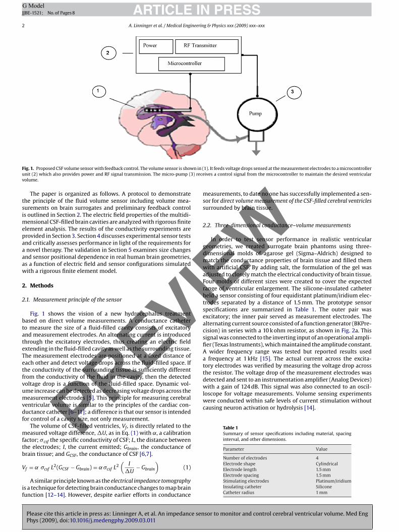
ig. 1. Proposed CSF volume sensor with feedback control. The volume sensor is shownit (2) which also provides power and RF signal transmission. The micro-pump (olume.
The paper is organized as follows. A protocol to demonstratehe principle of the fluid volume sensor including volume mea-urements on brain surrogates and preliminary feedback controls outlined in Section 2. The electric field properties of the multidi-
ensional CSF-filled brain cavities are analyzed with rigorous finitelement analysis. The results of the conductivity experiments arerovided in Section 3. Section 4 discusses experimental sensor testsnd critically assesses performance in light of the requirements fornovel therapy. The validation in Section 5 examines size changesnd sensor positional dependence in real human brain geometries,s a function of electric field and sensor configurations simulatedith a rigorous finite element model.
. Methods
.1. Measurement principle of the sensor
Fig. 1 shows the vision of a new hydrocephalus treatmentased on direct volume measurements. A conductance cathetero measure the size of a fluid-filled cavity consists of excitatorynd measurement electrodes. An alternating current is introducedhrough the excitatory electrodes, thus creating an electric fieldxtending in the fluid-filled cavity as well as the surrounding tissue.he measurement electrodes are positioned at a fixed distance ofach other and detect voltage drops across the fluid-filled space. Ifhe conductivity of the surrounding tissue is sufficiently differentrom the conductivity of the fluid in the cavity, then the detectedoltage drop is a function of the fluid-filled space. Dynamic vol-me increase can be detected as decreasing voltage drops across theeasurement electrodes [5]. This principle for measuring cerebral
entricular volume is similar to the principles of the cardiac con-uctance catheter [6–11]; a difference is that our sensor is intendedor control of a cavity size, not only measurement.
The volume of CSF-filled ventricles, Vf, is directly related to theeasured voltage difference, �U, as in Eq. (1) with ˛, a calibration
actor; �csf the specific conductivity of CSF; L, the distance betweenhe electrodes; I, the current emitted; Gbrain, the conductance ofrain tissue; and GCSF, the conductance of CSF [6,7].
( )
PREV
Please cite this article in press as: Linninger A, et al. An impedance senPhys (2009), doi:10.1016/j.medengphy.2009.03.011
f = ˛ �csf L2(GCSF − Gbrain) = ˛ �csf L2 I
�U− Gbrain (1)
A similar principle known as the electrical impedance tomographys a technique for detecting brain conductance changes to map brainunction [12–14]. However, despite earlier efforts in conductance
1). It feeds voltage drops sensed at the measurement electrodes to a microcontrollerives a control signal from the microcontroller to maintain the desired ventricular
measurements, to date no one has successfully implemented a sen-sor for direct volume measurement of the CSF-filled cerebral ventriclessurrounded by brain tissue.
2.2. Three-dimensional conductance–volume measurements
In order to test sensor performance in realistic ventriculargeometries, we created surrogate brain phantoms using three-dimensional molds of agarose gel (Sigma–Aldrich) designed tomatch the conductance properties of brain tissue and filled themwith artificial CSF. By adding salt, the formulation of the gel wasadjusted to closely match the electrical conductivity of brain tissue.Four molds of different sizes were created to cover the expectedrange of ventricular enlargement. The silicone-insulated catheterheld a sensor consisting of four equidistant platinum/iridium elec-trodes separated by a distance of 1.5 mm. The prototype sensorspecifications are summarized in Table 1. The outer pair wasexcitatory; the inner pair served as measurement electrodes. Thealternating current source consisted of a function generator (BKPre-cision) in series with a 10 k ohm resistor, as shown in Fig. 2a. Thissignal was connected to the inverting input of an operational ampli-fier (Texas Instruments), which maintained the amplitude constant.A wider frequency range was tested but reported results useda frequency at 1 kHz [15]. The actual current across the excita-tory electrodes was verified by measuring the voltage drop acrossthe resistor. The voltage drop of the measurement electrodes wasdetected and sent to an instrumentation amplifier (Analog Devices)
IEW
sor to monitor and control cerebral ventricular volume. Med Eng
Electrode length 1.5 mmElectrode spacing 1.5 mmStimulating electrodes Platinum/iridiumInsulating catheter SiliconeCatheter radius 1 mm

ARTICLE IN PRESSG ModelJJBE-1521; No. of Pages 8
A. Linninger et al. / Medical Engineering & Physics xxx (2009) xxx–xxx 3
F ts. Frc andT back c
2
twbioscripsfcptpwm
2
tufifiatottrobaAee
sue. In a set of separate experiments, the average conductivity of CSFwas found to be 2.01 S/m. This value is about twelve times higher thanthat of brain tissue. This contrast ratio between CSF and brain tissuewas found to be in good agreement with published data [16–18].
Table 2Boundary conditions and material properties used for computer simulations usingfinite element analysis.
I
ig. 2. Experimental setup of three-dimensional volume measurement experimenonductivity properties similar to the brain. B shows the conceptual circuit diagramhe reading was sent to a flow controller, FC, which controlled a micro-pump. Feed
.3. Experimental setting for the feedback control system
The feasibility of directly maintaining and restoring normal ven-ricular sizes was tested by means of a closed loop feedback systemith the volume sensor and a mini-pump actuator (Mk III, Hob-
ico Inc., Champaign, IL). The equivalent circuit diagram is shownn Fig. 2b. Input voltages ranging from 1 V to 5 V with a frequencyf 100 kHz generated an electric field of ∼0.0025 V/m in the fluidpace between the two measurement electrodes. 100 cc of artifi-ial CSF was initially filled into an elastic rubber balloon; a baselineeading for the calibration of the sensor was taken. Artificial CSFn the range of 5–350 cc volume was added by a second miniatureump to emulate CSF accumulation leading to ventricular expan-ion. The sensor processed voltage data with a sampling rate ofour readings per second to the controller. The digital controllerompared the actual volume with a desired set point to com-ute the desired control action. The controller output operatedhe pump actuator. Several control schemes including proportional,roportional integral, as well as on–off control were implementedith the virtual instrument panel of LabVIEW (National Instru-ents).
.4. Computer-aided sensor optimization
In order to estimate leakage currents and optimal distance ofhe sensor electrodes as well as to assess the dependence of vol-me measurements as a function of catheter position, a rigorousnite element analysis was conducted. The solution of the scalareld equations, V(−→x ), in the cavity and surrounding brain tissuellowed calculation of the equipotential lines and the prediction ofhe voltage drop at the measurement electrodes for any geometryf the cavities or different catheter configuration. In the simula-ions, electrical properties of fluid and brain tissue were taken fromhe experiments described above. The measured electric potentialesided in a computational domain with the electrical conductivityf the CSF, �1 = 2.01 S/m, and �2 = 0.172 S/m for the brain. Neumann
PREV
Please cite this article in press as: Linninger A, et al. An impedance senPhys (2009), doi:10.1016/j.medengphy.2009.03.011
oundary conditions as in Eq. (2) and in Table 2 were applied withcurrent density of 16.66 A/m at the upper excitatory electrode,, while the lower electrode, D, was grounded. The measurementlectrodes were represented as surfaces B and C; the measurementlectrodes were considered perfectly insulated. The finite element
ame A shows a CSF-filled three-dimensional cavity molded into agarose gel withthe control scheme for controlling the volume of fluid contained within the cavity.ontrol was implemented using an on–off algorithm and a flow transmitter, FT.
model in Eq. (2) was solved with ADINA [20].
�−→∇ 2
V(−→x ) = 0 (2)
with −→n · −→J = �
−→∇ V(−→x ) = 16.66 A/m2 at the excitatory electrodeA; V = 0 at electrode D.
3. Results
3.1. Static conductance–volume measurements
Before animal or human experimentation, we wanted to opti-mize the electronic properties and design parameters of the novelvolume sensor using expedient bench-testing on brain surrogateswith realistic geometry and electronic properties equal to realbrain tissue. In a series of experiments, we exactly determined theamount of salt needed to obtain the same contrast ratio of conduc-tivity between artificial CSF and gel as found between artificial CSFand real brain tissue. To emulate the complex shape of real ventri-cles in human brains, we cast three-dimensional ellipsoidal cavitieswith realistic shapes depicted in Fig. 3.
3.2. Dog brain conductivity experiments
Static experiments were performed to validate the sensor prin-ciple in real brain tissues. These experiments provided a means tovalidate the electrical conductivity ratio between CSF and brain tis-
EW
sor to monitor and control cerebral ventricular volume. Med Eng
Condition Gel phantom cavity Human brain hydrocephalus
Neumann −→n · −→J ∈ SA.D
−→n · −→J = 16.66 A/m2 −→n · −→
J = 0.88 A/m2
Dirichlet V = SB,C V = 0 V V = 0 VBrain tissue conductivity 0.172 S/m 0.172 S/mCSF conductivity 2.01 S/m 2.01 S/m

ARTICLE IN PRESSG ModelJJBE-1521; No. of Pages 8
4 A. Linninger et al. / Medical Engineering & Physics xxx (2009) xxx–xxx
F m 2 mo ms anc tial di
3
iaec1rmdt1c
dflvb
ume is reached in spite of the measurement inaccuracy. For control
Fres
ig. 3. A series of agarose gel cavities (Frames A–D) with increasing cavity size frof the cavity. Simulations of the electric field and conductance of the brain phantourrent density, represented as arrows in E–H, demonstrates that the voltage poten
.3. Monitoring and feedback control of ventricular sizes
The sensor performance in dynamically enlarging elastic cav-ties was examined. We used elastic rubber balloons to monitornd control the exact dynamic enlargement in bench-scalexperiments. Sensor calibration was performed through staticonductance–volume experiments as described in Section 2. Up to00 cc of artificial CSF were added and the total volume of fluid wasecorded and measured every 5 s. Through the LabVIEW controlodule, the sensor was able to measure and store the measurement
uring the fluid addition. Fig. 4a compares the volume inferred fromhe sensor reading with the actual fluid volume over a period of00 s. Speed and accuracy of the sensor in detecting the volumehange is suitable for active feedback control described next.
Closed-loop feedback control experiments were conducted toV
Please cite this article in press as: Linninger A, et al. An impedance senPhys (2009), doi:10.1016/j.medengphy.2009.03.011
ynamically measure and actively control the size of distensibleuid-filled spaces with feedback control. The enlargement of theentricle as it occurs in hydrocephalus was simulated as a distur-ance caused by fluid addition. The controller was programmed
ig. 4. Dynamic fluid volume measurements. Comparison of actual volume with volumesults demonstrate that fast volume expansion was detected reasonably well by the sensnlargement. The controller successfully maintained the volume at a desired volume by diimple on–off control scheme maintains the set point, but exhibits characteristic variatio
PRE
L to 16 mL. The volume sensor was placed vertically, collinear with the main axisd CSF were conducted using finite element analysis in Frames E–H. The simulatedfference strongly correlates with the cavity volume.
to operate the pump to remove the excess fluid and to restorethe volume desired set point. The fluid volume measurementswere automatically stored and sent to the controller four timesper second. The actual volume of balloon cavity was also carefullymeasured to quantify the sensor performance and the controllerresponse. The fluid volume was initially maintained at 150 cc forthe first 10 s. This phase was followed by CSF accumulation leadingto ventricular enlargement through the addition of 150–220 cc offluid over the next 10 s. The controller was then switched on and itsdynamic response activated the pump to restore the set point vol-ume of 150 cc in about 20 s. A typical controller response is shownin Fig. 4b. The readings show a slight offset between the actualand measured values. The simple on–off controller also indicatesa wavy response as expected. However, the desired set point vol-
IEW
sor to monitor and control cerebral ventricular volume. Med Eng
of ventricular sizes in hydrocephalus, only the restoration of thedesired ventricular size is important; larger measurement errorsin high volume ranges do not affect the accuracy close to the setpoint.
e sensing in elastic balloons in a dynamic experiment is shown in Frame A. Theseor. Frame B shows the response of the feedback controller to simulated ventricularscharging excess CSF through a micro-pump using an on–off control algorithm. Thens due to the simple control law.
ARTICLE IN PRESSG ModelJJBE-1521; No. of Pages 8
A. Linninger et al. / Medical Engineering & Physics xxx (2009) xxx–xxx 5
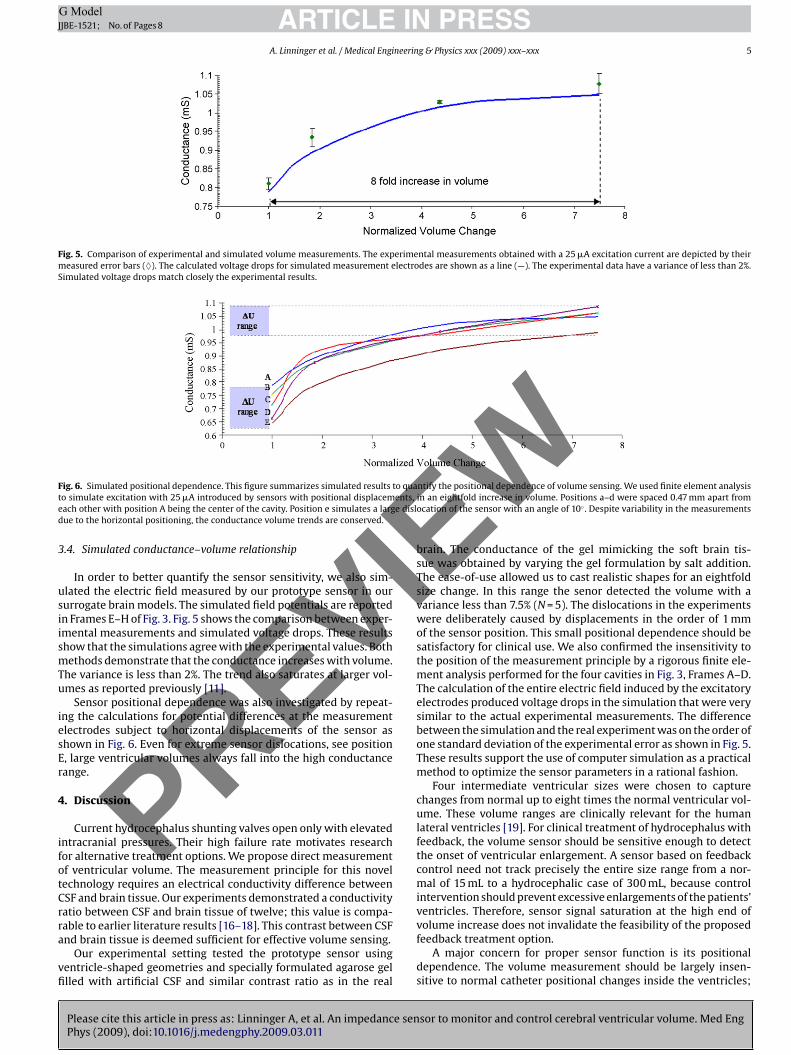
Fig. 5. Comparison of experimental and simulated volume measurements. The experimental measurements obtained with a 25 �A excitation current are depicted by theirmeasured error bars (♦). The calculated voltage drops for simulated measurement electrodes are shown as a line (—). The experimental data have a variance of less than 2%.Simulated voltage drops match closely the experimental results.
F to quat ents,e e disld
3
usiismTu
iesEr
4
ifotCrra
vfi
I
ig. 6. Simulated positional dependence. This figure summarizes simulated resultso simulate excitation with 25 �A introduced by sensors with positional displacemach other with position A being the center of the cavity. Position e simulates a largue to the horizontal positioning, the conductance volume trends are conserved..4. Simulated conductance–volume relationship
In order to better quantify the sensor sensitivity, we also sim-lated the electric field measured by our prototype sensor in oururrogate brain models. The simulated field potentials are reportedn Frames E–H of Fig. 3. Fig. 5 shows the comparison between exper-mental measurements and simulated voltage drops. These resultshow that the simulations agree with the experimental values. Bothethods demonstrate that the conductance increases with volume.
he variance is less than 2%. The trend also saturates at larger vol-mes as reported previously [11].
Sensor positional dependence was also investigated by repeat-ng the calculations for potential differences at the measurementlectrodes subject to horizontal displacements of the sensor ashown in Fig. 6. Even for extreme sensor dislocations, see position, large ventricular volumes always fall into the high conductanceange.
. Discussion
Current hydrocephalus shunting valves open only with elevatedntracranial pressures. Their high failure rate motivates researchor alternative treatment options. We propose direct measurementf ventricular volume. The measurement principle for this novelechnology requires an electrical conductivity difference betweenSF and brain tissue. Our experiments demonstrated a conductivityatio between CSF and brain tissue of twelve; this value is compa-
PREV
Please cite this article in press as: Linninger A, et al. An impedance senPhys (2009), doi:10.1016/j.medengphy.2009.03.011
able to earlier literature results [16–18]. This contrast between CSFnd brain tissue is deemed sufficient for effective volume sensing.
Our experimental setting tested the prototype sensor usingentricle-shaped geometries and specially formulated agarose gellled with artificial CSF and similar contrast ratio as in the real
ntify the positional dependence of volume sensing. We used finite element analysisin an eightfold increase in volume. Positions a–d were spaced 0.47 mm apart fromocation of the sensor with an angle of 10◦ . Despite variability in the measurements
brain. The conductance of the gel mimicking the soft brain tis-sue was obtained by varying the gel formulation by salt addition.The ease-of-use allowed us to cast realistic shapes for an eightfoldsize change. In this range the senor detected the volume with avariance less than 7.5% (N = 5). The dislocations in the experimentswere deliberately caused by displacements in the order of 1 mmof the sensor position. This small positional dependence should besatisfactory for clinical use. We also confirmed the insensitivity tothe position of the measurement principle by a rigorous finite ele-ment analysis performed for the four cavities in Fig. 3, Frames A–D.The calculation of the entire electric field induced by the excitatoryelectrodes produced voltage drops in the simulation that were verysimilar to the actual experimental measurements. The differencebetween the simulation and the real experiment was on the order ofone standard deviation of the experimental error as shown in Fig. 5.These results support the use of computer simulation as a practicalmethod to optimize the sensor parameters in a rational fashion.
Four intermediate ventricular sizes were chosen to capturechanges from normal up to eight times the normal ventricular vol-ume. These volume ranges are clinically relevant for the humanlateral ventricles [19]. For clinical treatment of hydrocephalus withfeedback, the volume sensor should be sensitive enough to detectthe onset of ventricular enlargement. A sensor based on feedbackcontrol need not track precisely the entire size range from a nor-mal of 15 mL to a hydrocephalic case of 300 mL, because controlintervention should prevent excessive enlargements of the patients’ventricles. Therefore, sensor signal saturation at the high end of
EW
sor to monitor and control cerebral ventricular volume. Med Eng
volume increase does not invalidate the feasibility of the proposedfeedback treatment option.
A major concern for proper sensor function is its positionaldependence. The volume measurement should be largely insen-sitive to normal catheter positional changes inside the ventricles;

INJ
6 neerin
rroodTttprwwc
tvidttAsrb
mwwapdtc
poWadewdi
Fits
ARTICLEG ModelJBE-1521; No. of Pages 8
A. Linninger et al. / Medical Engi
easonable dislocations should have a modest effect on the volumeeading. A rigorous estimate of expected positional dependencef the volume sensor technology inside an expanding ventricle inur surrogates reported in Fig. 6 showed that positional depen-ence was mild even with a single pair of measurement electrodes.he sensor design utilizes a ring electrode, thus making insensi-ive measurements of directional changes in volume. This supportshe mild dependence on catheter position. Initially, the sensor waslaced in the center of the cavity [8–11]. The experiments wereepeated by positioning the sensor gradually closer to the cavityall to assess positional dependency. Future sensor designs couldarrant an array of planar measurement electrodes if directional
hanges in volume are of interest.The envisioned feedback treatment was implemented concep-
ually to demonstrate the feasibility of maintaining desired fluidolume with the help of feedback based on dynamic volume sens-ng. A simple on–off control scheme was successful in restoringesired set point volume in response to CSF accumulation dis-urbance. In the experiment, CSF was inserted by a second pumpo emulate pathological CSF accumulation causing hydrocephalus.lthough the actual volume exhibited some fluctuation due to theimplicity of the control scheme, the sensor performed satisfacto-ily. The oscillations in the tracking response can be reduced easilyy implementing more advanced control schemes.
Saturation effects can be drastically reduced by adding moreeasurement electrodes with advanced calibration; an effort weill investigate in future work. The feedback control experimentsere performed with a higher excitation frequency than the
garose gel experiments. Frequency dependence was measured inrevious studies, and a frequency that provided the largest con-uctance measurement was implemented in our balloon model. Inhe agarose gel model, we used a lower frequency due to the largeonductivity ratio.
Our work is to improve existing therapy for hydrocephalicatients. However, there are limitations of our proposed technol-gy. Our sensor is intended for implantation into a single ventricle.e have focused on a design specifically for the lateral ventricle
nd frontal horn. Surgeons may want to implant the sensor into a
Please cite this article in press as: Linninger A, et al. An impedance senPhys (2009), doi:10.1016/j.medengphy.2009.03.011
ifferent part of the ventricular system that does not have as muchxpansion. The limitations on any particular site may be resolvedith patient-specific sensor design based on CT and MRI data. Ourevice is intended to reduce the number of shunt revisions. Exist-
ng shunts may malfunction due to excessive CSF removal, causing
ig. 7. Case study of conductance changes during the transition of hydrocephalus usingntermediate stages B–D (not shown) were artificially created by interpolation to emulateaken as size changes of the lateral ventricle with known volumes of the normal and hydrensor positions located between 1 and 5 mm away from the central axis. An eightfold in
PREV
PRESSg & Physics xxx (2009) xxx–xxx
over drainage with ventricular collapse. The proposed sensor needsto be tested clinically, to see if it prevents this from occurring.
In this article, we have assumed that the conductivity of CSF andbrain tissue remain constant throughout hydrocephalus. However,CSF conductivity might change due to hemorrhage or infection.The large contrast between CSF and brain tissue will diminish thechange in current density within the cavity if this occurs. There maybe a small percentage of leakage current as discussed earlier, butour calculations suggests its impact to be small compared to theconductance measurement due to volume enlargement. Our finiteelement model enables us to assess the effect of varying systemparameters such conductivity properties or leakage scenarios onsensor accuracy.
This work has focused on volume changes that occur in the ven-tricles. In instances such as normal pressure hydrocephalus, thesensor may not be useful if the ventricles do not change size. Never-theless, improvements to sensor design and instrumentation mayallow for greater sensitivity. Potentially, the sensor might be ableto detect periodic changes in the ventricular volume due to pulsepressure changes in the ventricles. This would reflect brain compli-ance, which changes in normal pressure hydrocephalus. Detectingof brain compliance changes might be another useful future appli-cation for the sensor technology introduced in this article.
5. Validation of sensor performance in simulated normaland hydrocephalic subjects
Even though at the early stages of the project we cannot justifyexperimentation on human subjects, we wished to assess theexpected sensor performance and positional dependence in realhuman brain geometries. To realize this assessment without harmto patients, we performed a rigorous finite element analysis of theinduced fields of different sensor configurations on real patientventricular models. The computations helped optimize our sensordesign parameters for best performance in hydrocephalus humans.Frame A of Fig. 7 shows normal ventricular spaces in a 32-year-oldnormal subject. An image of a fully developed hydrocephalicbrain is shown in Frame D. It was obtained from a 62-year-oldEW
sor to monitor and control cerebral ventricular volume. Med Eng
patient suffering from communicating hydrocephalus. The patientdata were collected according to IRB protocol at the Universityof Illinois at Chicago and the University of Chicago. The imageswere reconstructed using image reconstruction tools [21]. Theventricular sizes were determined using a snake algorithm where
our prototype design in simulations using finite element analysis. The sizes of thetransitions from normal to hydrocephalus. The projections of volume changes wereocephalic lateral ventricle. The simulations were repeated five times with differentcrease from normal size correlates with our experimental and simulated analysis.
I
ARTICLE IN PRESSG ModelJJBE-1521; No. of Pages 8
A. Linninger et al. / Medical Engineering & Physics xxx (2009) xxx–xxx 7
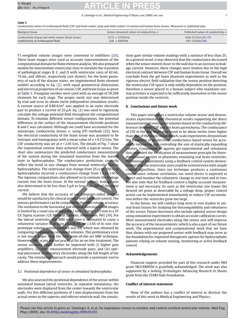
Table 3Conductivity values of cerebrospinal fluid (CSF) and brain matter (gray and white matter) in animal and human brain tissues. Measured vs. published data.
Biological tissue Sensor measured values of conductivity, � Published values of conductivity, �
C .078 SC .10 S/m
TTcmo7emaiebApcddBatiCtcosrhhTcac
wsTiGtvpciHsamca
5
sewa
I
onductivity of gray and white matter (brain tissue) 0.172 ± 0onductivity of cerebrospinal fluid 2.01 ± 0
1-weighted volume images were converted to milliliters [23].hese brain images were used as accurate representations of theomputational domain for finite element analysis. We also preparedodels for intermediate ventricular sizes to simulate the transition
f pathological stages B, C, and D with ventricular sizes of 42 mL,5 mL, and 160 mL, respectively (not shown). For the brain geom-try of each of the disease states, we implemented finite elementodels according to Eq. (2), with equal geometrical dimensions
nd electrical properties of our sensor, CSF, and brain tissue as givenn Table 3. Triangular meshes were used with an average of 59,568lements for each stage. The proper mesh size was determinedy trial and error to obtain mesh independent simulation results.current source of 0.88 A/m2 was applied to an outer electrode
air to produce a current of 25 �A. Eq. (2) was used to rigorouslyalculate the voltage potential field throughout the computationalomain. To emulate different sensor configurations, the potentialifference at the surface of the measurement electrode positionsand C was calculated. Although we could have accounted for the
nisotropic conductivity tensor, �, using DTI methods [22], herehe electrical conductivity of the brain tissue was assumed to besotropic and homogenous with a mean value of � = 0.172 S/m. TheSF conductivity was set at � = 2.01 S/m. The details of Fig. 7 showhe isopotential contour lines achieved with a typical sensor. Thehart also summarizes the predicted conductance measurementf the system during the simulated transition from the normaltate to hydrocephalus. The conductance predictions correctlyeflect the trend in size changes from normal ventricular size toydrocephalus. The ventricular size change from 25 cc to 250 cc inydrocephalus incurred a conductance change from 1 S to 1.45 S.he rigorous computations also allowed us to estimate the leakageurrent into the brain tissue. In the normal subject, leakage waslso determined to be less than 5 �A or less than 20% of the inputurrent.
We believe that the accuracy of our computer-aided analysisould be satisfactory for clinical sensors with feedback control. The
ensors performance can be compared to the MRI volume accuracy.he resolution in the horizontal and vertical direction in MR imagess limited by a voxel size of typically 1 mm × 1 mm × 1.5 mm in a 3TE Sigma scanner (GE Medical Systems, Milwaukee, WI) [19]. For
he lateral ventricles, this MRI resolution is estimated to cause aolumetric variance of at least ±0.276 mL, or ±3.6% of its size. Ourrototype volume sensor variance was 8%, which was obtained byomparing measured to calculated volumes. This preliminary errors not much larger than the best state-of-the-art MRI technique.owever, MRI scans are not practical for an on-line treatment. The
ensor accuracy could further be improved with (i) higher gainmplifiers, (ii) more measurement electrode pairs, and (iii) opti-al placement of excitatory electrodes along the full length of the
avity. The simulation approach would provide a systematic tool toddress these improvements.
.1. Positional dependence of sensor in simulated hydrocephalus
PREV
Please cite this article in press as: Linninger A, et al. An impedance senPhys (2009), doi:10.1016/j.medengphy.2009.03.011
We also assessed the positional dependence of the sensor withinimulated human lateral ventricles. In repeated simulations, thelectrodes were displaced from the center towards the ventricularalls. For five different positions of 1 mm displacements from the
ctual center to the superior and inferior ventricle wall, the simula-
/m 0.06–0.3 S/m [15–17]1.81–2.0 S/m [18]
tions gave similar volume readings with a variance of less than 2%.As a general trend, it was observed that the conductance decreasedwhen the sensor moved closer to the wall due to an increase in leak-age current. However, these changes were modest due to the highelectrical contrast between CSF and human brain tissue. Overall weconclude from the gel brain phantom experiments as well as therigorous electric field validation that the sensor position detectingthe ventricular CSF space is only mildly dependent on the position,therefore a sensor placed in a human subject who maintains nor-mal activities is expected to be sufficiently insensitive to the sensorposition inside the ventricle.
6. Conclusions and future work
This paper introduces a ventricular volume sensor and demon-strates experimental and theoretical results supporting the directmeasurement of ventricular volumes based on the conductivity dif-ferences between CSF and the brain parenchyma. The conductanceof CSF in the dog brain was found to be about twelve times higherthan that of the brain tissue. Bench-scale experiments demonstratethe feasibility of an electrical conductance-based sensor for accu-rately measuring and controlling the size of elastically expandingcavities. Comparison of agarose gel experimental and simulationdata provided the verification of measurement principle for three-dimensional cavities in phantoms emulating real brain ventricles.Preliminary experiments using a feedback control system demon-strated that the ventricular sizes could be restored even with simplecontrol algorithms. Since our technique produces a continuousconductance–volume correlation, our novel device is expected todetect and monitor the volumetric change in real time and in vivo.We also note that for feedback control a highly accurate measure-ment is not necessary. As soon as the ventricular size leaves thedesired set point as detectable by a voltage drop, proper controlaction can be implemented immediately to reduce CSF accumula-tion before the ventricles grow too large.
In the future, we will conduct long term in vivo studies in ani-mal brain tissues for studying the biocompatibility and robustnessof the sensor. Future directions also include optimal sensor designusing simulation experiments to obtain accurate calibration curves.More measurement electrodes along the sensor axis will improvethe accuracy of the measurements, which is also a part of our futurework. The experimental and computational work that we havethus shown with our proposed sensor with feedback may serve asthe foundation for improved therapeutic options for hydrocephalicpatients relying on volume sensing, monitoring or active feedbackcontrol.
Acknowledgements
Financial support provided for part of this research under NIHgrant 5R21EB4956 is gratefully acknowledged. The work was alsosupported by a Seeking Technologies Advancing Research in Shuntsgrant from the STARS Kids Foundation.
EW
sor to monitor and control cerebral ventricular volume. Med Eng
Conflict of interest statement
None of the authors has a conflict of interest to disclose theresults of this work to Medical Engineering and Physics.

INJ
8 neerin
R
[
[
[[
ARTICLEG ModelJBE-1521; No. of Pages 8
A. Linninger et al. / Medical Engi
eferences
[1] Smith JJ, Kampine JP. Circulatory physiology—the essentials. 2nd edn Baltimore:Williams & Wilkins; 1984. p. 180.
[2] Patwardhan RV, Nanda A. Implanted ventricular shunts in the United States: thebillion-dollar-a-year cost of hydrocephalus treatment. J Neurosurg 2005;56(1):139–45.
[3] Youmans JR. Neurological surgery: a comprehensive reference guide to thediagnosis and management of neurosurgical problems. 4th edn Philadelphia:Saunders; 1996. p. 890.
[4] Hydrocephalus Shunts. Shunts-treatment for hydrocephalus presented byinstitute from Neurology and Neurosurgery in New York City. NY: Hyman-Newman Institute for Neurology and Neurosurgery. Retrieved March 23, 2006,from http://nyneurosurgery.org/hydro shunt.html.
[5] Linninger A. Monitoring and controlling hydrocephalus. US Patent 005440 (10Jan. 2008).
[6] Baan J, Van der Velde ET, De Bruin HG, Smeenk GJ, Koops J, van Dijk AD, et al.Continuous measurement of left ventricular volume in animals and humans byconductance catheter. Circulation 1984;70:812–23.
[7] Burkhoff D, Van der Velde E, Kass D, Baan J, Maughan WL, Sagawa K. Accuracyof volume measurement by conductance catheter in isolated, ejecting caninehearts. Circulation 1985;72:440–7.
[8] Eriksson AB, Kronander H, Söderqvist E, Vaage J, Brodin LÅ. Correlation betweena mid-ventricular volume segment and global left ventricular volume measuredby the conductance catheter. Scand Cardiovasc J 2001;35:129–35.
[9] Wu CC, Schwenk TC, Mahler CM, Anne A, Finnerty PW, Haber HL, et al. Accu-
Please cite this article in press as: Linninger A, et al. An impedance senPhys (2009), doi:10.1016/j.medengphy.2009.03.011
racy of the conductance catheter for measurement of ventricular volumes seenclinically: effects of electric field homogeneity and parallel conductance. IEEETrans Biomed Eng 1997;44(4):266–77.
10] Steendijk P, Tulner SAF, Wiemer M, Bleasdale RA, Bax JJ, van der Wall EE, etal. Pressure–volume measurements by conductance catheter during cardiacresynchronization therapy. Eur Heart J Suppl 2004;6(Suppl D):D35–42.
[
[
PREV
PRESSg & Physics xxx (2009) xxx–xxx
[11] Wei C, Valvano J, Feldman M, Pearce J. Nonlinear conductance–volume relation-ship for murine conductance catheter measurement system. IEEE Trans BiomedEng 2005;52(10):1654–61.
12] Tidswell T, Gibson A, Bayford R, Holder D. Three-dimensional electricalimpedance tomography of human brain activity. NeuroImage 2001;13:283–94.
[13] Romsauerova A, McEwan A, Horesh L, Yerworth R, Bayford R. Multi-frequencyelectrical impedance tomography (EIT) of the human head: initial findings inbrain tumors, arteriovenous malformations and chronic stroke, developmentof an analysis method and calibration. Physiol Measure 2006;27:S147–61.
[14] Gilad O, Horesh L, Holder DS. Design of electrodes and current limits for lowfrequency electrical impedance tomography of the brain. Int Fed Med Biol Eng2007;45:621–33.
[15] Tsai J, Cao H, Tungjitkusolmun S, Woo E, Vorperian V, Webster J. Dependenceof apparent resistance of four-electrode probes on insertion depth. IEEE TransBiomed Eng 2000;47(1):41–8.
[16] Duck FA. Physical properties of tissue: a comprehensive reference book. Aca-demic Press, Harcourt Brace Jovanovich, Publishers; 1990.
[17] Foster KR, Schwan HP. Dielectric properties of tissues and biological materials:a critical review. Crit Rev Biomed Eng 1989;17(1):25–104.
[18] Schwan P. Linear and nonlinear electrode polarization and biological materials.Ann Biomed Eng 1992;20:269–88.
[19] Linninger A, Xenos M, Zhu D, Somayaji MR, Kondapalli S, Penn RD. Cerebrospinalfluid flow in the normal and hydrocephalic human brain. IEEE Trans Biomed Eng2007;54(2):291–302.
20] ADINA R&D Inc. 2008; http://www.adina.com/index.html.21] Materialize Inc. (MIMICS). 2008; http://www.materialise.be/mimics/main
sor to monitor and control cerebral ventricular volume. Med Eng
ENG.html.22] Linninger A, Somayaji MR, Mekarski M, Zhang L. Prediction of convection-
enhanced drug delivery to the human brain. J Theor Biol 2008;250:125–38.23] Zhu D, Xenos M, Linninger A, Penn R. Dynamics of lateral ventricle and cere-
brospinal fluid in normal and hydrocephalic brains. J Magn Reson Imaging2006;24:756–70.
IEW