g-csf protects motoneurons against axotomy-induced apoptotic death in neonatal mice
TRANSCRIPT

RESEARCH ARTICLE Open Access
G-CSF protects motoneurons against axotomy-induced apoptotic death in neonatal miceAlexandre Henriques1,2,3, Claudia Pitzer1*, Luc Dupuis2,3, Armin Schneider1*
Abstract
Background: Granulocyte colony stimulating factor (G-CSF) is a growth factor essential for generation ofneutrophilic granulocytes. Apart from this hematopoietic function, we have recently uncovered potentneuroprotective and regenerative properties of G-CSF in the central nervous system (CNS). The G-CSF receptorand G-CSF itself are expressed in a motoneurons, G-CSF protects motoneurons, and improves outcome in theSOD1(G93A) transgenic mouse model for amyotrophic lateral sclerosis (ALS). In vitro, G-CSF acts anti-apoptotically on motoneuronal cells. Due to the pleiotrophic effects of G-CSF and the complexity of the SOD1transgenic ALS models it was however not possible to clearly distinguish between directly mediated anti-apoptotic and indirectly protective effects on motoneurons. Here we studied whether G-CSF is able to protectmotoneurons from purely apoptotic cell death induced by a monocausal paradigm, neonatal sciatic nerveaxotomy.
Results: We performed sciatic nerve axotomy in neonatal mice overexpressing G-CSF in the CNS and found thatG-CSF transgenic mice displayed significantly higher numbers of surviving lumbar motoneurons 4 days followingaxotomy than their littermate controls. Also, surviving motoneurons in G-CSF overexpressing animals were larger,suggesting additional trophic effects of this growth factor.
Conclusions: In this model of pure apoptotic cell death the protective effects of G-CSF indicate direct actions ofG-CSF on motoneurons in vivo. This shows that G-CSF exerts potent anti-apoptotic activities towards motoneuronsin vivo and suggests that the protection offered by G-CSF in ALS mouse models is due to its directneuroprotective activity.
BackgroundGranulocyte-colony-stimulating factor is a cytokinethat stimulates the proliferation and the differentiationof myeloid precursors [1] and has been in clinical usefor more than 10 years in indications related to coun-teracting chemotherapy-induced neutropenia or forbone-marrow transplantations [2]. We and others dis-covered that G-CSF also acts as a growth factor in thebrain, and shows protective and regenerative proper-ties in a number of CNS disease models [3-6]. Themechanisms leading to these beneficial effects likelyinclude a combination of anti-apoptotic activity onneurons [5], stimulation of neurogenesis [5], enhance-ment of vessel formation [3], mobilization of bonemarrow derived cells [4] and systemic anti-inflamma-tory effects [5]. Recently we described that G-CSF was
protective in a mouse model of amyotrophic lateralsclerosis (ALS), the major adult-onset motoneurondisease. G-CSF treatment increased survival andvolume of spinal a motoneurons and protected moto-neuronal cell lines (NSC34) in vitro against apoptoticstimuli [6]. Importantly, while adult motoneurons pro-minently express the receptor for G-CSF [6,7] our stu-dies did not unequivocally show that G-CSF exertedprotection in this ALS model through its neuroprotec-tive activity.Here, we sought to determine whether G-CSF was
able to protect motoneurons in vivo in a model ofneonatal sciatic nerve axotomy. This experimentalparadigm has been largely described, is thought to relyalmost exclusively on the apoptotic machinery [8], andprovides a powerful tool to study the neuroprotectiveactivity of growth factors towards motoneurons in vivo[4,5,9-13].* Correspondence: [email protected]; [email protected]
1SYGNIS Bioscience, Im Neuenheimer Feld 515, 69120 Heidelberg, Germany
Henriques et al. BMC Neuroscience 2010, 11:25http://www.biomedcentral.com/1471-2202/11/25
© 2010 Henriques et al; licensee BioMed Central Ltd. This is an Open Access article distributed under the terms of the CreativeCommons Attribution License (http://creativecommons.org/licenses/by/2.0), which permits unrestricted use, distribution, andreproduction in any medium, provided the original work is properly cited.

ResultsThe G-CSF receptor is expressed on neonatalmotoneurons, and induced by axotomyWe have previously shown that the receptor of G-CSF(G-CSFR) is expressed on motoneurons in the adultspinal cord. We first determined whether the G-CSFreceptor was expressed on motoneurons during earlypostnatal development. At postnatal day 9 (P9) wenoted expression of the receptor on a motoneurons inthe spinal cord ventral horn, identified by the followingcriteria: Choline-Acetyltransferase (CHAT) expression,an identifiable nucleolus, and a size larger than 300 μm2
in the horizontal dimension (Figure 1). Invariably, allmotoneurons defined by these criteria expressed thereceptor.The expression of G-CSF receptor is induced by neu-
rons under neurodegenerative conditions such as aftercerebral ischemia or in ALS, presumably as an endogen-ous protective response [6,7]. We therefore examinedexpression of the G-CSF receptor by quantitative PCRof whole spinal cords 4 days following neonatal axotomyof the right sciatic nerve (i.e. on postnatal day 9).Indeed, we found that G-CSFR expression in the wholespinal cord increased by 55% (p < 0.005) (Figure 2A).To clarify whether this induction likely originated
from the injured motoneurons, we quantified fluorescentstaining intensities of ipsi- and contralateral motoneur-ons from the lumbar spinal cord region. We detectedsignificantly stronger protein expression on ipsi- versuscontralateral motoneurons, suggesting that axotomizedmotoneurons upregulate the receptor in response toinjury (p < 0,05) (Figure 2B).In conclusion, the G-CSF receptor is expressed at the
neonatal stage and its expression is increased inresponse to axotomy.
CNS- targeted overexpression of G-CSF protectsmotoneurons against cell death in neonatal pupsNeonatal motoneuron death following sciatic nerveaxotomy constitutes a widely used and well- establishedmodel of apoptotic motoneuron death that has beenused to examine protective effects of trophic factors.We chose to use transgenic mice that overexpress G-
CSF in the CNS as an endogenous “delivery system” tostudy effects of increased concentration of G-CSF onthe lesioned motoneurons. We used transgenic miceharbouring a bidirectional tta-responsive constructwith expression cassettes for murine G-CSF and EYFP(pBEG). These mice were crossed with mice expressingthe Tet-transactivator under control of the CaMKII a;-promoter, similar to the approach used in Pitzer et al.except for the use of the Thy1-driver used therein [7].In the following we compare three types of transgene
combinations in littermates derived from crosses ofmice heterozygous for the transgene CaMKII-tTA andheterozygous for pBEG. The three groups are a.) micenot harbouring any transgene ("wt”), b.) mice harbour-ing only the BEG2 transgene (which do not expressG-CSF), and c.) mice harbouring both the BEG2 trans-gene and the CaMKII-tTA driver, that do expressG-CSF.
merge
G-CSFr
G-CSFR
CHAT
A
B
C
Figure 1 The G-CSF receptor is expressed by motoneurons inthe spinal cord of wt neonatal mice. (A-C) Double fluorescenceimmunostaining of G-CSF receptor and CHAT (cholineacetyltransferase) in the neonatal mouse spinal cord ventral horn.CHAT is used as a marker for motoneurons. All defined a motoneuronsexpress G-CSFR (indicated by an arrow). All photomicrographs with40× original magnification (OM), size bar 25 μm.
Henriques et al. BMC Neuroscience 2010, 11:25http://www.biomedcentral.com/1471-2202/11/25
Page 2 of 9

Although expression via the CaMKII driver in ourtransgenes is mainly in the forebrain, release of G-CSFin the spinal cord is expected from long corticospinalaxonal connections. Indeed, the CaMKIIa;-promoterdirected G-CSF overexpression in the forebrain leads toa strong elevation of G-CSF levels in the neonatal brainand spinal cord (P9; 28.00 ± 11.60 vs 0.24 ± 0.23 pg/mgtissue protein; p < 0.05) (Figure 3).
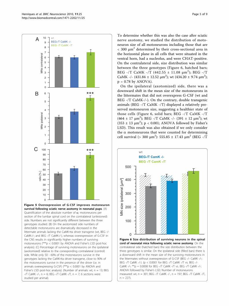
Offspring from crosses of BEG and CaMK-tta trans-genic mice were subjected to complete unilateral sciaticnerve axotomy at postnatal day 5 (P5). We analyzedeffects on motoneuron survival in the lumbar section ofthe spinal cord (L4/5 levels) after 4 days at P9. Examplesof the histological appearance of CHAT positive cells inthe ventral horn of the lumbar spinal cord of neonatalmice after sciatic nerve axotomy are given in figure 4.Our axotomy model results in a loss of approximately40 - 50% of motoneurons 4 days after the injury (Figure4A and 4B; Figure 5A and 5B), a result consistent withother studies [8,9]. Already apparent from the histologi-cal sections was that clearly more motoneurons survivein mice with the G CSF transgene activated by theCaMK-tta driver (BEG -/T CaMK -/T) (compare figure4B and 4F).We then quantified numbers of motoneurons in the
lumbar spinal cord of P9 neonatal mice contra- andipsilateral to the side of axotomy. The a motoneuronswere defined as (i) being situated in the ventral horn ofthe lumbar region; (ii) having a clearly identifiablenucleolus and staining positive for CHAT and (iii) hav-ing a minimum cross-sectional area of 300 μm2.There was no difference between the absolute num-
bers of motoneurons on the contralateral side betweenthe three genotypes used, although there was a non-sig-nificant trend for slightly higher numbers in the double-transgenic group (BEG -/T CaMK-/T (7.03 ± 0.39 moto-neurons/section) vs. BEG -/T CaMK-/- (5.98 ± 0.38) and
A
B
0
10
20
30
40
50
60
Contra Ipsi
Inte
gra
ted
den
sity
/ are
a
0
1
2
Control Axotomy
Fo
ldin
du
ctio
n(r
el. u
nit
s)
*
**
Figure 2 G-CSF receptor expression after sciatic nerveaxotomy. (A) G-CSF receptor is upregulated after sciatic nerveaxotomy in spinal cord of neonatal mice. Quantitative PCR for G-CSFreceptor of spinal cords of axotomized or not neonatal mice(induction of 55%; n = 3, **p < 0.005). (B) G-CSF receptor stainingintensity is stronger on the motoneurons after axotomy. Fluorescentintensity of G-CSF receptor staining on motoneurons was calculatedon the ipsi- and contralateral side with an image processingprogram (*p < 0.05, n = 49).
Figure 3 G-CSF concentration in the CNS. Levels of G-CSF arehigher in the brain and in the spinal cord of mice overexpressingG-CSF (BEG -/T CaMK -/T) compared to wild type littermate controls(BEG -/- CaMK -/-). G-CSF proteins were quantified by ELISA withprotein lysates from brain or spinal cord (n = 3, *p < 0.05).
Henriques et al. BMC Neuroscience 2010, 11:25http://www.biomedcentral.com/1471-2202/11/25
Page 3 of 9

wt (6.1 ± 0.28); p = 0.1 by ANOVA) (Figure 5A). On theaxotomized side, sciatic nerve axotomy dramaticallydecreased the numbers of motoneurons in animals lack-ing the CaMK-tta driver transgene, whereas doubletransgenic animals showed significantly higher numbersof surviving motoneurons (BEG -/T CaMK-/T: 5.43 ±0.28 motoneurons/section; BEG -/T CaMK-/-: 3.79 ±0.27 and wt: 3.15 ± 0.20; p < 0.0001 by ANOVA andFisher’s LSD post-hoc analysis) (Figure 5B). We alsodirectly compared the fraction of surviving motoneuronsrelative to the unlesioned side (Figure 5C). This analysisrevealed that only 50 - 60% of the motoneurons
survived in the genotypes lacking the CaMK-tta drivertransgene, whereas close to 90% of the motoneuronssurvived in the double transgenic mice (i.e. in animalsoverexpressing G-CSF) (BEG -/T CaMK-/T (88.76 ±4.32%) vs. BEG -/T CaMK-/- (62.14 ± 4.22%) and wt(51.93 ± 3.02%); p < 0.0001 by ANOVA and Fisher’sLSD post-hoc analysis).
G-CSF preserves motoneuron size after axotomyOur previous results in ALS mice had shown that theshrinking of motoneurons in the SOD1(G93A) trans-genic mice was partially restored by G-CSF treatment.
contralateral ipsilateral
BEG -/-CaMK -/-
50 µm
BEG -/-CaMK -/-
BEG -/T CaMK -/-
BEG -/T CaMK -/-
BEG -/T CaMK -/T
BEG -/T CaMK -/T
A
C
E
B
D
F
Figure 4 Examples of the histological evaluation of CHAT positive cells in the ventral horn of the lumbar spinal cord of neonatal miceafter sciatic nerve axotomy. Sections from the lumbar spinal cord were stained with CHAT antibodies. (A, B) Examples of motoneurons in theventral horn contralateral and ipsilateral to the lesion side in BEG -/- CaMK -/- mice (wild type littermates). (C, D) Examples of lumbar sectionscontra- and ipsilateral to the lesion from a BEG -/T CaMK -/- littermate (non-G-CSF expressing). (E, F) Examples of lumbar sections contra- andipsilateral to the lesion from a G-CSF overexpressor (BEG -/T CaMK -/T mouse). (B, D, F) There are more CHAT-positive cells detectable 4 daysafter axotomy on the ipsilateral side in mice overexpressing G-CSF (BEG -/T CaMK -/T). All photomicrographs with 20× original magnification(OM), size bar 50 μM.
Henriques et al. BMC Neuroscience 2010, 11:25http://www.biomedcentral.com/1471-2202/11/25
Page 4 of 9
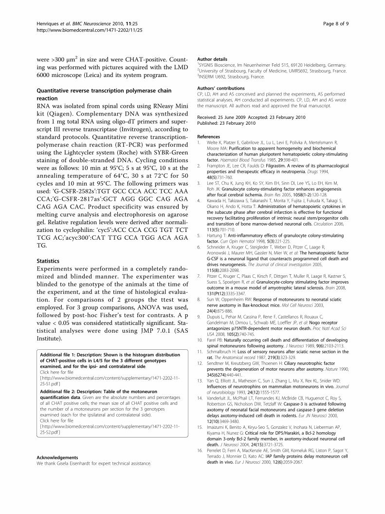
To determine whether this was also the case after sciaticnerve axotomy, we studied the distribution of moto-neuron size of all motoneurons including those that are< 300 μm2 determined by their cross-sectional area inthe horizontal plane in all cells that were situated in theventral horn, had a nucleolus, and were CHAT-positive.On the contralateral side, size distribution was similarbetween the three genotypes (Figure 6, hatched bars;BEG -/T CaMK -/T (442.55 ± 11.08 μm2); BEG -/TCaMK -/- (431.84 ± 12.52 μm2); wt (434.20 ± 9.74 μm2);p = 0.78 by ANOVA).On the ipsilateral (axotomized) side, there was a
downward shift in the mean size of the motoneurons inthe littermates that did not overexpress G-CSF (wt andBEG -/T CaMK-/-). On the contrary, double transgenicanimals (BEG -/T CaMK -/T) displayed a relatively pre-served motoneuron size, suggesting a healthier state ofthose cells (Figure 6, solid bars; BEG -/T CaMK -/T(464 ± 17 μm2); BEG -/T CaMK -/- (391 ± 12 μm2); wt(353 ± 13 μm2); p < 0.001; ANOVA followed by Fisher’sLSD). This result was also obtained if we only considerthe a motoneurons that were counted for determiningcell survival (> 300 μm2): 555.85 ± 17.43 μm2 (BEG -/T
0
1
2
3
4
5
6
7
8
9
10M
Ns
cont
rala
tera
l
0
1
2
3
4
5
6
MN
sip
sila
tera
l
0
10
20
30
40
50
60
70
80
90
ratio
[% ]
BEG -/T CaMK -/TBEG-/T CaMK -/-wt
100
A
B
C
***
***
Figure 5 Overexpression of G-CSF improves motoneuronsurvival following sciatic nerve axotomy in neonatal pups. (A)Quantification of the absolute number of a; motoneurons persection of the lumbar spinal cord on the contralateral (unlesioned)side. Numbers are not significantly different between the threegenotypes studied. (B) On the axotomized side numbers ofdetectable motoneurons are dramatically decreased in thelittermate animals lacking the CaMK-tta driver transgene (wt, BEG -/-CaMK-/-; and BEG -/T CaMK-/-), whereas overexpression of G-CSF inthe CNS results in significantly higher numbers of survivingmotoneurons (***p < 0.0001 by ANOVA and Fisher’s LSD post-hocanalysis). (C) Percentage of surviving motoneurons on the ipsilateral(axotomized) relative to the corresponding contralateral (control)side. While only 50 - 60% of the motoneurons survive in thegenotypes lacking the CaMK-tta driver transgene, close to 90% ofthe motoneurons survive in the presence of the driver (i.e. inanimals overexpressing G-CSF) (***p < 0.0001 by ANOVA andFisher’s LSD post-hoc analysis). (Number of animals: wt, n = 13; BEG-/T CaMK -/-, n = 6; BEG -/T CaMK -/T, n = 7; 6 sections werestudied per animal).
Figure 6 Size distribution of surviving neurons in the spinalcord of neonatal mice following sciatic nerve axotomy. On thecontralateral side (hatched bars) the size distribution between thethree genotypes is similar. On the ipsilateral side (filled bars) there isa downward shift in the mean size of the surviving motoneurons inthe littermates without overexpression of G-CSF (BEG -/- CaMK -/-;BEG -/T CaMK -/-). (p < 0.0001 for BEG -/T CaMK -/T vs. BEG -/-CaMK -/-; **p = 0.0008 for BEG -/T CaMK -/T vs. BEG -/T CaMK -/-;ANOVA followed by Fisher’s LSD; Number of motoneuronsmeasured: wt, n = 301; BEG -/T CaMK -/-, n = 197; BEG -/T CaMK -/T,n = 227).
Henriques et al. BMC Neuroscience 2010, 11:25http://www.biomedcentral.com/1471-2202/11/25
Page 5 of 9

CaMK -/T); 484.32 ± 19.73 μm2 (BEG -/T CaMK -/-);477.68 ± 17.93 μm2 (wt); p = 0.0029 by ANOVA. Addi-tional file 1 shows a histogram distribution of moto-neurons contra- and ipsilateral to the axotomy. Adetailed summary of all data measured is given in addi-tional file 2.
DiscussionHere we show that the receptor for G-CSF is expressedby neonatal motoneurons and that G-CSF protects lum-bar motoneurons in a model of neonatal sciatic nerveaxotomy. We have chosen this model because of theclarity of the involved pathophysiology to obtain moreinsight into the nature of G-CSF’s effect on motoneur-ons. Sendtner et al. first described rescue of motoneurondeath caused by neonatal axotomy by the growth factorCNTF [12]. This was followed by demonstration of anumber of other neurotrophic factors protective in thisparadigm. A series of papers then defined this axotomy-induced motoneuron death as apoptosis, with involve-ment of bcl proteins, AKT, IAPs, and caspases [14-16].Overexpression of G-CSF protects lumbar motoneuronsto an extent that is comparable to many of the classicneurotrophic factors. The experiments done here aretherefore a further step in redefining G-CSF as a neuro-trophic factor.On top of a relative increase of motoneuron numbers
following overexpression of G-CSF, we also noted anincrease of about 100 μm2 in the mean cross-sectionalarea of surviving motoneurons. This suggests a function-ally more active, or healthier state of those motoneur-ons. This has also been noted in adult SOD1(G93A)mice by us previously [7]. This finding might point toan additional trophic effect of G-CSF in addition tocounteracting apoptosis. Alternatively, this could signifya proportionally greater anti-apoptotic effect size of G-CSF on larger motoneurons which potentially expressmore receptors on the cell surface.We have studied motoneuron survival 4 days after
axotomy with the aim to demonstrate that G-CSF cancounteract apoptosis in motoneurons in vivo in a simpleparadigm as a mean to better dissect mechanisms ofaction in the more complex ALS mouse models. We donot know how transient this effect may be, and whetherincreased survival would still be detectable at 1 monthafter axotomy. All studies with growth factors in axot-omy models that have examined varied time points afteraxotomy found that the number of surviving neuronsdeclined with time to varying degrees in spite ofongoing treatment [17,18]. The “slope” of this declinemight give an indication of the relative potency ofgrowth factors in this paradigm (e.g [18]). However,there was always a lasting effect observed if growth fac-tors showed protection after shorter observation time
points. The purpose of our study was however not tocompare relative efficacies of growth factors in thisparadigm. We prefer to study questions of relative effi-cacy and effect stability in disease models closest and ofrelevance to the human indication targeted, which isALS and SOD1-mutant mice in our case.The observation that the G-CSF receptor was induced
by axotomy parallels findings from other neurologicaldisease models, such as stroke, spinal cord injury, orALS [6,7], and underlines the notion that the G-CSFsystem is part of an endogenous protective system forneurons. This is certainly an argument for the use of G-CSF as a pharmacological agent that adresses an endo-genously pre-specified mechanism. Indeed, G-CSF iscurrently under clinical investigation in ischemic stroke[19,20], and ALS [21-23].We have undertaken this experiment to better dissect
mechanisms responsible for the previously demonstratedbeneficial function of G-CSF in the SOD1(G93A) mousemodel for ALS. We have chosen the axotomy model toclearly answer the question whether G-CSF exerts adirect influence on motoneuron apoptosis in vivo. Wehave demonstrated potent anti-apoptotic properties ofthis “redefined” neurotrophic factor in vitro either incortical neurons [6], or in motoneurons [7], and wefavoured the anti-apoptotic hypothesis due to the strongeffect on motoneuron survival in vivo. However, ALS isa multifaceted disease, with many factors involved in thepathophysiology [24-26], and the SOD1(G93A) modelsreproduce this complexity with discussed involvementof the neuromuscular junction, the immune system, glialcells and others. Moreover, G-CSF is a pleiotropic factorthat for example also impacts on immunocompetenceand inflammatory reactions. The data presented hereclearly demonstrate a potent effect of G-CSF on moto-neuron apoptosis in vivo, and support our assumptionthat anti-apoptosis plays a central role in the observedeffects in the SOD1 transgenic ALS model.Since G-CSF has such a potent protective role in
apoptosis mediated by cell-inherent mechanisms as aresponse to nerve damage the interesting question ariseswhether the G-CSF system might also play a role indevelopmental death of motoneurons. This has indeedbeen shown for a number of neurotrophic factors thatprotect in the axotomy paradigm [27]. We do notbelieve that this is a confounding factor in our casesince the expression of the CaMKII driven tTA isswitched on during P2 and P5 [28-30], while the sensi-tive period for physiological motoneuron death isbetween E14 and P3 [27].Although we do not see significant effects on the
number of contralateral motoneurons in the differenttransgenic crosses, there is an interesting trend (p = 0.1)towards more motoneurons contralaterally in the G-CSF
Henriques et al. BMC Neuroscience 2010, 11:25http://www.biomedcentral.com/1471-2202/11/25
Page 6 of 9

overexpressors compared to littermates not transgenicfor the CaMKII tta driver. One might speculate that areason for this trend observation could be contralateraleffects of axotomy that are counteracted by G-CSF. Thisslight nominal difference on the contralateral side doeshowever not change our result, since the protectiveeffect of G-CSF is also highly significant when regardingipsi-/contralateral ratios instead of ipsilateral cellnumbers.The mechanisms responsible for counteraction of
apoptosis elicited by G-CSF in neurons have been pre-viously delineated by us: Activation of AKT in neuronsand induction of stat3 and bcl-proteins [6]. The AKTand stat3 pathways play a crucial role for the survival ofthe axotomized motoneurons [31,32]. It is thereforehighly likely that G-CSF mediates its protective effectvia these pathways in the axotomy paradigm as well.
ConclusionsHere we have shown that G-CSF counteracts moto-neuron death in a model of neonatal axotomy-inducedapoptosis. This is most likely mediated by direct actionsof the protein on motoneurons in vivo, and suggeststhat the protection offered by G-CSF in ALS mousemodels is due to its direct neuroprotective activity.Moreover, our data strengthen the overall evidence thatG-CSF is a candidate drug for the treatment of ALS.
MethodsGeneration of G-CSF-overexpressing miceGeneration of the BEG transgenic lines is also describedin [7]. We cloned the cDNA for murine G-CSF in a bi-directional Tet-transactivator (tta) responsive vector(pBI, Clontech). EYFP was inserted on the other side ofthe promoter for easy visualization of expression. Togenerate the pBI-EYFP-G-CSF ("pBEG”) plasmid, G-CSFwas amplified from a mouse brain cDNA library andinserted into the pBI Tet vector (Clontech) using therestriction sites NheI and EcoRV. EYFP was insertedinto the multiple cloning site of the vector. Transgenicmice ("BEG”) were generated, and selected for copynumber integration. Copy numbers were estimated byquantitative PCR on genomic DNA by comparingpBEG/cyclophilin ratios of wild type and founder mice.For generating CNS- target G-CSF overexpressing mice,mice of line BEG6 were crossed with mice expressingthe Tet-transactivator under control of the CaMKII-pro-moter (CaMKII-tta) [33], and successful activation ofthe construct was verified by EYFP imaging.
Sciatic nerve injuryNeonatal mice were anesthetized and immobilized byhypothermia after placement on ice, and unilateral scia-tic nerve section was performed on postnatal day 5 (P5).
The skin of the right leg was incised parallel to thefemur. To expose the sciatic nerve a longitudinal cutwas made through the biceps femoris muscle with asharpened forceps. The right sciatic nerve was trans-ected at midthighlevel, a small piece (about 1-2 mm inlength) of the nerve was removed to prevent reinnerva-tion and the overlying skin of the thigh was sutured.Pups were warmed and returned to the mother. All ani-mal manipulation followed current regulations of andwere approved by the Regierungspräsidium Karlsruhe,Germany.
ImmunohistochemistryAfter deep anesthesia spinal cords were removedrapidly, immersed in 4% PFA, and embedded in paraffin.For immunofluorescence, microtome sections of paraf-fin-embedded tissues (10 μm) were deparaffinated andmicrowaved (citrate buffer at 600 W for 15 min). Fordouble-fluorescence staining, sections were incubatedsimultaneously with the G-CSF receptor antiserum (SC-694; Santa Cruz Biotechnology, Santa Cruz, CA, USA;1:100) and the CHAT antibody (AB144P; ChemiconEurope Ltd., UK; 1:100) at 4°C overnight. After enhan-cing the G-CSF receptor staining by adding a biotiny-lated anti-rabbit secondary antibody (Dianova,Hamburg, Germany; 1:200), sections were incubatedwith a Streptavidin-coupled fluorophore (Invitrogen,Karlsruhe, Germany; 1:200) or the appropriate fluores-cence-coupled secondary antibody (Dianova, Hamburg,Germany; 1:200). The nuclei were counterstained withHoechst 33342 (Molecular Probes, 1:10000). Controlsincluded omission of primary antibodies, fluorescenceswapping and single-fluorescence stainings. Pictureswere captured by video camera (Olympus DP71)coupled to a fluorescent microscope (Olympus IX80).To quantify staining intensity for the G-CSF receptor,pictures were analyzed with an image processing pro-gram (ImageJ). Intensity of grey levels were calculatedfor each motoneuron and normalized to its area.For light-microscopic CHAT-staining coronal paraffin
sections from the lumbar spinal cord (P9) were stainedwith the CHAT antibody using the avidin-biotin com-plex technique with DAB as chromogen (DakoCytoma-tion), and counterstained with hemalaun.
Counting of motoneuronsCounting of motoneurons was performed at postnatalday 9 (P9), i.e. 4 days following axotomy at P5. Spinalcord paraffin sections of a thickness of 10 μm, spacedby 100 μm over a length of 0.5 mm, were counted atthe spinal level L4-L5. Motor axons that constitute thesciatic nerve in C57/bl6 mice originate from L3 to L5[34]. All neurons in the ventral horn were counted as amotoneurons if they had a clearly identifiable nucleolus,
Henriques et al. BMC Neuroscience 2010, 11:25http://www.biomedcentral.com/1471-2202/11/25
Page 7 of 9
were >300 μm2 in size and were CHAT-positive. Count-ing was performed with pictures acquired with the LMD6000 microscope (Leica) and its system program.
Quantitative reverse transcription polymerase chainreactionRNA was isolated from spinal cords using RNeasy Minikit (Qiagen). Complementary DNA was synthesizedfrom 1 mg total RNA using oligo-dT primers and super-script III reverse transcriptase (Invitrogen), according tostandard protocols. Quantitative reverse transcription-polymerase chain reaction (RT-PCR) was performedusing the Lightcycler system (Roche) with SYBR-Greenstaining of double-stranded DNA. Cycling conditionswere as follows: 10 min at 95°C; 5 s at 95°C, 10 s at theannealing temperature of 64°C, 30 s at 72°C for 50cycles and 10 min at 95°C. The following primers wasused: ‘G-CSFR-2582s’:TGT GCC CCA ACC TCC AAACCA;’G-CSFR-2817as’:GCT AGG GGC CAG AGACAG AGA CAC. Product specificity was ensured bymelting curve analysis and electrophoresis on agarosegel. Relative regulation levels were derived after normali-zation to cyclophilin: ‘cyc5’:ACC CCA CCG TGT TCTTCG AC;’acyc300’:CAT TTG CCA TGG ACA AGATG.
StatisticsExperiments were performed in a completely rando-mized and blinded manner. The experimenter wasblinded to the genotype of the animals at the time ofthe experiment, and at the time of histological evalua-tion. For comparisons of 2 groups the ttest wasemployed. For 3 group comparisons, ANOVA was used,followed by post-hoc Fisher’s test for contrasts. A pvalue < 0.05 was considered statistically significant. Sta-tistical analyses were done using JMP 7.0.1 (SASInstitute).
Additional file 1: Description: Shown is the histogram distributionof CHAT-positive cells in L4/5 for the 3 different genotypesexamined, and for the ipsi- and contralateral side.Click here for file[ http://www.biomedcentral.com/content/supplementary/1471-2202-11-25-S1.pdf ]
Additional file 2: Description: Table of the motoneuronquantification data. Given are the absolute numbers and percentagesof all CHAT positive cells; the mean size of all CHAT positive cells andthe number of a motoneurons per section for the 3 genotypesexamined (each for the ipsilateral and contralateral side).Click here for file[ http://www.biomedcentral.com/content/supplementary/1471-2202-11-25-S2.pdf ]
AcknowledgementsWe thank Gisela Eisenhardt for expert technical assistance.
Author details1SYGNIS Bioscience, Im Neuenheimer Feld 515, 69120 Heidelberg, Germany.2University of Strasbourg, Faculty of Medicine, UMRS692, Strasbourg, France.3INSERM U692, Strasbourg, France.
Authors’ contributionsCP, LD, AH and AS conceived and planned the experiments, AS performedstatistical analyses, AH conducted all experiments. CP, LD, AH and AS wrotethe manuscript. All authors read and approved the final manuscript.
Received: 25 June 2009 Accepted: 23 February 2010Published: 23 February 2010
References1. Welte K, Platzer E, Gabrilove JL, Lu L, Levi E, Polivka A, Mertelsmann R,
Moore MA: Purification to apparent homogeneity and biochemicalcharacterization of human pluripotent hematopoietic colony-stimulatingfactor. Haematol Blood Transfus 1985, 29:398-401.
2. Frampton JE, Lee CR, Faulds D: Filgrastim. A review of its pharmacologicalproperties and therapeutic efficacy in neutropenia. Drugs 1994,48(5):731-760.
3. Lee ST, Chu K, Jung KH, Ko SY, Kim EH, Sinn DI, Lee YS, Lo EH, Kim M,Roh JK: Granulocyte colony-stimulating factor enhances angiogenesisafter focal cerebral ischemia. Brain Res 2005, 1058(1-2):120-128.
4. Kawada H, Takizawa S, Takanashi T, Morita Y, Fujita J, Fukuda K, Takagi S,Okano H, Ando K, Hotta T: Administration of hematopoietic cytokines inthe subacute phase after cerebral infarction is effective for functionalrecovery facilitating proliferation of intrinsic neural stem/progenitor cellsand transition of bone marrow-derived neuronal cells. Circulation 2006,113(5):701-710.
5. Hartung T: Anti-inflammatory effects of granulocyte colony-stimulatingfactor. Curr Opin Hematol 1998, 5(3):221-225.
6. Schneider A, Kruger C, Steigleder T, Weber D, Pitzer C, Laage R,Aronowski J, Maurer MH, Gassler N, Mier W, et al: The hematopoietic factorG-CSF is a neuronal ligand that counteracts programmed cell death anddrives neurogenesis. The Journal of clinical investigation 2005,115(8):2083-2098.
7. Pitzer C, Kruger C, Plaas C, Kirsch F, Dittgen T, Muller R, Laage R, Kastner S,Suess S, Spoelgen R, et al: Granulocyte-colony stimulating factor improvesoutcome in a mouse model of amyotrophic lateral sclerosis. Brain 2008,131(Pt12):3335-3347.
8. Sun W, Oppenheim RW: Response of motoneurons to neonatal sciaticnerve axotomy in Bax-knockout mice. Mol Cell Neurosci 2003,24(4):875-886.
9. Dupuis L, Pehar M, Cassina P, Rene F, Castellanos R, Rouaux C,Gandelman M, Dimou L, Schwab ME, Loeffler JP, et al: Nogo receptorantagonizes p75NTR-dependent motor neuron death. Proc Natl Acad SciUSA 2008, 105(2):740-745.
10. Farel PB: Naturally occurring cell death and differentiation of developingspinal motoneurons following axotomy. J Neurosci 1989, 9(6):2103-2113.
11. Schmalbruch H: Loss of sensory neurons after sciatic nerve section in therat. The Anatomical record 1987, 219(3):323-329.
12. Sendtner M, Kreutzberg GW, Thoenen H: Ciliary neurotrophic factorprevents the degeneration of motor neurons after axotomy. Nature 1990,345(6274):440-441.
13. Yan Q, Elliott JL, Matheson C, Sun J, Zhang L, Mu X, Rex KL, Snider WD:Influences of neurotrophins on mammalian motoneurons in vivo. Journalof neurobiology 1993, 24(12):1555-1577.
14. Vanderluit JL, McPhail LT, Fernandes KJ, McBride CB, Huguenot C, Roy S,Robertson GS, Nicholson DW, Tetzlaff W: Caspase-3 is activated followingaxotomy of neonatal facial motoneurons and caspase-3 gene deletiondelays axotomy-induced cell death in rodents. Eur JN Neurosci 2000,12(10):3469-3480.
15. Imaizumi K, Benito A, Kiryu-Seo S, Gonzalez V, Inohara N, Lieberman AP,Kiyama H, Nunez G: Critical role for DP5/Harakiri, a Bcl-2 homologydomain 3-only Bcl-2 family member, in axotomy-induced neuronal celldeath. J Neurosci 2004, 24(15):3721-3725.
16. Perrelet D, Ferri A, MacKenzie AE, Smith GM, Korneluk RG, Liston P, Sagot Y,Terrado J, Monnier D, Kato AC: IAP family proteins delay motoneuron celldeath in vivo. Eur J Neurosci 2000, 12(6):2059-2067.
Henriques et al. BMC Neuroscience 2010, 11:25http://www.biomedcentral.com/1471-2202/11/25
Page 8 of 9

17. Baumgartner BJ, Shine HD: Neuroprotection of spinal motoneuronsfollowing targeted transduction with an adenoviral vector carrying thegene for glial cell line-derived neurotrophic factor. Exp Neurol 1998,153(1):102-112.
18. Matheson CR, Wang J, Collins FD, Yan Q: Long-term survival effects ofGDNF on neonatal rat facial motoneurons after axotomy. Neuroreport1997, 8(7):1739-1742.
19. Schabitz WR, Schneider A: Developing granulocyte-colony stimulatingfactor for the treatment of stroke: current status of clinical trials. Stroke2006, 37(7):1654, author reply 1655..
20. Schabitz WR, Schneider A: New targets for established proteins: exploringG-CSF for the treatment of stroke. Trends Pharmacol Sci 2007,28(4):157-161.
21. Nefussy B, Artamonov I, Deutsch V, Naparstek E, Nagler A, Drory VE:Recombinant human granulocyte-colony stimulating factoradministration for treating amyotrophic lateral sclerosis: A pilot study.Amyotroph Lateral Scler 2009, 1-7.
22. Tarella C, Rutella S, Gualandi F, Melazzini M, Scime R, Petrini M, Moglia C,Ulla M, Omede P, Bella VL, et al: Consistent bone marrow-derived cellmobilization following repeated short courses of granulocyte-colony-stimulating factor in patients with amyotrophic lateral sclerosis: resultsfrom a multicenter prospective trial. Cytotherapy 2009, 12(1):50-59.
23. Zhang Y, Wang L, Fu Y, Song H, Zhao H, Deng M, Zhang J, Fan D:Preliminary investigation of effect of granulocyte colony stimulatingfactor on amyotrophic lateral sclerosis. Amyotroph Lateral Scler 2008, 1-2.
24. Blackburn D, Sargsyan S, Monk PN, Shaw PJ: Astrocyte function and role inmotor neuron disease: a future therapeutic target?. Glia 2009,57(12):1251-1264.
25. Xiao Q, Zhao W, Beers DR, Yen AA, Xie W, Henkel JS, Appel SH: MutantSOD1(G93A) microglia are more neurotoxic relative to wild-typemicroglia. J Neurochem 2007, 102(6):2008-2019.
26. Fischer LR, Culver DG, Tennant P, Davis AA, Wang M, Castellano-Sanchez A,Khan J, Polak MA, Glass JD: Amyotrophic lateral sclerosis is a distalaxonopathy: evidence in mice and man. Exp Neurol 2004, 185(2):232-240.
27. Sendtner M, Pei G, Beck M, Schweizer U, Wiese S: Developmentalmotoneuron cell death and neurotrophic factors. Cell Tissue Res 2000,301(1):71-84.
28. Bayer KU, Lohler J, Schulman H, Harbers K: Developmental expression ofthe CaMkinase IIisoforms: ubiquitous gamma- and delta-CaM kinase IIare the early isoforms and most abundant in the developing nervoussystem. Brain Res Mol Brain Res 1999, 70(1):147-154.
29. Petralia RS, Sans N, Wang YX, Wenthold RJ: Ontogeny of postsynapticdensity proteins at glutamatergic synapses. Mol Cell Neurosci 2005,29(3):436-452.
30. Gross C, Zhuang X, Stark K, Ramboz S, Oosting R, Kirby L, Santarelli L,Beck S, Hen R: Serotonin1A receptor acts during development toestablish normal anxiety-like behaviour in the adult. Nature 2002,416(6879):396-400.
31. Namikawa K, Honma M, Abe K, Takeda M, Mansur K, Obata T, Miwa A,Okado H, Kiyama H: Akt/protein kinase B prevents injury-inducedmotoneuron death and accelerates axonal regeneration. J Neurosci 2000,20(8):2875-2886.
32. Schweizer U, Gunnersen J, Karch C, Wiese S, Holtmann B, Takeda K, Akira S,Sendtner M: Conditional gene ablation of Stat3 reveals differentialsignaling requirements for survival of motoneurons during developmentand after nerve injury in the adult. J Cell Biol 2002, 156(2):287-297.
33. Mansuy IM, Winder DG, Moallem TM, Osman M, Mayford M, Hawkins RD,Kandel ER: Inducible and reversible gene expression with the rtTAsystem for the study of memory. Neuron 1998, 21(2):257-265.
34. Rigaud M, Gemes G, Barabas ME, Chernoff DI, Abram SE, Stucky CL,Hogan QH: Species and strain differences in rodent sciatic nerveanatomy: implications for studies of neuropathic pain. Pain 2008, 136(1-2):188-201.
doi:10.1186/1471-2202-11-25Cite this article as: Henriques et al.: G-CSF protects motoneuronsagainst axotomy-induced apoptotic death in neonatal mice. BMCNeuroscience 2010 11:25.
Submit your next manuscript to BioMed Centraland take full advantage of:
• Convenient online submission
• Thorough peer review
• No space constraints or color figure charges
• Immediate publication on acceptance
• Inclusion in PubMed, CAS, Scopus and Google Scholar
• Research which is freely available for redistribution
Submit your manuscript at www.biomedcentral.com/submit
Henriques et al. BMC Neuroscience 2010, 11:25http://www.biomedcentral.com/1471-2202/11/25
Page 9 of 9