further evidence concerning macrophages producing 19 s-antibody in mice
TRANSCRIPT
EXPERIENTIA 25/4 Specialia 401
Further Evidence Concerning Macrophages Producing 19 S-Antibody in Mice
I n a p rev ious c o m m u n i c a t i o n we descr ibed macro- phages which p roduced in mice 19 S - A n t i b o d y aga ins t S R B C L This resu l t was o b t a i n e d b y a mod i f i ca t ion of t he p l aque t e c h n i q u e of J~RNX and NORI)IN ~ a n d a morpholog ica l s t u d y of t he p l aque - fo rming cells 1. In f u r t h e r e x p e r i m e n t s we c o m b i n e d cytological observa- t ions w i th func t iona l s tudies on these p l aque - fo rming cells.
The morpholog ica l a spec t of t he p l aque - fo rming cells ind ica tes t h a t some of these cells are macrophages . I f i t is so, t h e n these cells should d e m o n s t r a t e phagocy t i c ac t iv i ty . The pu rpose of th i s s t u d y is there fore to show
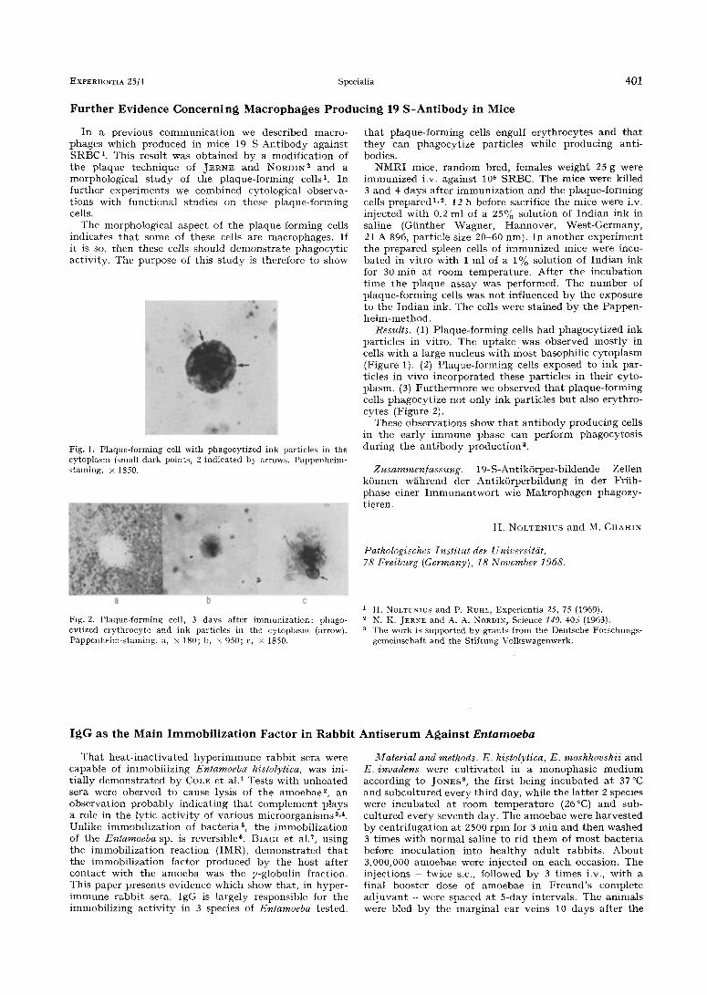
Fig. 1. Plaque-forming cell with phagocytized ink particles in the cytoplasnl (small dark points, 2 indicated by arrows. Pappenheim- staining. • 1850.
t h a t p l aq u e - fo rmi n g cells engulf e r y t h r o c y t e s a n d t h a t t h e y can phagocy t i ze par t ic les whi le p r o d u c i n g an t i - bodies.
N M R I mice, r a n d o m bred , females w e i g h t 25 g were i m m u n i z e d i.v. aga ins t 108 SRBC. The mice were kil led 3 a n d 4 days a f t e r i m m u n i z a t i o n a n d t h e p l aque - fo rming cells p r e p a r e d 1,2. 12 h before sacrifice t h e nlice were i.v. in jec ted w i t h 0.2 ml of a 25% so lu t ion of I n d i a n ink in sal ine (Gi in the r Wagner , H a n n o v e r , W e s t - G e r m a n y , 21 A 896, par t ic le size 20-60 nm). I n a n o t h e r e x p e r i m e n t t h e p r e p a r e d spleen cells of i m m u n i z e d mice were incu- b a t e d in v i t ro w i th 1 ml of a 1% so lu t ion of I n d i a n ink for 30 m i n a t room t e m p e r a t u r e . Af te r t h e i n c u b a t i o n t i m e t h e p l a q u e assay was per formed. T h e n u m b e r of p l aq u e - fo rmi n g cells was n o t in f luenced b y t i le exposure to t h e I n d i a n ink. The cells were s t a ined b y t h e P a p p e n - h e l m - m e t h o d .
Results. (1) P l aq u e - fo rmi n g cells h a d p h a g o c y t i z e d ink par t ic les in vi t ro . The u p t a k e was obse rved m o s t l y in cells w i t h a large nucleus w i t h mos t basophi l i c c y t o p l a s m (Figure 1). (2) P l aque - fo rming cells exposed to ink par- t icles in v ivo i nco rpo ra t ed these par t ic les in t h e i r cyto- p lasm. (3) F u r t h e r m o r e we obse rved t h a t p l aq u e - fo rming cells phagocy t i ze no t on ly ink par t ic les b u t also e ry th ro - cy tes (Figure 2).
These o b s e rv a t i o n s show t h a t a n t i b o d y p r o d u c i n g cells in t h e ea r ly i m m u n e p h a s e can pe r fo rm phagocy tos i s d u r i n g t h e a n t i b o d y p r o d u c t i o n 3.
Zusammen/assung. 19-S-Ant ik6 rpe r -b i ldende Zellen k 6 n n e n w~ihrend der A n t i k 6 r p e r b i l d u n g in der Fr i ih- phase e iner I m m u n a n t w o r t wie M a k r o p h a g e n phagozy- t ieren.
H. NOLTENIUS a n d M. CHAHIN
Pathologisches Institut der Universitdt, 78 Freiburg (Germany), 18 November 1968.
Fig. 2. Plaque-forming cell, 3 days after immunization: phago- cytized erythrocyte and ink particles in the cytoplasm (arrow). Pappenheim-staining. a, • 180; b, • 950; c, • 1850.
1 H. NOLTENIUS and P. RUHL, Experientia 25, 75 (1969). 2 N. K. JERNE and A. A. NORtoN, Science 140, 405 (1963). a The work is supported by grants from the Deutsche Forschungs-
gemeinschaft and the Stiftung Volkswagenwerk.
IgG as the Main Immobil izat ion Factor in Rabbit Antiserum Against Entamoeba
T h a t h e a t - i n a c t i v a t e d h y p e r i m m u n e r a b b i t sera were capab le of immob i l i z i ng Enlamoeba histolytica, was ini- t ia l ly d e m o n s t r a t e d b y COLE e t al. 1 Tests w i th u n h e a t e d sera were obe rved to cause lysis of t he a m o e b a e 2, a n obse rva t i on p r o b a b l y i nd i ca t i ng t h a t c o m p l e m e n t p lays a role in the lyric a c t i v i t y of va r ious mic roo rgan i sms 3A. Unl ike i m m o b i l i z a t i o n of bac t e r i a 5, t he immob i l i z a t i o n of t he Entamoeba sp. is revers ib le 6. BIAGI et al. 7, us ing t he immob i l i z a t i on reac t ion (IMR), d e m o n s t r a t e d t h a t t he immob i l i z a t i on fac to r p roduced b y t he hos t a f t e r c o n t a c t w i th t he a m o e b a was t he 7-globul in f ract ion. This pape r p resen t s ev idence wh ich show tha t , in hype r - i m m u n e r a b b i t sera, IgG is largely respons ib le for t h e immobi l i z ing ac t i v i t y in 3 species of Entamoeba tes ted .
Material and methods. E. histolytica, E. moshkovskii and E. invadens were c u l t i v a t e d in a m o n o p h a s i c m e d i u m accord ing to JONES s, t h e f i rs t be ing i n c u b a t e d a t 37~ an d s u b c u l t u r e d eve ry t h i r d day, whi le the l a t t e r 2 species were i n c u b a t e d a t room t e m p e r a t u r e (26~ a n d sub- cu l tu red eve ry s ev en t h day. The a m o e b a e were h a r v e s t e d b y cen t r i f u g a t i o n a t 2500 r p m for 3 ra in a n d t h e n washed 3 t imes w i t h n o r m a l sal ine to r id t h e m of m o s t bac t e r i a before inocu la t ion in to h e a l t h y adu l t r abb i t s . A b o u t 3,000,000 a m o e b a e were in jec ted on each occasion. The in jec t ions - twice s.c., followed b y 3 t imes i.v., w i t h a f inal boos t e r dose of a m o e b a e in F r e u n d ' s comple te a d j u v a n t - were spaced a t 5-day in te rva ls . T h e an i ma l s were bled b y t h e m a r g i n a l ear ve ins 10 days a f t e r t he