fungi, protozoa, and helminthes. fungi kingdom fungi divided into 2 groups: macroscopic fungi...
TRANSCRIPT
Fungi, Protozoa, and Helminthes
Fungi
Kingdom Fungi
Divided into 2 groups: macroscopic fungi
(mushrooms, puffballs, gill fungi)
microscopic fungi (molds, yeasts)
Majority are unicellular or colonial
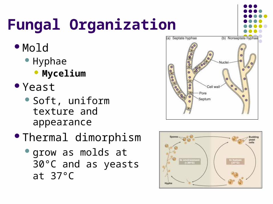
Fungal OrganizationMold
Hyphae Mycelium
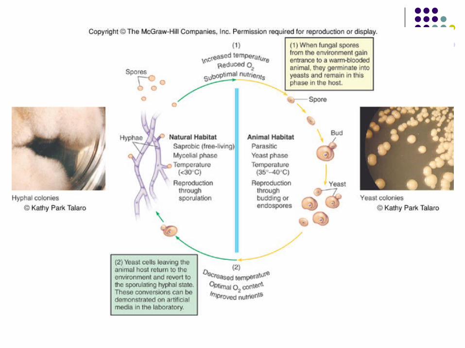
Yeast Soft, uniform texture and
appearance Thermal dimorphism
grow as molds at 30°C and as yeasts at 37°C
Fungal Organization - Mold
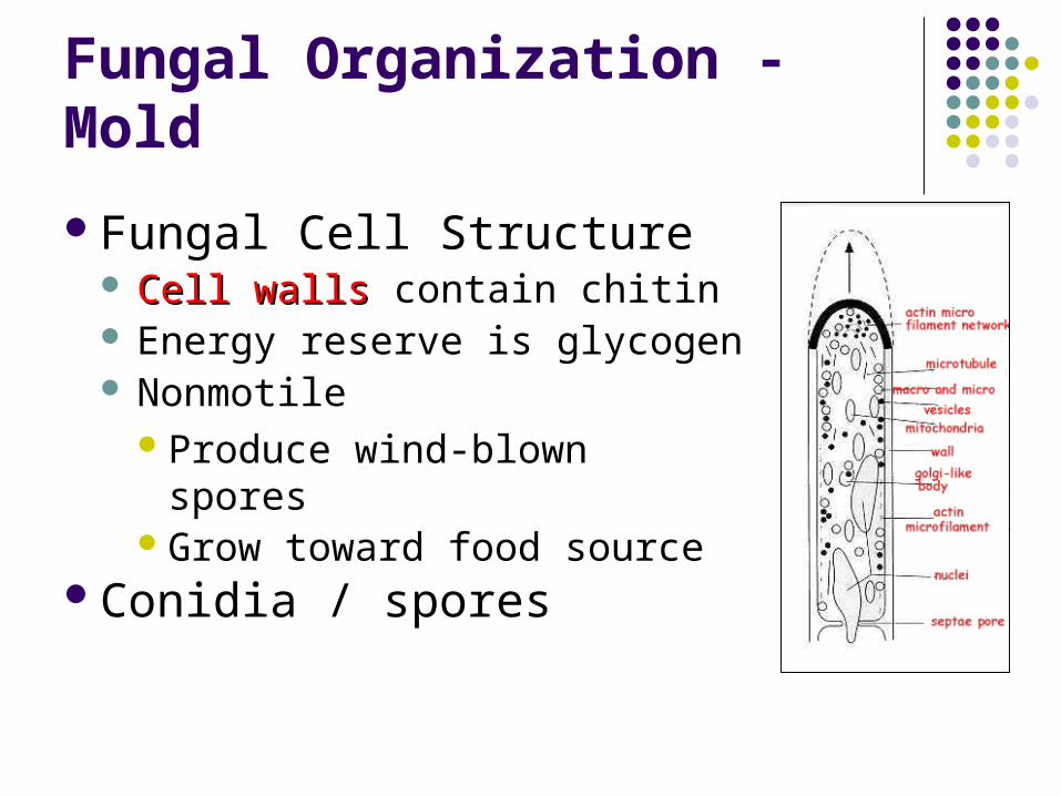
Fungal Cell Structure Cell wallsCell walls contain chitin Energy reserve is glycogen Nonmotile
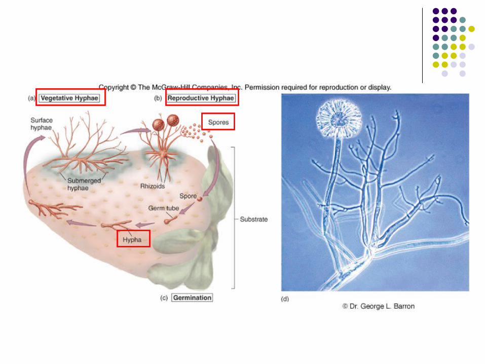
Produce wind-blown sporesGrow toward food source
Conidia / spores
Fungal Organization
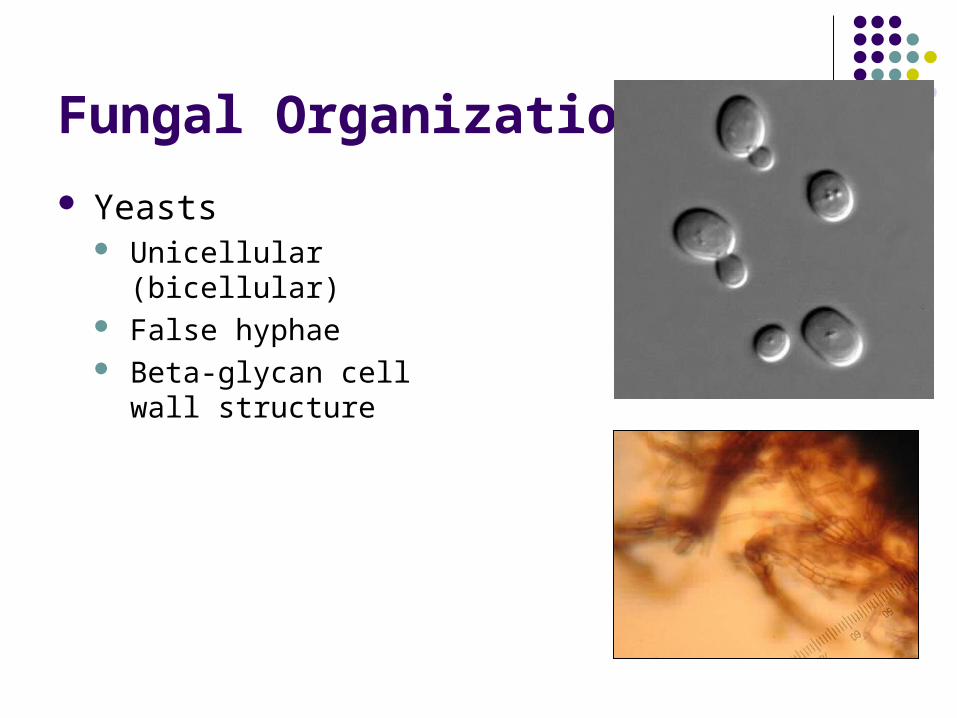
Yeasts Unicellular (bicellular) False hyphae Beta-glycan cell wall
structure
Fungal Nutrition
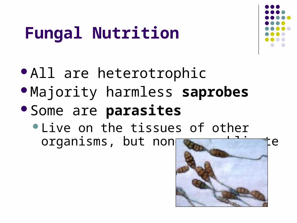
All are heterotrophicMajority harmless saprobes Some are parasites
Live on the tissues of other organisms, but none are obligate
Fungal Reproduction
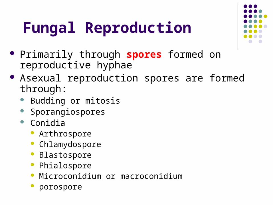
Primarily through spores formed on reproductive hyphae
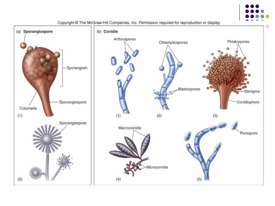
Asexual reproduction spores are formed through: Budding or mitosis Sporangiospores Conidia
Arthrospore Chlamydospore Blastospore Phialospore Microconidium or macroconidium porospore
Reproductive strategies
Sexual reproduction Spores are formed following fusion of male and
female strains and formation of sexual structure Sexual spores and spore-forming structures
are one basis for classification Zygospores Ascospores Basidiospores
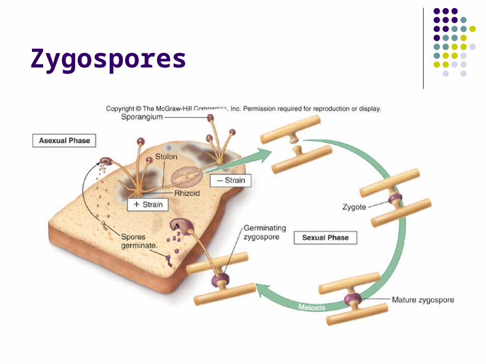
Zygospores
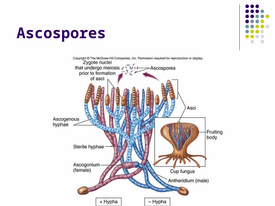
Ascospores
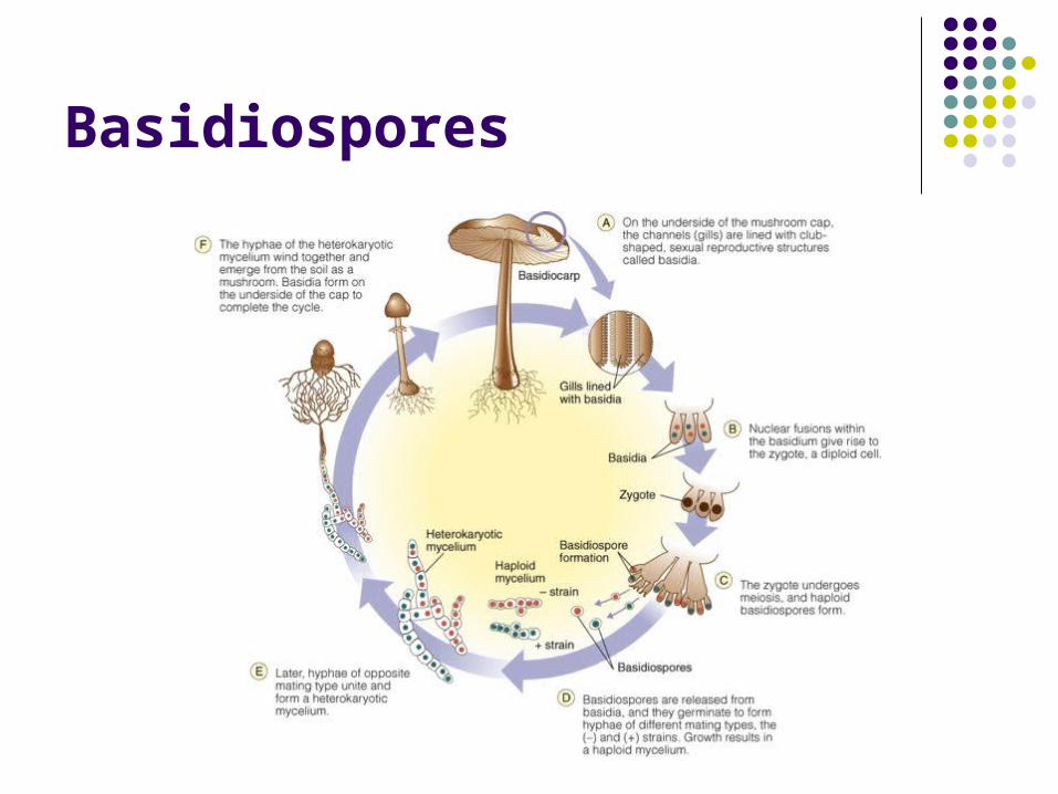
Basidiospores
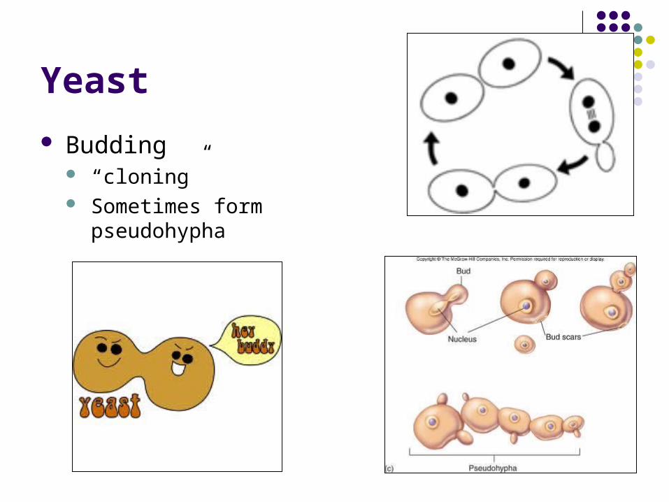
Yeast
Budding “cloning” Sometimes form
pseudohypha
Fungal Classification
Yeast verse Mold Asexual verse Sexual (reproductive strategies) Terrestrial or water
Fungal Classification
Subkingdom Amastigomycota Terrestrial inhabitants including those of medical
importance:
1. Zygomycota – zygospores; sporangiospores and some conidia
2. Ascomycota – ascospores; conidia
3. Basidiomycota – basidiospores; conidia
4. Deuteromycota*** – majority are yeasts and molds; no sexual spores known; conidia
Fungal Classification
Subkingdom Mastigomycota
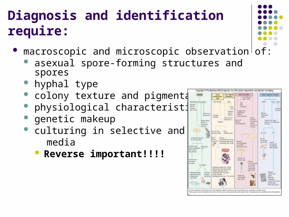
Diagnosis and identification require: macroscopic and microscopic observation of:
asexual spore-forming structures and spores hyphal type colony texture and pigmentation physiological characteristics genetic makeup culturing in selective and enriched media
Reverse important!!!!
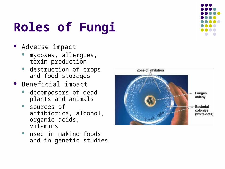
Roles of Fungi Adverse impact
mycoses, allergies, toxin production
destruction of crops and food storages
Beneficial impact decomposers of dead
plants and animals sources of antibiotics,
alcohol, organic acids, vitamins
used in making foods and in genetic studies
Characterization of Fungal Infections
Systemic Subcutaneous Cutaneous Superficial Opportunistic
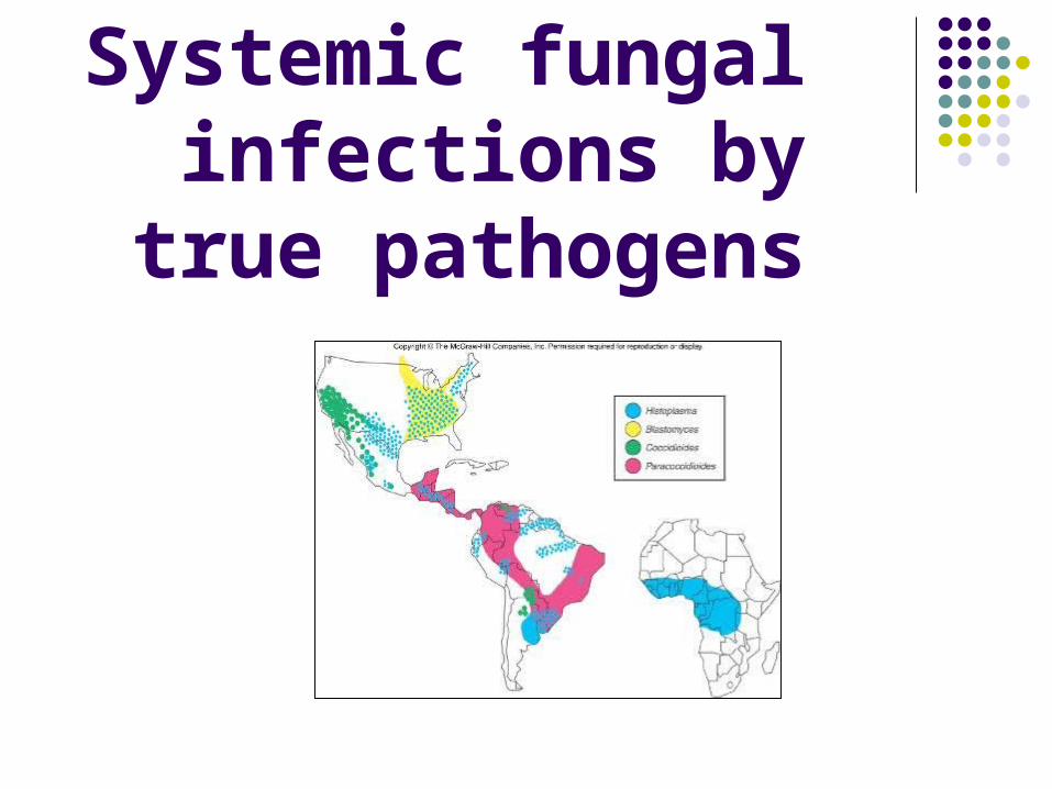
Systemic fungal infections by true
pathogens
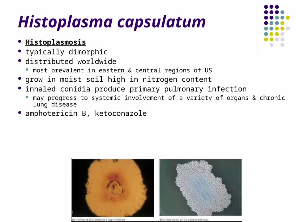
Histoplasma capsulatum Histoplasmosis typically dimorphic distributed worldwide
most prevalent in eastern & central regions of US grow in moist soil high in nitrogen content inhaled conidia produce primary pulmonary infection
may progress to systemic involvement of a variety of organs & chronic lung disease amphotericin B, ketoconazole

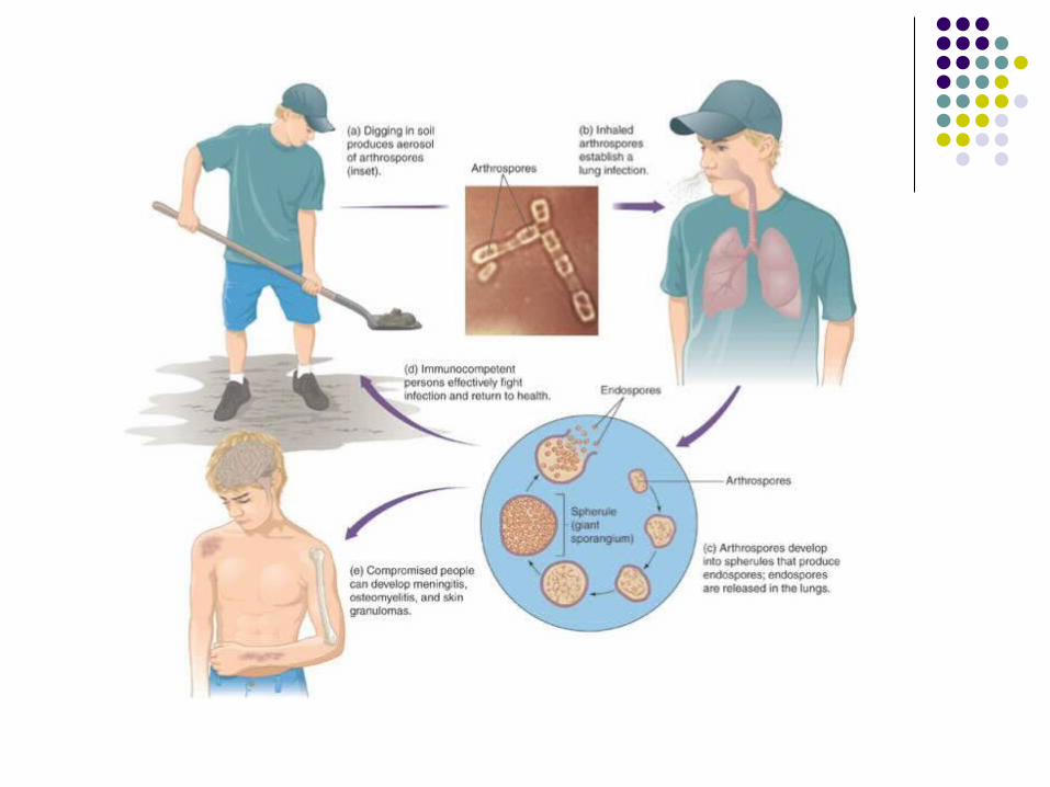
Coccidioides immitis Coccidioidomycosis distinctive morphology
blocklike arthroconidia in the free-living stage
arthrospores inhaled from dust Creates spherules and
nodules in the lungs lives in alkaline soils in
semiarid, hot climates endemic to southwestern US amphotericin B treatment
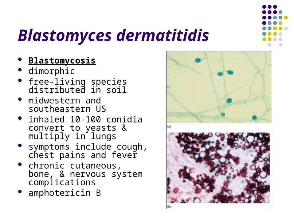
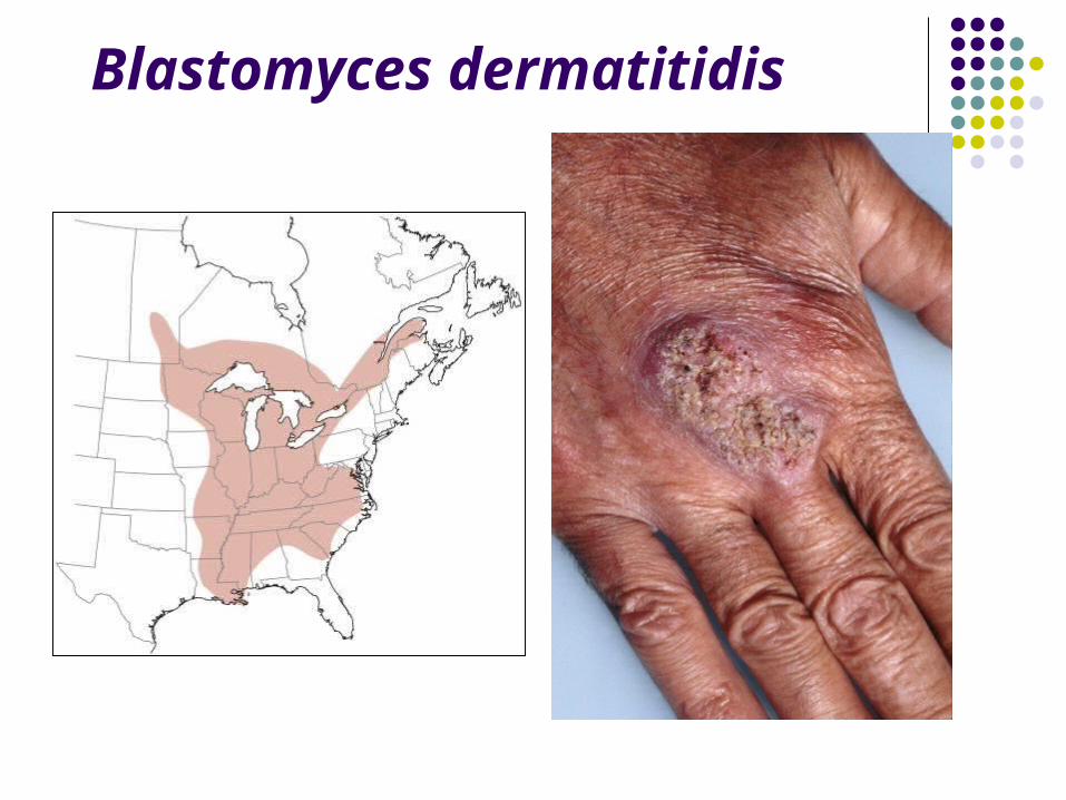
Blastomyces dermatitidis Blastomycosis dimorphic free-living species distributed
in soil midwestern and southeastern
US inhaled 10-100 conidia
convert to yeasts & multiply in lungs
symptoms include cough, chest pains and fever
chronic cutaneous, bone, & nervous system complications
amphotericin B
Blastomyces dermatitidis
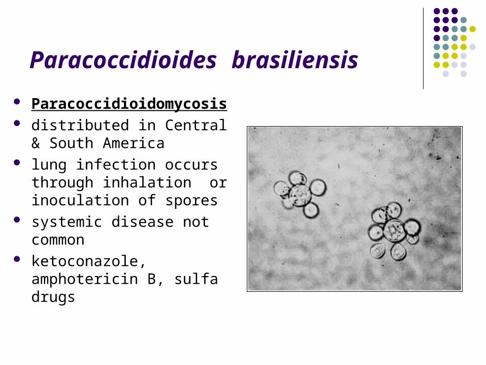
Paracoccidioides brasiliensis
Paracoccidioidomycosis distributed in Central & South
America lung infection occurs through
inhalation or inoculation of spores
systemic disease not common ketoconazole, amphotericin B,
sulfa drugs
Subcutaneous Mycoses
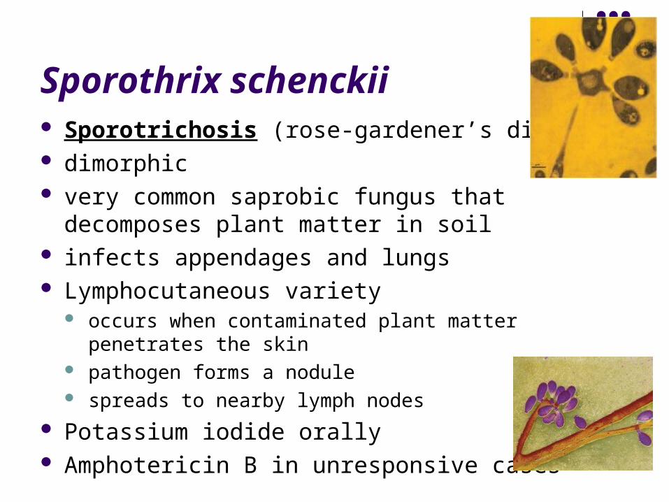
Sporothrix schenckii Sporotrichosis (rose-gardener’s disease) dimorphic very common saprobic fungus that decomposes
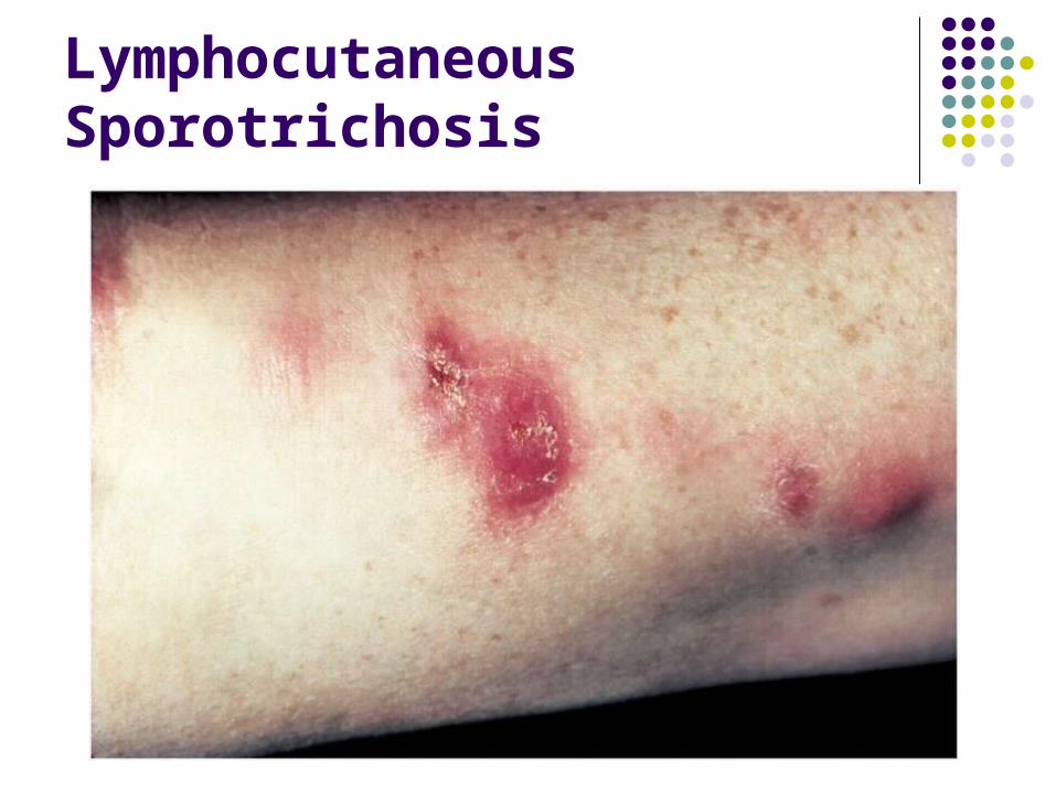
plant matter in soil infects appendages and lungs Lymphocutaneous variety
occurs when contaminated plant matter penetrates the skin pathogen forms a nodule spreads to nearby lymph nodes
Potassium iodide orally Amphotericin B in unresponsive cases
Lymphocutaneous Sporotrichosis
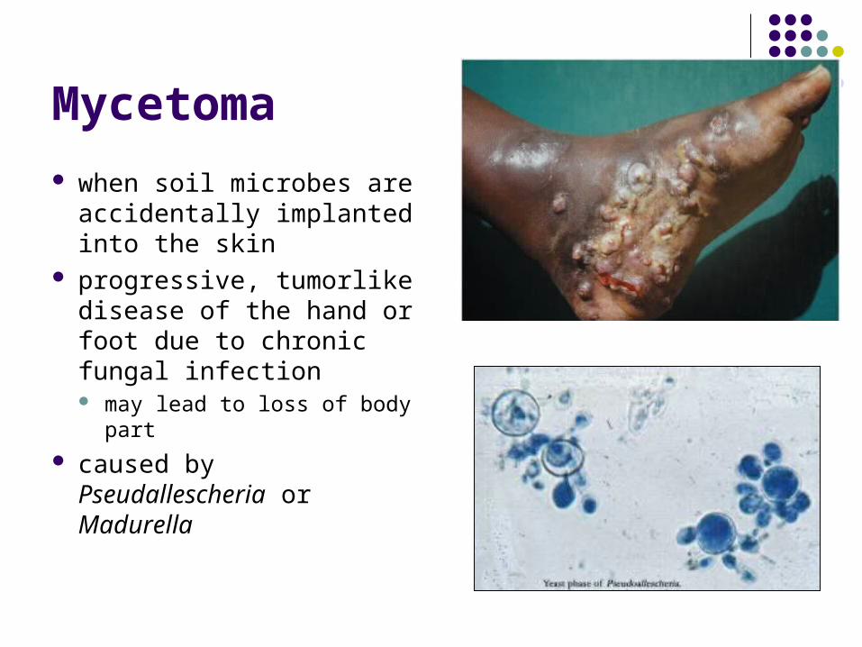
Mycetoma
when soil microbes are accidentally implanted into the skin
progressive, tumorlike disease of the hand or foot due to chronic fungal infection may lead to loss of body
part caused by
Pseudallescheria or Madurella
Cutaneous Mycoses
Cutaneous Mycoses
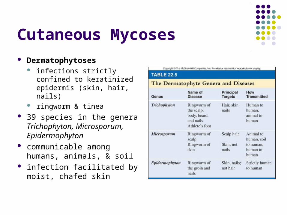
Dermatophytoses infections strictly confined
to keratinized epidermis (skin, hair, nails)
ringworm & tinea 39 species in the genera
Trichophyton, Microsporum, Epidermophyton
communicable among humans, animals, & soil
infection facilitated by moist, chafed skin
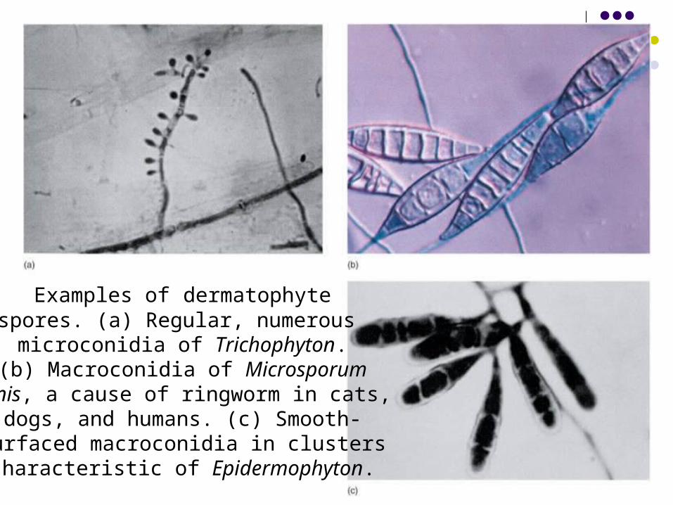
Examples of dermatophytespores. (a) Regular, numerous microconidia of Trichophyton.
(b) Macroconidia of Microsporumcanis, a cause of ringworm in cats,
dogs, and humans. (c) Smooth-surfaced macroconidia in clusterscharacteristic of Epidermophyton.

Dermatophytoses Ringworm of scalp
tinea capitis affects scalp & hair-bearing regions of head hair may be lost
Ringworm of body tinea corporis occurs as inflamed, red ring lesions anywhere on smooth skin
Ringworm of groin tinea cruris “jock itch” affects groin & scrotal regions
Ringworm or foot & hand tinea pedis & tinea manuum spread by exposure to public surfaces; occurs between digits & on soles
Ringworm of nails tinea unguium persistent colonization of the nails of the hands & feet that distorts the nail
bed
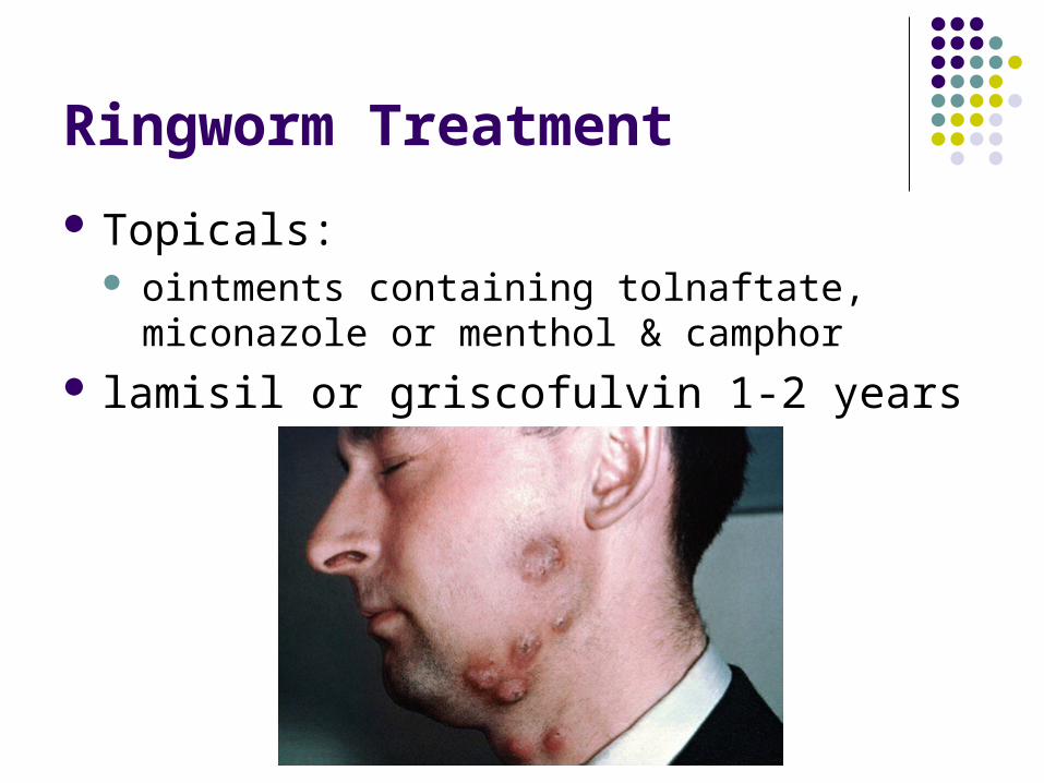
Ringworm Treatment
Topicals: ointments containing tolnaftate, miconazole or
menthol & camphor lamisil or griscofulvin 1-2 years
Superficial Mycoses
Superficial Mycoses
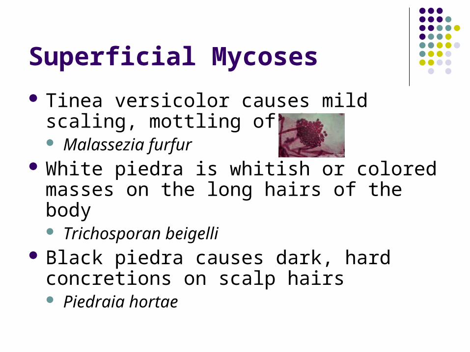
Tinea versicolor causes mild scaling, mottling of skin Malassezia furfur
White piedra is whitish or colored masses on the long hairs of the body Trichosporan beigelli
Black piedra causes dark, hard concretions on scalp hairs Piedraia hortae
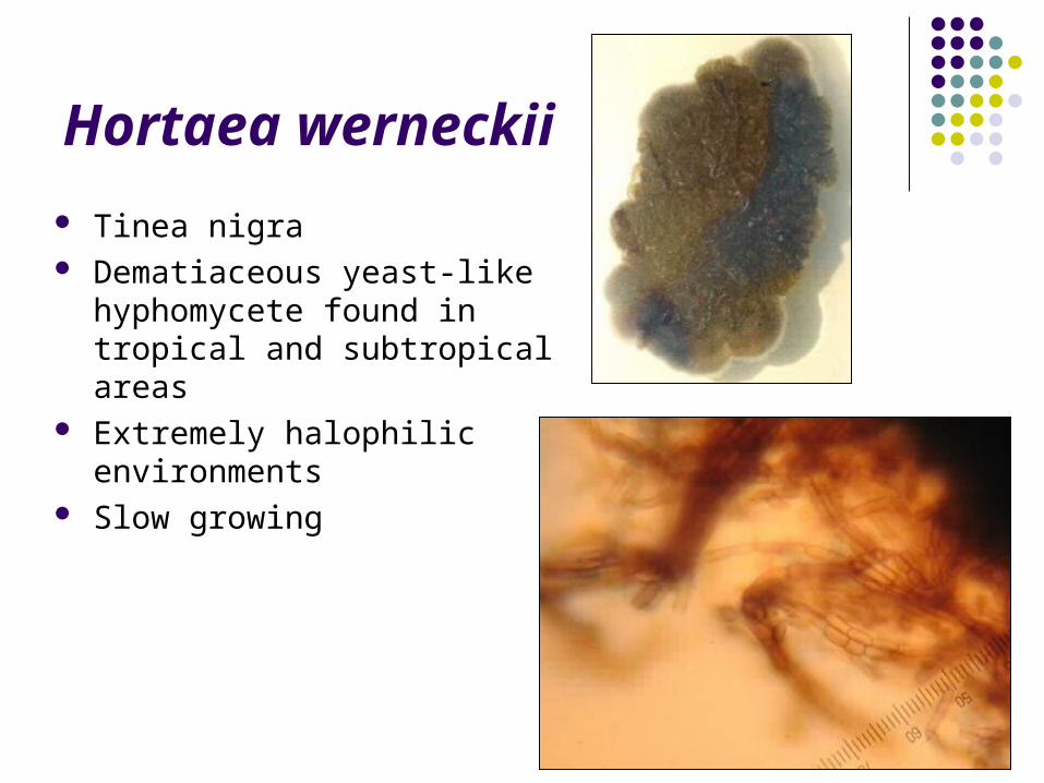
Hortaea werneckii
Tinea nigra Dematiaceous yeast-like
hyphomycete found in tropical and subtropical areas
Extremely halophilic environments
Slow growing
Opportunistic Pathogens
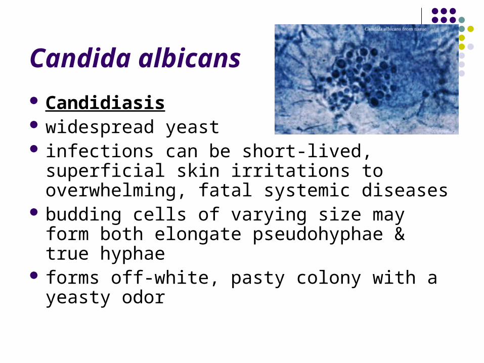
Candida albicans
Candidiasis widespread yeast infections can be short-lived, superficial skin
irritations to overwhelming, fatal systemic diseases
budding cells of varying size may form both elongate pseudohyphae & true hyphae
forms off-white, pasty colony with a yeasty odor
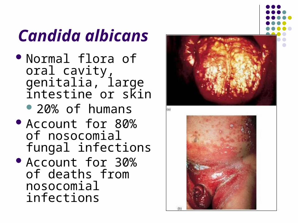
Candida albicans Normal flora of oral
cavity, genitalia, large intestine or skin 20% of humans
Account for 80% of nosocomial fungal infections
Account for 30% of deaths from nosocomial infections
Candida albicans
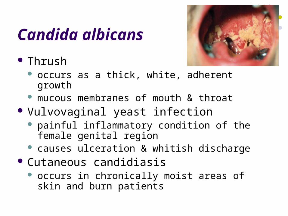
Thrush occurs as a thick, white, adherent growth mucous membranes of mouth & throat
Vulvovaginal yeast infection painful inflammatory condition of the female
genital region causes ulceration & whitish discharge
Cutaneous candidiasis occurs in chronically moist areas of skin and burn
patients
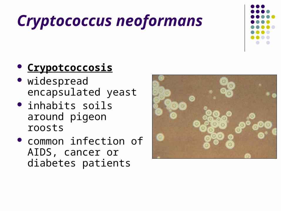
Cryptococcus neoformans
Crypotcoccosis widespread encapsulated
yeast inhabits soils around
pigeon roosts common infection of
AIDS, cancer or diabetes patients
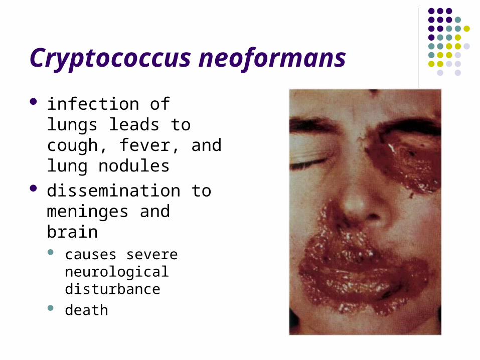
Cryptococcus neoformans
infection of lungs leads to cough, fever, and lung nodules
dissemination to meninges and brain causes severe
neurological disturbance death
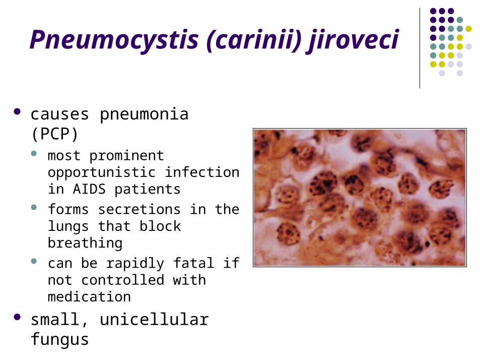
Pneumocystis (carinii) jiroveci
causes pneumonia (PCP) most prominent opportunistic
infection in AIDS patients forms secretions in the lungs
that block breathing can be rapidly fatal if not
controlled with medication
small, unicellular fungus
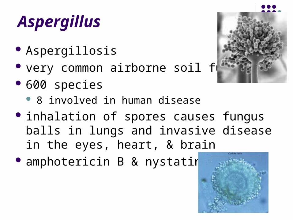
Aspergillus
Aspergillosis very common airborne soil fungus 600 species
8 involved in human disease inhalation of spores causes fungus balls in
lungs and invasive disease in the eyes, heart, & brain
amphotericin B & nystatin
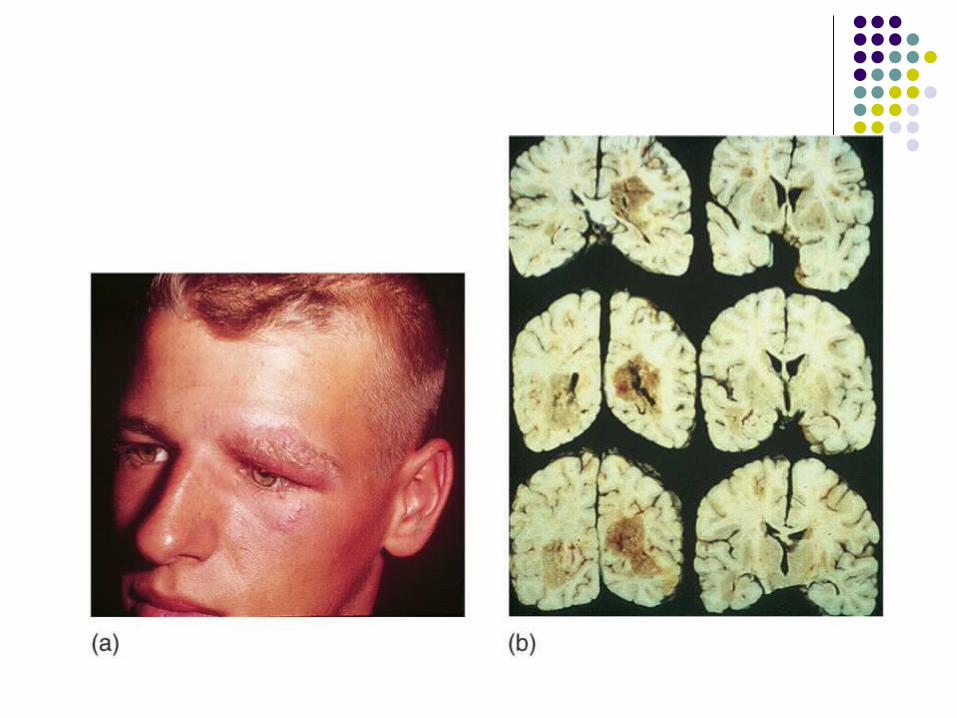
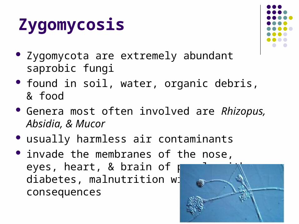
Zygomycosis
Zygomycota are extremely abundant saprobic fungi
found in soil, water, organic debris, & food Genera most often involved are Rhizopus, Absidia,
& Mucor usually harmless air contaminants invade the membranes of the nose, eyes, heart, &
brain of people with diabetes, malnutrition with severe consequences
Mycotoxicoses
Fungal toxins lead to mycotoxicoses usually caused by eating poisonous or
hallucinogenic mushrooms aflatoxin toxic and carcinogenic
grains, corn peanuts lethal to poultry and livestock
Stachybotrys chartarum sick building syndrome severe hematologic and neurological damage
Parasites
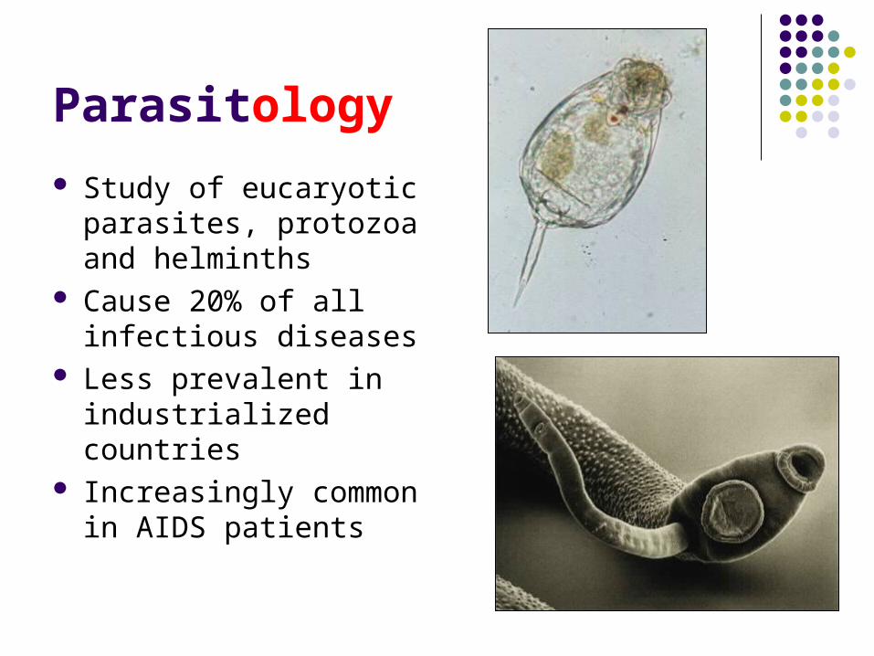
Parasitology
Study of eucaryotic parasites, protozoa and helminths
Cause 20% of all infectious diseases
Less prevalent in industrialized countries
Increasingly common in AIDS patients
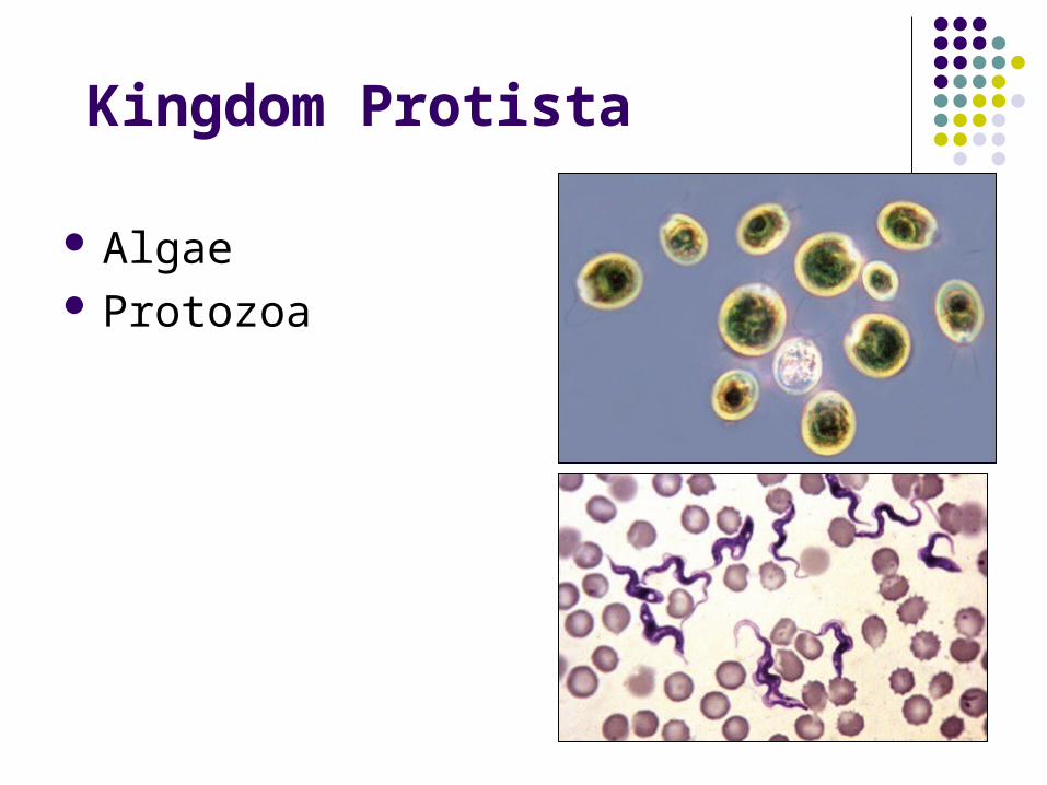
Kingdom Protista
Algae Protozoa
Protozoa
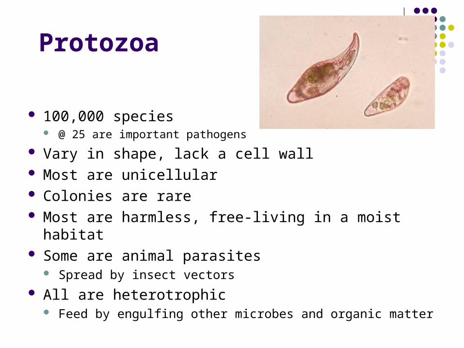
100,000 species @ 25 are important pathogens
Vary in shape, lack a cell wall Most are unicellular Colonies are rare Most are harmless, free-living in a moist habitat Some are animal parasites
Spread by insect vectors
All are heterotrophic Feed by engulfing other microbes and organic matter
Protozoa
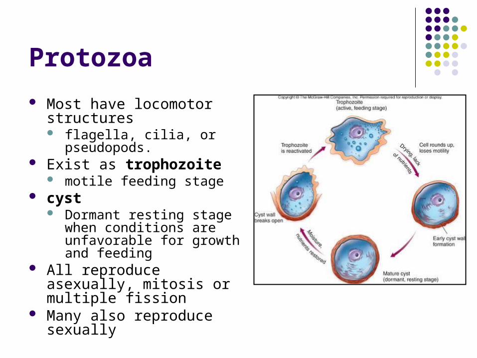
Most have locomotor structures flagella, cilia, or pseudopods.
Exist as trophozoite motile feeding stage
cyst Dormant resting stage when
conditions are unfavorable for growth and feeding
All reproduce asexually, mitosis or multiple fission
Many also reproduce sexually
Protozoan Classification
Difficult because of diversity Simple grouping is based on method of
motility, reproduction, and life cycle
Protozoan Classification
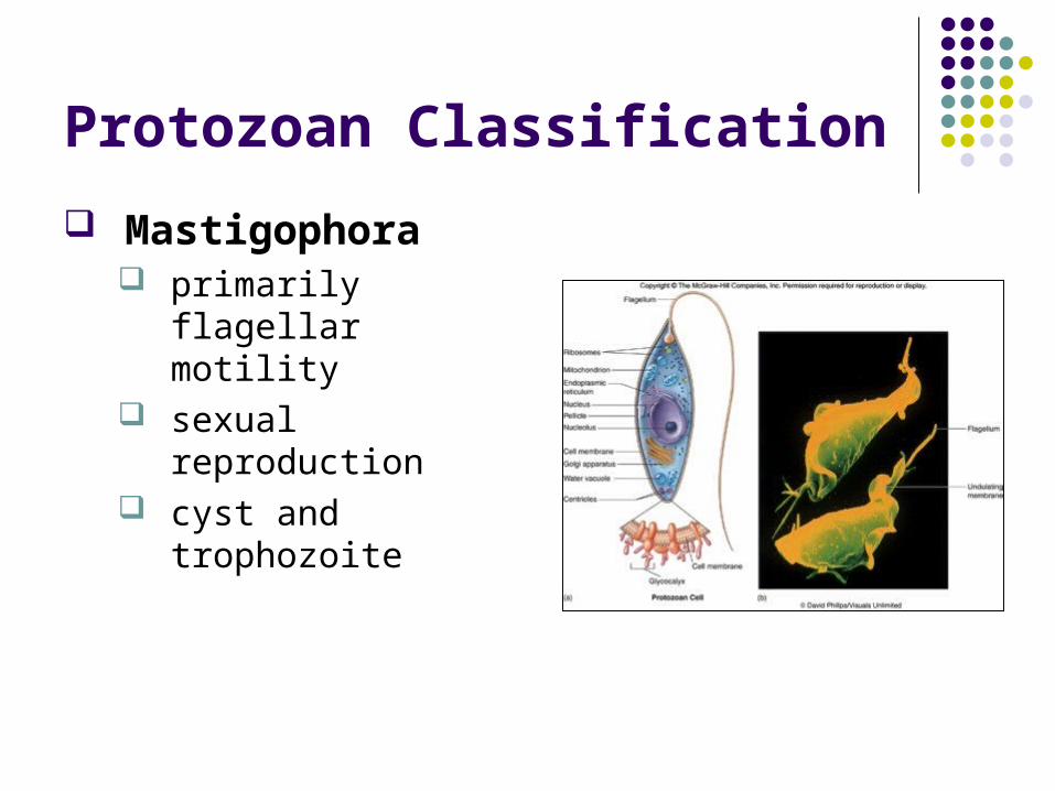
Mastigophora primarily flagellar
motility sexual reproduction cyst and trophozoite
Protozoan Classification
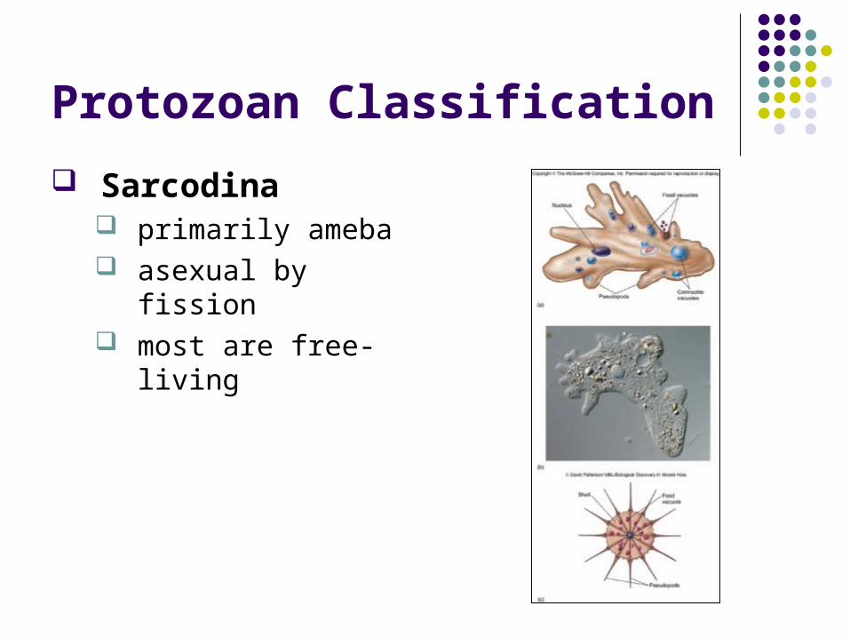
Sarcodina primarily ameba asexual by fission most are free-living
Protozoan Classification
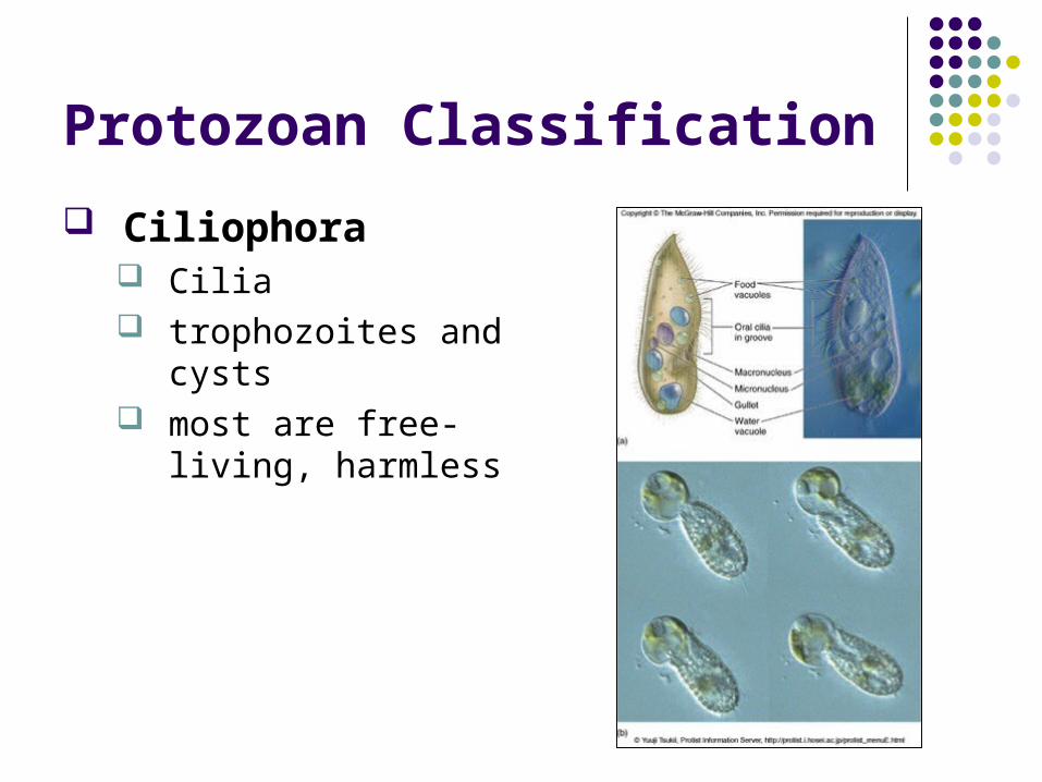
Ciliophora Cilia trophozoites and
cysts most are free-living,
harmless
Protozoan Classification
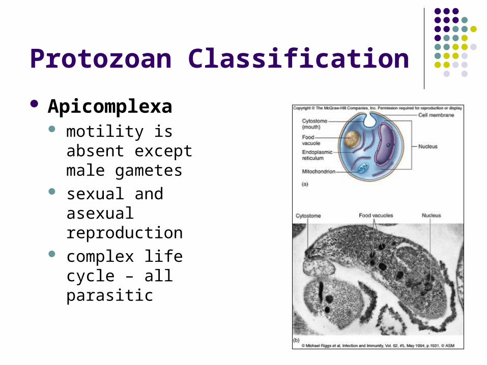
Apicomplexa motility is absent
except male gametes sexual and asexual
reproduction complex life cycle – all
parasitic
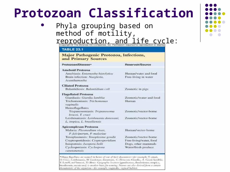
Protozoan Classification Phyla grouping based on method of
motility, reproduction, and life cycle:
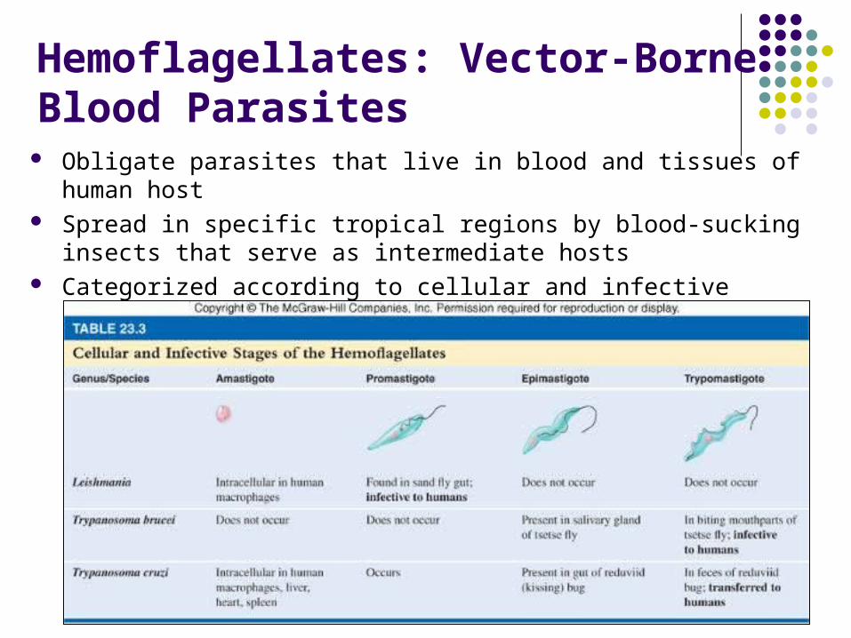
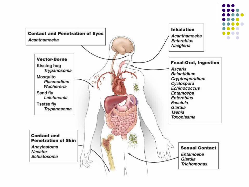
Hemoflagellates: Vector-Borne Blood Parasites
Obligate parasites that live in blood and tissues of human host Spread in specific tropical regions by blood-sucking insects that serve
as intermediate hosts Categorized according to cellular and infective stages
Protozoal Diseases
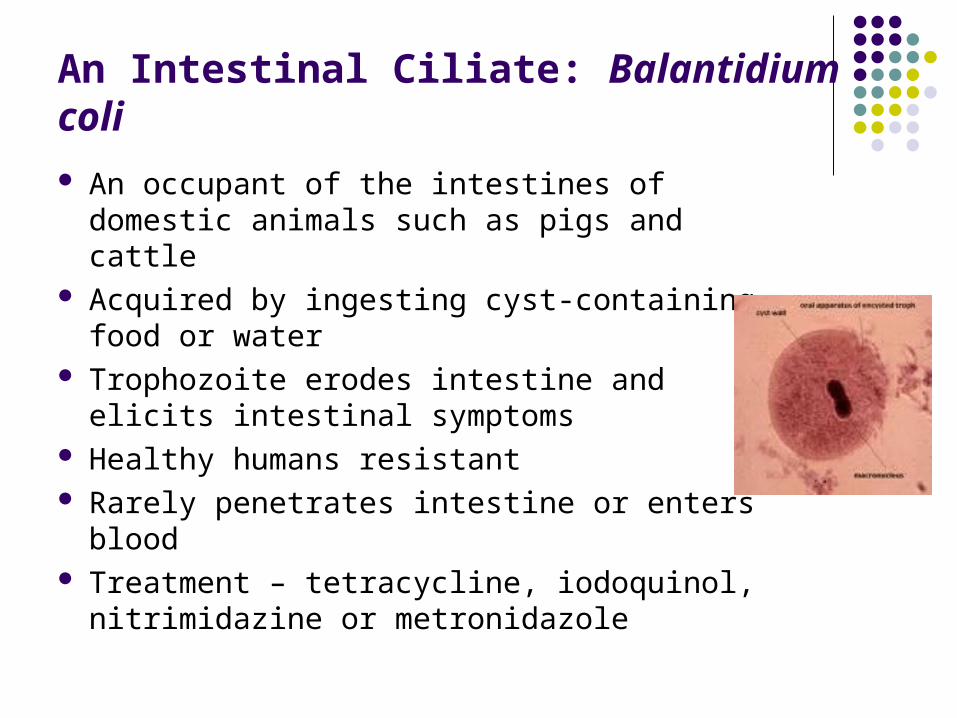
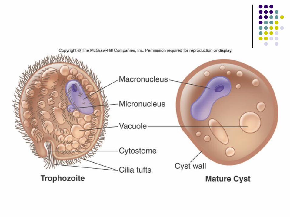
An Intestinal Ciliate: Balantidium coli
An occupant of the intestines of domestic animals such as pigs and cattle
Acquired by ingesting cyst-containing food or water
Trophozoite erodes intestine and elicits intestinal symptoms
Healthy humans resistant Rarely penetrates intestine or enters blood Treatment – tetracycline, iodoquinol,
nitrimidazine or metronidazole
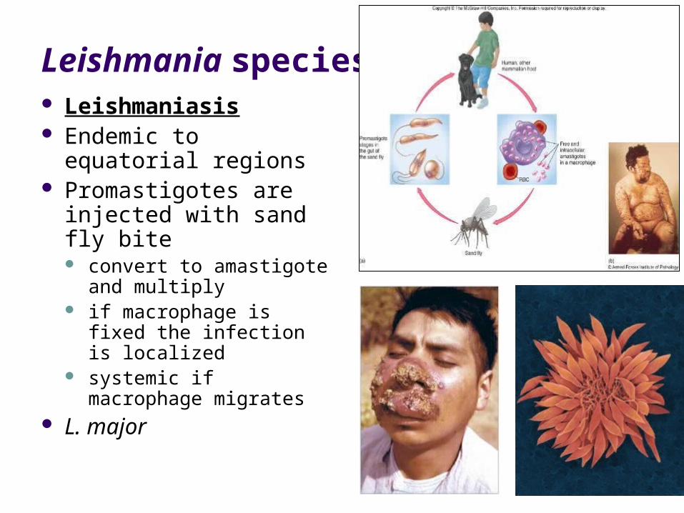
Leishmania species Leishmaniasis Endemic to equatorial
regions Promastigotes are
injected with sand fly bite convert to amastigote
and multiply if macrophage is fixed
the infection is localized systemic if macrophage
migrates L. major

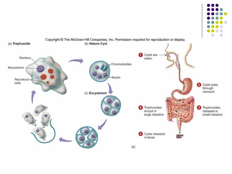
Entamoeba histolytica
Amebiasis Alternates between a large trophozoite Motile by means of pseudopods and a smaller
nonmotile cyst Humans are the primary hosts Ingested Carried by 10% of world population
Entamoeba histolytica
Cysts swallowed and travel to small intestine alkaline pH and digestive juices stimulate cysts to release 4
trophozoites
Trophozoites attach, multiply, actively move about and feed
Asymptomatic in 90% of patients Ameba may secrete enzymes that dissolve tissues
and penetrate deeper layers of the mucosa Causing dysentery, abdominal pain, fever, diarrhea
and weight loss
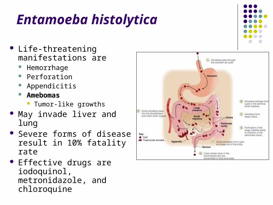
Entamoeba histolytica
Life-threatening manifestations are Hemorrhage Perforation Appendicitis Amebomas
Tumor-like growths May invade liver and lung Severe forms of disease
result in 10% fatality rate Effective drugs are
iodoquinol, metronidazole, and chloroquine
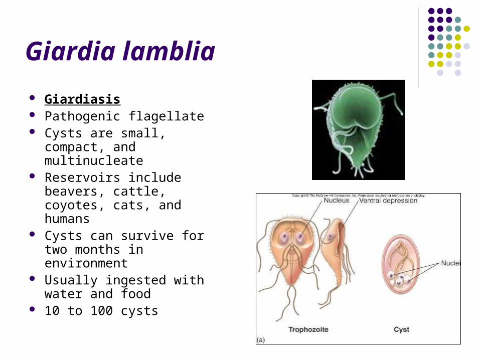
Giardia lamblia
Giardiasis Pathogenic flagellate Cysts are small, compact,
and multinucleate Reservoirs include beavers,
cattle, coyotes, cats, and humans
Cysts can survive for two months in environment
Usually ingested with water and food
10 to 100 cysts
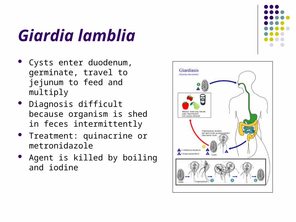
Giardia lamblia Cysts enter duodenum, germinate,
travel to jejunum to feed and multiply
Diagnosis difficult because organism is shed in feces intermittently
Treatment: quinacrine or metronidazole
Agent is killed by boiling and iodine
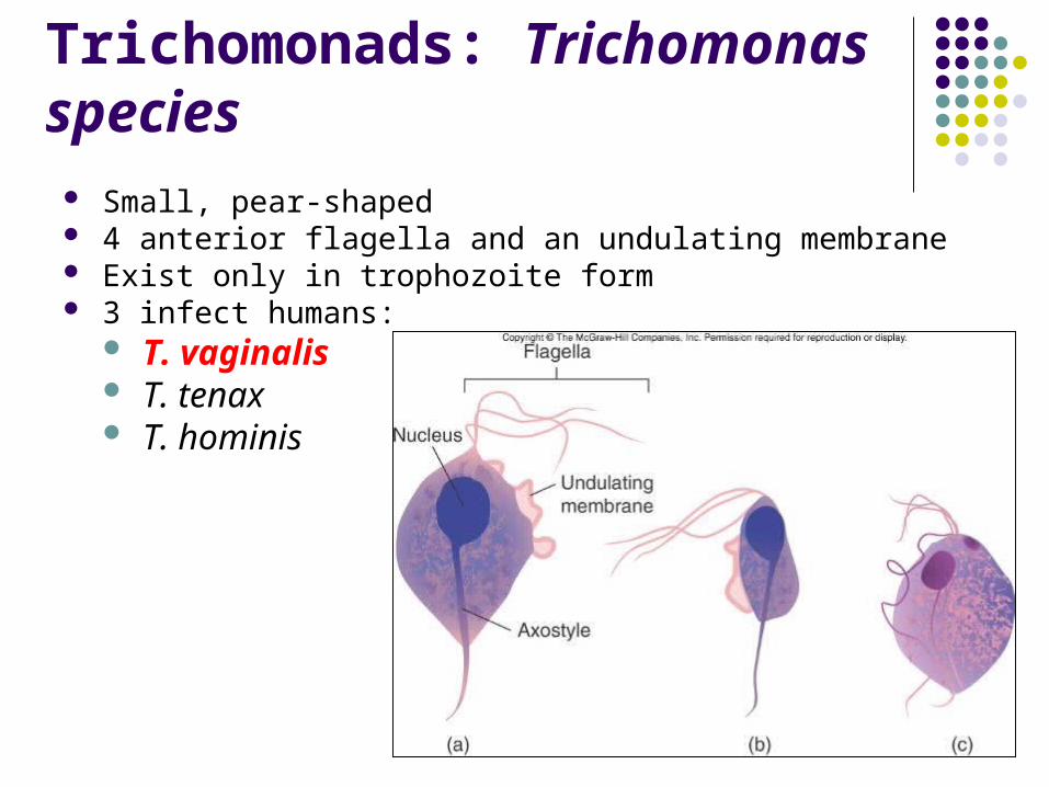
Trichomonads: Trichomonas species
Small, pear-shaped 4 anterior flagella and an undulating membrane Exist only in trophozoite form 3 infect humans:
T. vaginalis T. tenax T. hominis
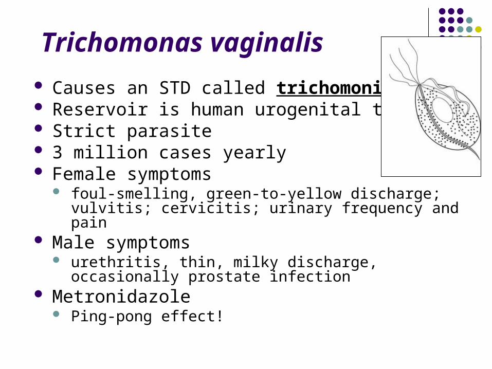
Trichomonas vaginalis
Causes an STD called trichomoniasis Reservoir is human urogenital tract Strict parasite 3 million cases yearly Female symptoms
foul-smelling, green-to-yellow discharge; vulvitis; cervicitis; urinary frequency and pain
Male symptoms urethritis, thin, milky discharge, occasionally prostate
infection Metronidazole
Ping-pong effect!
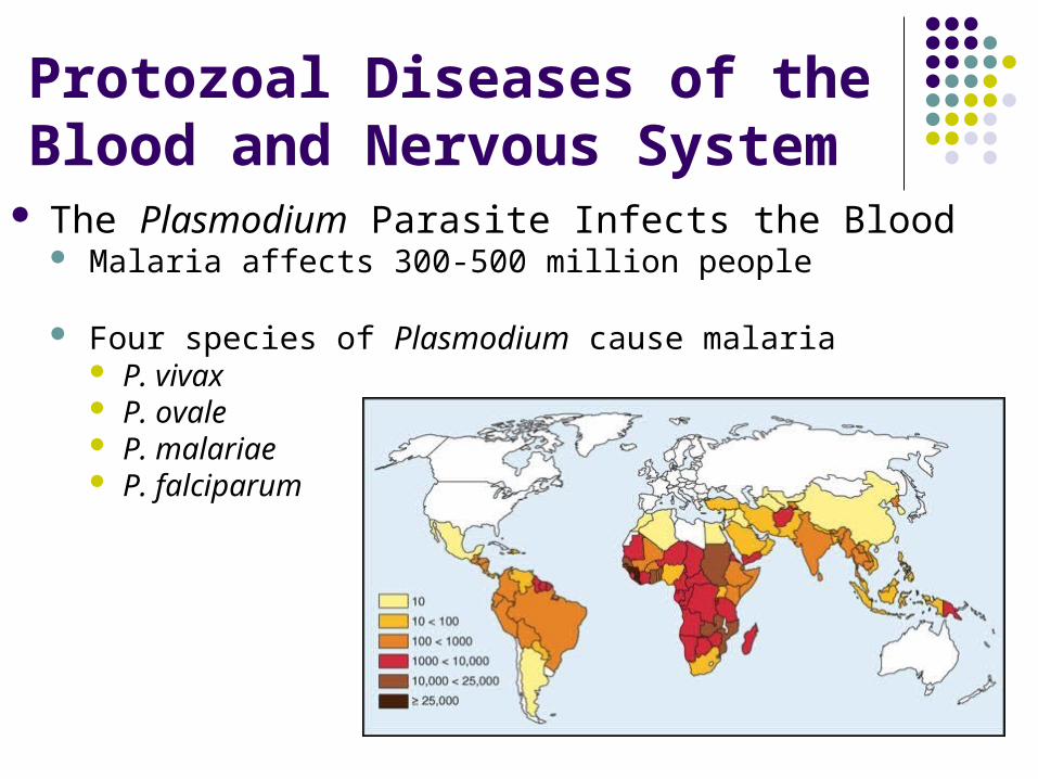
Protozoal Diseases of the Blood and Nervous System
The Plasmodium Parasite Infects the Blood Malaria affects 300-500 million people
Four species of Plasmodium cause malaria P. vivax P. ovale P. malariae P. falciparum
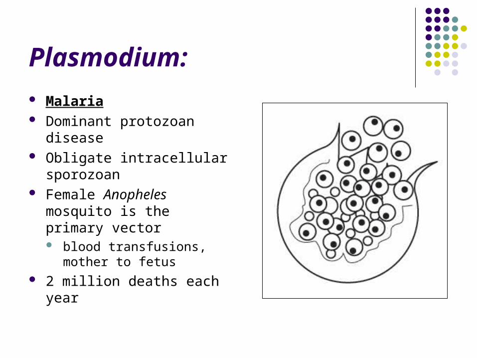
Plasmodium:
Malaria Dominant protozoan
disease Obligate intracellular
sporozoan Female Anopheles
mosquito is the primary vector blood transfusions, mother
to fetus 2 million deaths each year
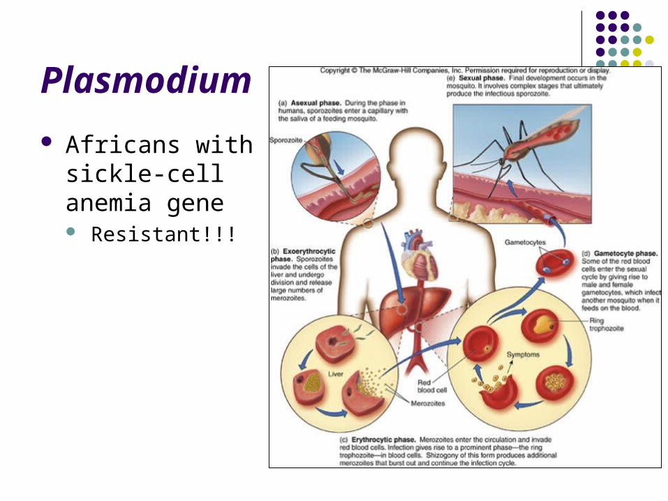
Plasmodium
Africans with sickle-cell anemia gene Resistant!!!
Trypanosoma species and Trypanosomiasis
Distinguished by their infective stage trypomastigote
elongate, spindle-shaped cell with tapered ends, eel-like motility
2 types of trypanosomiasis: T. brucei
African sleeping sickness T. cruzi
Chagas disease
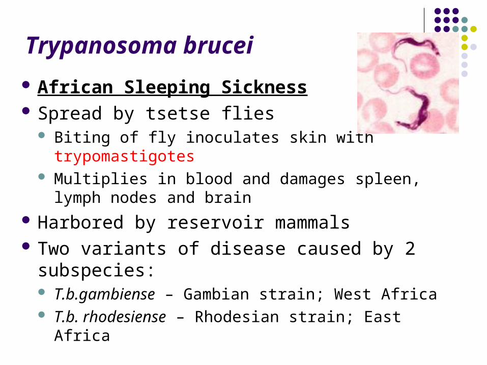
Trypanosoma brucei
African Sleeping Sickness Spread by tsetse flies
Biting of fly inoculates skin with trypomastigotes Multiplies in blood and damages spleen, lymph
nodes and brain Harbored by reservoir mammals Two variants of disease caused by 2
subspecies: T.b.gambiense – Gambian strain; West Africa T.b. rhodesiense – Rhodesian strain; East Africa
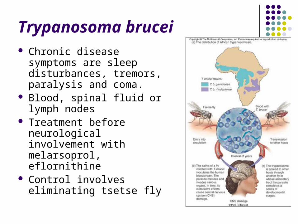
Trypanosoma brucei Chronic disease symptoms
are sleep disturbances, tremors, paralysis and coma.
Blood, spinal fluid or lymph nodes
Treatment before neurological involvement with melarsoprol, eflornithine
Control involves eliminating tsetse fly
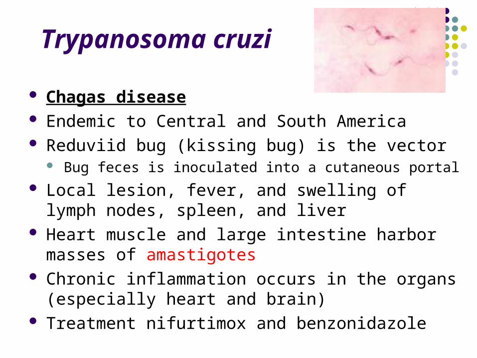
Trypanosoma cruzi
Chagas disease Endemic to Central and South America Reduviid bug (kissing bug) is the vector
Bug feces is inoculated into a cutaneous portal
Local lesion, fever, and swelling of lymph nodes, spleen, and liver
Heart muscle and large intestine harbor masses of amastigotes
Chronic inflammation occurs in the organs (especially heart and brain)
Treatment nifurtimox and benzonidazole
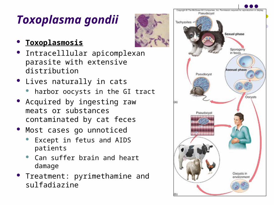
Toxoplasma gondii
Toxoplasmosis Intracelllular apicomplexan
parasite with extensive distribution Lives naturally in cats
harbor oocysts in the GI tract Acquired by ingesting raw meats
or substances contaminated by cat feces
Most cases go unnoticed Except in fetus and AIDS patients Can suffer brain and heart damage
Treatment: pyrimethamine and sulfadiazine
Parasitic Helminths
Parasitic Helminths
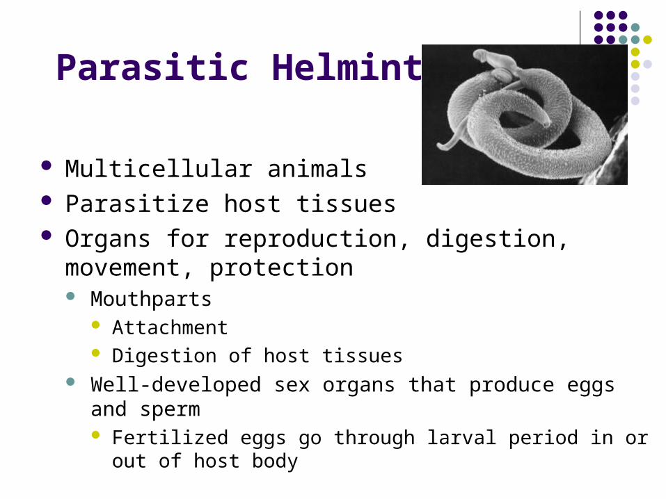
Multicellular animals Parasitize host tissues Organs for reproduction, digestion, movement,
protection Mouthparts
Attachment Digestion of host tissues
Well-developed sex organs that produce eggs and sperm Fertilized eggs go through larval period in or out of host body
Helminths
Flatworms (Phylum Platyhelminthes) do not have respiratory or circulatory
structures, or a digestive tractCestodes (tapeworms)Trematodes or flukes
Roundworms (Phylum Nematoda)
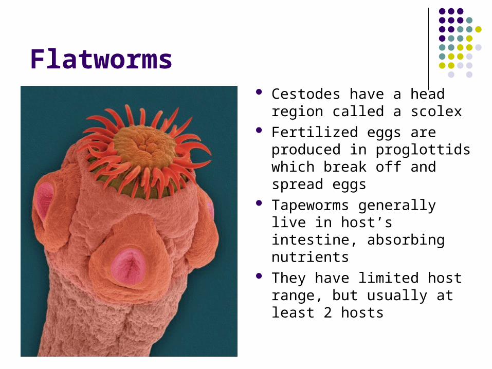
Flatworms Cestodes have a head
region called a scolex Fertilized eggs are
produced in proglottids which break off and spread eggs
Tapeworms generally live in host’s intestine, absorbing nutrients
They have limited host range, but usually at least 2 hosts
Flatworms
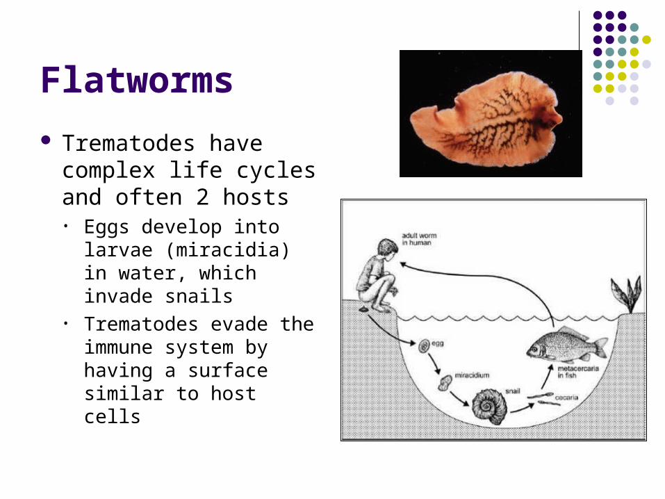
Trematodes have complex life cycles and often 2 hosts• Eggs develop into
larvae (miracidia) in water, which invade snails
• Trematodes evade the immune system by having a surface similar to host cells
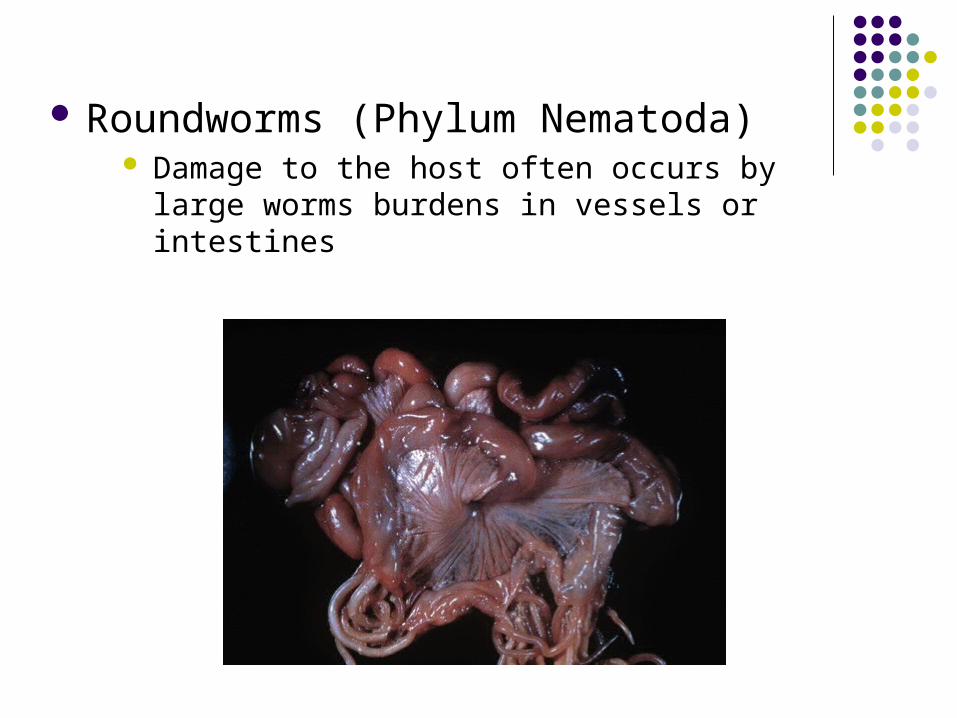
Roundworms (Phylum Nematoda) Damage to the host often occurs by large worms
burdens in vessels or intestines
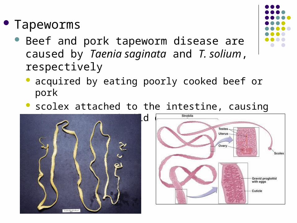
Tapeworms Beef and pork tapeworm disease are caused by
Taenia saginata and T. solium, respectively acquired by eating poorly cooked beef or pork scolex attached to the intestine, causing obstruction and
mild diarrhea
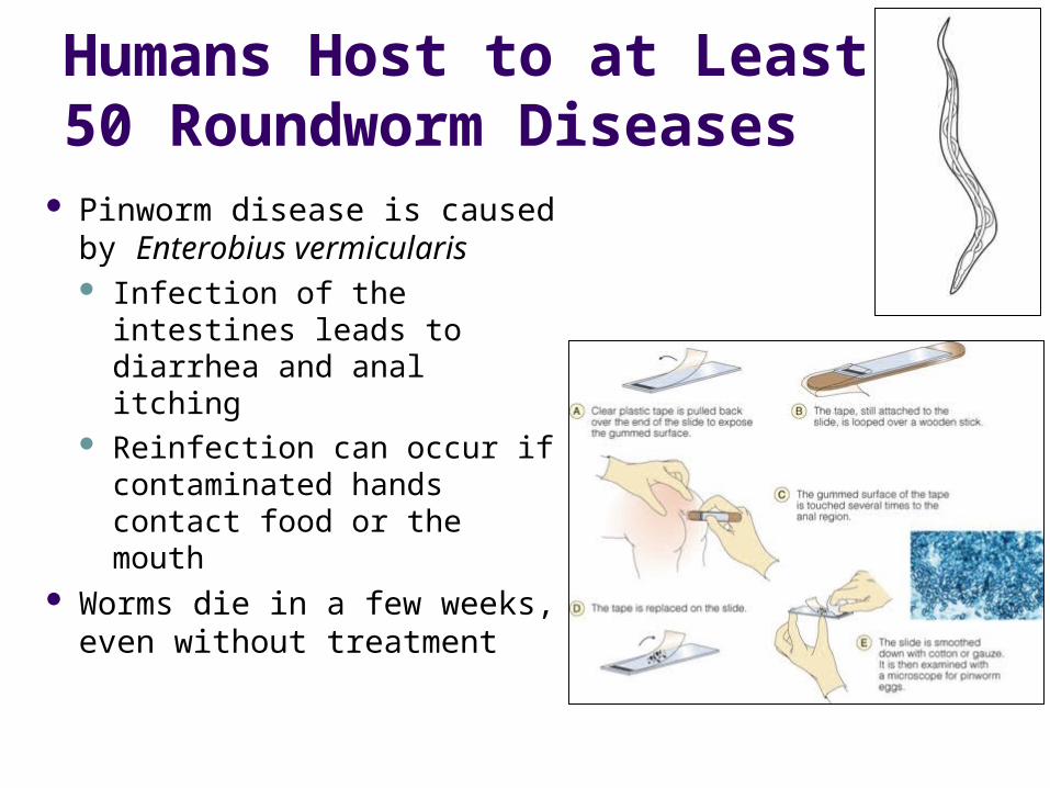
Humans Host to at Least 50 Roundworm Diseases
Pinworm disease is caused by Enterobius vermicularis Infection of the intestines
leads to diarrhea and anal itching
Reinfection can occur if contaminated hands contact food or the mouth
Worms die in a few weeks, even without treatment
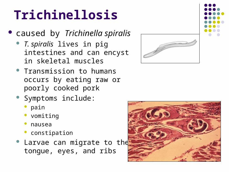
Trichinellosis caused by Trichinella spiralis
T. spiralis lives in pig intestines and can encyst in skeletal muscles
Transmission to humans occurs by eating raw or poorly cooked pork
Symptoms include: pain vomiting nausea constipation
Larvae can migrate to the tongue, eyes, and ribs
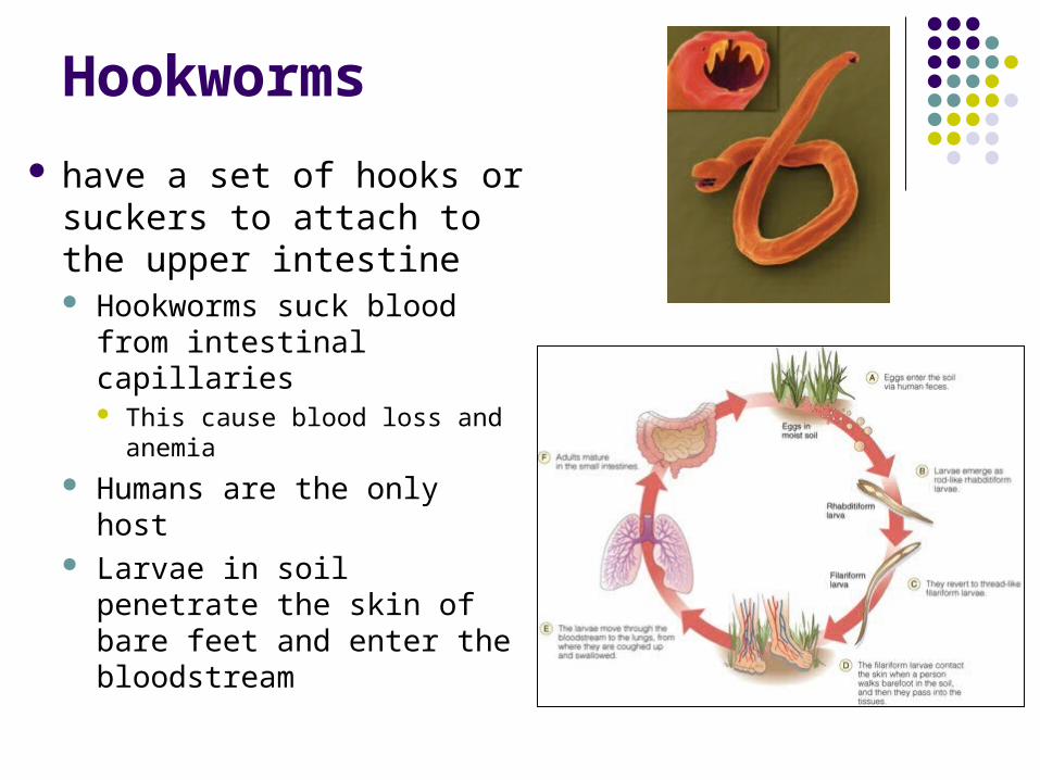
Hookworms
have a set of hooks or suckers to attach to the upper intestine Hookworms suck blood
from intestinal capillaries This cause blood loss and
anemia
Humans are the only host Larvae in soil penetrate the
skin of bare feet and enter the bloodstream