fungal and protozoal mini-case history studies for courses
TRANSCRIPT

Fungal and Protozoal Mini-Case History Studies for Courses InvolvingMedical Microbiology
Resource Type: Curriculum: Classroom
Publication Date: 10/12/2000
AuthorsDiana CundellPhiladelphia UniversityPhiladelphia, PA 19144USAEmail: [email protected]
Abstract
26 fungal and protozoal mini-case history studies are designed as problem solving exercises to allow students to test theirpowers of deduction in identifying and treating important human pathogens, using clear cut cases involving "at risk"individuals wherever possible.
Activity
Invitation for User Feedback. If you have used the activity and would like to provide feedback, please send an e-mail [email protected]. Feedback can include ideas which complement the activity and new approaches forimplementing the activity. Your comments will be added to the activity under a separate section labeled "Feedback."Comments may be edited.
Editor’s Note (2008): This Curriculum Resource was published prior to establishment of current criteria of submission, andas such, does not contain all criteria required of current publications. However, the Editorial Committee felt that the activityitself remained worthwhile and relevant, and encourages potential users to contact the authors for clarification as needed. If you do update this activity for use with your students, and are interested in updating the resource for distribution in thelibrary, please contact ASM at [email protected].
INTRODUCTION
Background.
Mini-case history studies can be used as classroom exercises or given to students to work through as part of a writtenexamination. In this activity, 26 mini-case history studies divided into two sections, fungi and protozoa, are presented.Suggested answer content for each component of the mini-case histories is italicized next to the question.
To incorporate these mini-case history studies into the curriculum, begin by presenting them to the students as a classroomor laboratory exercise. Divide students into groups of two to four individuals, assign one mini-case history study to eachgroup, and allow them 15 to 20 minutes to work through it. This gives adequate time to discuss the problem and determineanswers to the questions.
Subsequently, when each new sub-section of mycology and protozoology is covered, use one or two mini-case histories toaccompany the lectures as problem-based learning exercises. Assign the mini-case history studies once the sub-section hasbeen completed and give the students a few days to prepare a paragraph, which shows their conclusions and method ofarriving at each conclusion, for each mini-case history study.
[Note: My classes are usually a mixture of Physician Assistant, Biology/Pre-Med., and Environmental Science majors andbetween 80 and 90% of all types of students enjoy these "sleuthing" exercises. All mini-case histories here have also beena component of my students’ examinations at some point (usually a maximum of two mini-case histories each for mycologyand protozoology) and this has helped in ironing out any confusing descriptions or sayings.]
PROCEDURE
Materials.Fungal and Protozoal Mini-Case History Studies for courses involving Medical Microbiology--Answer KeyFungal and Protozoal Mini-Case History Studies for courses involving Medical Microbiology–A list of mini-cases provided foreach classroom or laboratory exercise, grouped as fungi and protozoa.
Instructor Version.The mini-case history studies may be used in several ways.
A. Possible applications.
The mini-case histories included in this activity have been utilized successfully as classroom exercises, take homeassignments, and examination material. The exercises tend to work best when a time limit is identified and when studentsare encouraged to try and work from memory rather than scanning through their notes.
MicrobeLibrary http://archive.microbelibrary.org/edzine/details_print.asp?id=497&lang=
1 of 3 3/20/2012 4:17 PM

Classroom exercise. The classroom exercise is useful, especially to the professor with a big class, since it encouragesstudents' participation and critical thinking without being overly time consuming. Groups of two to four students appear tooperate the most effectively for this exercise; lone students may feel somewhat "swamped" by the information, while largergroups may encourage one or two individuals to analyze the mini-case history with the remainder of the group not becomingtruly involved. A time limit of 15 to 20 minutes for each mini-case history has been found to be optimal, allowing studentssufficient time to digest the data but not too long to make them doubt their conclusions or "over-analyze" the information(often the case with the very bright student). Varying the students within each group and the spokesperson for each groupalso adds challenge to the exercise.
Take home assignment. Once the students are familiar with the steps needed to work through a mini-case history, they areready to tackle one on their own. The ability to do this outside class and without help or hindrance from other groupmembers may encourage the quieter student to perform more efficiently. In addition, many students, especially foreignlanguage students, may be more able to put their ideas into writing rather than into an oral presentation. This kind ofassignment, unlike the classroom exercise, allows more time and imposes less pressure. Again, it is advisable to encouragestudents to set some kind of time limit to prevent over-analysis, and to try and work as much from memory as possible toencourage the development of microbiological algorithms.
Examination material. The microbiological algorithms generated by the classroom and take home assignments may be testedas a component of the microbiology examination. Ideally, a student should be able to use the series of steps they havememorized to successfully analyze a mini-case history under examination conditions. However, many students, in the "heatof the moment" have trouble doing this, so a professor might want to allow the use of a limited amount of notes made bythe student to be used in the examination (similar to formulae used in mathematics and physics examinations).
B. How I use these studies.
Since the process of working through a mini-case history is a novel skill, I usually begin with the classroom or laboratoryexercise; at least 50 to 75% of the group grasp the procedure on the first occasion and can help teach the remaining classmembers. I give the students guidance as to how the process works by presenting two example mini-case histories andshowing them each step taken to answer the associated questions. Each group of two to four students is then given thesame two mini-case histories and asked to work through them for 15 to 20 minutes. They are asked to follow four steps:
Underline all components of the mini-case history that are important in reaching a diagnosis (e.g. laboratory data andrisk factors such as vacations recently taken, occupations, or lifestyle practices).
1.
Make a list of all appropriate species that fall into the category being studied and produce diseases of the kindpresented in the mini-case history. For example, in the case of a gram-positive coccus causing a skin infection write alist of all gram-positive cocci capable of causing this condition.
2.
Take the list and begin to eliminate candidates from it by using the information from the mini-case history. Discusseach candidate with your classmates until you reach a consensus.
3.
Once you have identified the responsible organism, go on to determine a treatment strategy, other at-riskindividuals, and methods of infection prevention, as appropriate.
4.
I ask the students to use their notes as little as possible during this exercise to allow them to develop a "feel" for thesubject and improve their critical thinking skills. They are not, however, prevented from using their notes at this point. Oncethey have seen the method for processing the mini-case history data, each group in the class is assigned a different, thirdmini-case history analysis and given 15 to 20 minutes to repeat the steps.
When the groups have finished deliberating, each member answers one of the questions associated with their mini-casehistory. This ensures that all students understand the process and could be assumed to be able to repeat it on their own. Inaddition, students are asked to comment on the ease with which they could identify the organism: Was the identityobvious? What pieces of information would they take away from the mini-case history to make it more difficult? How wouldthey improve the mini-case history? Asking students to comment on the histories helps them focus on the concepts beingdemonstrated. Many brighter students generate ideas on the subject even during the first mini-case history session.
Once students understand how to proceed with a mini-case history, they can be encouraged to pursue them alone on a takehome assignment. Students are asked to use a similar time limit as during class (15 to 20 minutes) to analyze the mini-casehistory and then write a paragraph explaining their method of reaching conclusions, any further thoughts they have aboutthe condition the patient is suffering from and the "story" accompanying it. They are essentially asked to critique themini-case, looking for loopholes, ascertaining the ease with which it could be solved, and sharing any ideas that mightimprove it.
By the time my students reach the examination stage, they have already processed many of these mini-case histories. Forthe microbiology course taught at this institution, four microbiology examinations are given each semester with the last twocontaining a maximum of two mini-case histories in any particular sub-section of microbiology. This has been successful inthe past, since the combination of mini-case history analyses, short answer questions, fill-in-the-blank, and multiplematching in my examinations has allowed me to evaluate a wide variety of students’ skills.
C. The concept of a mini-case history.
These mini-case histories were generated over a period of six weeks during the summer of 1998 using clinical andmicrobiological information gleaned from four separate microbiology textbooks. In making each mini-case, I initially focusedon the symptoms and manifestation of the disease and the microscopic appearance of the organism causing it–informationthat the students gained in lectures and from the course textbook. By gradually adding pieces of information–such as theage of the patient, a risk factor, or a laboratory finding–other similar organisms were eliminated as potential causativeagents so that the identity of the pathogen became obvious. The only difficulty was to balance how much information togive, so the answer wasn’t totally obvious or completely obscure.
In formulating the mini-cases, I used risk factors including lifestyle, vacations, and age as much as possible to try and instillin the students a sense that these could provide real pointers as to the identity of a microorganism in a sick patient. I felt it
MicrobeLibrary http://archive.microbelibrary.org/edzine/details_print.asp?id=497&lang=
2 of 3 3/20/2012 4:17 PM

was important to make the situations as realistic as possible so that patients used "everyday" language. In addition, I triedto show that diagnostic mistakes are made, e.g. that conditions which appeared to be child abuse, irritable bowel disease,or lactose intolerance may actually be microbial in origin and that all information, no matter how apparently trite, may beimportant. Finally, the idea of the mini-case history was that it really would appear as a kind of "detective story" with a"plot," so students would enjoy "sleuthing" out the "culprit"–and learn new skills in the process.
Student Version.
Classroom Exercise: You have been given a mini-case history in the area of ___________, which was covered in yourlecture sections during the past week(s). You have been assigned to a group of four students and, as a group, youneed to work through mini-case history number ____ from the list given, during the next 15 to 20 minutes. Use thefollowing strategy to diagnose the mini-case. First, underline all components of the mini-case history that you thinkare important in reaching a diagnosis. Second, make a list of all fungal/protozoal species that fall into the designatedcategory and produce diseases of the kind described in the mini-case history. Third, eliminate organisms from yourlist by using the additional information presented in the mini-case history. Once you have identified the organisminvolved, go on to determine treatment strategy, other at-risk individuals, and methods of infection prevention, asappropriate. Once you have completed the mini-case history, please raise your hand. As a group, you will be asked topresent to the class your list of "suspect" organisms, and explain how you eliminated each one and arrived at theanswers to the questions.
1.
Homework Exercise: You have been given a mini-case history in the area of ___________, which was covered in yourlecture sections during the past week(s). Use the following strategy to diagnose the mini-case. First, underline allcomponents of the mini-case history that you think are important in reaching a diagnosis. Second, make a list of allfungal/protozoal species that fall into the designated category and produce diseases of the kind described in themini-case history. Third, eliminate organisms from your list by using the additional information presented in themini-case history. Once you have identified the organism involved, go on to determine treatment strategy, otherat-risk individuals, and methods of infection prevention, as appropriate. Present, in paragraph form, your list of"suspect" organisms, and explain how you eliminated each one and arrived at the answers to the questions.
2.
Safety Issues. Not applicable.
SUPPLEMENTARY MATERIALS
Possible modifications.In my course, bacteria, fungi, protozoa, and viruses are taught as separate and sequential subjects in a similar fashion tothe way they are set-out in most microbiology textbooks. The mini-case histories are therefore presented in a similar,sequential way and are somewhat easier to diagnose since they are associated with each sub-section. Mini-case historystudies could also be assigned at the end of a course as a method of outcome assessment to see how good the studentsare at going back and identifying the organisms, when presented with a whole series of them.
Reference Textbooks Used to Generate Mini-Case History Studies
Black, J. G. 1999. Microbiology principles and explorations, 4th ed., Prentice-Hall Inc., Englewood Cliffs, N.J.1.Jensen, M. J. and D. N. Wright. 1997. Microbiology for the health sciences, 4th ed., Prentice-Hall Inc., EnglewoodCliffs, N.J.
2.
Prescott, L. M., J. P. Harley, and D. A. Klein. 1999. Microbiology, 4th ed., McGraw-Hill Book Co., New York.3.Talaro, K. and A. Talaro. 1999. Foundations in microbiology, 3rd ed., McGraw-Hill Book Co., New York.4.
Related ActivitiesBacterial Mini-Case History Studies for Courses Involving Medical MicrobiologyViral Mini-Case History Studies for Courses Involving Medical Microbiology
MicrobeLibrary http://archive.microbelibrary.org/edzine/details_print.asp?id=497&lang=
3 of 3 3/20/2012 4:17 PM
Fungal diseases
A birdwatcher, returning from a visit to the Ozarks in Missouri, complains to his primary carephysician of weakness and lethargy. On examination, he is found to have lymphoadenopathy anda distended abdomen. Blood work reveals that the patient is anemic and an X-ray confirmshepatosplenomegaly. A mass, which was not nodular, was observed in both the liver and spleen aswere a series of lesions. A few days later, the patient was admitted to the hospital in a confusedstate and with severe shortness of breath. X-rays revealed a "bird shot" appearance to the lungsand sputum and bone marrow cultures were positive for small budding intracellular yeasts. (4points)The related image is from a room temperature culture of the organism - View a relatedimage
1.
What do you think that the patient is suffering from? Histoplasma capsulatum.
How did he contract it? Contact with bird guano.
Name another group of individuals who are at risk? Any individuals with defective cellularimmunity.
Name another U.S. state in which the disease is common. Illinois, Tennessee, Michigan, andOhio.
A male patient is admitted to the emergency room in a weakened state with a persistent fever andweight loss. His history includes the fact that he is HIV-positive and, until recently, lived inPanama. Blood work demonstrates reduced white cell, platelet, and red cell numbers. Bonemarrow examination reveals a decreased number of white cells and the presence of anascomycete yeast. The yeast produces numerous macroconidia when cultured on an agar plate. (5points)View a related image
2.
What is wrong with this patient? Explain your reasoning. (3 points) Histoplasma capsulatum. Thepatient is HIV-positive; plus blood and bone marrow work suggests severeimmunocompromisation. An ascomycete means the organism could be Histoplasma orBlastomyces, both of which can cause disseminated infection in this patient group. The fact thathe lived in Panama, however, points towards H. capsulatum.
With what environmental source would you associate this organism? Bird guano.
Name two states in the U.S. in which this organism commonly causes infection? (2 points) Illinois,Tennessee, Michigan, and Ohio.
A male patient returns from a fall trip to southern Canada, where he went to visit his African-American relatives. He presents at the clinic with "flu-like" symptoms of hoarseness and coughing
3.
Fungal and Protozoal Mini-Case History Studies http://archive.microbelibrary.org/edzine/curriculum/Toolbox/toolbox7c.htm
1 of 10 3/20/2012 4:18 PM

but is also experiencing chest pain. A chest X-ray reveals small nodules in his lungs that mayrepresent tumors, sputum specimens stained with calcofluor dye showed large broad-based yeast.(10 points)View a related image
What organism do you think this patient may be infected with based on the above events? (3points) Blastomyces dermatiditis. Of the four true pathogens possibly causing these symptoms,the patient is at risk of Histoplasma capsulatum because he is an adult male. But, the areasuggests B. dermatiditis, plus he is African-American, a second risk factor. Nodules in the lungsalso suggests B. dermatiditis, as do the ovoid yeast (H. capsulatum is a fish-eye yeast). It is alsounlikely to be C. immitis since this is dormant during cooler times of the year, i.e. fall.
Which states of the U.S. would you associate with this organism? It has been seen mostly inIllinois and the Ohio River basin area.
Name a group of people at risk of infection with this organism? African-Americans, pregnantwomen, middle-aged people, and males.
What would you expect to see if you cultured the yeast from the lung nodules in the laboratory? Ifgrown at 25ºC you would see fungi, and if grown at 37ºC yeast.
Name two other dimorphic yeasts and explain how they differ in appearance from the organismresponsible for this case. (4 points) Histoplasma are usually small, intracellular yeasts whileCoccidiodes are large spherule yeasts.
A 45-year-old African-American man from Georgia was in good health until 2 months ago whenhe developed a low grade fever, myalgia, and a non-productive cough. On admission to thehospital, he was feverish, had breathing difficulties, and stated he had lost 5 pounds in weight overthe past 2 months. The patient tested as HIV-negative, had not traveled recently, and was anon-smoker. He worked as a gardener for a local city park. A chest X-ray showed that the patienthad consolidation of the middle lobe of his right lung with a small TB-like mass at the samelocation. An observant physician also noticed that the patient had a raised lesion on the bridge ofthe nose, which was tender to the touch. Bronchoscopy yielded a biopsy sample, which containeda large, round, budding yeast. Culture of the yeast yielded an ascomycete producing numerousmacro- and microconidia. (6 points)View a related image
4.
What is wrong with this patient? What led you to this diagnosis? (3 points) Blastomycesdermatiditis infection. Several risk factors are identified; the patient is a middle-aged African-American male living in Georgia who deals with plants, soil, and wood in a city park where hemay also encounter animal feces from pigeons, ducks, geese, etc. The culture yielded anascomycete which is either Histoplasma or Blastomyces. Histoplasma could also come from birdfeces but does not produce ovoid yeast.
What complications of this disease can occur? (2 points) Abscesses, nodules, bone lesions,prostate and epididymis infection.
How would you treat this patient? Dihydroxystilbamine therapy.
A 25-year-old male presents to his primary care physician with a neck mass. He has movedaround the U.S. a lot, most recently from Arizona. Samples of purulent material drained from the
5.
Fungal and Protozoal Mini-Case History Studies http://archive.microbelibrary.org/edzine/curriculum/Toolbox/toolbox7c.htm
2 of 10 3/20/2012 4:18 PM

mass contained fungal arthrospores and giant sporangia (spherules). Culture of the arthrosporesyielded a fluffy, white mold. (4 points)View a related image
What is wrong with this patient? What led you to this diagnosis? (2 points) Coccidiodes immitis.The finding of spherules and arthrospores and also the patient’s most recent residence inArizona, where the organism is common, points to this diagnosis.
A technician did not notice the mold immediately on the culture plates and opened them to lookfor bacteria. Was this dangerous and if so, why? (2 points) Yes, opening the plates could releasethe arthrospores for inhalation.
A male patient arrived back in the United States from a tour of Australia a week ago after a shortvacation. He presents to his primary care physician with a small, hard, non-tender nodule on hishand. He thinks he may have "something in it" but doesn’t remember how. He woke up andnoticed it after a particularly extended 48-hour drinking session. The patient is a self-confessedalcoholic. (5 points)View a related image A labeled view of this image
6.
What organism is this? Sporothrix schenckii. This organism lives in warm climates and isassociated with roses ("rose gardener’s disease").
How did the patient contract it? Came into contact with rose bark or thorns.
How could he have prevented it? Covered up bare skin.
How would you treat it? Iodine, heat packs.
Can you anticipate any long-term problems? Since the patient is an alcoholic, the organism couldinvade the lymphatics after the nodule necroses.
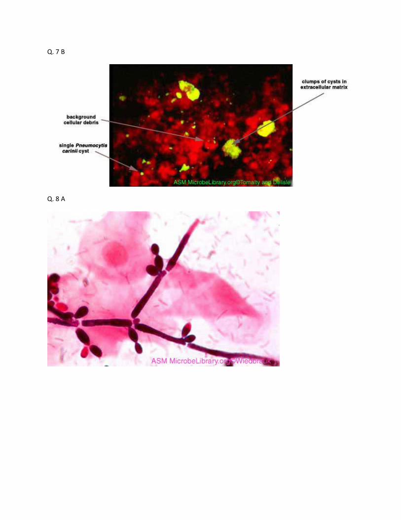
A 29-year-old, HIV-positive male was admitted to the hospital with a two-week history ofnon-productive cough, fever, and shortness of breath. On examination, he had a mild fever, wasbreathing rapidly, and had significantly reduced arterial oxygen levels. A chest X-raydemonstrated bilateral pulmonary infiltration. A sample of sputum was obtained bybronchoalveolar lavage (BAL). An unusual organism was obtained from the BAL, whichdisplayed the characteristics of both a protozoan and a fungus. This organism has cyst andtrophozoite phases but is genetically similar to the yeast Saccharomyces. (5 points)View a related image A labeled view of this image
7.
What is wrong with this patient? Pneumocystis carinii.
How did he contract this organism? Exposure is thought to occur during childhood with thehealthy immune system holding it in check. Immunocompromisation due to HIV has allowed it tore-emerge.
What kinds of patients are at risk from infection by this organism? Any patients with defectivecell-mediated immunity either genetic or acquired.
How would you treat this patient? Pentamidine and cotrimoxazole for 10 days.
What is the outlook for his condition? Untreated patients have a 50-100% mortality; treatedmuch better.
Fungal and Protozoal Mini-Case History Studies http://archive.microbelibrary.org/edzine/curriculum/Toolbox/toolbox7c.htm
3 of 10 3/20/2012 4:18 PM

A female patient underwent surgery to clear a blocked ureter 3 days ago and is now experiencingvaginal discomfort, with a milky white discharge, local inflammation, and ulceration. An ovoidyeast recovered from the discharge produced white colonies on sheep blood agar, reproduced bybudding, and subsequently produced germ tubes. (5 points)View a related image View a related image A labeled view of this image
8.
What is wrong with this patient? What led you to this diagnosis? (2 points) Candida albicans. C.albicans is the yeast most commonly causing vulvovaginal infection, plus the production of germtubes indicates albicans yeast.
Is this organism life-threatening? Yes, for AIDS or other immunocompromised patients.
How did the patient contract the infection? The likelihood is that the in-dwelling cannula allowedthe endogenous yeast to invade the body.
A male patient presents with a 3-year history of pain and itching of the toes of both feet and of hisleft palm and fingers. Small red lesions were visible on his left fingers. He is currently in goodhealth, otherwise, and in training for his college baseball, volleyball, and swimming teams. Duringthe 3 years, he has been stopping and starting with his medication and has now returned to theclinic since the condition does not appear to be responding to his current medication any more.Direct examination of skin scrapings with the calcofluor dye revealed what is seen in the relatedimage below. A culture of skin scrapings from the patient’s feet grew a white, fuzzy fungus,which demonstrated thin-walled macroconidia and numerous microconidia. (5 points)View a related image
9.
What is wrong with this patient? Athlete’s foot.
How is he likely to have contracted it? (2 points) Contact with infected skin or hair. Theorganism survives in moist environments, e.g., pool decks and shower rooms.
To which group of organisms does the fungus belong? Why is this group so named? (2 points)Dermatophytes. Organisms that live on the non-living tissues of keratin (nails, skin, hair).
A patient is admitted to the emergency room with seizures and in a confused state. His wife tellsthe physicians that he has had a fever and cough for the past week but was convinced it was justthe "flu" and so did not consult anyone. The physician tests for HIV but the patient isHIV-negative. (3 points)
10.
One of the other things the patient’s wife told the physicians is that her husband keeps pigeons asa hobby. What disease does this now suggest he has? Cryptococcus neoformans, pigeon fancier’sdisease.
Why did the physician suggest testing for HIV? (2 points) The disease is a hallmark of AIDS andcauses death in these patients. Finding of infection is usually associated with bird contact orimmunocompromisation.
A male with full-blown AIDS is admitted to the emergency room in a confused state, disoriented,and lethargic. The patient had a one-week history of progressively worsening headache,photophobia, lethargy, and fever. On admission, CT scans and chest X-rays were performed. Bothappeared normal, however a lumbar puncture revealed aseptic meningitis. Direct examination ofthe cerebrospinal fluid (CSF) with India ink showed encapsulated yeasts. Putting the CSF sampleon sheep blood agar revealed pink colonies of a round yeast which was gram-variable. (5 points)
11.
Fungal and Protozoal Mini-Case History Studies http://archive.microbelibrary.org/edzine/curriculum/Toolbox/toolbox7c.htm
4 of 10 3/20/2012 4:18 PM

View a related image
What organism is causing the patient’s illness? What led you to this diagnosis? (2 points) In AIDS,mental changes are usually associated with either Toxoplasma or Cryptococcal infection. This isa fungal infection, therefore Cryptococcus neoformans.
How is the patient likely to have become infected? Bird guano, especially pigeons.
How would you treat this patient? Amphoteracin B, flucytosine or miconazole for weeks ormonths.
What is the outlook for the patient? Usually fatal.
A 60-year-old woman with noninsulin-dependent diabetes mellitus (NIDDM) presents with a4-day history of left eye swelling and frontal headache. On examination, she has a fever and aseverely distended left eye. A CT scan of the nasal sinuses and eye shows fluid accumulation.Following surgery to drain the fluid, a fungus was isolated from it which demonstrated broad,aseptate hyphae with right angle branching. The fungus showed characteristic zygote fruitingbodies. (5 points)
12.
What is wrong with this patient? What led you to this diagnosis? (3 points) Rhinocerebralzygomycosis. Zygote formation means a zygomycete. Infection occurs inimmunocompromised and NIDDM.
How is the disease managed? (2 points) Debridement and IV antifungals. Can be fatal.
Protozoa
A young, male university student returned from a trip to Hawaii at the end of October and wasadmitted to the emergency room with convulsions. The student had been on vacation with anumber of his classmates and had been ill with cold-like symptoms and headaches since hisreturn. The vacation consisted of a hiking tour inland and visits to a number of wooded areas.When questioned, his classmates said they had all eaten and drunk roughly the same things onvacation. One of the female students said that the male patient had been "acting strange" eversince they visited an inland pool, which had its own underground spring as a water supply, wherehe showed off his diving skills. (The patient was a member of the university diving team.) Spinalfluid was cultured on non-nutrient media with a lawn of E. coli. Organisms were visualized with amethylene blue stain on the agar. (5 points)View a related image
1.
Based on the above history, what do you think was wrong with the male patient? Naegleriafowlderi or Acanthamoeba infection of the brain (primary acute meningoencephalitis).
How did he acquire the infection? Diving into contaminated water forced the amoeba throughthe nasal passages and they then attacked the brain.
What treatment would you have prescribed? (2 points) Miconazole and/or Amphoteracin B.Usually cases are too far advanced to do anything however.
What other steps would you take to prevent additional cases of infection with this organism?Clean out water and don’t dive in any water that is contaminated.
A male patient, recently returned from a year spent with the Peace Corps in Kenya, presents to2.
Fungal and Protozoal Mini-Case History Studies http://archive.microbelibrary.org/edzine/curriculum/Toolbox/toolbox7c.htm
5 of 10 3/20/2012 4:18 PM

his primary care physician with fever and a distended abdomen. On examination the young man isjaundiced and an X-ray of his abdomen reveals opaque, nodular growths in the large intestine.Liver biopsy recovers large trophozoites containing a prominent and distinctive nucleolus called akaryosome. (4 points)View a related image
Which organism is this patient infected with? Entamoeba histolytica.
How did he contract it? Food or water contaminated by cysts which then hatched and invadedhis large intestine.
What other bodily targets does this organism have? Cecum, appendix, colon, and rectum.
Could this infection have been prevented? How? Yes, by using boiled, chlorinated or filteredwater.
A female patient presents to her primary care physician with pain radiating from her groin and alow-grade fever. On examination she is found to have a purulent vaginal discharge, which isgreen, foul smelling and contains motile, flagellated protozoa. Her physician tells her he suspectsan STD but she denies this, saying that her boyfriend has "no problems" and she is "faithful tohim." (4 points)View a related image
3.
What disease does this patient have? Is it an STD? Trichomonas vaginalis infection; yes.
Where has the organism come from? Explain your answer. (2 points) Trichomonas infection canbe either asymptomatic or sub-clinical. The woman’s boyfriend may have such an infection orshe may herself have had the infection for some time without realizing it.
How would you treat this patient? To avoid "ping-pong" infections both partners must be treatedwith metronidazole for at least one week.
A 24-year-old woman is referred to a public health clinic as a result of contact tracing from a caseof gonorrhea. She recently had unprotected sex with her infected male partner but showed nosymptoms. A pelvic examination recovers a greenish discharge containing protozoa withcharacteristic "jerky" motility. The woman tested negative for gonorrhea. (6 points)View a related image
4.
What is wrong with this patient? How common is her infection? Trichomonas vaginalis. Thisorganism affects between 3 and 15% of the U.S. population as a sub-clinical infection.
What symptoms does it produce in women and men? (2 points) Women can have dysuria ordischarge with some being asymptomatic. Men can have prostate or seminal vesicle infectionwith most being asymptomatic.
What treatment would you prescribe for this patient? (2 points) To avoid "ping-pong" infectionsboth partners must be treated with oral and vaginal metronidazole for at least one week.
An anthropologist has just returned to Philadelphia after an extensive study of the NativeAmerican peoples of New Mexico. He presents at his primary care physician with diarrhea,abdominal pain, and fever. Stool samples reveal protozoal cysts and motile trophozoites andgastroscopy reveals more protozoa attached to the jejunum. Further confirmation of the diagnosis
5.
Fungal and Protozoal Mini-Case History Studies http://archive.microbelibrary.org/edzine/curriculum/Toolbox/toolbox7c.htm
6 of 10 3/20/2012 4:18 PM

is obtained by performing an ELISA on the stool to detect specific antigen. (4 points)View a related imageView a related image
Which organism is this? Explain your answer. (3 points) Giardia lamblia. Gastroenteritis suggestseither Entamoeba histolytica or G. lamblia. E. histolytica would have attacked the largeintestine, however, and this is attacking the small intestine suggesting G. lamblia as a morelikely candidate.
How did the anthropologist get the infection? Water contaminated with cysts and/or trophozoites.
What treatment would you recommend? Metranidazole.
A male patient, recently returned from a visit to Africa (two weeks ago), is admitted to thehospital with an intermittent fever, swollen lymph nodes, and joint pain. His wife brings him to theemergency room and says she is having trouble "waking him during the day" whereas he "staysawake all night." The admitting physician notices that the man is slurring his words and his handsare shaking. Tests reveal the presence of a tropomastigote in the patient’s bloodstream togetherwith IgM raised against the organism. (5 points)View a related image A labeled view of this image
6.
What do you think is wrong with this man? Trypanosoma brucei. This is rhodesiense due to thespeed of attack of the organism on the brain.
What area of the body is affected by the disease? It has attacked the brain affecting speech andcoordination.
How and where did he contract the disease and what is its prognosis? Bitten by a tsetse fly andwill probably die in 2 to 3 months because of speed of onset of brain involvement.
What is the life cycle of this organism? Natural hosts are wild animals. It is transmitted from aninfected host to man via the bite of a tsetse fly.
How would you treat this condition? Metapryosol or pentamidine.
A young male patient, who has just returned from 2 years of service in the Peace Corps in Brazil,presents at his primary care physician with fever, chest pain, breathlessness, and a distendedabdomen. He was in a car accident about 6 months ago, while on active duty, and received ablood transfusion in a local Rio de Janeiro hospital. The physician refers him to the hospital wherehe is admitted for observation. A chest X-ray shows both hepatosplenomegaly and heartenlargement and an EKG shows a systolic murmur. The patient does not remember being bittenby anything during his time in the Peace Corps, as all Peace Corps volunteers were advised bytheir commanding officers to use netting and insect repellents and to remain "covered up"whenever possible. The patient has also had no sexual intercourse over the past 6 months. Thepatient died 2 years later from heart damage caused by the hemoflagellate trypanosomal protozoainfecting him. (5 points)
7.
What is wrong with this patient? How did you arrive at this diagnosis? (2 points) Chagas diseasecaused by Trypanosoma cruzi. This disease targets the heart.
How can this disease be contracted? (2 points) Through a blood transfusion or from being bittenby reduviid bugs.
Fungal and Protozoal Mini-Case History Studies http://archive.microbelibrary.org/edzine/curriculum/Toolbox/toolbox7c.htm
7 of 10 3/20/2012 4:18 PM
How do you think this patient contracted the disease? Probably from the blood transfusionassociated with his car accident.
A recent immigrant to the United States from Brazil, who has been in the country for 3 months,presents to his primary care physician suffering from an intense spiking fever. His weight hasdropped from 170 to 120 pounds and he shows a distended stomach. On X-ray, his spleen andliver are enlarged and he is also somewhat anemic. The physician also notes that the man’s skin isextensively scarred as a result of ulcers, which have subsequently healed. The patient isprescribed amphoteracin B and his physician notes that unless the situation improves he will needto be given pentamidine or pentavalent antimony. (4 points)
8.
Which disease and organism are causing the patient’s condition? (2 points) Leishmania donovani,kala azar.
Name another area of the world to which it is endemic. China, Latin America.
How likely is it that the patient will die, in spite of the physician’s best efforts? 75-100%.
Simon, a student, presents with ulcerated, raised lesions on his neck, calves, and feet. Theselesions have drained, ulcerated, healed, and then broken open again. He traveled to the MiddleEast last summer (4 months ago) and spent June and July in Israel followed by several weeks inEgypt. During this time he remembers being bitten by numerous "small black flies." While inEgypt, he noticed the first neck lesion. Blood work shows a normal white cell count and culture ofthe neck lesion reveals a hemoflagellate protozoan. (5 points)
9.
What is wrong with Simon? What has led you to this diagnosis? (2 points) Hemoflagellates meanTrypanosomes or Leishmania. Trypanosomes do not produce skin lesions, therefore it iscutaneous Leishmania. Given the region of world it is Leishmania tropicana.
What are the "black flies" the patient referred to? Sandflies.
Why are infections typically seen on the head and neck? (2 points) The fly can’t bite throughclothing.
A man returned two weeks ago from a vacation in Africa and presents in the emergency roomwith a severe, spiking fever and chills. Observation in the hospital shows that he exhibits thesesymptoms every 2 to 3 days, thereafter appearing to recover. His liver and spleen are enlarged andthe patient also demonstrates a reduced red cell count due to hemolytic anemia. Blood analysisconfirms the presence of trophozoite forms of a sporozoan protozoan. (8 points)View a related image
10.
What tropical disease is the patient suffering from? Malaria/Plasmodium.
What aspects of the disease are causing his symptoms? Erythrocytic bursting every 2 to 3 days.
The patient remembers being bitten by an insect while in Africa. What kind of insect acts as avector for this disease? Are there any other ways of being infected? The Anopheles mosquito actsas a vector for this disease. Contaminated needles are another way of becoming infected.
This is a mild form of the disease. What kind of prognosis can the patient expect? Recovery in 5years or so.
How would you recommend treating this patient? Quinine derivatives (mepacrine/ chloroquine).
Fungal and Protozoal Mini-Case History Studies http://archive.microbelibrary.org/edzine/curriculum/Toolbox/toolbox7c.htm
8 of 10 3/20/2012 4:18 PM
What two simple methods could this man have used to reduce his risk of contracting this disease?Select from: Used insect repellent, stayed indoors at night, or used netting over windows.
David returned from Africa 12 days prior to his admission to the hospital. He had been in Africafor 6 months working as a civil engineer on road building projects in Rwanda, Zaire, and Uganda.Seven days prior to admission he began suffering from fevers with chills, cough, and myalgia.Three days ago he had episodes of fevers and chills, three times a day. On admission, the patientwas stuporous and showed mental changes. Blood work showed lowered white cell and plateletcounts. He also had a hematocrit of approximately half that of normal levels with grosslyprolonged bleeding times. A sporozoan parasite was recovered from the blood, which had acharacteristic "ring" form. Despite treatment, the patient eventually went into a coma and died. (6points)View a related image
11.
What was wrong with David? What led you to this diagnosis? You should be able to identify thisorganism to the species level. (2 points) Symptoms point to malaria. Severity and speed of attacksuggests falciparum. Sporozoans mean Toxoplasma or Plasmodium.
Why is the observation of mental changes significant? Cerebral malaria.
What treatment would you prescribe for David, if I told you he has parasitemia levels of 25%?Exchange transfusion of his red blood cells (RBC).
Could David’s condition have been prevented? (2 points) Prophylaxis by use of anti-malarialsand insect repellents; currently there is no vaccine.
Andrea, a one-week-old child, has hydrocephalus, hepatosplenomegaly, and is blind. Blood andtissue analyses from affected sites show no evidence of protozoa. (4 points)
12.
What caused the baby’s condition? Toxoplasma gondii exposure during pregnancy.
What is this kind of effect on a fetus called? Teratogenic.
How could Andrea’s condition have been prevented? Get rid of the cat or don’t clean the litterbox during pregnancy.
Name one source of infection from this organism. Cats and birds.
A HIV-positive male with a T-helper cell count of 60/ml presents with headaches, weakness, anddifficulty maintaining his balance when walking. His friend who brought him to the emergencyroom stated that the patient had been quite agitated over the previous few days and that hisspeech was slurred and "rambling." On physical examination, the patient was afebrile, but notoriented towards space or time, and unable to recognize his friend. Imaging of the brain revealedmultiple ring-enhancing lesions containing oocytes of a sporozoan parasite. (5 points)
13.
What organism do you think is causing the patient’s condition? How do you arrive at thisdiagnosis? (2 points) Toxoplasma gondii. Brain lesions in an HIV-positive patient meansCryptococcus or Toxoplasma.
How is he likely to have become infected? Contact with oocysts via cats.
Name another group of patients at risk of serious infection from this organism. Pregnant womenand their fetuses.
Fungal and Protozoal Mini-Case History Studies http://archive.microbelibrary.org/edzine/curriculum/Toolbox/toolbox7c.htm
9 of 10 3/20/2012 4:18 PM
What is the outlook for this patient? Poor, the disease is usually fatal.
Nicola, a 3-year-old child who began attending a day care center two weeks ago, was admitted tothe hospital following a 3-day history of increasingly severe diarrhea with episodes of vomiting. Inthe previous 24 hours she had 12 bowel movements. Prior to this, the child had been healthy andno other illnesses were reported for any other members of the family. The child’s mother, whoaccompanied her, was concerned that it might be Salmonella infection since the pets in the daycare center included turtles and an iguana. On examination, the child was lethargic, dehydrated,and had an elevated heart rate. A stool specimen contained no white or red cells, but yeast-like,non-motile, oocyst sporozoan parasites were recovered. Her diarrhea resolved after a furtherweek. (6 points)View a related image
14.
What protozoa are able to cause diarrhea? (3 points) Entamoeba histolytica, Giardialamblia, and Cryptospiridium parvum.
Of these, which is the most likely to be causing Nicola’s diarrhea and why? (2 points) C.parvum is most likely to be causing the diarrhea. G. lamblia and C. parvum are mostlikely in the U.S., since E. histolytica is tropical. G. lamblia does not have oocysts and ismotile.
How is Nicola likely to have contracted the infection? Is there any significance to the daycare center’s choice of pets? Day-care, oral-fecal spread; yes, both pets carry theorganism in their intestines.
Fungal and Protozoal Mini-Case History Studies http://archive.microbelibrary.org/edzine/curriculum/Toolbox/toolbox7c.htm
10 of 10 3/20/2012 4:18 PM

Fungal diseases
A birdwatcher, returning from a visit to the Ozarks in Missouri, complains to his primary carephysician of weakness and lethargy. On examination, he is found to have lymphoadenopathy anda distended abdomen. Blood work reveals that the patient is anemic and an X-ray confirmshepatosplenomegaly. A mass, which was not nodular, was observed in both the liver and spleen aswere a series of lesions. A few days later, the patient was admitted to the hospital in a confusedstate and with severe shortness of breath. X-rays revealed a "bird shot" appearance to the lungsand sputum and bone marrow cultures were positive for small budding intracellular yeasts. (4points)The related image is from a room temperature culture of the organism - View a relatedimage
1.
What do you think that the patient is suffering from?
How did he contract it?
Name another group of individuals who are at risk?
Name another U.S. state in which the disease is common.
A male patient is admitted to the emergency room in a weakened state with a persistent fever andweight loss. His history includes the fact that he is HIV-positive and, until recently, lived inPanama. Blood work demonstrates reduced white cell, platelet, and red cell numbers. Bonemarrow examination reveals a decreased number of white cells and the presence of anascomycete yeast. The yeast produces numerous macroconidia when cultured on an agar plate. (5points)View a related image
2.
What is wrong with this patient? Explain your reasoning. (3 points)
With what environmental source would you associate this organism?
Name two states in the U.S. in which this organism commonly causes infection? (2 points)
A male patient returns from a fall trip to southern Canada, where he went to visit his African-American relatives. He presents at the clinic with "flu-like" symptoms of hoarseness and coughingbut is also experiencing chest pain. A chest X-ray reveals small nodules in his lungs that mayrepresent tumors, sputum specimens stained with calcofluor dye showed large broad-based yeast.(10 points)View a related image
3.
What organism do you think this patient may be infected with based on the above events? (3points)
Fungal and Protozoal Mini-Case History Studies http://archive.microbelibrary.org/edzine/curriculum/Toolbox/toolbox7c1.htm
1 of 8 3/20/2012 4:18 PM

Which states of the U.S. would you associate with this organism?
Name a group of people at risk of infection with this organism?
What would you expect to see if you cultured the yeast from the lung nodules in the laboratory?
Name two other dimorphic yeasts and explain how they differ in appearance from the organismresponsible for this case. (4 points)
A 45-year-old African-American man from Georgia was in good health until 2 months ago whenhe developed a low grade fever, myalgia, and a non-productive cough. On admission to thehospital, he was feverish, had breathing difficulties, and stated he had lost 5 pounds in weight overthe past 2 months. The patient tested as HIV-negative, had not traveled recently, and was anon-smoker. He worked as a gardener for a local city park. A chest X-ray showed that the patienthad consolidation of the middle lobe of his right lung with a small TB-like mass at the samelocation. An observant physician also noticed that the patient had a raised lesion on the bridge ofthe nose, which was tender to the touch. Bronchoscopy yielded a biopsy sample, which containeda large, round, budding yeast. Culture of the yeast yielded an ascomycete producing numerousmacro- and microconidia. (6 points)View a related image
4.
What is wrong with this patient? What led you to this diagnosis? (3 points)
What complications of this disease can occur? (2 points)
How would you treat this patient?
A 25-year-old male presents to his primary care physician with a neck mass. He has movedaround the U.S. a lot, most recently from Arizona. Samples of purulent material drained from themass contained fungal arthrospores and giant sporangia (spherules). Culture of the arthrosporesyielded a fluffy, white mold. (4 points)View a related image
5.
What is wrong with this patient? What led you to this diagnosis? (2 points)
A technician did not notice the mold immediately on the culture plates and opened them to lookfor bacteria. Was this dangerous and if so, why? (2 points)
A male patient arrived back in the United States from a tour of Australia a week ago after a shortvacation. He presents to his primary care physician with a small, hard, non-tender nodule on hishand. He thinks he may have "something in it" but doesn’t remember how. He woke up andnoticed it after a particularly extended 48-hour drinking session. The patient is a self-confessedalcoholic. (5 points)View a related image
6.
What organism is this?
How did the patient contract it?
How could he have prevented it?
How would you treat it?
Fungal and Protozoal Mini-Case History Studies http://archive.microbelibrary.org/edzine/curriculum/Toolbox/toolbox7c1.htm
2 of 8 3/20/2012 4:18 PM

Can you anticipate any long-term problems?
A 29-year-old, HIV-positive male was admitted to the hospital with a two-week history ofnon-productive cough, fever, and shortness of breath. On examination, he had a mild fever, wasbreathing rapidly, and had significantly reduced arterial oxygen levels. A chest X-raydemonstrated bilateral pulmonary infiltration. A sample of sputum was obtained bybronchoalveolar lavage (BAL). An unusual organism was obtained from the BAL, whichdisplayed the characteristics of both a protozoan and a fungus. This organism has cyst andtrophozoite phases but is genetically similar to the yeast Saccharomyces. (5 points)View a related image
7.
What is wrong with this patient?
How did he contract this organism?
What kinds of patients are at risk from infection by this organism?
How would you treat this patient?
What is the outlook for his condition?
A female patient underwent surgery to clear a blocked ureter 3 days ago and is now experiencingvaginal discomfort, with a milky white discharge, local inflammation, and ulceration. An ovoidyeast recovered from the discharge produced white colonies on sheep blood agar, reproduced bybudding, and subsequently produced germ tubes. (5 points)View a related image View a related image
8.
What is wrong with this patient? What led you to this diagnosis? (2 points)
Is this organism life-threatening?
How did the patient contract the infection?
A male patient presents with a 3-year history of pain and itching of the toes of both feet and of hisleft palm and fingers. Small red lesions were visible on his left fingers. He is currently in goodhealth, otherwise, and in training for his college baseball, volleyball, and swimming teams. Duringthe 3 years, he has been stopping and starting with his medication and has now returned to theclinic since the condition does not appear to be responding to his current medication any more.Direct examination of skin scrapings with the calcofluor dye revealed what is seen in the relatedimage below. A culture of skin scrapings from the patient’s feet grew a white, fuzzy fungus,which demonstrated thin-walled macroconidia and numerous microconidia. (5 points)View a related image
9.
What is wrong with this patient?
How is he likely to have contracted it? (2 points)
To which group of organisms does the fungus belong? Why is this group so named? (2 points)
A patient is admitted to the emergency room with seizures and in a confused state. His wife tellsthe physicians that he has had a fever and cough for the past week but was convinced it was justthe "flu" and so did not consult anyone. The physician tests for HIV but the patient is
10.
Fungal and Protozoal Mini-Case History Studies http://archive.microbelibrary.org/edzine/curriculum/Toolbox/toolbox7c1.htm
3 of 8 3/20/2012 4:18 PM

HIV-negative. (3 points)
One of the other things the patient’s wife told the physicians is that her husband keeps pigeons asa hobby. What disease does this now suggest he has?
Why did the physician suggest testing for HIV? (2 points)
A male with full-blown AIDS is admitted to the emergency room in a confused state, disoriented,and lethargic. The patient had a one-week history of progressively worsening headache,photophobia, lethargy, and fever. On admission, CT scans and chest X-rays were performed. Bothappeared normal, however a lumbar puncture revealed aseptic meningitis. Direct examination ofthe cerebrospinal fluid (CSF) with India ink showed encapsulated yeasts. Putting the CSF sampleon sheep blood agar revealed pink colonies of a round yeast which was gram-variable. (5 points)View a related image
11.
What organism is causing the patient’s illness? What led you to this diagnosis? (2 points)
How is the patient likely to have become infected?
How would you treat this patient?
What is the outlook for the patient?
A 60-year-old woman with noninsulin-dependent diabetes mellitus (NIDDM) presents with a4-day history of left eye swelling and frontal headache. On examination, she has a fever and aseverely distended left eye. A CT scan of the nasal sinuses and eye shows fluid accumulation.Following surgery to drain the fluid, a fungus was isolated from it which demonstrated broad,aseptate hyphae with right angle branching. The fungus showed characteristic zygote fruitingbodies. (5 points)
12.
What is wrong with this patient? What led you to this diagnosis? (3 points)
How is the disease managed? (2 points)
Protozoa
A young, male university student returned from a trip to Hawaii at the end of October and wasadmitted to the emergency room with convulsions. The student had been on vacation with anumber of his classmates and had been ill with cold-like symptoms and headaches since hisreturn. The vacation consisted of a hiking tour inland and visits to a number of wooded areas.When questioned, his classmates said they had all eaten and drunk roughly the same things onvacation. One of the female students said that the male patient had been "acting strange" eversince they visited an inland pool, which had its own underground spring as a water supply, wherehe showed off his diving skills. (The patient was a member of the university diving team.) Spinalfluid was cultured on non-nutrient media with a lawn of E. coli. Organisms were visualized with amethylene blue stain on the agar. (5 points)View a related image
1.
Based on the above history, what do you think was wrong with the male patient?
How did he acquire the infection?
What treatment would you have prescribed? (2 points)
Fungal and Protozoal Mini-Case History Studies http://archive.microbelibrary.org/edzine/curriculum/Toolbox/toolbox7c1.htm
4 of 8 3/20/2012 4:18 PM
What other steps would you take to prevent additional cases of infection with this organism?
A male patient, recently returned from a year spent with the Peace Corps in Kenya, presents tohis primary care physician with fever and a distended abdomen. On examination the young man isjaundiced and an X-ray of his abdomen reveals opaque, nodular growths in the large intestine.Liver biopsy recovers large trophozoites containing a prominent and distinctive nucleolus called akaryosome. (4 points)View a related image
2.
Which organism is this patient infected with?
How did he contract it?
What other bodily targets does this organism have?
Could this infection have been prevented? How?
A female patient presents to her primary care physician with pain radiating from her groin and alow-grade fever. On examination she is found to have a purulent vaginal discharge, which isgreen, foul smelling and contains motile, flagellated protozoa. Her physician tells her he suspectsan STD but she denies this, saying that her boyfriend has "no problems" and she is "faithful tohim." (4 points)View a related image
3.
What disease does this patient have? Is it an STD?
Where has the organism come from? Explain your answer. (2 points)
How would you treat this patient?
A 24-year-old woman is referred to a public health clinic as a result of contact tracing from a caseof gonorrhea. She recently had unprotected sex with her infected male partner but showed nosymptoms. A pelvic examination recovers a greenish discharge containing protozoa withcharacteristic "jerky" motility. The woman tested negative for gonorrhea. (6 points)View a related image
4.
What is wrong with this patient? How common is her infection?
What symptoms does it produce in women and men? (2 points)
What treatment would you prescribe for this patient? (2 points)
An anthropologist has just returned to Philadelphia after an extensive study of the NativeAmerican peoples of New Mexico. He presents at his primary care physician with diarrhea,abdominal pain, and fever. Stool samples reveal protozoal cysts and motile trophozoites andgastroscopy reveals more protozoa attached to the jejunum. Further confirmation of the diagnosisis obtained by performing an ELISA on the stool to detect specific antigen. (4 points)View a related imageView a related image
5.
Which organism is this? Explain your answer. (3 points)
Fungal and Protozoal Mini-Case History Studies http://archive.microbelibrary.org/edzine/curriculum/Toolbox/toolbox7c1.htm
5 of 8 3/20/2012 4:18 PM

How did the anthropologist get the infection?
What treatment would you recommend?
A male patient, recently returned from a visit to Africa (two weeks ago), is admitted to thehospital with an intermittent fever, swollen lymph nodes, and joint pain. His wife brings him to theemergency room and says she is having trouble "waking him during the day" whereas he "staysawake all night." The admitting physician notices that the man is slurring his words and his handsare shaking. Tests reveal the presence of a tropomastigote in the patient’s bloodstream togetherwith IgM raised against the organism. (5 points)View a related image
6.
What do you think is wrong with this man?
What area of the body is affected by the disease?
How and where did he contract the disease and what is its prognosis?
What is the life cycle of this organism?
How would you treat this condition?
A young male patient, who has just returned from 2 years of service in the Peace Corps in Brazil,presents at his primary care physician with fever, chest pain, breathlessness, and a distendedabdomen. He was in a car accident about 6 months ago, while on active duty, and received ablood transfusion in a local Rio de Janeiro hospital. The physician refers him to the hospital wherehe is admitted for observation. A chest X-ray shows both hepatosplenomegaly and heartenlargement and an EKG shows a systolic murmur. The patient does not remember being bittenby anything during his time in the Peace Corps, as all Peace Corps volunteers were advised bytheir commanding officers to use netting and insect repellents and to remain "covered up"whenever possible. The patient has also had no sexual intercourse over the past 6 months. Thepatient died 2 years later from heart damage caused by the hemoflagellate trypanosomal protozoainfecting him. (5 points)
7.
What is wrong with this patient? How did you arrive at this diagnosis? (2 points)
How can this disease be contracted? (2 points)
How do you think this patient contracted the disease?
A recent immigrant to the United States from Brazil, who has been in the country for 3 months,presents to his primary care physician suffering from an intense spiking fever. His weight hasdropped from 170 to 120 pounds and he shows a distended stomach. On X-ray, his spleen andliver are enlarged and he is also somewhat anemic. The physician also notes that the man’s skin isextensively scarred as a result of ulcers, which have subsequently healed. The patient isprescribed amphoteracin B and his physician notes that unless the situation improves he will needto be given pentamidine or pentavalent antimony. (4 points)
8.
Which disease and organism are causing the patient’s condition? (2 points)
Name another area of the world to which it is endemic.
How likely is it that the patient will die, in spite of the physician’s best efforts?
Fungal and Protozoal Mini-Case History Studies http://archive.microbelibrary.org/edzine/curriculum/Toolbox/toolbox7c1.htm
6 of 8 3/20/2012 4:18 PM

Simon, a student, presents with ulcerated, raised lesions on his neck, calves, and feet. Theselesions have drained, ulcerated, healed, and then broken open again. He traveled to the MiddleEast last summer (4 months ago) and spent June and July in Israel followed by several weeks inEgypt. During this time he remembers being bitten by numerous "small black flies." While inEgypt, he noticed the first neck lesion. Blood work shows a normal white cell count and culture ofthe neck lesion reveals a hemoflagellate protozoan. (5 points)
9.
What is wrong with Simon? What has led you to this diagnosis? (2 points)
What are the "black flies" the patient referred to?
Why are infections typically seen on the head and neck? (2 points)
A man returned two weeks ago from a vacation in Africa and presents in the emergency roomwith a severe, spiking fever and chills. Observation in the hospital shows that he exhibits thesesymptoms every 2 to 3 days, thereafter appearing to recover. His liver and spleen are enlarged andthe patient also demonstrates a reduced red cell count due to hemolytic anemia. Blood analysisconfirms the presence of trophozoite forms of a sporozoan protozoan. (8 points)View a related image
10.
What tropical disease is the patient suffering from?
What aspects of the disease are causing his symptoms?
The patient remembers being bitten by an insect while in Africa. What kind of insect acts as avector for this disease? Are there any other ways of being infected?
This is a mild form of the disease. What kind of prognosis can the patient expect?
How would you recommend treating this patient?
What two simple methods could this man have used to reduce his risk of contracting this disease?
David returned from Africa 12 days prior to his admission to the hospital. He had been in Africafor 6 months working as a civil engineer on road building projects in Rwanda, Zaire, and Uganda.Seven days prior to admission he began suffering from fevers with chills, cough, and myalgia.Three days ago he had episodes of fevers and chills, three times a day. On admission, the patientwas stuporous and showed mental changes. Blood work showed lowered white cell and plateletcounts. He also had a hematocrit of approximately half that of normal levels with grosslyprolonged bleeding times. A sporozoan parasite was recovered from the blood, which had acharacteristic "ring" form. Despite treatment, the patient eventually went into a coma and died. (6points)View a related image
11.
What was wrong with David? What led you to this diagnosis? You should be able to identify thisorganism to the species level. (2 points)
Why is the observation of mental changes significant?
What treatment would you prescribe for David, if I told you he has parasitemia levels of 25%?
Could David’s condition have been prevented? (2 points)
Fungal and Protozoal Mini-Case History Studies http://archive.microbelibrary.org/edzine/curriculum/Toolbox/toolbox7c1.htm
7 of 8 3/20/2012 4:18 PM

Andrea, a one-week-old child, has hydrocephalus, hepatosplenomegaly, and is blind. Blood andtissue analyses from affected sites show no evidence of protozoa. (4 points)
12.
What caused the baby’s condition?
What is this kind of effect on a fetus called?
How could Andrea’s condition have been prevented?
Name one source of infection from this organism.
A HIV-positive male with a T-helper cell count of 60/ml presents with headaches, weakness, anddifficulty maintaining his balance when walking. His friend who brought him to the emergencyroom stated that the patient had been quite agitated over the previous few days and that hisspeech was slurred and "rambling." On physical examination, the patient was afebrile, but notoriented towards space or time, and unable to recognize his friend. Imaging of the brain revealedmultiple ring-enhancing lesions containing oocytes of a sporozoan parasite. (5 points)
13.
What organism do you think is causing the patient’s condition? How do you arrive at thisdiagnosis? (2 points)
How is he likely to have become infected?
Name another group of patients at risk of serious infection from this organism.
What is the outlook for this patient?
Nicola, a 3-year-old child who began attending a day care center two weeks ago, was admitted tothe hospital following a 3-day history of increasingly severe diarrhea with episodes of vomiting. Inthe previous 24 hours she had 12 bowel movements. Prior to this, the child had been healthy andno other illnesses were reported for any other members of the family. The child’s mother, whoaccompanied her, was concerned that it might be Salmonella infection since the pets in the daycare center included turtles and an iguana. On examination, the child was lethargic, dehydrated,and had an elevated heart rate. A stool specimen contained no white or red cells, but yeast-like,non-motile, oocyst sporozoan parasites were recovered. Her diarrhea resolved after a furtherweek. (6 points)View a related image
14.
What protozoa are able to cause diarrhea? (3 points)
Of these, which is the most likely to be causing Nicola’s diarrhea and why? (2 points)
How is Nicola likely to have contracted the infection? Is there any significance to the daycare center’s choice of pets?
Fungal and Protozoal Mini-Case History Studies http://archive.microbelibrary.org/edzine/curriculum/Toolbox/toolbox7c1.htm
8 of 8 3/20/2012 4:18 PM
Fungal and Protozoal Mini-Case History Studies for Courses Involving Medical Microbiology -- Answer Key
Fungal diseases Q. 1
Q. 2

Q. 3
Q. 4

Q. 5
Q. 6 A

Q. 6 B
Q. 7 A
Q. 7 B
Q. 8 A

Q. 8 B
Q. 8 C
Q. 9
Q. 11

Protozoa
Q. 1
Q. 2

Q. 3
Q. 4
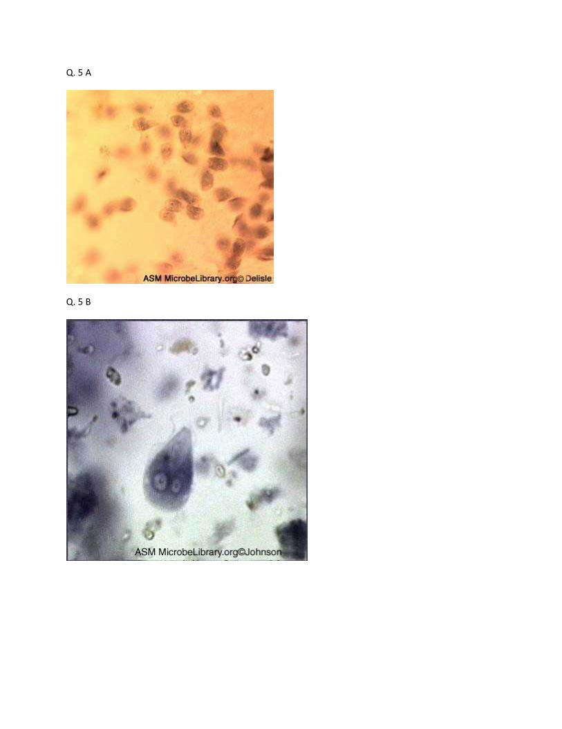
Q. 5 A
Q. 5 B

Q. 6 A
Q. 6 B

Q. 10
Q. 11

Q. 14