functions of the cerebral hemisphere dr fawzia alrouq medical college ksu
TRANSCRIPT

• Functions of The Cerebral Hemisphere
• Dr Fawzia AlRouq• Medical college • KSU

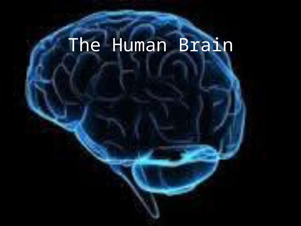
The Human Brain
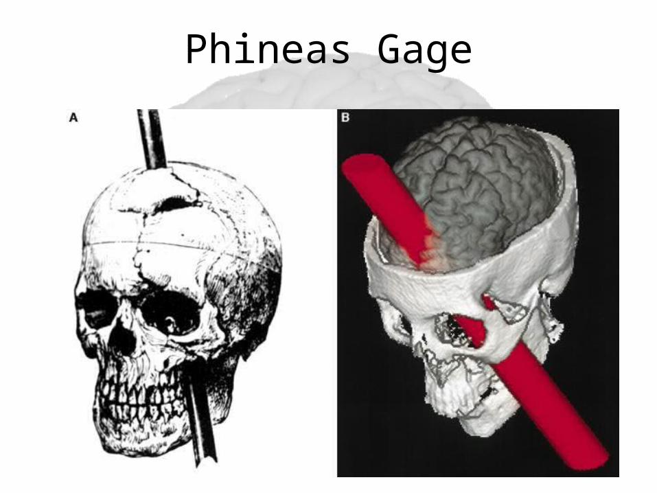
Phineas Gage
Phineas Gage• In 1848 in Vermont, had a 3.5-foot-long, 13 lb. metal rod blown into his skull, through his brain, and out of the top of his head. Gage survived. In fact, he never even lost consciousness.• Friends reported a complete change in his personality after the incident. He lost all impulse control.
Overview of the Brain
6
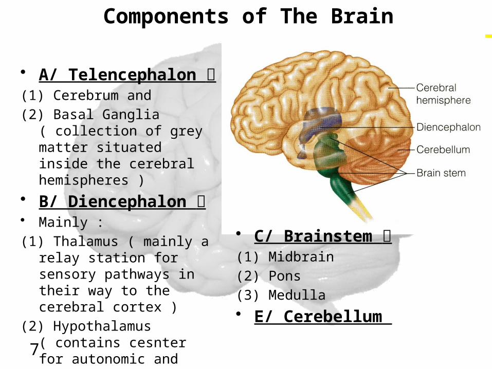
Components of The Brain
• A/ Telencephalon (1) Cerebrum and(2) Basal Ganglia ( collection
of grey matter situated inside the cerebral hemispheres )
• B/ Diencephalon • Mainly :(1) Thalamus ( mainly a
relay station for sensory pathways in their way to the cerebral cortex )
(2) Hypothalamus ( contains cesnter for autonomic and endocrine control )
7
• C/ Brainstem (1) Midbrain(2) Pons(3) Medulla• E/ Cerebellum
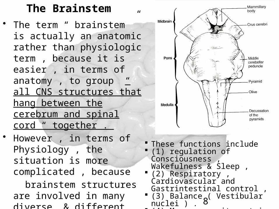
The Brainstem • The term “ brainstem ” is
actually an anatomic rather than physiologic term , because it is easier , in terms of anatomy , to group “ all CNS structures that hang between the cerebrum and spinal cord “ together .
• However , in terms of Physiology , the situation is more complicated , because
brainstem structures are involved in many diverse & different bodily functions .
8
These functions include (1) regulation of
Consciousness , Wakefulness & Sleep ,
(2) Respiratory , Cardiovascular and Gastrintestinal control ,
(3) Balance ( Vestibular nuclei ) .
(4) Moreover , it contain several Cranial Nerve nuclei .
,
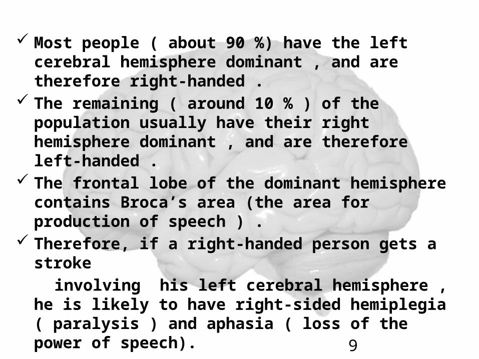
Most people ( about 90 %) have the left cerebral hemisphere dominant , and are therefore right-handed .
The remaining ( around 10 % ) of the population usually have their right hemisphere dominant , and are therefore left-handed .
The frontal lobe of the dominant hemisphere contains Broca’s area (the area for production of speech ) .
Therefore, if a right-handed person gets a stroke
involving his left cerebral hemisphere , he is likely to have right-sided hemiplegia ( paralysis ) and aphasia ( loss of the power of speech). 9

Lobes, the Cerebral Cortex, and Cortical Regions of the Brain
Objectives:• Students will be able to describe the general structure of the
Cerebrum and Cerebral Cortex.
• Students will be able to identify the Cerebrum, the Lobes of the Brain, the Cerebral Cortex, and its major regions/divisions.
• Students will be able to describe the primary functions of the Lobes and the Cortical Regions of the Brain.
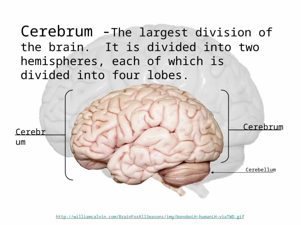
Cerebrum -The largest division of the brain. It is divided into two hemispheres, each of which is divided into four lobes.
CerebrumCerebrum
Cerebellum
http://williamcalvin.com/BrainForAllSeasons/img/bonoboLH-humanLH-viaTWD.gif
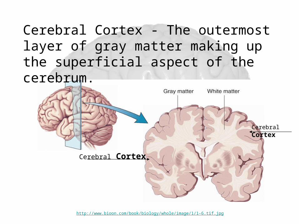
Cerebral Cortex
Cerebral Cortex
Cerebral Cortex - The outermost layer of gray matter making up the superficial aspect of the cerebrum.
http://www.bioon.com/book/biology/whole/image/1/1-6.tif.jpg
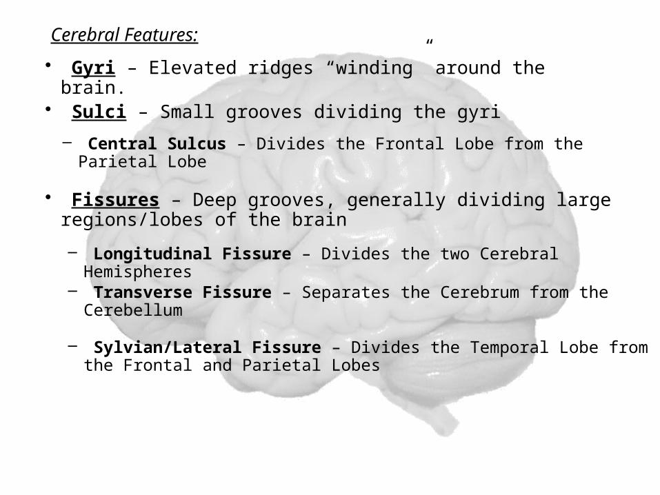
Cerebral Features:
• Sulci – Small grooves dividing the gyri
– Central Sulcus – Divides the Frontal Lobe from the Parietal Lobe
• Fissures – Deep grooves, generally dividing large regions/lobes of the brain
– Longitudinal Fissure – Divides the two Cerebral Hemispheres
– Transverse Fissure – Separates the Cerebrum from the Cerebellum
– Sylvian/Lateral Fissure – Divides the Temporal Lobe from the Frontal and Parietal Lobes
• Gyri – Elevated ridges “winding” around the brain.

Gyri (ridge)
Fissure
(deep groove)
Sulci (groove)
http://williamcalvin.com/BrainForAllSeasons/img/bonoboLH-humanLH-viaTWD.gif
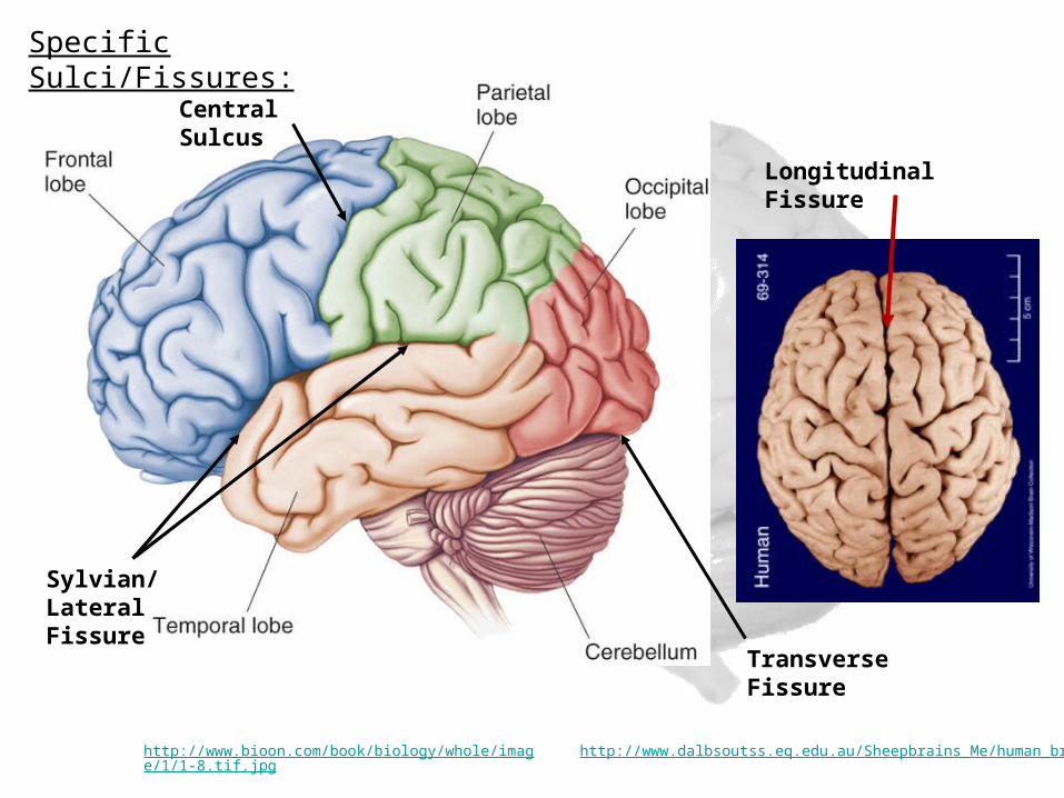
Longitudinal Fissure
Transverse Fissure
Sylvian/Lateral Fissure
Central Sulcus
http://www.bioon.com/book/biology/whole/image/1/1-8.tif.jpg http://www.dalbsoutss.eq.edu.au/Sheepbrains_Me/human_brain.gif
Specific Sulci/Fissures:

Lobes of the Brain (4)
• Frontal• Parietal• Occipital• Temporal
* Note: Occasionally, the Insula is considered the fifth lobe. It is located deep to the Temporal Lobe.
http://www.bioon.com/book/biology/whole/image/1/1-8.tif.jpg
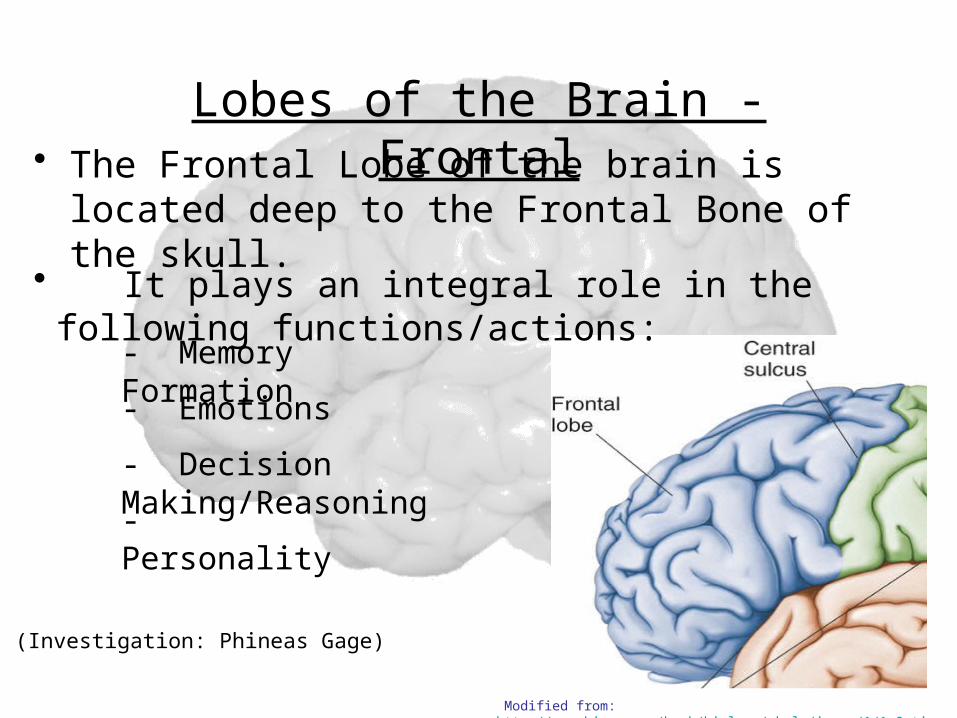
Lobes of the Brain - Frontal• The Frontal Lobe of the brain is located deep to the
Frontal Bone of the skull.
(Investigation: Phineas Gage)
• It plays an integral role in the following functions/actions:
- Memory Formation
- Emotions
- Decision Making/Reasoning
- Personality
Modified from: http://www.bioon.com/book/biology/whole/image/1/1-8.tif.jpg
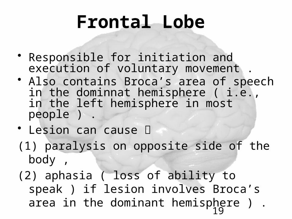
Frontal Lobe
• Responsible for initiation and execution of voluntary movement .
• Also contains Broca’s area of speech in the dominnat hemisphere ( i.e., in the left hemisphere in most people ) .
• Lesion can cause (1) paralysis on opposite side of the body , (2) aphasia ( loss of ability to speak ) if
lesion involves Broca’s area in the dominant hemisphere ) .
19
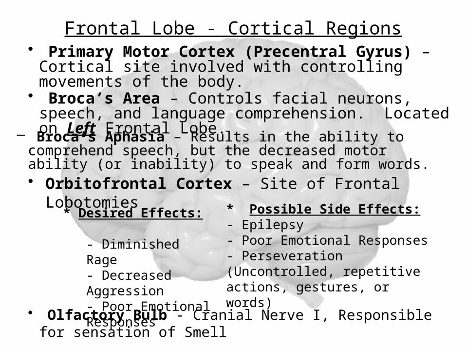
Frontal Lobe - Cortical Regions
• Orbitofrontal Cortex – Site of Frontal Lobotomies
• Primary Motor Cortex (Precentral Gyrus) – Cortical site involved with controlling movements of the body.
• Broca’s Area – Controls facial neurons, speech, and language comprehension. Located on Left Frontal Lobe.
– Broca’s Aphasia – Results in the ability to comprehend speech, but the decreased motor ability (or inability) to speak and form words.
• Olfactory Bulb - Cranial Nerve I, Responsible for sensation of Smell
* Desired Effects:- Diminished Rage- Decreased Aggression- Poor Emotional Responses
* Possible Side Effects:- Epilepsy- Poor Emotional Responses- Perseveration (Uncontrolled, repetitive actions, gestures, or words)

Primary Motor Cortex/ Precentral Gyrus
Broca’s Area
Orbitofrontal Cortex
Olfactory Bulb
Modified from: http://www.bioon.com/book/biology/whole/image/1/1-8.tif.jpg
Regions
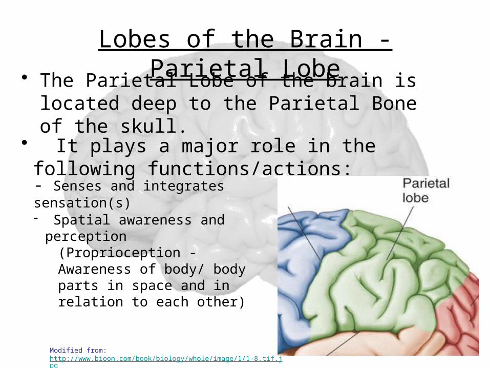
Lobes of the Brain - Parietal Lobe
• The Parietal Lobe of the brain is located deep to the Parietal Bone of the skull.
• It plays a major role in the following functions/actions:
- Senses and integrates sensation(s)
- Spatial awareness and perception
(Proprioception - Awareness of body/ body parts in space and in relation to each other)
Modified from: http://www.bioon.com/book/biology/whole/image/1/1-8.tif.jpg
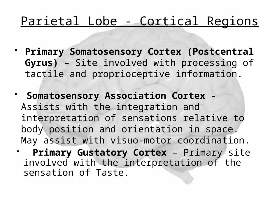
Parietal Lobe - Cortical Regions
• Primary Somatosensory Cortex (Postcentral Gyrus) – Site involved with processing of tactile and proprioceptive information.
• Somatosensory Association Cortex - Assists with the integration and interpretation of sensations relative to body position and orientation in space. May assist with visuo-motor coordination.
• Primary Gustatory Cortex – Primary site involved with the interpretation of the sensation of Taste.
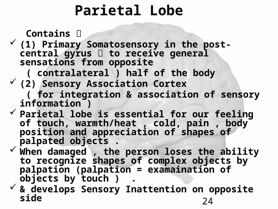
Parietal Lobe
Contains (1) Primary Somatosensory in the post-
central gyrus to receive general sensations from opposite
( contralateral ) half of the body (2) Sensory Association Cortex ( for integration & association of sensory
information ) Parietal lobe is essential for our feeling of
touch, warmth/heat , cold, pain , body position and appreciation of shapes of palpated objects .
When damaged , the person loses the ability to recognize shapes of complex objects by palpation (palpation = examaination of objects by touch ) .
& develops Sensory Inattention on opposite side 24
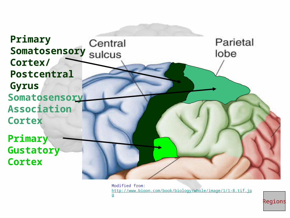
Primary Somatosensory Cortex/ Postcentral Gyrus
Primary Gustatory Cortex
Somatosensory Association Cortex
Regions
Modified from: http://www.bioon.com/book/biology/whole/image/1/1-8.tif.jpg
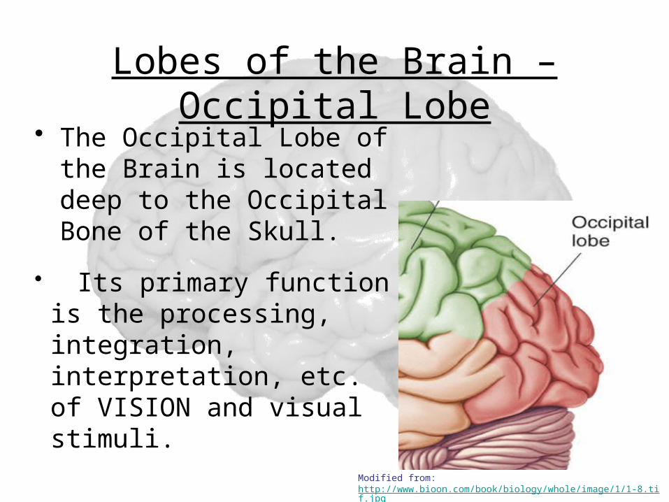
Lobes of the Brain – Occipital Lobe
• The Occipital Lobe of the Brain is located deep to the Occipital Bone of the Skull.
• Its primary function is the processing, integration, interpretation, etc. of VISION and visual stimuli.
Modified from: http://www.bioon.com/book/biology/whole/image/1/1-8.tif.jpg
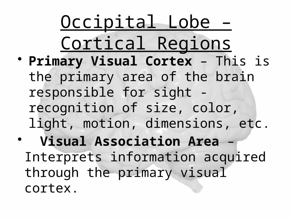
Occipital Lobe – Cortical Regions
• Primary Visual Cortex – This is the primary area of the brain responsible for sight -recognition of size, color, light, motion, dimensions, etc.
• Visual Association Area – Interprets information acquired through the primary visual cortex.
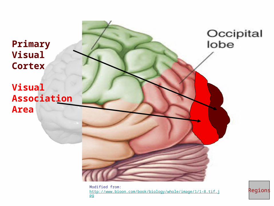
Primary Visual Cortex
Visual Association Area
RegionsModified from: http://www.bioon.com/book/biology/whole/image/1/1-8.tif.jpg
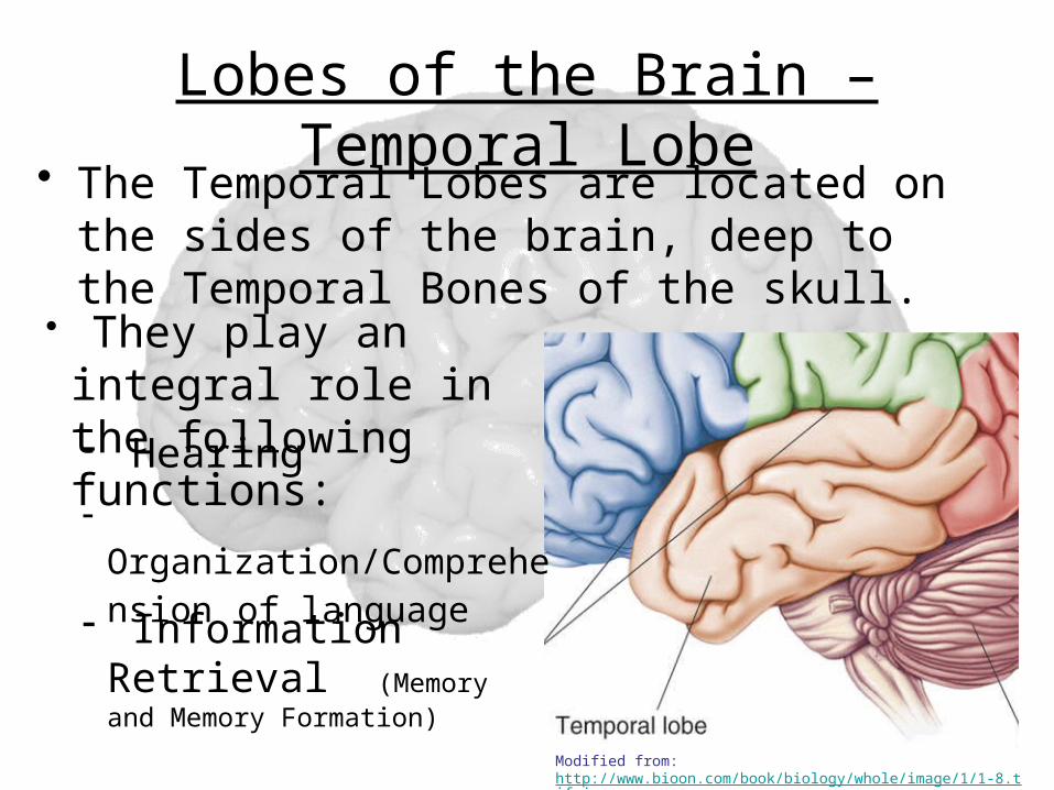
Lobes of the Brain – Temporal Lobe
• The Temporal Lobes are located on the sides of the brain, deep to the Temporal Bones of the skull.
• They play an integral role in the following functions:- Hearing
- Organization/Comprehension of language
- Information Retrieval (Memory and Memory Formation)
Modified from: http://www.bioon.com/book/biology/whole/image/1/1-8.tif.jpg
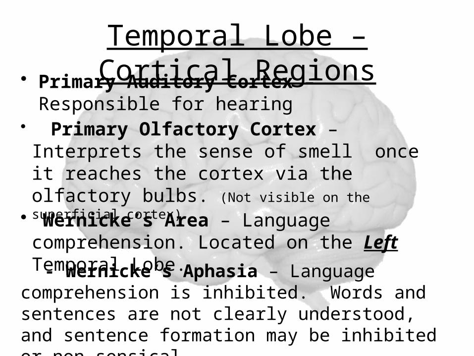
Temporal Lobe – Cortical Regions• Primary Auditory Cortex – Responsible for hearing
• Primary Olfactory Cortex – Interprets the sense of smell once it reaches the cortex via the olfactory bulbs. (Not visible on the superficial cortex)
• Wernicke’s Area – Language comprehension. Located on the Left Temporal Lobe.
- Wernicke’s Aphasia – Language comprehension is inhibited. Words and sentences are not clearly understood, and sentence formation may be inhibited or non-sensical.
Temporal Lobe
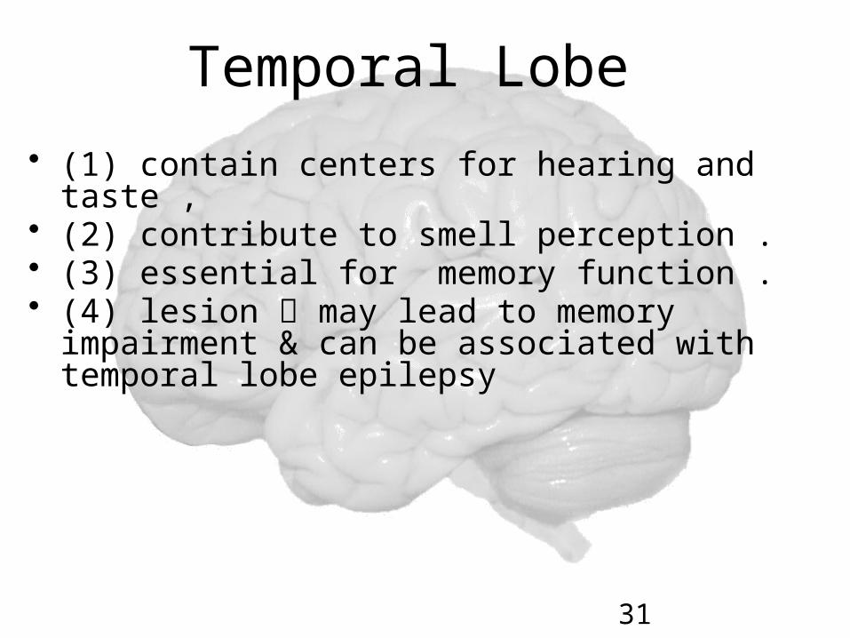
• (1) contain centers for hearing and taste ,• (2) contribute to smell perception . • (3) essential for memory function . • (4) lesion may lead to memory
impairment & can be associated with temporal lobe epilepsy
31
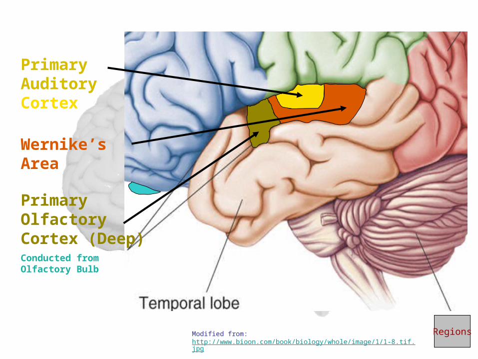
Primary Auditory Cortex
Wernike’s Area
Primary Olfactory Cortex (Deep)Conducted from Olfactory Bulb
RegionsModified from: http://www.bioon.com/book/biology/whole/image/1/1-8.tif.jpg
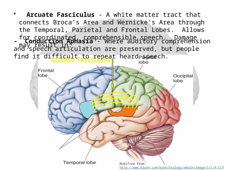
• Arcuate Fasciculus - A white matter tract that connects Broca’s Area and Wernicke’s Area through the Temporal, Parietal and Frontal Lobes. Allows for coordinated, comprehensible speech. Damage may result in:
- Conduction Aphasia - Where auditory comprehension and speech articulation are preserved, but people find it difficult to repeat heard speech.
Modified from: http://www.bioon.com/book/biology/whole/image/1/1-8.tif.jpg
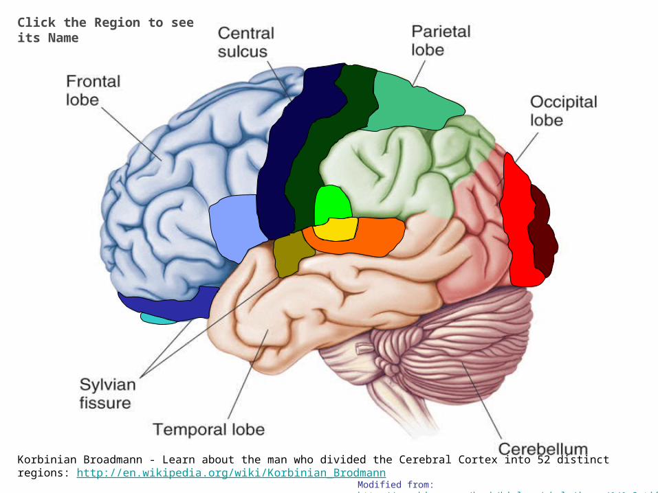
Click the Region to see its Name
Korbinian Broadmann - Learn about the man who divided the Cerebral Cortex into 52 distinct regions: http://en.wikipedia.org/wiki/Korbinian_Brodmann
Modified from: http://www.bioon.com/book/biology/whole/image/1/1-8.tif.jpg
Functional Principles of the Cerebral hemispheres
1. Each cerebral hemisphere receives sensory information from, and sends motor commands to, the opposite side of body2. The 2 hemispheres have somewhat different functions although their structures are alike3. Correspondence between a specific function and a specific region of cerebral cortex is not precise4. No functional area acts alone; conscious behavior involves the entire cortex
Higher level: Prefrontal Cortex• Most complicated region, coordinates info from all other association areas• Important in intellect, planning, reasoning, mood, abstract ideas, judgement, conscience, and accuratley predicting consequences• Phineas Gage?
Hemispheric Lateralization• Functional differences between left and right hemispheres• In most people, left hemisphere (dominant hemisphere) controls:
– reading, writing, and math, decision-making, logic, speech and language (usually)• Right cerebral hemisphere relates to:
– recognition (faces, voice inflections), affect, visual/spatial reasoning, emotion, artistic skills
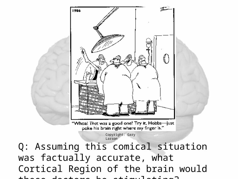
Q: Assuming this comical situation was factually accurate, what Cortical Region of the brain would these doctors be stimulating?
Copyright: Gary Larson
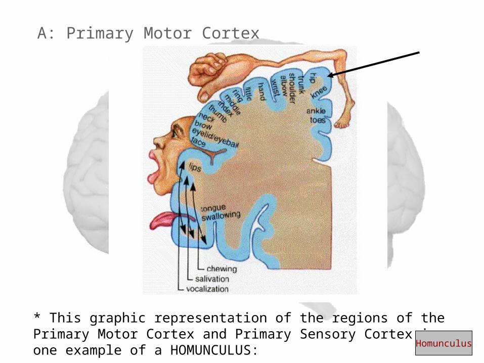
A: Primary Motor Cortex
* This graphic representation of the regions of the Primary Motor Cortex and Primary Sensory Cortex is one example of a HOMUNCULUS:
Homunculus