functionalization of chitosan/poly(lactic acid-glycolic acid) sintered microsphere scaffolds via...
TRANSCRIPT
Functionalization of chitosan/poly(lactic acid-glycolic acid)sintered microsphere scaffolds via surface heparinizationfor bone tissue engineering
Tao Jiang,1 Yusuf Khan,2 Lakshmi S. Nair,3 Wafa I. Abdel-Fattah,4 Cato T. Laurencin2,3
1Department of Chemical Engineering, University of Virginia, Charlottesville, Virginia 229042Department of Chemical, Materials and Biomolecular Engineering, University of Connecticut, Storrs,Connecticut 062693Department of Orthopaedic Surgery, University of Connecticut Health Center, Farmington, Connecticut 060304Biomaterials Department, National Research Centre, Cairo, 12311, Egypt
Received 23 January 2009; revised 14 May 2009; accepted 29 July 2009Published online 23 September 2009 in Wiley InterScience (www.interscience.wiley.com). DOI: 10.1002/jbm.a.32615
Abstract: Scaffolds exhibiting biological recognition andspecificity play an important role in tissue engineering andregenerative medicine. The bioactivity of scaffolds in turninfluences, directs, or manipulates cellular responses. Inthis study, chitosan/poly(lactic acid-co-glycolic acid) (chi-tosan/PLAGA) sintered microsphere scaffolds were func-tionalized via heparin immobilization. Heparin was suc-cessfully immobilized on chitosan/PLAGA scaffolds withcontrollable loading efficiency. Mechanical testing showedthat heparinization of chitosan/PLAGA scaffolds did notsignificantly alter the mechanical properties and porousstructures. In addition, the heparinized chitosan/PLAGAscaffolds possessed a compressive modulus of 403.98 619.53 MPa and a compressive strength of 9.83 6 0.94 MPa,which are in the range of human trabecular bone. Further-more, the heparinized chitosan/PLAGA scaffolds hadan interconnected porous structure with a total porevolume of 30.93 6 0.90% and a median pore size of
172.33 6 5.89 lm. The effect of immobilized heparin onosteoblast-like MC3T3-E1 cell growth was investigated.MC3T3-E1 cells proliferated three dimensionally through-out the porous structure of the scaffolds. Heparinized chi-tosan/PLAGA scaffolds with low heparin loading (1.7 lg/scaffold) were shown to be capable of stimulating MC3T3-E1 cell proliferation by MTS assay and cell differentiationas evidenced by elevated osteocalcin expression whencompared with nonheparinized chitosan/PLAGA scaffoldand chitosan/PLAGA scaffold with high heparin loading(14.1 lg/scaffold). This study demonstrated the potentialof functionalizing chitosan/PLAGA scaffolds via heparin-ization with improved cell functions for bone tissue engi-neering applications. � 2009 Wiley Periodicals, Inc.J Biomed Mater Res 93A: 1193–1208, 2010
Key words: bone tissue engineering; chitosan; poly(lacticacid-glycolic acid); heparin; bioactive scaffold
INTRODUCTION
Tissue and organ failure of the human body hasbeen a worldwide health care problem. Transplanta-tion of autogenous or allogenous tissues for thereplacement of diseased organs, although still a val-uable medical therapy, suffers from a number of lim-itations such as donor shortage, transplant rejection,and disease transmission.1 Tissue engineering repre-sents a novel, promising strategy that applies theprinciples of engineering and life sciences towardthe development of biological substitutes thatrestore, maintain, or improve tissue function.2 In the
realm of tissue engineering, a biodegradable, cyto-compatible biomaterial is one of the central elementsthat is critical for successful tissue regeneration inmany applications.3–5
Both natural and synthetic materials are widelyused in research and for clinical applications nowa-days. Most biomaterials used in medical deviceselicit a nonspecific response or foreign body reactionfrom biological systems upon implantation. Torender specificity to an implantable biomaterial, oneof the most important advances in biomaterials sci-ence has been to engineer biomaterials with appro-priate bulk and especially surface properties to con-trol the cell and tissue reactions to biomaterials.These finely designed biomaterials should satisfy anumber of criteria. For instance, the biomaterialsshould have recognition sites and demonstrate speci-ficity; the biomaterials should induce specific healing
Correspondence to: C. T. Laurencin; e-mail: [email protected]
� 2009 Wiley Periodicals, Inc.

pathways; and the biomaterials should not elicitnonspecific body responses.
The synthetic biodegradable polymer poly(lacticacid-glycolic acid) (PLAGA) has been investigatedfor various biomedical applications due to the itscell and tissue compatibility, resorbability, and excel-lent mechanical strength.6–8 Nevertheless, PLAGAencounters the problem of lacking reactive sites towhich more biological features could be added on.The lack of suitable functional groups of PLAGAmakes it less feasible for the absorption, physicalcomplexation, and/or covalent conjugation of spe-cific bioactive ligands. Chitosan, a natural polysac-charide derived from crustacean chitin, is a biocom-patible, enzymatically degradable biomacromoleculereceiving much research interest.9–11 It has structuralsimilarity to glycosaminoglycans, making it a poten-tial candidate for connective tissue engineering. Inaddition, chitosan possesses free amine andhydroxyl groups on its molecular chain. These reac-tive sites make it possible to further physically orchemically modify chitosan to impart bioactivity.Research has shown that RGDS (Arg-Gly-Asp-Ser)containing peptide sequences or bone morphogeneticprotein-2 (BMP-2) can be covalently conjugated tochitosan.12–14 These modified-chitosan substratesshowed enhanced cell attachment, proliferation, andmineralization.
Our laboratory initially developed a sinteredpolymeric microsphere technique to create three-dimensional (3D) porous scaffolds for bone regenera-tion.15–18 Using this technique, we previously devel-oped a chitosan/PLAGA sintered microspheresystem that has an interconnected porous structureand is mechanically competent suitable as scaffoldsfor load-bearing bone tissue engineering applica-tions.19 In addition, we proposed that the aminegroups of chitosan on the microsphere surface couldbe readily protonated and subsequently beneficialfor the introduction of certain bioactive molecules.
Heparin is a linear highly sulfated glycosamino-glycan consisting of sulfated repeating disaccharideunit, which has long been used clinically as an anti-coagulant and is also widely recognized to be a bio-logically important biomolecule. Studies have shownthat freely present heparin could affect cellular func-tions of MC3T3-E1 cells.20,21 In addition, heparin isable to bind with a number of growth factors suchas fibroblast growth factors (FGFs), transforminggrowth factor–b (TGF–b), platelet-derived growthfactors (PDGF), insulin-like growth factor II (IGF-II),and bone morphogenetic proteins-2 and -4 (BMP-2and -4) with heparin-binding domains which areinvolved in the regulation of many cell fate proc-esses of the osteoblast lineage.22 Studies have investi-gated the capability of heparin-containing substratesto interact with and deliver growth factors for
enhanced cell functions.23,24 Furthermore, heparinand its structurally similar heparan sulfate wereindicated to be involved in the signaling pathwaysof certain growth factors.25,26 Therefore, the effect ofheparin directly on osteoblastic cell growth and itsinteraction with most skeletal growth factors makesit an interesting biomolecule for applications in bonetissue engineering.
The objective of this study was to functionalize thecomposite chitosan/PLAGA sintered microspherescaffolds with the biomolecule heparin, characterizethe heparinized chitosan/PLAGA scaffolds, andinvestigate the effect of immobilized heparin on thegrowth of the osteoblast-like MC3T3-E1 cells.
MATERIALS AND METHODS
Materials
PLAGA (85:15 lactide to glycolide ratio, MW 5 120kDa) was obtained from Alkermes, Wilmington, OH. Chi-tosan (derived from chitin extracted from crab andshrimp shells, 83.3% degree of deacetylation, viscosity of1% solution in 1% (v/v) acetic acid 5 115 cps) was pur-chased from Vanson HaloSources, Redmond, WA andwas pulverized to randomly shaped microparticles withsizes of 25 6 16 lm (by ICO Polymers, Bloombury, NJ).Methylene chloride, ascorbic acid, bovine serum albumin,and toluidine blue O were obtained from Fisher Scientific(Hampton, NH). Poly(vinyl alcohol) (PVA), b-glycero-phosphate, and heparin sodium salt (180 USP units/mg)were obtained from Sigma (St. Louis, MO). Fluoresceinconjugate heparin was procured from Molecular Probes,Eugene, OR. Alpha minimum essential medium (a-MEM), fetal bovine serum (FBS), and penicillin–strepto-mycin for cell culture were purchased from InvitrogenCorporation (Carlsbad, CA). 1,25-dihydroxyvitamin D3was purchased from EMD Biosciences (San Diego, CA)and was reconstituted in 100% ethanol at a concentrationof 50 lg/mL.
Fabrication of composite chitosan/PLAGA sinteredmicrosphere scaffold
Chitosan/PLAGA composite microspheres were pre-pared using the solvent evaporation technique as previ-ously described.19 Briefly, milled chitosan microparticleswere mixed with 20% (w/v) PLAGA solution in methyl-ene chloride according to a 1:4 chitosan to PLAGA weightratio and vortexed for 12 h until a homogeneous suspen-sion was obtained. Chitosan/PLAGA suspension was thenpoured into 1% PVA solution under a stirring speed of 210rpm for 24 h to allow the methylene chloride to evaporate.The resultant chitosan/PLAGA composite microsphereswere washed with distilled deionized (DI) water, filtered,lyophilized, sieved to different sizes, and then stored in adessicator for future use.
1194 JIANG ET AL.
Journal of Biomedical Materials Research Part A
Cylindrical (5 mm diameter 3 10 mm length and 5 mmdiameter 3 15 mm length) or disk-like (10 mm diameter 32 mm thickness) chitosan/PLAGA composite scaffoldswere fabricated by packing composite microspheres meas-uring 500–710 lm in diameter, into either a stainless steel(for fabrication of cylindrical scaffolds) or a brass mold(for fabrication of disk-like scaffolds). The mold washeated at 1128C (for fabrication of cylindrical scaffolds) or1068C (for fabrication of disk-like scaffolds) for 4 h toachieve bonding between neighboring microspheres. Dif-ferent temperatures were to achieve comparable degree ofsintering in molds with different heat transfer properties.Once heating was completed, the mold was allowed tocool down to room temperature before the scaffolds wereremoved. These scaffolds are referred to as chitosan/PLAGA scaffolds.
Immobilization of heparin on chitosan/PLAGAscaffold
The disk-like chitosan/PLAGA composite scaffoldswere placed in 24-well plates and soaked in 1 mL of0.5% acetic acid (HAc) for 5 min. After being washedthree times with DI water and air-dried, the HAc-treatedchitosan/PLAGA scaffolds (designated as HAc-chitosan/PLAGA) were immersed in 1 mL of 4 lg/mL or 40 lg/mL or 200 lg/mL aqueous heparin solution for 2 h. Thescaffolds were then washed three times with DI water,air-dried, and rehydrated using ethanol series of 100%,70%, 50%, and DI water for 30 min in each step and air-dried. Only the 4 lg/mL and 40 lg/mL heparin-treatedHAc-chitosan/PLAGA scaffolds were used for cell studyand they were designated as HAc-chitosan/PLAGA lowheparin and HAc-chitosan/PLAGA high heparin, respec-tively. Heparin concentrations in solution before and afterscaffold immersion were quantified using the Chromoge-nix Coatest1 heparin kit (DiaPharma, West Chester, OH)according to the manufacturer’s instruction. This colori-metric assay is based on the ability of heparin binding toAntithrombin (AT). The heparin-AT complex subse-quently neutralizes excess Factor Xa (FXa), while theremaining FXa hydrolyses a chromogenic substrate togive color. The absorbance is then read photometricallyat 405 nm. The amount of heparin immobilized onscaffolds was determined by the concentration difference(n 5 4).
Heparin adsorption and dissolution
The kinetics of heparin adsorption on chitosan/PLAGAscaffolds and heparin adsorption capacity were investi-gated using a toluidine blue assay. For heparin adsorp-tion study, the disk-like chitosan/PLAGA composite scaf-folds were placed in 24-well plates and soaked in 1 mLof 0.5% acetic acid (HAc) for 5 min. After being washedthree times with DI water and air-dried, the HAc-chito-san/PLAGA scaffolds were immersed in 1 mL of 200 lg/mL heparin aqueous solutions for various times upto 8 h.At predetermined time points, 250 lL of the left-overheparin solution was assayed. In addition, various hepa-
rin concentrations 4 lg/mL, 40 lg/mL, and 200 lg/mLwere chosen. The adsorption time was then fixed to 2 hto investigate the relationship between the original hepa-rin concentration and the amount of heparin adsorbed onscaffolds. The reaction between toluidine blue and hepa-rin allows for the determination of heparin content insolution. Briefly, a 0.005% solution of toluidine blue wasprepared in 0.01N HCl containing 0.2% NaCl. Standardheparin solution was prepared by dissolving 10 mg hepa-rin in 100 mL 0.2% NaCl aqueous solution. Next, 2.5 mLof 0.005% toluidine blue solution was pipetted into testtubes. Test samples, heparin standards (10–70 lg heparin)along with a blank solution (0.2% NaCl) were added anddiluted with 0.2% NaCl to a total volume of 5 mL. Allthe tubes were agitated for 30 s. Hexane (5 mL) was thenadded to each tube, and the tubes were shaken vigo-rously for another 30 s to separate the heparin dye com-plex. The aqueous layer from all the tubes was removedand the absorbance was measured at 631 nm within 30min using a Shimadzu UV-1201 spectrophotometer. Thedifference in the absorbance of the original heparin solu-tion versus the left-over heparin solution provided theamount of heparin that is adsorbed on to a particularscaffold.
For heparin dissolution, Chitosan/PLAGA scaffolds(5 mm diameter 3 15 mm length) were heparinized with2.5 mL of 4 lg/mL or 200 lg/mL heparin solution asdescribed earlier. The heparinized scaffolds were placed in15-mL plastic centrifuge tubes containing 3 mL phosphatebuffered saline (PBS), pH 7.4 and were placed in a shakingwater bath at 378C. At appropriate intervals, namely 4, 8,12, 24 h; 3, 9, 14, 21, 28 and 37 days, 500 lL of the dissolu-tion medium was withdrawn, filtered through 0.22 lmmembrane, and assayed for heparin content using a Coat-est1 Heparin chromogenic assay kit (DiaPharma Group,West Chester, OH) according to the manufacturer’sinstruction.
Characterization of heparinized chitosan/PLAGAscaffold
Scanning electron microscopy (SEM)
Surface morphology of the chitosan/PLAGA scaffold,HAc-chitosan/PLAGA scaffold, and heparinized chitosan/PLAGA scaffold as well as pure PLAGA scaffold wasvisualized using a JEOL JSM-6400 scanning electron micro-scope.
X-ray photoelectron spectroscopy (XPS)
The surface chemical characteristics of the HAc-chito-san/PLAGA and HAc-chitosan/PLAGA low heparin andhigh heparin scaffolds were evaluated by XPS (ESCA/SAM, Physical Electronics, (PHI) 560) using Mg X-ray radi-ation (300 W, 16 kV). The porous scaffolds were mountedonto a copper sample stage and were analyzed at a pres-sure of 1028 Torr with pass energy of 200 eV.
FUNCTIONALIZATION OF CHITOSAN/PLAGA SINTERED MICROSPHERE SCAFFOLDS 1195
Journal of Biomedical Materials Research Part A

Confocal microscopy
The disk-like chitosan/PLAGA composite scaffolds weretreated with 1 mL of 4 lg/mL or 40 lg/mL or 200 lg/mLfluorescence conjugated heparin solution according to theimmobilization method described earlier. Heparinized chi-tosan/PLAGA scaffolds were further mounted onto glassslides with anti-fade mounting solution (CHEMICONInternational, Temecula, CA). Fluorescence was subse-quently visualized using a Zeiss LSM 510-UV confocallaser scanning microscope (CLSM, Carl Zeiss MicroImag-ing, Thornwood, NY).
Mechanical testing
The chitosan/PLAGA scaffolds and HAc-chitosan/PLAGA low heparin scaffolds (5 mm diameter 3 10 mmlength) were tested using an Instron mechanical testingmachine (Instron model 5544, Canton, MA). Compressivetesting on scaffolds was conducted at ambient temperatureand humidity with a crosshead speed of 5 mm/min. Com-pressive modulus and compressive strength were deter-mined using Merlin software associated with Instron (n5 6).
Mercury intrusion porosimetry
The total pore volume and median pore size of the chi-tosan/PLAGA scaffolds and HAc-chitosan/PLAGA lowheparin scaffolds (5 mm diameter 3 10 mm length) weredetermined using a mercury intrusion porosimeter (Micro-meritics Autopore III, Norcross, GA) (n 5 3).
In vitro cell proliferation on heparinized chitosan/PLAGA scaffold
Cell seeding and cell culture
Disk-like HAc-chitosan/PLAGA scaffolds, HAc-chito-san/PLAGA low heparin scaffolds, HAc-chitosan/PLAGAhigh heparin scaffolds, and control sintered PLAGA micro-sphere scaffolds for in vitro cell study were sterilized byimmersing in 70% ethanol for 10 min, washed three timeswith sterile DI water for 30 min, and exposed to UV lightfor 30 min on each side. Osteoblast-like MC3T3-E1 cells(ATCC, Manassas, VA) were seeded onto the aforemen-tioned scaffolds at a density of 5 3 104 cells per scaffold.Cells were cultured in a-MEM supplemented with 10%FBS, 1% penicillin–streptomycin, 3 mM b-glycerophos-phate and 10 lg/mL ascorbic acid, and maintained in anincubator at 378C with 5% CO2 and 95% humidified air for21 days.
Cell proliferation
At predetermined time points, cell attachment and pro-liferation were visualized qualitatively using SEM. Cellson scaffolds were fixed at room temperature in 1% and 3%gluteraldehyde for 1 h and 24 h, respectively. The cell-scaf-
fold constructs were then dehydrated sequentially usingethanol series (50%, 70%, 80%, 90%, 95%, and 100%) for 10min each. Cell-scaffolds were allowed to dry overnight,coated with gold/palladium, and cell proliferation wasevaluated under SEM.
Cell proliferation was also quantitatively analyzed byutilizing 3-(4,5-dimethylthiazol-2-yl)-5-(3-carboxymethoxy-phenyl)-2-(4-sulfophenyl)-2H-tetrazolium (MTS, Promega,Madison, WI) mitochondrial reduction according to themanufacturer’s instructions. In brief, scaffolds at predeter-mined time points were washed with PBS, transferred intoa new well plate containing 1 mL culture medium and200 lL MTS solution, and incubated at 378C for 2 h. At theend of the incubation time, the reaction was stopped byadding 250 lL of 10% sodium dodecyl sulfate (SDS) solu-tion. The resulting solution was diluted in a 4:1 ratio usingdistilled DI water and the absorbance was read at 490 nmusing a Tecan SpectroFluo Plus reader (TECAN USA,Boston, MA).
Cell nuclei and cytoskeletal actin staining
Cell migration and cell ingrowth into the porous scaf-folds were evaluated by nucleic and actin staining. Atpredetermined time points (day 7, 11, and 14), cells onscaffolds were washed once with PBS and subsequentlyfixed with 4% paraformaldehyde for 20 min. Cells werethen permeabilized with 0.1% Triton-100 for 5 min andblocked with bovine serum albumin for 30 min at roomtemperature. The cytoskeletal protein actin was thenstained with TRITC-conjugated phalloidin (1:100 dilution)(CHEMICON International, Temecula, CA) for 60 min atroom temperature. Simultaneously, nuclei counterstainingwere performed by incubating cell-scaffold constructswith 40-6-diamidino-2-phenylindole (1:300 dilution) (DAPI,CHEMICON International, Temecula, CA) for 5 min atroom temperature. Cell-scaffold constructs were furthermounted onto glass slides with anti-fade mounting solu-tion (CHEMICON International, Temecula, CA). Fluores-cence was subsequently visualized using a Zeiss LSM510-UV confocal laser scanning microscope (CLSM, CarlZeiss MicroImaging, Thornwood, NY).
Alkaline phosphatase activity
Alkaline phosphatase (ALP) activity of the cells wasmeasured as an early marker of the maintenance of theosteoblastic phenotype using an alkaline phosphatase sub-strate kit (Bio-Rad, Hercules, CA). This colorimetric assayis based on the conversion of p-nitrophenyl phosphate (P-NPP) into p-nitrophenol (P-NP) in the presence of ALP,where the rate of P-NP production is proportional to ALPactivity. Briefly, cells were lysed with 1% Triton X-100 inDEPC-treated water and the cell lysates were collected andstored at 2708C in a freezer. On thawing, 100 lL samplewas added to 400 lL of P-NPP substrate and buffer solu-tion mixture and incubated at 378C for 30 min. The reac-tion was then stopped by adding 500 lL of 0.4N sodiumhydroxide. The production of P-NP was determined by theabsorbance at 405 nm using TECAN. The results of ALP
1196 JIANG ET AL.
Journal of Biomedical Materials Research Part A

activity were normalized by the number of cells on respec-tive scaffolds.
Osteocalcin expression
For the measurement of osteocalcin expression, cells werecultured in a-MEM supplemented with 10% FBS, 1% peni-cillin–streptomycin, 3 mM b-glycerophosphate, 10 lg/mLascorbic acid, and freshly dissolved 0.01 nM 1,25-dihydroxy-vitamin D3. One day before the predetermined time points,scaffolds were transferred into a new well plate and freshmedia without 1,25-dihydroxyvitamin D3 were supplied.After 24 h, cell culture media were withdrawn and frozen at2708C. After 21 days, all sample media were thawed andthe osteocalcin expression levels in the media were quanti-fied using a mouse osteocalcin ELISA kit (Biomedical Tech-nologies, Stoughton, MA) according to the manufacturer’sinstruction. The osteocalcin expression levels in media werenormalized by the number of cells on respective scaffolds.
Statistical analysis
Quantitative data were reported as mean 6 standarddeviation. Statistical analysis was performed using a one-
way analysis of variance (one-way ANOVA). Comparisonbetween the two means was determined using the Tukeytest.
RESULTS
Heparin immobilization on chitosan/PLAGAscaffold
Figure 1 shows the scanning electron micrographsof the surface morphology of sintered PLAGA scaf-fold, acetic acid (HAc) treated PLAGA scaffold.Figure 2 shows the SEM micrographs of the surfacemorphology of chitosan/PLAGA scaffold, aceticacid-treated chitosan/PLAGA scaffold (HAc-chito-san/PLAGA), and 40 lg/mL heparin-treated HAc-chitosan/PLAGA scaffold. HAc treatment did notchange the surface morphology of PLAGA scaffold[Fig. 1(a–d)], indicating that the PLAGA phase wasmorphologically stable during the short term HActreatment. For chitosan/PLAGA scaffold, chitosanmicroparticles were found to be evenly distributedand embedded in the PLAGA phase on the micro-
Figure 1. Scanning electron micrographs indicating the effect of acetic acid treatment on the morphology of PLAGA scaf-folds. No obvious morphological change was observed on PLAGA scaffold before (a and b) and after (c and d) acetic acidtreatment. Magnifications: 350 (a and c) and 3200 (b and d).
FUNCTIONALIZATION OF CHITOSAN/PLAGA SINTERED MICROSPHERE SCAFFOLDS 1197
Journal of Biomedical Materials Research Part A

sphere surfaces [Fig. 2(a,b)]. HAc treatment of thechitosan/PLAGA scaffold partially dissolved chito-san on the microsphere surface, thus created aninterconnecting chitosan network on the scaffold sur-face based on the SEM observation [Fig. 2(c,d)]. Fur-ther heparin modification did not change the surfacemorphology [Fig. 2(e,f)].
The presence of heparin on the surface of chitosan/PLAGA scaffolds after heparinization was demon-
strated in Figure 3. XPS spectra of HAc-chitosan/PLAGA and HAc-chitosan/PLAGA high heparinscaffolds are shown in Figure 3(a). The XPS spectrumof HAc-chitosan/PLAGA scaffold showed abundantcarbon and oxygen peaks due to both of the poly-mers, chitosan and PLAGA. A nitrogen peak at thebinding energy of �400 eV was attributed to the pres-ence of chitosan on microsphere surface. After hepa-rinization, the element sulfur peak appeared at the
Figure 2. Scanning electron micrographs indicating the effect of acetic acid treatment on the morphology of chitosan/PLAGA scaffolds. For the chitosan/PLAGA scaffold (a and b), chitosan particles were embedded in the PLAGA compo-nent forming a discontinuous phase; in comparison, acetic acid treatment created an interconnecting chitosan network onthe surface of HAc-chitosan/PLAGA scaffold (c and d); further heparin immobilization (e and f) does not change the sur-face characteristics when compared with HAc-chitosan/PLAGA scaffold (g and h). Magnifications: 350 (a, c, and e), and3300 (b and d), and 3200 (f).
1198 JIANG ET AL.
Journal of Biomedical Materials Research Part A

binding energy of 168–170 eV [see insets on Fig. 3(a)]which indicated the presence of heparin on the sur-face of the heparinized scaffold. The presence of hep-arin on the surface of chitosan/PLAGA scaffolds wasfurther confirmed by immobilization of fluorescenceconjugated heparin and visualized under a confocalmicroscope. Figure 3(b–e) shows the confocal micro-graphs of heparin immobilized chitosan/PLAGA sin-tered microsphere scaffolds. The green fluorescenceindicated the uniform distribution of heparin mole-cules on the scaffolds. Nonheparinzed HAc-chitosan/PLAGA scaffold [Fig. 3(b)] appeared black undermicroscope (negative control). On the heparinizedHAc-chitosan/PLAGA scaffolds [Fig. 3(c–e)], thegreen fluorescence intensity increased with theincrease of the original heparin solution, indicatingmore heparin was adsorbed onto scaffold surface.
Heparin adsorption and dissolution
Heparin adsorption on chitosan/PLAGA scaffoldwas evaluated over an 8 h period of time and the
results are shown in Figure 4(a). When the scaffoldswere treated with 1 mL of 200 lg/mL heparin solu-tion, the amount of heparin that was adsorbed onscaffold increased with the adsorption time initially.After the first 2 h, a saturation of adsorption wasreached, after which point, the adsorption/dissolu-tion process was in equilibrium and no furtherincrease in adsorbed heparin was observed. By treat-ing with 200 lg heparin, �30% of heparin can beadsorbed by the disk-like scaffold.
In addition, the relationship between heparinadsorption and the initial heparin solution concentra-tion was investigated with a fixed immobilizationtime of 2 h [Fig. 4(b)]. It was found that enhancedheparin adsorption was achieved when increasingthe initial concentration of the heparin solution. Spe-cifically, it was calculated that 1.7 6 0.1 lg, 14.1 64.8 lg, and 66.3 6 22.8 lg of heparin was immobi-lized onto the chitosan/PLAGA scaffold after beingplaced in 4 lg/mL, 40 lg/mL, or 200 lg/mL heparinsolutions for 2 h, respectively.
For heparin dissolution, it was determined that�10.5 6 0.8 lg heparin was immobilized on chitosan/
Figure 3. (a) X-ray photoelectron spectroscopy spectra of HAc-chitosan/PLAGA scaffold and HAc-chitosan/PLAGAhigh heparin scaffold. Insets: enlarged XPS spectra between 100 ev and 200 ev; the peak at 168–170 ev (dotted circles)belonging to S 2p electrons suggests the presence of heparin, indicating the immobilization of heparin on HAc-chitosan/PLAGA scaffold. (b–e) Confocal fluorescent micrographs of fluorescein heparin immobilized chitosan/PLAGA scaffolds af-ter treated with heparin solutions at the concentration of (b) 0 lg/mL (negative control); (c) 4 lg/mL; (d) 40 lg/mL; and(e) 200 lg/mL. Green fluorescence was visible on 4 lg/mL heparin-treated scaffold indicating the presence of heparin.Heparin distribution was more uniform and abundant throughout the scaffolds when treated with 40 lg/mL heparin orhigher concentration. Fluorescence intensity increased with the increase of heparin concentration indicating higher amountof immobilized heparin on scaffolds. Magnification: 310. [Color figure can be viewed in the online issue, which isavailable at www.interscience.wiley.com.]
FUNCTIONALIZATION OF CHITOSAN/PLAGA SINTERED MICROSPHERE SCAFFOLDS 1199
Journal of Biomedical Materials Research Part A

PLAGA scaffolds (5 mm 3 15 mm) when treated with2.5 mL of 4 lg/mL heparin solution. Chitosan/PLAGA scaffolds treated with 4 lg/mL heparin solu-tion showed no detectable heparin release over the 37days period. In addition, �236.9 6 15.0 lg heparinwas immobilized on chitosan/PLAGA scaffolds (5mm 3 15 mm) when treated with 2.5 mL of 200 lg/mL heparin solution. Cumulative heparin release pro-file from these scaffolds during the first 24 h is shownin Figure 4(c). It was found that only a small amountof heparin (0.2 lg) was released from the scaffoldwithin 24 h. After the first day, no detectable heparinrelease was observed.
Characterization of heparinized chitosan/PLAGAscaffold
The compressive properties, total pore volume, andmedian pore size of the chitosan/PLAGA scaffoldsand HAc-chitosan/PLAGA high heparin scaffoldsare shown in Figure 5. Chitosan/PLAGA scaffoldsshowed a compressive modulus of 418.16 6 14.72MPa and a compressive strength of 10.26 6 0.65 MPa.In addition, the scaffolds had a total pore volume of31.48 6 0.79% with a median pore size of 173.92 63.33 lm. On the other hand, HAc-chitosan/PLAGAhigh heparin scaffolds showed a compressive modu-lus of 403.98 6 19.53 MPa and a compressive strengthof 9.83 6 0.94 MPa. The total pore volume and me-dian pore size of the heparinized scaffolds were 30.936 0.90% and 172.33 6 5.89 lm, respectively. Therewas no statistically significant difference in the afore-mentioned properties between the chitosan/PLAGAscaffolds and heparinized chitosan/PLAGA scaffolds.Thus, it was demonstrated that the heparinizationprocess did not affect the mechanical properties andporous structures of scaffolds.
Cell proliferation
Cell proliferation on HAc-chitosan/PLAGA low/high scaffolds, control HAc-chitosan/PLAGA scaf-folds, and PLAGA scaffolds was visualized usingSEM and is shown in Figure 6. Based on SEM observa-tion, the trend of cell proliferation and migration onall scaffolds is similar. Cells were found to attach andproliferate on the scaffolds primarily around themicrosphere adjoining areas after 7 days [Fig.6(a,d,g,j)]. Only a small amount of cells were found inthese areas on either heparinized or nonheparinizedchitosan/PLAGA scaffolds, whereas a great amountof cells were found on PLAGA scaffolds at day 7. Cellproliferation propagated to microsphere surfaces andcells migrated from one microsphere to another or tothe porous structure after 14 days of culture on allscaffolds [Fig. 6(b,e,h,k)]. At day 14, dramatic cell pro-liferation was observed on both heparinized and non-heparinized chitosan/PLAGA scaffolds when com-pared with day 7. At this time point, PLAGA scaffoldsstill show a higher cell population than other types ofscaffolds. After 21 days of culture, cells fully spreadon all scaffolds and proliferated prominently both atthe adjoining areas and on the microsphere surfaces[Fig. 6(c,f,i,l)]. Cell migration toward the void spacesof the scaffolds was also observed.
Cell proliferation was quantitatively measured byMTS assay and shown in Figure 7(a). HAc-chitosan/PLAGA, HAc-chitosan/PLAGA low heparin, HAc-chitosan/PLAGA high heparin, and PLAGA scaf-folds supported MC3T3-E1 cell proliferation as indi-
Figure 4. (a) Heparin adsorption on chitosan/PLAGAscaffold (2 mm 3 10 mm) with time. Original heparin solu-tion concentration was 200 lg/mL. After 2 h, the adsorptionreached maximum and continued to stay at the same level.(b) Heparin adsorption on chitosan/PLAGA scaffold (2 mm3 10 mm) with different concentrations of heparin solu-tions. Adsorption time was fixed to 2 h. Increasing the con-centration of the heparin solution resulted in the increase ofheparin that immobilized on scaffolds. (*) indicates a signifi-cant difference (p < 0.05). Increase of heparin concentrationresults in significant increase of heparin immobilized onHAc-chitosan/PLAGA scaffolds. (c) Cumulative heparinrelease from chitosan/PLAGA scaffolds (5 mm 3 15 mm)treated with 200 lg/mL heparin solution in first 24 h.
1200 JIANG ET AL.
Journal of Biomedical Materials Research Part A

cated by the increasing cell number during the 21-day study period. Cell proliferation on PLAGA scaf-folds was significantly higher than heparinized andnonheparinized chitosan/PLAGA scaffolds duringthe 21 days of study. In addition, at day 21, cell pro-liferation on HAc-chitosan/PLAGA low heparinscaffold is significantly higher than HAc-chitosan/PLAGA and HAc-chitosan/PLAGA high heparinscaffolds. This observation at 21 days was furtherconfirmed by SEM in Figure 7(b–d), showing greatercell proliferation on HAc-chitosan/PLAGA lowheparin scaffold than HAc-chitosan/PLAGA andHAc-chitosan/PLAGA high heparin scaffolds.
Cell nuclei and cytoskeletal actin staining
3D cell proliferation and migration on HAc-chito-san/PLAGA low heparin scaffold was visualized viastaining with DAPI for cell nuclei. Osteoblast cytos-keletal protein actin was also stained and examinedusing CLSM and actin dynamics was monitoredover a 14-day time period. Figure 8 shows the dualstaining for cell nuclei and actin. Cells were found toattach at the joint areas between adjacent micro-spheres at day 7 [Fig. 8(a)]. After 11 days, cells pro-liferated dramatically and covered the surfaces ofmicrospheres [Fig. 8(d)]. At day 14, higher levels of
cell proliferation was observed on the microspheresurfaces. Moreover, cells were found to migrate to-ward and bridge the gaps among microspheres [Fig.8(g)]. Actin filaments were noticed to align them-selves along the circumferences of microspheres atearly time point [Fig. 8(b)]. As cells proliferated, newactin filaments were nucleated and polymerized intolonger filaments. The leading edge of actin filamentsmoved toward the entire microsphere surface fromthe microsphere–microsphere joint area, whichbegan to form an actin filament network on themicrosphere surface at day 11 [Fig. 8(e)]. This labilestructure of actin filaments was responsible for themovement and proliferation of cells toward micro-sphere surface. With the substantial cell proliferationafter 14 days, basal actin filaments were entirelyinterconnected and formed an orthogonal networkon the microsphere surfaces as well as in the gapsbetween microspheres [Fig. 8(h)].
Alkaline phosphatase activity
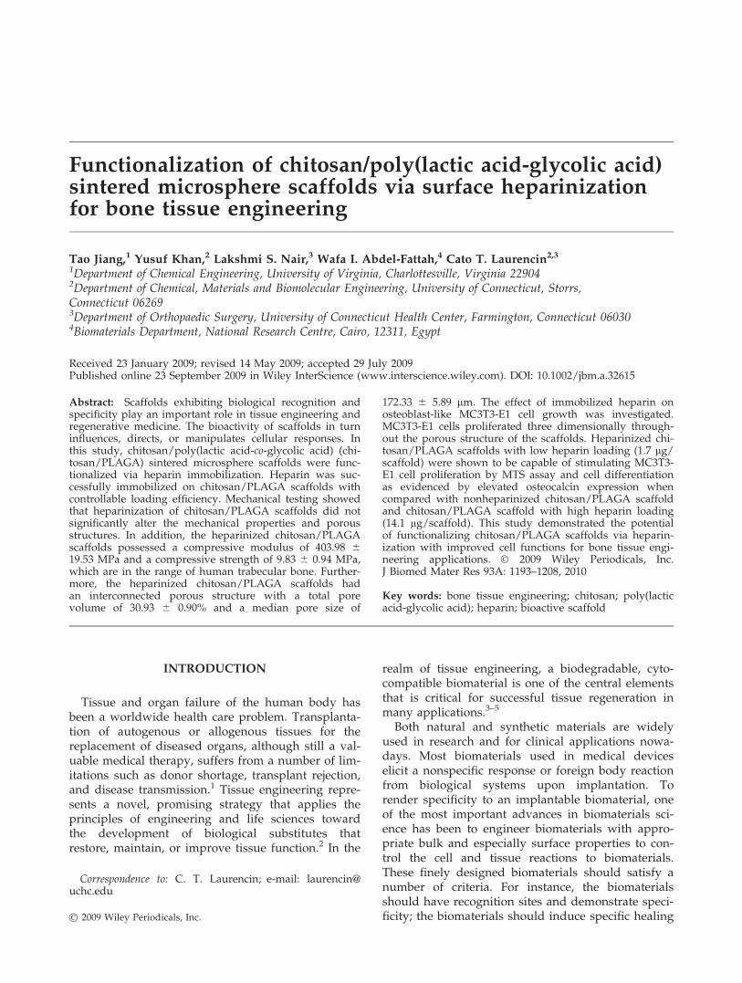
Figure 9 shows the ALP activity of the MC3T3-E1cells cultured on different scaffolds at day 7, 14, and21. ALP of cells continued to be expressed on all sub-strates during the period of study. The ALP activityof cells peaked at 21 days after culture on HAc-chito-
Figure 5. Effect of heparinization process on the compressive properties and porous structure of chitosan/PLAGA scaf-folds: (a) compressive modulus, (b) compressive strength, (c) total pore volume, and (d) median pore size of chitosan/PLAGA scaffolds and HAc-chitosan/PLAGA low heparin scaffolds. Results suggest that the compressive properties, totalpore volume, and median pore size of scaffolds are not significantly affected by scaffold heparinization.
FUNCTIONALIZATION OF CHITOSAN/PLAGA SINTERED MICROSPHERE SCAFFOLDS 1201
Journal of Biomedical Materials Research Part A

san/PLAGA low heparin scaffolds, HAc-chitosan/PLAGA high heparin scaffolds, and PLAGA scaf-folds. On HAc-chitosan/PLAGA scaffolds, the ALPactivity at day 14 was significantly higher than day 7.There was no further significant difference betweenday 21 and day 14. At day 21, cells on HAc-chito-san/PLAGA low and high heparin scaffolds showedelevated ALP activity than cells on PLAGA scaffolds.
Osteocalcin expression
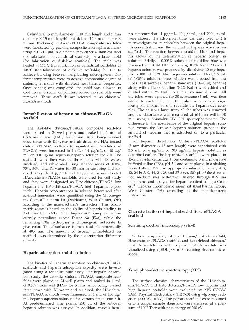
Figure 10 shows the extracellular osteocalcinexpression of MC3T3-E1 cells cultured on differentscaffolds. At day 7, the cells on HAc-chitosan/PLAGA low heparin scaffolds showed significantlyhigher osteocalcin expression than all the other scaf-folds. At day 14, osteocalcin expression of cells onHAc-chitosan/PLAGA scaffolds picked up andshowed significantly elevated level when comparedwith day 7. Cells on HAc-chitosan/PLAGA low hep-arin scaffolds showed the highest osteocalcin expres-
sion level and it was significantly higher than HAc-chitosan/PLAGA high heparin scaffolds andPLAGA scaffolds. At day 21, a significantly elevatedosteocalcin expression level was noticed on HAc-chi-tosan/PLAGA high heparin scaffolds and PLAGAscaffolds when compared with day 14. The averageosteocalcin expression level on HAc-chitosan/PLAGA low heparin scaffolds remained to be thehighest among the groups.
DISCUSSION
Our earlier work has shown that chitosan/PLAGAscaffolds possessed excellent mechanical propertiesand appropriate porous structures suitable for bonetissue engineering applications.19 One of the mainbenefits of introducing chitosan into PLAGA micro-spheres and developing chitosan/PLAGA compositesystem is that chitosan imparts functionality due tothe reactive amino groups. Therefore, the objective
Figure 6. Scanning electron micrographs showing MC3T3-E1 cell proliferation on (a–c) HAc-chitosan/PLAGA scaffolds,(d–f) HAc-chitosan/PLAGA low heparin scaffolds, (g–i) HAc-chitosan/PLAGA high heparin scaffolds, and (j–l) PLAGAscaffolds at (a, d, g, j) 7 days; (b, e, h, k) 14 days; and (c, f, i, l) 21 days after cell seeding. Magnification: 3100.
1202 JIANG ET AL.
Journal of Biomedical Materials Research Part A

of this study was to immobilize a biomolecule hepa-rin onto the sintered chitosan/PLAGA scaffolds dueto the important biological activity of heparin mole-cules.
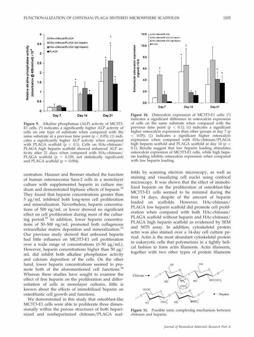
In this study, an ionic interaction was used to im-mobilize heparin onto the sintered chitosan/PLAGAscaffolds due to the negatively charged feature ofheparin. For this purpose, the surface of chitosan/PLAGA scaffolds was protonated by treating with adilute acetic acid, which converted the NH2 groups
of chitosan on the surface of scaffold to positivelycharged NH3
þ groups. Heparin, on the other hand,has high negative charge density, and thus is capa-ble of interacting with protonated chitosan. The pos-sible underlying complexing mechanism betweenchitosan and heparin is demonstrated in Figure 11.The presence of heparin on heparinized chitosan/PLAGA scaffolds was ascertained by performingXPS on the surface of the scaffolds. XPS spectrum ofHAc-chitosan/PLAGA high heparin scaffold clearlyshowed a peak at the binding energy of 168–170eVwhich is a characteristic peak of the 2p electrons ofelement sulfur. It was also noticed that this sulfurpeak did not appear on the XPS spectrum of HAc-chitosan/PLAGA low heparin scaffold (data notshown). It might be because that the amount of hep-arin immobilized on HAc-chitosan/PLAGA low hep-arin scaffold is below the detection limit of XPS. XPSis a quantitative spectroscopic surface chemistryanalysis technique. However, it is not an idealmethod in this situation to quantify or even qualita-tively compare the amount of immobilized heparinwith various loadings. The reasons might reside intwo aspects. Since the scaffold has a 3D porousstructure, a great amount of heparin would possiblyreside inside of scaffolds and could not be detectedby XPS. Therefore, heparin present on the surfacecould not stand for the amount of heparin immobi-lized on scaffolds. In addition, the intensity of thesulfur peak was comparatively weak on the spec-trum, and the quantitative accuracy for the weakerXPS signals is usually deteriorated and less than theactual values.27 In this study, a colorimetric assaywas adopted to measure the heparin solution con-centrations before and after heparin treatment. Itwas found that the amount of heparin on HAc-chito-san/PLAGA low heparin scaffold was approxi-mately one eighth of the amount on HAc-chitosan/PLAGA high heparin scaffold. It has also been foundthat the mechanical properties and the porous struc-tures were not affected by the heparinization pro-cess. Heparinized scaffolds remained to be �30% po-rous and mechanically competent for load-bearingbone tissue engineering applications.
Chitosan/PLAGA scaffolds were investigated forionic adsorption of heparin and its release. Heparin-ization was achieved by the ionic interactionbetween positively charged chitosan and negativelycharged heparin. The presence of heparin on thescaffold surface was confirmed by different techni-ques such as XPS and fluorescent staining. The hepa-rin uptake was shown to be nonlinear for the first 2h, after which equilibrium was reached. The heparindissolution study showed that no heparin releasewas detected from the scaffolds with low heparinloading. In addition, only a tiny amount of heparinwas released from the scaffolds with high heparin
Figure 7. (a) MTS assay for MC3T3-E1 cell proliferation.(*) and (y) indicate significant difference when comparedwith other groups at the same time point at significancelevels of p < 0.05 and p < 0.1, respectively. At day 21, cellproliferation on HAc-chitosan/PLAGA low heparin scaf-fold is significantly higher than HAc-chitosan/PLAGAscaffold (p 5 0.012) and HAc-chitosan/PLAGA high scaf-fold (p 5 0.053). (b–e) Scanning electron micrographsshowing MC3T3-E1 cell proliferation at day 21 on (b)HAc-chitosan/PLAGA scaffolds, (c) HAc-chitosan/PLAGAlow heparin scaffolds, (d) HAc-chitosan/PLAGA high hep-arin scaffolds, and (e) PLAGA scaffolds after 21 days ofcell culture. Greater cell proliferation was observed onHAc-chitosan/PLAGA low heparin scaffold than HAc-chi-tosan/PLAGA and HAc-chitosan/PLAGA high heparinscaffolds. Magnification: 350.
FUNCTIONALIZATION OF CHITOSAN/PLAGA SINTERED MICROSPHERE SCAFFOLDS 1203
Journal of Biomedical Materials Research Part A

loading over a 37-day period. The heparin releasedfrom the scaffolds may be mainly attributed to thattrapped within scaffolds but not ionically bound.The majority of the heparin that is ionically attachedto chitosan was presumably not released. This maybe explained by the relatively strong interactionbetween chitosan and heparin, considering that bothchitosan and heparin are highly charged and possi-bly allow multi-site binding. It is then presumed thatheparin may only be released upon scaffold degra-dation. Therefore, the chitosan/PLAGA scaffoldsmay serve as a heparin reservoir. The immobilizedheparin on chitosan/PLAGA scaffolds undergoes lit-tle loss and may be able to continuously serve asbinding sites for certain growth factors.
Heparin, except for its widely known anticoagu-lant activity, is also recognized to be an importantbioactive molecule capable of interacting with a largenumber of growth factors with heparin-binding
domains such as FGFs, TGF–b, and BMP-2 and 4.These growth factors are involved in the regulationof cell fate processes of osteoprogenitor cells andcommitted osteoblastic cells.28 In addition, they playa central role in bone formation and bone fracturehealing by acting through autocrine or paracrinemechanisms on bone cell proliferation and differen-tiation.28 Therefore, heparinization of bone tissue en-gineering scaffolds is a potential strategy to developscaffolds that are capable of locally absorbing/sequestering heparin-binding growth factors in an invivo environment for accelerated bone regeneration.Heparin by itself also plays a role in cell prolifera-tion, differentiation, and mineralization. Bothstrongly inhibitory and stimulatory effects of heparinon the proliferation of osteoblastic cells werereported.29–32 These different observations of heparineffect on osteoblastic cell functions may reflect thefact that the effect is dependent on the heparin con-
Figure 8. Confocal fluorescence microscopy of cell nuclei (a, d, g), cytoskeletal protein actin (b, e, h), and dual stainingwith TRITC-conjugated phalloidin and DAPI (c, f, i) on HAc-chitosan/PLAGA low heparin scaffolds at day 7 (a, b, c), day11 (d, e, f), and day 14 (g, h, i). Cell nuclei and actin filament alignment were observed along the adjoining area betweenmicrospheres at day 7. Comparing to day 7, greater cell proliferation toward microsphere surface and porous structurewas noticed after day 11 and interconnected orthogonal actin network was formed after 14 days. Cell migration into thevoid space of the scaffolds was also observed at day 14 (white arrow). [Color figure can be viewed in the online issue,which is available at www.interscience.wiley.com.]
1204 JIANG ET AL.
Journal of Biomedical Materials Research Part A
centration. Hausser and Brenner studied the functionof human osteosarcoma Saos-2 cells in a monolayerculture with supplemented heparin in culture me-dium and demonstrated biphasic effects of heparin.33
They found that heparin concentrations greater than5 lg/mL inhibited both long-term cell proliferationand mineralization. Nevertheless, heparin concentra-tions of 500 ng/mL or lower showed no significanteffect on cell proliferation during most of the cultur-ing period.33 In addition, lower heparin concentra-tions of 50–500 ng/mL promoted both collagenousextracellular matrix deposition and mineralization.33
Our previous study showed that unbound heparinhad little influence on MC3T3-E1 cell proliferationover a wide range of concentrations (0–50 lg/mL).However, heparin concentrations higher than 50 lg/mL did inhibit both alkaline phosphatase activityand calcium deposition of the cells. On the otherhand, lower heparin concentrations seemed to pro-mote both of the aforementioned cell functions.34
Whereas these studies have sought to examine theeffect of free heparin on the proliferation and differ-entiation of cells in monolayer cultures, little isknown about the effects of immobilized heparin onosteoblastic cell growth and functions.
We demonstrated in this study that osteoblast-likeMC3T3-E1 cells were able to proliferate three dimen-sionally within the porous structures of both hepari-nized and nonheparinized chitosan/PLAGA scaf-
folds by scanning electron microscopy, as well asstaining and visualizing cell nuclei using confocalmicroscopy. It was shown that the effect of immobi-lized heparin on the proliferation of osteoblast-likeMC3T3-E1 cells seemed to be minimal during thefirst 14 days, despite of the amount of heparinloaded on scaffolds. However, HAc-chitosan/PLAGA low heparin scaffold did promote cell prolif-eration when compared with both HAc-chitosan/PLAGA scaffold without heparin and HAc-chitosan/PLAGA high heparin scaffold as evidenced by SEMand MTS assay. In addition, cytoskeletal proteinactin was also stained over a 14-day cell culture pe-riod. Actin is the most abundant cytoskeletal proteinin eukaryotic cells that polymerizes in a tightly heli-cal fashion to form actin filaments. Actin filaments,together with two other types of protein filaments
Figure 9. Alkaline phosphatase (ALP) activity of MC3T3-E1 cells. (*) indicates a significantly higher ALP activity ofcells on one type of substrate when compared with thesame substrate at a previous time point (p < 0.05); (y) indi-cates a significantly higher ALP activity when comparedwith PLAGA scaffold (p < 0.1). Cells on HAc-chitosan/PLAGA high heparin scaffold showed enhanced ALP ac-tivity after 21 days when compared with HAc-chitosan/PLAGA scaffold (p 5 0.139, not statistically significant)and PLAGA scaffold (p 5 0.094).
Figure 10. Osteocalcin expression of MC3T3-E1 cells. (*)indicates a significant difference in osteocalcin expressionof cells on the same substrate when compared with theprevious time point (p < 0.1); (y) indicates a significanthigher osteocalcin expression than other groups at day 7 (p< 0.05); ({) indicates a significant higher osteocalcinexpression when compared with HAc-chitosan/PLAGAhigh heparin scaffold and PLAGA scaffold at day 14 (p <0.1); Results suggest that low heparin loading stimulatesosteocalcin expression of MC3T3-E1 cells, while high hepa-rin loading inhibits osteocalcin expression when comparedwith low heparin loading.
Figure 11. Possible ionic complexing mechanism betweenchitosan and heparin.
FUNCTIONALIZATION OF CHITOSAN/PLAGA SINTERED MICROSPHERE SCAFFOLDS 1205
Journal of Biomedical Materials Research Part A

known as microtubules and intermediate filaments,form the cytoskeleton. Actin filaments provide me-chanical support for the cell, determine the cellshape, and enable coordinated and directed cellmovements.35 Using confocal laser scanning micros-copy, the dynamics of the 3D formation of actin cy-toskeleton were visualized. Results indicated thatcells had adhered to the scaffold surface and prolif-erated through the porous structures of HAc-chito-san/PLAGA low heparin scaffolds after being cul-tured for 14 days. The actin cytoskeleton was wellorganized and distributed within the 3D structuresof HAc-chitosan/PLAGA low heparin scaffolds in-dicative of cell movement and migration into the po-rous structures.
It has been well established that long-term andhigh-dose of heparin administration causes heparin-induced osteoporosis.36–38 The underlining mecha-nisms of heparin-induced osteoporosis are yet unde-fined; however, it may be due to the fact that hepa-rin results in impaired osteoblast and enhancedosteoclast activity and functions.36–38 Although highdoses of heparin treatment seemed to be detrimentalto osteoblastic cells, low doses of heparin, on thecontrary, were shown to be able to promote osteo-blast cell differentiation, extracellular matrix deposi-tion, and mineralization as described.34 Due to itswidely recognized interaction with a large numberof growth factors, heparin is considered to be biolog-ically important in the bone healing process. There-fore, heparin immobilization on bone tissue engi-neering scaffolds is a promising approach to developbioactive matrices for bone repair; however, caremust be taken to avoid adverse effects of heparin-associated situations. In this study, a low dose ofheparin (1.7 6 0.1 lg, HAc-chitosan/PLAGA lowheparin scaffolds) and a relatively high dose of hep-arin (14.1 6 4.8 lg, HAc-chitosan/PLAGA high hep-arin scaffolds) were immobilized onto chitosan/PLAGA scaffolds. Heparin effects on osteoblast-likeMC3T3-E1 cell differentiation and mineralizationwere investigated by monitoring two bone cell dif-ferentiation markers, alkaline phosphatase (ALP)and osteocalcin. It was found that cells on hepari-nized chitosan/PLAGA scaffolds showed elevatedALP activity when compared with nonheparinizedchitosan/PLAGA scaffolds and PLAGA scaffolds af-ter 21 days of culture. There was no significant dif-ference between HAc-chitosan/PLAGA low heparinscaffolds and HAc-chitosan/PLAGA high heparinscaffolds. On the other hand, immobilized heparinshowed an effect on osteocalcin expression in adose-dependent manner. Osteocalcin expression ofcells on HAc-chitosan/PLAGA low heparin scaffoldswas significantly higher than HAc-chitosan/PLAGAscaffolds at day 7; however, no significant differencewas observed between HAc-chitosan/PLAGA high
heparin scaffolds and HAc-chitosan/PLAGA scaf-folds. In addition, comparing to HAc-chitosan/PLAGA low heparin scaffolds, osteocalcin expressionof cells on HAc-chitosan/PLAGA high heparin scaf-folds was inhibited with significantly reducedexpression levels at day 7 and day 14. At day 21, theaverage osteocalcin expression level on HAc-chito-san/PLAGA low heparin scaffold was higher thanHAc-chitosan/PLAGA scaffold (p 5 0.121), HAc-chi-tosan/PLAGA high heparin scaffold (p 5 0.254), andPLAGA scaffold (p 5 0.152), but not statistically sig-nificant. Results suggest that low heparin loadingstimulates osteocalcin expression of MC3T3-E1 cells,whereas high heparin loading inhibits osteocalcinexpression when compared with low heparin load-ing. The mechanisms of the stimulatory effect of lowdoses heparin and inhibitory effect of high doses ofheparin on MC3T3-E1 cell differentiation were notclear, however, may be related to the interaction ofheparin to heparin-binding growth factors. It wasdemonstrated that heparin and heparin-like poly-mers were able to stabilize heparin-binding growthfactors and protect them from pH, thermal, proteo-lytic degradations, or forming inactive complex.39–42
Heparin and heparin-like polysaccharides such asheparan sulfate and dextran sulfate were alsoreported to stimulate ligand binding to the signalingreceptors43,44 or facilitate to continuously serve theligands to their receptors45 and thus potentiate theirbiological activities. We then presume that the hepa-rin-mediated enhanced activities of local growth fac-tors contributed to the enhanced cell differentiation. Ithas also been proposed that excess heparin and hepa-rin-like polymers may excessively bond to growthfactors, and in turn restrict their availability, or com-pete with growth factors for their available bindingsites on cell membranes.46 We believe that one ofthese mechanisms may have accounted for the low-ered osteocalcin expression of MC3T3-E1 cells onHAc-chitosan/PLAGA high heparin scaffolds.
CONCLUSIONS
This work demonstrated the feasibility of modify-ing chitosan/PLAGA sintered microsphere scaffoldswith bioactive glycosaminoglycan heparin and eval-uated the heparinized chitosan/PLAGA scaffolds forload-bearing bone tissue engineering applications.The biomolecule heparin was successfully immobi-lized onto the chitosan/PLAGA scaffolds via anionic interaction between heparin and chitosan mole-cules. The heparinization process was found to haveno influence on the mechanical properties and po-rous structures of the scaffolds. The uptake of hepa-rin by scaffolds was shown to be manipulated byvarying either heparin concentration or the time for
1206 JIANG ET AL.
Journal of Biomedical Materials Research Part A

immobilization. It was shown that only very mini-mal amount of heparin could be released from theheparinized chitosan/PLAGA scaffolds, and it wasthus presumed that the immobilized heparin wasretained on the scaffolds instead of being released. Itwas also shown that heparinized chitosan/PLAGAscaffolds supported MC3T3-E1 osteoblast cell prolif-eration. Immobilized heparin at a low dosageshowed a stimulatory effect on MC3T3-E1 cell prolif-eration, whereas a high dosage of heparin does notshow such similar effect on cell proliferation. Inaddition, low heparin loading showed a stimulatoryeffect on cell differentiation as indicated by theenhanced osteocalcin expression, whereas high hepa-rin loading, on the contrary, did not show such stim-ulatory effect. Future work will focus on in vivo eval-uation of the heparinized chitosan/PLAGA scaffoldsfor bone regeneration using a load-bearing defectmodel.
This work is supported by the National Science Founda-tion Grant INT0115595. Dr. Laurencin was previously therecipient of a Presidential Faculty Fellow Award from theNational Science Foundation.
References
1. Weiss MJ, Ng CY, Madsen JC. Tolerance, xenotransplanta-tion: Future therapies. Surg Clin North Am 2006;86:1277–1296.
2. Langer R, Vacanti JP. Tissue engineering. Science 1993;260:920–926.
3. Kidane AG, Punshon G, Salacinski HJ, Ramesh B, Dooley A,Olbrich M, Heitz J, Hamilton G, Seifalian AM. Incorporationof a lauric acid-conjugated GRGDS peptide directly into thematrix of a poly(carbonate-urea)urethane polymer for use incardiovascular bypass graft applications. J Biomed Mater ResA 2006;79:606–617.
4. Hammond JS, Beckingham IJ, Shakesheff KM. Scaffolds forliver tissue engineering. Expert Rev Med Devices 2006;3:21–27.
5. Lu HH, Cooper JA, Manuel S, Freeman JW, Attawia MA, KoFK, Laurencin CT. Anterior cruciate ligament regenerationusing braided biodegradable scaffolds: In vitro optimizationstudies. Biomaterials 2005;26:4805–4816.
6. Borden M, El-Amin SF, Attawia M, Laurencin CT. Structuraland human cellular assessment of a novel microsphere-basedtissue engineered scaffold for bone repair. Biomaterials 2003;24:597–609.
7. Thissen H, Chang KY, Tebb TA, Tsai WB, Glattauer V, Ram-shaw JAM, Werkmeister JA. Synthetic biodegradable micro-particles for articular cartilage tissue engineering. J BiomedMater Res A 2006;77:590–598.
8. Sarkar S, Lee GY, Wong JY, Desai TA. Development andcharacterization of a porous micro-patterned scaffold for vas-cular tissue engineering applications. Biomaterials 2006;27:4775–4782.
9. Muzzarelli RAA, Mattiolibelmonte M, Tietz C, Biagini R, Fer-ioli G, Brunelli MA, Fini M, Giardino R, Ilari P, Biagini G.Stimulatory effect on bone-formation exerted by a modifiedchitosan. Biomaterials 1994;15:1075–1081.
10. Vandevord PJ, Matthew HWT, Desilva SP, Mayton L, Wu B,Wooley PH. Evaluation of the biocompatibility of a chitosanscaffold in mice. J Biomed Mater Res 2002;59:585–590.
11. Tomihata K, Ikada Y. In vitro and in vivo degradation offilms of chitin and its deacetylated derivatives. Biomaterials1997;18:567–575.
12. Ho MH, Wang DM, Hsieh HJ, Liu HC, Hsien TY, Lai JY,Hou LT. Preparation and characterization of RGD-immobi-lized chitosan scaffolds. Biomaterials 2005;26:3197–3206.
13. Mochizuki M, Kadoya Y, Wakabayashi Y, Kato K, Okazaki I,
Yamada M, Sato T, Sakairi N, Nishi N, Nomizu M. Laminin-
1 peptide-conjugated chitosan membranes as a novel
approach for cell engineering. FASEB J 2003;17:875–877.14. Park YJ, Kim KH, Lee JY, Ku Y, Lee SJ, Min BM, Chung CP.
Immobilization of bone morphogenetic protein-2 on a nanofi-
brous chitosan membrane for enhanced guided bone regener-ation. Biotechnol Appl Biochem 2006;43:17–24.
15. Borden M, Attawia M, Khan Y, Laurencin CT. Tissue engi-neered microsphere-based matrices for bone repair: Designand evaluation. Biomaterials 2002;23:551–559.
16. Borden M, Attawia M, Laurencin CT. The sintered micro-sphere matrix for bone tissue engineering: In vitro osteocon-ductivity studies. J Biomed Mater Res 2002;61:421–429.
17. Borden M, Attawia M, Khan Y, El-Amin SE, Laurencin CT.Tissue-engineered bone formation in vivo using a novel sin-tered polymeric microsphere matrix. J Bone Joint Surg Br2004;86:1200–1208.
18. Khan YM, Katti DS, Laurencin CT. Novel polymer-synthe-sized ceramic composite-based system for bone repair: An invitro evaluation. J Biomed Mater Res A 2004;69:728–737.
19. Jiang T, Abdel-Fattah WI, Laurencin CT. In vitro evaluationof chitosan/poly(lactic acid-glycolic acid) sintered micro-sphere scaffolds for bone tissue engineering. Biomaterials2006;27:4894–4903.
20. Sakamoto S, Sakamoto M, Goldberg L, Colarusso L, Gotoh Y.Mineralization induced by beta-glycerophosphate in culturesleads to a marked increase in collagenase synthesis by mouseosteogenic MC3T3-E1 cells under subsequent stimulationwith heparin. Biochem Biophys Res Commun 1989;162:773–780.
21. Shibata Y, Abiko Y, Goto K, Moriya Y, Takiguchi H. Heparinstimulates the collagen-synthesis in mineralized cultures ofthe osteoblast-like cell-line, MC3T3-E1. Biochem Int 1992;28:335–344.
22. Capila I, Linhardt RJ. Heparin-protein interactions. AngewChem Int Ed 2002;41:391–412.
23. Liu LS, Ng CK, Thompson AY, Poser JW, Spiro RC. Hyal-uronate-heparin conjugate gels for the delivery of basic fibro-blast growth factor (FGF-2). J Biomed Mater Res 2002;62:128–135.
24. Benoit DSW, Anseth KS. Heparin functionalized PEG gels
that modulate protein adsorption for hMSC adhesion and dif-
ferentiation. Acta Biomater 2005;1:461–470.25. Andress DL. Heparin modulates the binding of insulin-like
growth-factor (IGF) binding protein-5 to a membrane-protein
in osteoblastic cells. J Biol Chem 1995;270:28289–28296.26. Irie A, Habuchi H, Kimata K, Sanai Y. Heparan sulfate is
required for bone morphogenetic protein-7 signaling. Bio-chem Biophys Res Commun 2003;308:858–865.
27. Barr TL. Modern ESCA: The Principles and Practice of X-rayPhotoelectron Spectroscopy. Boca Raton, FL: CRC Press; 1994.
28. Canalis E. Skeletal Growth Factors. Philadelphia, PA: Lip-pincott Williams & Wilkins; 2000.
29. Matziolis G, Erli HJ, Rau HM, Klever P, Bosserhoff AK, Paar
O. Heparin improves human osteoblast proliferation in vitro.
Orthopade 2002;31:575–581.
30. Nikitovic D, Zafiropoulos A, Tzanakakis GN, Karamanos
NK, Tsatsakis AM. Effects of glycosaminoglycans on cell pro-
liferation of normal osteoblasts and human osteosarcoma
cells depend on their type and fine chemical compositions.
Anticancer Res 2005;25:2851–2856.
FUNCTIONALIZATION OF CHITOSAN/PLAGA SINTERED MICROSPHERE SCAFFOLDS 1207
Journal of Biomedical Materials Research Part A

31. Yang LF, Butcher M, Simon RR, Osip SL, Shaughnessy SG.
The effect of heparin on osteoblast differentiation and activity
in primary cultures of bovine aortic smooth muscle cells.
Atherosclerosis 2005;179:79–86.32. Osip SL, Butcher M, Young E, Yang LF, Shaughnessy SG.
Differential effects of heparin and low molecular weight hep-arin on osteoblastogenesis and adipogenesis in vitro. ThrombHaemost 2004;92:803–810.
33. Hausser HJ, Brenner RE. Low doses and high doses of hepa-rin have different effects on osteoblast-like Saos-2 cells invitro. J Cell Biochem 2004;91:1062–1073.
34. Jiang T, Laurencin CT. Transactions of the 31st Annual Meet-ing of the Society for Biomaterials. Pittsburgh, Pennsylvania.2006. p 407.
35. Alberts B, Bray D, Lewis J, Raff M, Roberts K, Watson JD.Molecular Biology of the Cell, 3rd ed, New York/London:Garland publishing, Inc.; 1994.
36. Muir JM, Hirsh J, Weitz JI, Andrew M, Young E, Shaugh-nessy SG. A histomorphometric comparison of the effects ofheparin and low-molecular-weight heparin on cancellousbone in rats. Blood 1997;89:3236–3242.
37. Folwarczna J, Janiec W, Sliwinski L. Effects of heparin andlow-molecular-weight heparins on bone mechanical proper-ties in rats. Thromb Haemost 2004;92:940–946.
38. Nelson-Piercy C. Heparin-induced osteoporosis. Scand JRheumatol Suppl 1998;107:68–71.
39. Gospodarowicz D, Cheng J. Heparin protects basic and acidicFGF from inactivation. J Cell Physiol 1986;128:475–484.
40. Sommer A, Rifkin DB. Interaction of heparin with humanbasic fibroblast growth-factor—Protection of the angiogenicprotein from proteolytic degradation by a glycosaminoglycan.J Cell Physiol 1989;138:215–220.
41. Mccaffrey TA, Falcone DJ, Brayton CF, Agarwal LA, WeltFGP, Weksler BB. Transforming growth factor-beta activity ispotentiated by heparin via dissociation of the transforminggrowth factor-beta alpha-2-macroglobulin inactive complex.J Cell Biol 1989;109:441–448.
42. Mccaffrey TA, Falcone DJ, Vicente D, Du BH, Consigli S,Borth W. Protection of transforming growth factor-beta-1activity by heparin and fucoidan. J Cell Physiol 1994;159:51–59.
43. Yayon A, Klagsbrun M, Esko JD, Leder P, Ornitz DM. Cell-surface, heparin-like molecules rre required for binding of ba-sic fibroblast growth-factor to its high-affinity receptor. Cell1991;64:841–848.
44. Rapraeger AC, Krufka A, Olwin BB. Requirement of hepa-ran-sulfate for bFGF-mediated fibroblast growth and myo-blast differentiation. Science 1991;252:1705–1708.
45. Takada T, Katagiri T, Ifuku M, Morimura N, Kobayashi M,Hasegawa K, Ogamo A, Kamijo R. Sulfated polysaccharidesenhance the biological activities of bone morphogenetic pro-teins. J Biol Chem 2003;278:43229–43235.
46. Blanquaert F, Barritault D, Caruelle JP. Effects of heparan-likepolymers associated with growth factors on osteoblast prolif-eration and phenotype expression. J Biomed Mater Res 1999;44:63–72.
1208 JIANG ET AL.
Journal of Biomedical Materials Research Part A