functional studies of rna polymerase ii-dependent ...pub.epsilon.slu.se/1266/1/avh-st.pdf · 1...
TRANSCRIPT
1
Functional Studies of RNA Polymerase II-dependent Transcription in Yeast
Saccharomyces cerevisiae
Susanna Tronnersjö Faculty of Natural Resources and Agricultural Sciences
Department of Plant Biology and Forest Genetics Uppsala
Doctoral thesis Swedish University of Agricultural Sciences
Uppsala 2006

2
Acta Universitatis Agriculturae Sueciae 2006: 109 ISSN 1652-6880 ISBN 91-576-7258-x © 2006 Susanna Tronnersjö, Uppsala Tryck: SLU Service/Repro, Uppsala 2006
3
Abstract
Tronnersjö, S. 2006. Functional studies of RNA polymerase II-dependent transcription in yeast Saccharomyces cerevisiae Doctor’s dissertation. ISSN 1652-6880, ISBN 91-576-7258-x Nutrient availability is one of the environmental conditions that have the largest effect on cell growth. The nutrient status is transmitted by several signal transduction pathways, providing an essential framework for the coordination of cell growth. These pathways have been shown to be remarkably well conserved among eukaryotes. Transcriptional initiation is the endpoint for many signal transduction pathways and thus the final answer to an environmental change. The control of gene expression through transcription initiation is therefore one of the most fundamental processes in biology, containing many layers of complexity. I have used the model organism Saccharomyces cerevisiae to study different mechanisms involved in the control of gene expression. The mediator protein complex is required for regulated transcription of nearly all RNA polymerase II-dependent genes. Studies of the Mediator subunit Med21 revealed that it genetically and physically interacts with two other Mediator subunits, Med7 and Med10. A strong Med21 self-interaction was also identified, suggesting a possible dimerisation of Med21. The second part of the study focused on elucidating the function of the split jumonji domain, comprising the jmjN and jmjC subdomains, that has been identified in many transcription factors, by studying its physical interactions. I identified 19 different yeast proteins that could interact either directly or indirectly with the jumonji domain of the zinc finger protein Gis1 in a complex interaction pattern. Gis1 is known to be involved in nutrient dependent regulation of gene expression. Genome-wide microarray analysis was used to investigate the role of Gis1 and the related Rph1 protein in the growth phase-dependent gene regulation. This resulted in the identification of both redundant and unique roles of Gis1 and Rph1 in regulation of genes with an enrichment of STRE and PDS elements in their promoters. One of the proteins that interact directly with the jumonji domain of Gis1 is the unconvential prefoldin-like protein Bud27. Genetic and biochemical methods were used to study the roles of Bud27 and Gis1 in nutrient dependent regulation of gene expression by the TOR signaling pathway. Keywords: Bud27, diauxic shift, Gis1, jmjC, jumonji, Mediator, nutrient signaling, PDS, rapamycin, Rph1, STRE, TOR, URI Author’s address: Susanna Tronnersjö, Department of Plant Biology and Forest genetics, Uppsala Genetic Centre, Swedish University of Agricultural Sciences, Box 7080, S-750 07 Uppsala, Sweden. E-mail: Susanna. Tonnersjö@imbim.uu.se

4

5
Contents
Abbreviations Introduction, 9 Yeast as a model system, 9 Nutrient dependent regulation of growth, 10
The different growth phases of Saccharomyces cerevisiae, 10 The lag phase and logaritmic growth, 10 The diauxic shift and the post-diauxic growth phase, 11 The stationary phase and quiescence, 12
Glucose sensing signaling pathways, 14 Glucose induction, 15 Glucose repression, 16 The Ras/cAMP pathway, 16
Other nutrient sensing signaling pathways,19 The TOR pathway, 19 The fermentable growth medium induced pathway, 21 The PHO pathway, 21
The convergence of nutrient signaling pathways on Rim15, 22 General transcription, 26
Promoters and promoter sites, 27 The RNA polymerase II enzyme, 27 Assembly of the pre-initiation complex, 28 DNA-binding transcription factors,29 Cofactors, 30 The Mediator, 31
Chromatin dynamics and gene regulation, 33
Chromatin organisation, 33 Covalent modifications of histones, 33 Chromatin remodeling, 36
Regulation of biological processes by sumoylation,38 The Gis1 and Rph1 proteins, 40 The Bud27 protein, 42 Aims of the present investigation, 43
6
Results and discussion, 43 Genetic and physical interactions of Med21 (I), 43 Identification of Gis1 jumonji domain interacting proteins (II), 45 Global expression analysis of Gis1 and Rph1 function in different growth phases (III), 47 The roles of Bud27 and Gis1 in TOR-dependent regulation (IV), 48 Conclusions, 49 Future perspectives, 50 References, 52 Acknowledgements, 66
7
Appendix
Papers I-IV The present thesis is based on the following papers, which will be referred to by their Roman numerals. I. Hallberg, M., Hu, G. Z., Tronnersjö, S., Shaikhibrahim, Z., Balciunas, S. and Ronne, H. (2006) Functional and physical interactions within the middle domain of the yeast mediator. Mol. Gen. Genomics, 276:197-210 II. Tronnersjö, S., Hanefalk, C., Balciunas, D., Hu, G. Z., Nordberg, N., Murén, E. and Ronne, H. The jmjN and jmjC domains of the yeast zinc finger protein Gis1 interact with 19 proteins involved in transcription, sumoylation and DNA-repair. Mol. Gen. Genomics (in press). III. Orzechowski-Westholm, J., Tronnersjö, S., Nordberg, N., Komorowski, J. and Ronne, H. Role of the jmjC-containing zinc finger proteins Gis1 and Rph1 in growth phase-dependent gene expression in yeast. IV. Tronnersjö, S., Hu, G. Z., Nordberg, N. and Ronne, H. Functional studies of Bud27 and Gis1, two yeast proteins involved in TOR signaling. Papers I and II are reproduced with the due permission from the publishers.

8
Abbreviations
List for abbreviations used in the text: ATP adenosine triphosphate cAMP cyclic adenosine monophosphate CTD carboxy-terminal domain GAPs GTPase-activating proteins GEFs GTP-exchange factors GPCR G-protein-coupled receptor GTF general transcription factor HAT histone acetyltransferase HDAC histone deacetylase HDM histone demethylase HMT histone methyltransferase INR initiator element JMJ jumonji PKA protein kinase A PDS post-diauxic shift PIC pre-initiation complex STRE stress responsive element SUMO small ubiquitin like modifier TBP TATA box binding protein TCA Tri carboxylic acid TOR target of rapamycin

9
Introduction
Yeast as a model system As researchers try to dissect the complex patterns of molecular biology, the use of different model organisms has become one way to break down more difficult systems into simpler ones. The rationale for using model organisms is the fact that the main pathways and regulatory systems have been strongly conserved between different organisms during evolution. Hence, understanding a simple organism can provide a low resolution map that is useful for understanding the more complex patterns of a higher organism. Therefore, though most research within molecular biology is focused on understanding higher organisms like humans and plants, many researchers still use simple model organisms.
The yeast Saccharomyces cerevisiae, also called budding yeast or baker's yeast, has been in the service of humans for thousands of years. Its ability to produce carbon dioxide and alcohol were used in baking and brewing long before S. cerevisiae was actually recognised as a living organism. Schwann was, in 1837, the first scientist to look at yeast in the microscope, and described it as a budding organism. However, the major breakthrough in the recognition of yeast and its properties came in 1859, when Pasteur proved that it is essential for fermentation.
Quite apart from its usefulness in baking and brewing, S. cerevisiae is one of the oldest and most thoroughly studied model organisms due to its many genetic and practical advantages. Among these advantages is the fact that it is a unicellular organism which is very easy and cheap to grow. It can be grown as single colonies on agar plates, or in liquid culture. It grows rapidly, with a doubling time of 90 minutes, and in many ways its culturing resembles the handling of bacteria. However, yeast has the great advantage of being an eukaryote, and is therefore more similar to higher organisms such as animals and plants. It is therefore frequently possible to express recombinant proteins from higher eukaryotes which are functional in yeast. A memorable quote in this context is that of Boris Magasanik who once said: "yeast should be seen not as a large bacterium, but as a small cow".
The genetic advantages of using S. cerevisiae in research are many. Its 12.5 Mb genome, not including the rDNA which is highly variable, is very compact, with genes representing ~72% of the total DNA. The genome is divided into 16 chromosomes, and its full sequence was published as a supplement to Nature in 1997 (Clayton et al., 1997). The yeast genome contains ~6,200 genes, and only a handful of them, mostly those encoding ribosomal proteins, have introns. Compared to higher organisms, there are also fewer gene duplications. This, in combination with the fact that recombinant transformed DNA is easily integrated into the yeast genome by homologous recombination, makes it easy to study gene function by targeting specific yeast genes for disruptions (gene knockouts) or introducing other well-defined mutations into the genome.

10
The existence of both a haploid and diploid state of an organism facilitates genetic studies. S. cerevisiae has this advantage, and can in the haploid state exist as one of two mating types, a or α. Most research is made using the haploid state, as this makes it easy perform genetic manipulations and to directly see the effect of knockouts and other mutations. The diploid state adds the possibility of crossing different mutated strains to each other in order to generate new haploid strains with combinations of several knockouts.
Although many technical obstacles in working with higher, multicellular organisms have been overcome during the last years, S. cerevisiae has retained its importance in molecular genetic research due to the combined power of its closer relatedness to higher organisms, as compared to bacteria, and the simplicity of working with yeast.
Nutrient dependent regulation of cell growth The different growth phases of Saccharomyces cerevisiae Unicellular organisms such as yeast have during evolution been forced to develop mechanisms that can deal with constantly changing environmental conditions. One type of environmental change that causes dramatic effects on microorganisms is a change in nutrient availability. When yeast cells are grown in a liquid culture, on rich media and with constant shaking, the culture shows changes in growth rate over time as a response to the exhaustion of different nutrients. The growth curve can be divided into five different growth phases marked by differences in the growth rate, but more importantly representing differences in metabolism and in gene expression patterns. The five phases are the lag phase, the exponential growth phase, the diauxic shift, the post-diauxic growth phase, and the stationary phase (Figure1). The lag phase and exponential growth When glucose is added to glucose-deprived yeast, the cells rapidly adapt their metabolism to fermentation of glucose during a short lag phase to ensure optimal and exclusive use of this rich carbon source. After this adaptation, the cells start to grow exponentially, reaching their highest growth rate with a doubling time of 90 min in rich media. During this exponential growth phase the yeast cells only metabolise glucose even if other carbon sources are present (see further in section Glucose sensing signaling pathways). The glucose is fed into the glycolytic pathway forming non fermentable carbon compounds, particularly ethanol. While glucose uptake and the flow through glycolysis is stimulated, gluconeogenesis is inhibited. In this first phase of growth, much more ethanol is produced than can be oxidised in the TCA cycle. This occurs because the respiratory activity is inhibited by high concentrations of glucose, a phenomenon originally called the Crabtree effect (Ephrussi et al., 1956). Thus, a concentration of more than 0.9% glucose represses the formation of mitochondrial structures and respiratory enzymes (Polakis et al., 1964). Therefore, S. cerevisiae, in contrast to most other yeasts, produces high levels of ethanol even under fully aerobic conditions. This effect is
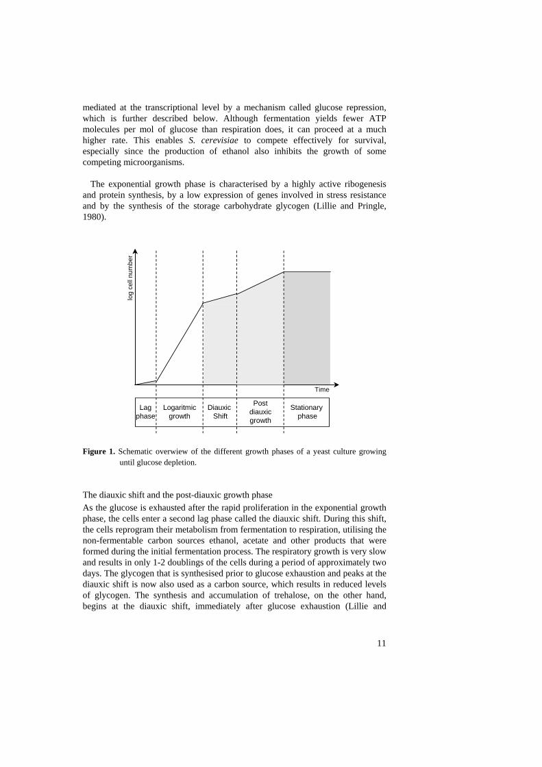
mediated at the transcriptional level by a mechanism called glucose repression, which is further described below. Although fermentation yields fewer ATP molecules per mol of glucose than respiration does, it can proceed at a much higher rate. This enables S. cerevisiae to compete effectively for survival, especially since the production of ethanol also inhibits the growth of some competing microorganisms.
The exponential growth phase is characterised by a highly active ribogenesis and protein synthesis, by a low expression of genes involved in stress resistance and by the synthesis of the storage carbohydrate glycogen (Lillie and Pringle, 1980).
Time
log
cell
num
ber
Logaritmicgrowth
Diauxic Shift
Postdiauxic growth
Stationaryphase
Lagphase
Figure 1. Schematic overwiew of the different growth phases of a yeast culture growing
until glucose depletion. The diauxic shift and the post-diauxic growth phase As the glucose is exhausted after the rapid proliferation in the exponential growth phase, the cells enter a second lag phase called the diauxic shift. During this shift, the cells reprogram their metabolism from fermentation to respiration, utilising the non-fermentable carbon sources ethanol, acetate and other products that were formed during the initial fermentation process. The respiratory growth is very slow and results in only 1-2 doublings of the cells during a period of approximately two days. The glycogen that is synthesised prior to glucose exhaustion and peaks at the diauxic shift is now also used as a carbon source, which results in reduced levels of glycogen. The synthesis and accumulation of trehalose, on the other hand, begins at the diauxic shift, immediately after glucose exhaustion (Lillie and
11

12
Pringle, 1980). Trehalose functions partly as a carbohydrate reserve but mainly in stress related protection of the cell (Panek and Panek, 1990; Wiemken, 1990).
Characteristic for the diauxic shift and the post-diauxic growth is the induction
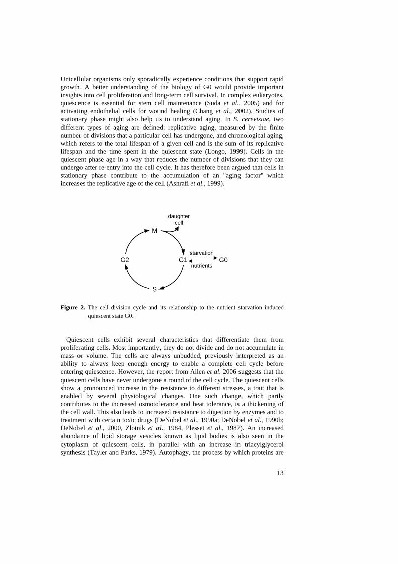
of a broad range of genes involved in stress resistance. The most well studied protein induced under these conditions is the Hsp70 related Ssa3 protein (Werner-Washburn et al., 1987), which therefore often is used as a marker for the diauxic shift. The Hsp70 proteins are highly conserved throughout evolution, and are encoded by multigene families in eukaryotes (Ingolia et al., 1982; Mues et al., 1986). Interestingly, the genes in this gene family show different expression patterns, some are constitutively expressed, while others are expressed specifically as a response to stresses like heat shock. The Hsp70 proteins are involved in precursor polypeptide import into both mitochondria and the ER (Deshaies et al., 1988). In vitro evidence also suggests that they are necessary for recycling of coated vesicles (Chappell et al., 1986; Ungewickel, 1985). The HSP70 gene SSA3 in S. cerevisiae was first shown to be strongly induced by heat shock, but it is also induced in response to low cAMP levels, and is therefore highly induced at the diauxic shift where cAMP levels drop drastically (Werner-Washburn et al., 1989). The stationary phase and quiescence If cells are maintained in liquid culture, they are finally triggered to enter a non-proliferating resting state, the stationary phase, also called G0, as a result of carbon starvation after the diauxic growth (Figure 2). It should be noted that yeast cells also enter a stationary non-proliferating state when starved for other nutrients, such as nitrogen, phosphorous or sulphur (Lillie and Pringle, 1980). Whether there is only one, or several different stationary phases is not yet fully understood (Werner-Washburn et al., 1996, Granot and Snyder, 1991). Not all types of starvation lead to stationary phase. For example, starvation for inositol causes rapid cell death (Keith et al., 1977) and most laboratory strains lose viability when grown to saturation in a synthetic defined medium (Bitterman et al., 2003). After many years of research a distinction was made between the state of the cell culture and the states of individual cells. Stationary phase is thus used to refer to a saturated cell culture, while the constituent cells in such a culture which have entered G0 are referred to as being in quiescence (Gray et al., 2004).
Until recently, it was thought that most of the cells in a stationary phase culture are indeed in a quiescent state. However, Allen et al. (2006) showed that a stationary phase culture can be separated into two cell fractions where only one fraction, representing 50% of the cells, homogeneously contains cells with quiescent characteristics, while the other fraction is heterogeneous including both budding and non budding cells. Interestingly, this study also showed that over 90% of the quiescent cells are cells with no bud scars, which suggests that the only cells that can enter quiescence are the daughter cells that are produced during the 1-2 cell doublings that occur during the post-diauxic growth phase.
Most eukaryotic cells, whether from unicellular or multicellular organisms, spend most of their life span in a quiescence state (Lewis and Gattie, 1980).
Unicellular organisms only sporadically experience conditions that support rapid growth. A better understanding of the biology of G0 would provide important insights into cell proliferation and long-term cell survival. In complex eukaryotes, quiescence is essential for stem cell maintenance (Suda et al., 2005) and for activating endothelial cells for wound healing (Chang et al., 2002). Studies of stationary phase might also help us to understand aging. In S. cerevisiae, two different types of aging are defined: replicative aging, measured by the finite number of divisions that a particular cell has undergone, and chronological aging, which refers to the total lifespan of a given cell and is the sum of its replicative lifespan and the time spent in the quiescent state (Longo, 1999). Cells in the quiescent phase age in a way that reduces the number of divisions that they can undergo after re-entry into the cell cycle. It has therefore been argued that cells in stationary phase contribute to the accumulation of an "aging factor" which increases the replicative age of the cell (Ashrafi et al., 1999).
G0G1
S
M
G2
daughtercell
nutrients
starvation
Figure 2. The cell division cycle and its relationship to the nutrient starvation induced
quiescent state G0.
Quiescent cells exhibit several characteristics that differentiate them from proliferating cells. Most importantly, they do not divide and do not accumulate in mass or volume. The cells are always unbudded, previously interpreted as an ability to always keep enough energy to enable a complete cell cycle before entering quiescence. However, the report from Allen et al. 2006 suggests that the quiescent cells have never undergone a round of the cell cycle. The quiescent cells show a pronounced increase in the resistance to different stresses, a trait that is enabled by several physiological changes. One such change, which partly contributes to the increased osmotolerance and heat tolerance, is a thickening of the cell wall. This also leads to increased resistance to digestion by enzymes and to treatment with certain toxic drugs (DeNobel et al., 1990a; DeNobel et al., 1990b; DeNobel et al., 2000, Zlotnik et al., 1984, Plesset et al., 1987). An increased abundance of lipid storage vesicles known as lipid bodies is also seen in the cytoplasm of quiescent cells, in parallel with an increase in triacylglycerol synthesis (Tayler and Parks, 1979). Autophagy, the process by which proteins are
13
14
recycled into amino acids through vacuolar degradation, is induced, and electron-dense material (probably polyphosphate) accumulates in the vacuoles (Noda and Oshumi, 1998; Matile et al., 1969). As a cell enters the quiescent state, the levels of glycogen are very low, as quiescent cells instead use trehalose as their main carbohydrate reserve. The trehalose levels peaks as cells enter quiescence and thereafter decrease gradually as the cell stays in this state (Lillie and Pringle, 1980).
The overall transcription rate is three to five times lower in quiescent cells than in cells growing exponentially (Boucherie, 1985; Choder, 1991). This together with the drastic decrease in the expression of genes encoding ribosomal proteins and a drop in translational initiation results in a reduction in total protein abundance in quiescent cells. There is still some transcription and translation going on but there seems to be a preference for certain groups of genes and mRNAs, some of which are actually increased rather than decreased in the quiescence phase. For example, some genes which show almost undetectable expression levels in exponentially growing cells are found to be induced during or after the diauxic shift, for example CYC7 (Pillar and Bradshaw, 1991), HSP26 (Petko and Lindquist, 1986), HSP12 (Praekelt and Meacock, 1990) and the previously mentioned SSA3. Even though the functional relevance for survival in stationary phase for many of the induced genes has not yet been determined, the large proportion of genes encoding proteins required for proteolysis, or proteins involved in protein stabilisation and transport suggest that protein stability and turnover are likely to be important for survival during stationary phase.
There is still an ongoing discussion as to whether the quiescent state called G0 is in fact a specific phase, distinct from G1. An alternative hypothesis is that the stationary phase represents an extended G1 phase, where the cells are exhibiting an especially slow rate of growth. This alternative was raised as many of the morphological changes that are seen in stationary phase cells are also observed in slowly growing but mitotically active cells. The morphological, biochemical and physiological changes associated with stationary phase are thus not unique, but are seen in other cells, particularly in response to stress. To establish that the stationary phase is indeed a distinct out-of-cycle phase of growth, it will be necessary to identify genes specifically required for the transition between stationary phase and the cell cycle or to identify a biochemical activity that is specific for stationary phase. However, a compelling argument for quiescence as a distinct growth phase comes from studies of the gsc1 mutant. Thus, it was shown that cells impaired in Gsc1 activity are unable to exit from stationary phase and resume proliferation after addition of glucose, but show no defect in mitotic growth (Drebot et al., 1987). (See also Werner-Washburn et al., 1993; Herman, 2002; Grey et al., 2004 and references therein). Glucose sensing signaling pathways Within 20 min after glucose addition ~20% of the roughly 6,200 genes in yeast are up- or down-regulated threefold or more and ~40% of the genes show at least a twofold change in expression (Wang et al., 2004). Virtually all living cells possess
15
a sophisticated genetic program that responds to the presence of glucose, which is a rich and therefore preferred source of energy and carbon for many cells. Studies of the glucose response in eukaryotes have gained great insights from work using S. cerevisiae as a model organism. However, whereas downstream components and their functions often have been elucidated in great detail, studies of the initial glucose-sensing mechanisms have proven to be more difficult. As described above, S. cerevisiae preferentially uses glucose as its sole carbon source, even under aerobic conditions and in the presence of alternative carbon sources. This preference for glucose is mediated by several glucose sensing and signaling mechanisms that ensure its optimal use. Discussed below are the three main glucose-responsive pathways in yeast: glucose induction, glucose repression and the Ras/cAMP pathway. Glucose induction As mentioned above, yeast cells growing on glucose obtain their energy mainly through fermentation. Since fermentation is a relatively inefficient way of generating energy, a high glycolytic flux is essential. Yeast cells are able to increase their glycolytic capacity by the induction of a number of glycolytic genes (Rolland et al., 2002). Studies have shown that it is the increased levels of different glycolytic metabolites that trigger the induction of glycolytic genes. These different intermediates seem to function as metabolic messengers that regulate the glycolytic activity in response to changes in substrate concentrations (Boles et al., 1993). How these metabolic signals are transmitted is still uncertain, but various regulatory elements that are mainly found in the promoters of glycolytic genes, and DNA-binding proteins that bind to these elements have been identified. In particular, Gcr1, which binds to the CTTCC motif, seems to be of central importance (Uemura et al., 1997).
An important part of the glucose response is regulation of the glucose uptake. It has been shown that the expression and function of the seven genes encoding functional hexose transporters in yeast (the HXT genes) are regulated to optimise the glucose uptake at any time. This is possible due to the fact that the different transporters exhibit either high, low or intermediate affinity for glucose. In the total absence of glucose, the expression of all HXT genes is repressed. This repression is relieved in the presence of glucose but different HXT genes are derepressed depending on the glucose concentration. Thus, high glucose concentrations mediate the repression of genes encoding transporters with high and intermediate affinity while at the same time the expression of genes for low-affinity transporters is induced (Rolland et al., 2002 and references therein).
The main glucose induction pathway in short terms triggers expression of glucose induced genes by Mth1-dependent binding of Rtg1 to glucose-induced promoters. Low concentrations of glucose are sensed by the Snf3/Rtg2 transmembrane complex, leading to ubiquitin-dependent proteolytic degradation of Mth1. Loss of Mth1 in turn triggers phosphorylation of Rtg1, which prevents its binding to the glucose induced promoters (Özcan and Johnston, 1999; Rolland et al., 2002 and references therein).

16
Glucose repression When cells are grown on glucose, a large number of proteins become dispensable for S. cerevisiae. To limit carbohydrate usage to only glucose, a variety of genes are therefore subjected to a transcriptional down-regulation referred to as glucose repression. The main groups of genes that are subjected to glucose repression are the genes encoding enzymes involved in respiration and gluconeogenesis, proteins necessary for the uptake and metabolism of alternative carbon sources and, as mentioned above, also the genes encoding the high affinity hexose transporters (Ronne, 1995, Rolland et al., 2002 and references therein).
Glucose repression is mediated by the transcription factor Mig1 (and to some extent also by the related Mig2 protein) which in the presence of glucose is in a non phosphorylated state where it can repress its target genes by recruiting of Ssn6/Tup1 co-repressor complex to their promoters (Nehlin and Ronne, 1990; Östling and Ronne, 1998; Keleher et al., 1992; Treitel and Carlson, 1998). The key regulator in glucose repression is the Snf1 kinase which in the absence of glucose is activated and phosphorylates Mig1, thereby inhibiting its transfer into the nucleus and thus relieving the repression of its target genes (De Vit et al., 1997). Previous studies have shown that inhibition of Snf1 requires glucose uptake, but that this is in itself not sufficient to generate the signal that inhibits the Snf1-dependent phosphorylation of Mig1. It has therefore been suggested that the glucose concentration is sensed intracellularly by other mechanisms than those involved in triggering of the glucose induction pathway (Reifenberger et al., 1997). Significantly, epistasis experiments have shown that hexokinase is required for the glucose-dependent inhibition of Snf1 that triggers translocation of Mig1 to the nucleus (Rolland et al., 2002 and references therein). In addition, the finding that the major hexokinase, Hxk2, can itself localise to the nucleus, where it associates with promoters of glucose-repressed genes, has prompted the suggestion that it has a role in the glucose repression response beyond its role in regulating Snf1 activity (Moreno and Herrero, 2002). The expression and activity of Hxk2 is itself dependent on glucose levels, and the partial nuclear localisation of Hxk2 has been shown to be mediated by Mig1 (Ahuatzi et al., 2004). Furthermore, during growth on high glucose media the Hxk2 kinase is highly expressed, while it is only weakly expressed on low glucose media (Herrero et al., 1995). This expression pattern is regulated by the main glucose induction pathway in an Rtg1 dependent manner, as described above (Mosley et al., 2003; Kim et al., 2003). The Ras/cAMP pathway The Ras/cAMP pathway is required for proper regulation of growth and cell cycle progression in response to glucose. Cells which are deficient in this pathway show physiological changes normally associated with nutrient deprivation, like cell cycle arrest in G1, accumulation of trehalose and glycogen, enhanced expression of various stress-related genes, and increased resistance to stress. In contrast, cells exhibiting an increased Ras/cAMP signaling and thus an increased protein kinase A (PKA) activity fail to arrest in G1, are defective for glycogen and trehalose synthesis, rapidly loose viability and remain highly sensitive to stress even after
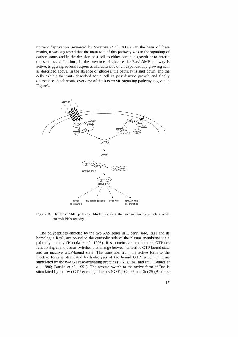
nutrient deprivation (reviewed by Swinnen et al., 2006). On the basis of these results, it was suggested that the main role of this pathway was in the signaling of carbon status and in the decision of a cell to either continue growth or to enter a quiescent state. In short, in the presence of glucose the Ras/cAMP pathway is active, triggering several responses characteristic of an exponentially growing cell, as described above. In the absence of glucose, the pathway is shut down, and the cells exhibit the traits described for a cell in post-diauxic growth and finally quiescence. A schematic overview of the Ras/cAMP signaling pathway is given in Figure3.
Cyr1
Gpr1
Glucose
Gpa2
Gpa2
cAMP
GDPRas
Ras
GDP
GEFs
GAPs
Tpk1,2,3Bcy1
active PKA
inactive PKA
stressresistance
gluconeogenesis glycolysis growth andproliferation
GTP
GAP
Tpk1,2,3
Bcy1 cAMP
GTP
Figure 3. The Ras/cAMP pathway. Model showing the mechanism by which glucose
controls PKA activity.
The polypeptides encoded by the two RAS genes in S. cerevisiae, Ras1 and its homologue Ras2, are bound to the cytosolic side of the plasma membrane via a palmitoyl moiety (Kuroda et al., 1993). Ras proteins are monomeric GTPases functioning as molecular switches that change between an active GTP-bound state and an inactive GDP-bound state. The transition from the active form to the inactive form is stimulated by hydrolysis of the bound GTP, which in turnis stimulated by the two GTPase-activating proteins (GAPs) Ira1 and Ira2 (Tanaka et al., 1990; Tanaka et al., 1991). The reverse switch to the active form of Ras is stimulated by the two GTP-exchange factors (GEFs) Cdc25 and Sdc25 (Broek et
17
18
al., 1987; Damak et al., 1991). The active GTP bound Ras, which is found in the presence of glucose, stimulates the adenylate cyclase Cyr1 to produce cAMP (Uno et al., 1987), which in turn activates PKA (see below).
The Ras/cAMP pathway responds to extracellular glucose availability, which is sensed by the GPCR (G-protein-coupled receptor) system. Gpa2, a member of the Gα protein family of heterotrimeric G proteins, has an activity which, as for Ras, depends on the binding of GTP. The Gpb1 and Gpb2 proteins were identified as interactors to Gpa2, and are both G-protein β subunits, while no putative γ subunit has yet been described. The activity of Gpa2 is negatively regulated by the GAP Rgs1, and both Gpa2 and Rgs1 have been shown to be involved in glucose sensing (Kraakman et al., 1999; Versele et al., 1999). The Gpr1 protein is a member of the seven transmembrane receptor superfamily which is located on the yeast cell surface. Genetic analysis suggests that Gpr1 functions as a glucose sensor upstream of the Gpa2 protein, as overexpression of GPA2 suppresses the defect of a gpr1Δ ras2Δ strain (Xue et al., 1994). Further, a deletion of GPR1 attenuates but does not eliminate the cAMP-dependent response to the addition of glucose, indicating that Gpr1 is indeed involved in cAMP-dependent glucose signaling, though it is not the sole mediator of that signaling. These findings led to the suggestion that Gpr1 functions as a glucose receptor which via Gpa2 contributes to the increased activation of the adenylate cyclase encoded by CYR1, and thus to an increased production of cAMP (Santangelo, 2006 and references therein).
There is a direct link between the intracellular cAMP level and the growth rate in yeast. The addition of glucose results in a rapid but transient spike in the level of intracellular cAMP, which increases 5- to 50-fold within 1-2 minutes of glucose addition and then returns to near basal levels within 20 minutes (van der Plaat, 1974). Genetic results suggest that all effects of cAMP in yeast are mediated by the cAMP-dependent protein kinase, PKA. It therefore seems likely that the sole purpose of the spike in cAMP levels that is produced after glucose addition is to activate PKA.
Protein kinase A, PKA, consists of a catalytic subunit encoded by the partially redundant TPK1, TPK2 and TPK3 genes, and a negative regulatory subunit encoded by the BCY1 gene. In the absence of nutrients, when the cAMP levels in the cell are low, Bcy1 binds to and inhibits the catalytic subunit, forming a heterotetrameric complex. The addition of glucose instantly increases the level of cAMP in the cell through GPCR-dependent activation of the Ras/cAMP pathway. The free cAMP binds to Bcy1, which then dissociates from the catalytic PKA subunit, resulting in an activated form of PKA (reviewed by Thevelein and de Winde 1999). The subcellular localisation of PKA also changes as a result of glucose signaling. Thus, the inactive Bcy1-bound form of PKA is localised both in the cytoplasm and in the nucleus, while the activated PKA is only found in the nucleus (Griffioen et al., 2000).
19
Other nutrient sensing signaling pathways As described above, a broad range of phenotypic changes occur as yeast cells are subjected to changes in glucose availability. Similar responses as to glucose deprivation also occur as a response to starvation for other essential nutrients like nitrogen. These nutrient responses are transmitted by several signal transduction pathways, providing an essential framework for coordination of cellular responses to external stimuli. These different pathways have been shown to be remarkably well conserved in eukaryotes. Adding an extra layer to the complexity, there is an extensive crosstalk and also a partial redundancy between some of these pathways. Three of these pathways are briefly discussed below, the TOR-pathway, the fermentable growth medium induced pathway and the PHO pathway. The TOR pathway When added to eukaryotic cells, the immunosuppressive rapamycin triggers a reaction corresponding to the response to nutrient deprivation, a response which is conserved from yeast to human cells. For its biological activity, rapamycin needs an intracellular cofactor, Fpr1, together with which it can target and inhibit the TOR kinases (Heitman et al., 1992). All eukaryotic genomes examined encode at least one TOR kinase, which belongs to a group of kinases known as the phophatidylinositol kinase related kinase family (PIKK family).
The TOR signaling pathway is a conserved network that links cell growth to nutrient availability in eukaryotic cells. S. cerevisiae has two TOR kinases, Tor1 and Tor2, which though highly similar in sequence show some differences in function. Thus, while Tor1 has only one function, which is redundantly shared with Tor2, Tor2 also has a unique function not shared with Tor1. The distinct and partially overlapping functions of Tor1 and Tor2 are mediated by two distinct complexes, TORC1 and TORC2 (Loewith et al., 2002). The TORC1 complex consists of either Tor1 or Tor2, Tco89, Kog1 and Lst8. This complex mediates the shared function of Tor1 and Tor2, which is to regulate ribosome biogenesis, translation, sorting and turnover of nutrient permeases, specific changes in gene transcription, induction of autophagy and cell cycle arrest in response to nutrient, mainly nitrogen, availability. The TORC2 complex consists of the Tor2, Lst8, Avo3, Avo1, Bit61 and Avo2 proteins, and mediates the unique function of Tor2 as a regulator of actin organisation in response to nutrient availability. While the shared function of Tor1 and Tor2 is sensitive to rapamycin, the unique function of Tor2 is not (Reinke et al., 2004).
In short, when growth conditions are favourable, the TOR pathway is active, maintaining conditions that are necessary for growth, i.e. high ribogenesis, efficient translational initiation, rapid nutrient import and polarisation of the actin cytoskeleton to the bud. In contrast, when the cell is starved for nitrogen or treated with rapamycin, the resulting inhibition of the TOR pathway causes a downregulation of protein synthesis, an upregulation of stress related genes and an upregulation of autophagy, in summary traits typical of the G0 state.
20
The TOR pathway mainly responds to nitrogen availability, but the upstream regulators still remain elusive. Some evidence exists suggesting that certain amino acids, specifically glutamate and glutamine, may be important signaling nutrients (Crespo et al., 2002, Komeili et al., 2000), but only some of the TOR targets answer to glutamine depletion while others do not. Thus, while glutamine depletion activates the TOR-controlled transcription factors Gln3, Rtg1 and Rtg3, the TOR-regulated Msn2/4 localisation and the down regulation ribosomal proteins are not effected (Crespo et al., 2002). This suggests that there are other nutrient regulators affecting TOR signaling that remain to be identified. Transcriptional profiling has, however, confirmed that rapamycin treatment mimics the response to nitrogen and glutamine depletion rather than the response to glucose depletion (Peng et al., 2002).
Many processes downstream of TOR are regulated by a switch in phosphatase activity mediated by the phosphatase Sit4 and its two regulatory proteins Tap42 and Tip41 (Beck and Hall, 1999; Jacinto et al., 2001). Under nutrient rich conditions, TOR kinase activity promotes the binding of Sit4 to Tap42, thus maintaining Sit4 inactive. When nutrients are limited or as a response to rapamycin treatment, Tap42 dissociates from Sit4 which then in its active form activates different targets such as the transcription factor Gln3, the kinase Npr1 and Tip41 (Beck and Hall, 1999; Schmidt et al., 1998). Also the two Sit4-related type 2A phosphatases Pph21 and Pph22 are TOR regulated (Sneddon et al., 1990). Thus, TOR activity leads to the phosphorylation of Tap42 thereby promoting the binding of Tap42 to Pph21 or Pph22. This small Tap42-Pph21/22 complex is only seen in exponentially growing cells and has a positive effect on protein synthesis. There is also negative regulation by dephosphorylation of Tap42 by Pph21/22 in complex with the two positive regulatory type 2A phosphatase subunits, Cdc55 and Tpd3 (Jiang and Broach, 1999). This phosphatase network is also important for TOR-mediated repression of the RTG target genes that encode structural enzymes involved in the production of α-ketoglutarate used for synthesis of glutamate and glutamine (Butow and Avadhani, 2004; Düvel et al., 2003).
The TOR pathway has a prominent role in the regulation of several metabolic pathways. Rapamycin treatment modulates primarily the expression of genes involved in nitrogen metabolism, the glycolytic pathway and the tricarboxylic acid pathway. Most strikingly regulated is the group of genes involved in the uptake and assimilation of different nitrogen sources. Thus, rapamycin treatment downregulates genes involved in the uptake and metabolism of preferred nitrogen sources such as glutamate and ammonia, and upregulates genes involved in the uptake and use of poor nitrogen sources such as proline and urea. (Crespo and Hall, 2002) The negative regulation of certain genes is achieved by sequestering their transcriptional activators in the cytoplasm (Beck and Hall, 1999).
The unique function of Tor2 and the TORC2 complex is in the regulation of the spatial direction of yeast cell growth (Loewith et al., 2002). In S. cerevisiae, most of the growth occurs in the bud. To accomplish this, yeast cells polarise their actin cables and actin patches towards the bud, and intracellular vesicle transport is thereby directed towards the bud. This polarization is essential for targeted
21
membrane growth at the bud tip, where transport vesicles fuse with the plasma membrane. It is also essential for targeted secretion of macromolecules at the bud tip, and for targeted transport of other macromolecules from the mother cell to the bud (reviewed by Pruyne and Bretscher, 2000).
The fermentable growth medium induced pathway As mentioned above, the addition of glucose to derepressed stationary phase cells triggers a rapid increase in the intracellular cAMP level resulting in a transient increase in PKA activity. Cells that are grown in the presence of glucose but lack some other essential nutrient arrest in G1. After addition of the missing nutrient, the cells revert back to normal growth, but, notably, this is achieved without any peak in cAMP (Hirimburgegama et al., 1992). Hence, this response is not mediated by the Ras/cAMP pathway, but it still requires PKA activity. The finding of a cAMP independent but PKA-dependent nutrient response led to the suggestion that after the transient cAMP increase that occurs upon addition of glucose to derepressed cells, another pathway is responsible for maintaining the high PKA activity. This new pathway was named the fermentable growth medium induced pathway because its activation requires both a fermentable carbon source and other nutrients needed for growth (Thevelein, 1995).
Sch9 is a protein kinase which is distantly related to the catalytic subunits of PKA encoded by the three TPK genes. Overexpresssion of SCH9 causes similar phenotypic effects as overexpression of the TPK genes, such as heat shock sensitivity (Toda et al., 1988). Cells that are grown on abundant glucose but are deprived of nitrogen express starvation specific genes, and the Sch9 kinase is essential for the reversion of this response after addition of nitrogen to the cells. This suggests that the Sch9 kinase is involved in nitrogen dependent signaling (Crauwels et al., 1997). The fact that a deletion of Sch9 also causes a slow growth phenotype which can be overcome by an elevated PKA activity further suggest that the two kinases might act redundantly (Toda et al., 1988). In 2005, Roosen et al. presented data supporting this notion but also showed that this redundancy is only partial, which suggests that the two kinases act in distinct signal transduction pathways. The PHO pathway Phosphate is an essential nutrient that is needed for nucleic acid and phospholipid biosynthesis, as well as for energy metabolism. When phosphate becomes limiting, the first response of S. cerevisiae cells is to increase the production of high affinity phosphate transporters and secreted phosphatases which can scavenge phosphate from the environment. Similar to depletion of other essential nutrients, phosphate starvation causes S. cerevisiae to arrest in G0. The molecular components of the phosphate sensing machinery are still unidentified, but some findings suggest that phosphate levels are sensed both on the outside and on the inside of the cell.
The signaling of the phosphate levels is mediated by the PHO pathway, which regulates the expression of several phosphate-responsive genes that encode proteins involved in the acquisition and specific uptake of phosphate from
22
extracellular sources. The system for phosphate uptake is divided into low affinity and high affinity transport. The low affinity transport satisfies the cellular need for phosphate at normal or high concentrations of external phosphate, while the high affinity transport system is induced in response to phosphate starvation (Persson et al., 2003). Under high phosphate conditions, the cyclin dependent kinase (CDK)-cyclin complex Pho85-Pho80 phosphorylates the Pho4 transcription factor, thereby inactivating it by causing its exclusion from the nucleus (Kaffman et al., 1994; O'Neill et al., 1996). When cells are starved for phosphate, Pho81 inhibits the Pho85-Pho80 kinase, thus allowing the non-phosphorylated Pho4 to associate with the nuclear import receptor Pse1 and enter the nucleus where it induces the expression of several genes (Kaffman et al., 1998; O'Shea, 1996). The targets for Pho4 are the genes encoding the high affinity phosphate transporters Pho84 and Pho89, and the secreted acid phosphatases Pho5, Pho11, Pho12 (reviewed by Oshima, 1997; Mouillon and Persson, 2006).
In short, phosphate starvation promotes an increased uptake of phosphate from the surrounding medium by inducing the expression of high affinity phosphate transporters and acid phosphatases. When phosphate is added to phosphate-deprived cells, the PHO pathway is rapidly shut down and phosphate uptake is reduced mainly as a consequence of a rapid proteolytic Pho84 degradation (Mouillon and Persson, 2006). The convergence of nutrient signaling pathways on Rim15 The fact that the G0-related changes that occur as a result of a decrease in PKA activity following nutrient limitation are strikingly similar to the changes observed after rapamycin treatment, suggested that the PKA and TOR signaling pathways might converge on a common downstream component. Furthermore, the transcriptional targets of the FMG pathway show considerable overlap with those of the PKA pathway, indicating that these two pathways also might share a common downstream effector. Finally, several studies indicate that Pho85 negatively regulates the expression of an additional set of genes that are not directly involved in phosphate uptake, which include genes encoding glycogen and trehalose synthesis enzymes and stress related genes typically induced under glucose-limiting conditions prior to entry into G0. (DeRisi et al., 1997; Timblin and Bergman, 1997; Ogawa et al., 2000; Carrol et al., 2001; Nishizawa et al., 2004). These findings suggest that proper execution of the G0 program also includes integration of the Pho85-mediated phosphate signal.
Indeed, in 2003 Pedruzzi et al. identified the protein kinase Rim15, previously known as a downstream target of the PKA pathway, as a target for TOR and FMG signaling. A few years later additional information also showed that the Rim15 kinase is regulated by the PHO pathway (Wanke et al., 2005). Although the complex regulatory pattern is not yet fully understood, and the pathways also have non-overlapping functions, the Rim15 kinase obviously plays a key role as the protein on which the four pathways converge. In short, downregulation of the TORC1, PKA, PHO and FGM pathways all lead to activation of several G0-specific effects, which are largely Rim15-dependent.
23
Rim15 was originally cloned as a protein involved in meiosis, where it was
shown to stimulate the removal of repressive regulators at the promoters of early meiosis-specific genes, leading to their induction (Su and Mitchell, 1993). Subsequently, the Rim15 protein was shown to physically interact with the trehalose synthase complex protein Tps1, and a deletion of Rim15 was shown to exhibit physiological defects when grown to stationary phase, including defects in the accumulation of glycogen, in the acquisition of thermotolerance and in the ability to arrest properly in G1 (Reinders et al., 1998). The overlap with effects caused by mutations that constitutively activate PKA prompted a study of the effect of the rim15 deletion on transcripts known to be negatively regulated by PKA. This resulted in the finding that the rim15 deletion strain failed to induce SSA3, HSP12 and HSP26 after the diauxic shift. Finally, the kinase activity of Rim15 was also shown to be inhibited by PKA (Reinders et al., 1998).
Synthesis of cAMP is induced by glucose, but not by any other nutrients. Still, the response to other types of nutrient limitation, which do not cause reduced cAMP levels, does in fact result in many of the same G0 phenotypes as are seen after glucose deprivation. Rapamycin treatment, which mimics nutrient deprivation by causing an inactivation of the TORC1 complex, exhibits the same pattern. However, in a rim15 mutant, rapamycin fails to induce SSA3, HSP12 and HSP26, nor does the mutant accumulate glycogen or trehalose. In fact, it was subsequently shown that the rapamycin-dependent inactivation of TORC1 leads to hyperphosphorylation of Rim15 and its relocalisation from the cytoplasm to the nucleus (Pedruzzi et al., 2003).
The key to the regulation of Rim15-dependent G0 traits was shown to be this nuclear relocalisation of the kinase. Its cytoplasmic retention in cells where the PKA, TOR, PHO and FGM pathways are active is dependent on its binding to the 14-3-3 anchor protein Bmh2. Bmh2 can in turn only bind to Rim15 that has been phosphorylated at Threonin-1075, a phosphorylation which is dependent on both TORC1 and PKA (Wanke et al., 2005).
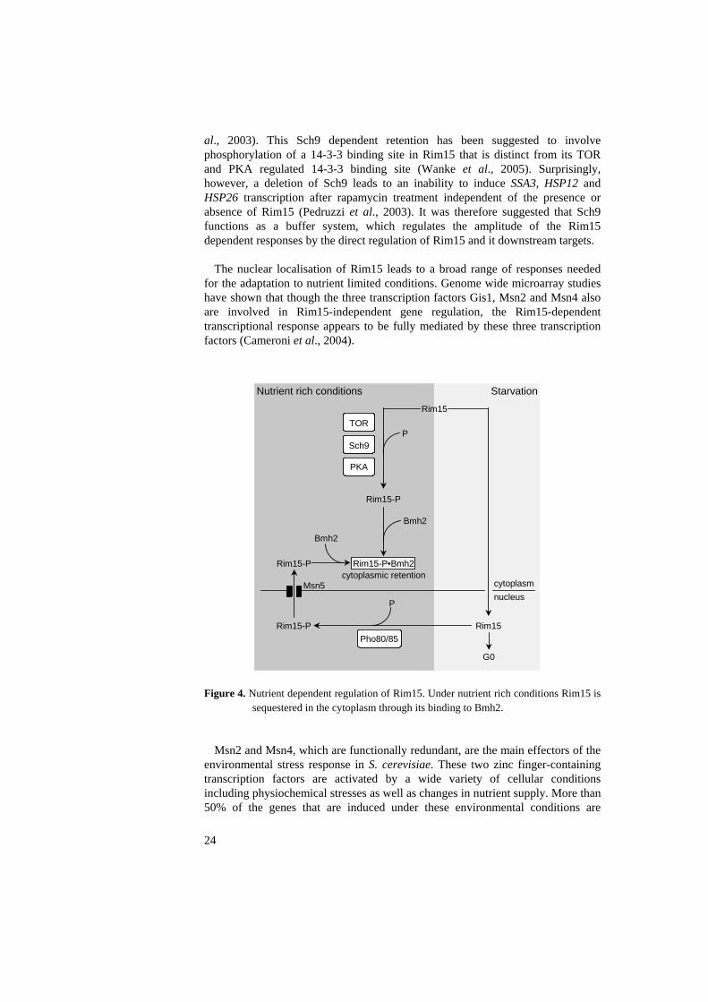
Inactivation of TORC1 or PKA eventually leads to dephosphorylation of the 14-3-3-binding site of Rim15, thereby releasing the kinase from the anchor protein, and enabling its transfer into the nucleus where it is able to activate its downstream targets. The regulation of Rim15 by the PHO pathway is different in that it targets not the cytoplasmic but the nuclear pool of the kinase. Thus, the active Pho85 kinase phosphorylates nuclear Rim15 at its Bmh2 target site, triggering the nuclear export of Rim15 which is mediated by the exportin Msn5. Accordingly, activation of the PHO pathway causes a transfer of Rim15 out of the nucleus, thereby preventing the activation of its downstream targets (Wanke et al., 2005). A model for the nutrient dependent regulation of Rim15 is presented in Figure4.
Cells that lack Sch9 accumulate Rim15 in the nucleus under exponential growth, when Rim15 is normally found in the cytoplasm. Thus, it appears that the Sch9 kinase is needed for cytoplasmic retention of Rim15 in log phase cells (Pedruzzi et
al., 2003). This Sch9 dependent retention has been suggested to involve phosphorylation of a 14-3-3 binding site in Rim15 that is distinct from its TOR and PKA regulated 14-3-3 binding site (Wanke et al., 2005). Surprisingly, however, a deletion of Sch9 leads to an inability to induce SSA3, HSP12 and HSP26 transcription after rapamycin treatment independent of the presence or absence of Rim15 (Pedruzzi et al., 2003). It was therefore suggested that Sch9 functions as a buffer system, which regulates the amplitude of the Rim15 dependent responses by the direct regulation of Rim15 and it downstream targets.
The nuclear localisation of Rim15 leads to a broad range of responses needed for the adaptation to nutrient limited conditions. Genome wide microarray studies have shown that though the three transcription factors Gis1, Msn2 and Msn4 also are involved in Rim15-independent gene regulation, the Rim15-dependent transcriptional response appears to be fully mediated by these three transcription factors (Cameroni et al., 2004).
Rim15-P
TOR
Sch9
PKA
P
Bmh2
Rim15-P
Rim15-PPho80/85
Rim15
Rim15
cytoplasmnucleus
P
Bmh2
Msn5
Nutrient rich conditions Starvation
G0
Rim15-P•Bmh2cytoplasmic retention
Figure 4. Nutrient dependent regulation of Rim15. Under nutrient rich conditions Rim15 is
sequestered in the cytoplasm through its binding to Bmh2.
Msn2 and Msn4, which are functionally redundant, are the main effectors of the environmental stress response in S. cerevisiae. These two zinc finger-containing transcription factors are activated by a wide variety of cellular conditions including physiochemical stresses as well as changes in nutrient supply. More than 50% of the genes that are induced under these environmental conditions are
24

25
regulated by Msn2/4 (Gasch et al., 2000; Causton et al., 2001). The Msn2/4-induced genes belong to several functional classes including enzymes involved in carbohydrate metabolism, antioxidant proteins and proteins involved in protein degradation (Boy-Marcotte et al., 1998; Gasch et al., 2000; Causton et al., 2001; Hasan et al., 2002). The importance of Msn2/4 in cellular adaptation to less favourable growth conditions has further been shown by the fact that a double deletion of the MSN2 and MSN4 genes causes sensitivity to various forms of stress. These cells also fail to accumulate stress-regulated messages following heat stress, osmotic stress, nutrient starvation, and DNA damage (Martinez-Pastor et al., 1996; Schmitt and McEntee, 1996). Msn2/4 bind specifically to a DNA motif, referred to as the stress response element STRE (AGGGG), which is found in the promoters of Msn2/4-induced genes like HSP12, HSP26, CTT1 and DDR2 (Martinez-Pastor et al., 1996; Schmitt and McEntee, 1996).
Msn2/4-mediated gene expression is regulated by the nutrient dependent shuttling of the proteins over the nuclear membrane. Under normal growth conditions, Msn2 and Msn4 are localised in the cytoplasm, but upon stress or nutritional depletion they are transported to the nucleus. If the stress continues, they display an oscillatory behaviour, shuttling back and forth between the nucleus and the cytoplasm (Görner et al., 1998; Jaquet et al., 2003). As for Rim15, the sequestering of Msn2 in the cytoplasm under rich nutrient conditions is controlled by its binding to the 14-3-3 protein Bmh2 (Beck and Hall, 1999). Furthermore, in the absence of the Msn5 exportin, Msn2 and Msn4 are restricted to the nucleus, but this nuclear localisation is not sufficient for induction of the STRE-dependent genes. This finding indicates that subcellular localisation is only one level of control in the response of Msn2/4 to stress (Estruch, 2000). Another control operates on the level of the binding of the Msn2 to DNA, which is reported to be controlled by stress and is possibly dependent on the protein kinase Gsk3 (Görner et al., 1998; Hirata et al., 2003).
The Gis1 protein is a zinc finger transcription factor which can function as an activator upon binding to the post diauxic shift element PDS (T(T/A)AGGGAT) known to mediate transcriptional activation in response to nutritional limitation (Boorstein and Craig, 1990). This function of Gis1 as an activator in the post-diauxic shift response was initially identified for the PDS driven activation of SSA3, where Gis1 in turn is positively regulated by Rim15 and Sch9 (Pedruzzi et al., 2000, Roosen et al., 2005). Subsequent studies have also identified PDS element dependent Gis1 repression of the DPP1 gene, encoding diacylglycerol pyrophosphate phosphatase, and the GRE1 gene, encoding a hydrophilin known to be induced in response to stress (Oshiro et al., 2003; Roosen et al., 2005). Gis1 is also involved, together with its close homologue Rph1, with which it shares identical zinc fingers, in the repression of the PHR1 gene encoding photoreactivation lyase. Rph1 is known to mediate this repression via a sequence motif that overlaps with a STRE element (Jang et al., 1999). Furthermore, Gis1 has an effect, although weak, on the induction of the STRE driven genes HSP12 and HSP26 (Pedruzzi et al., 2000). Hence, it was suggested that Gis1 also may act through STRE elements. Further evidence for this came from the finding that the while an msn2 msn4 double deletion still is capable of a low induction of the
STRE-driven genes HSP12 and DDR1, an msn2 msn4 gis1 triple deletion completely abolishes this induction (Roosen et al., 2005). Furthermore, the msn2 msn4 mutant shows impaired induction of the PDS-regulated GRE1 gene, a response which is completely abolished in the msn2 msn4 gis1 background. Hence, Gis1 and Msn2/4 appear to regulate STRE-mediated transcription of a subset of genes in a cooperative manner (Cameroni et al., 2004; Roosen et al., 2005).
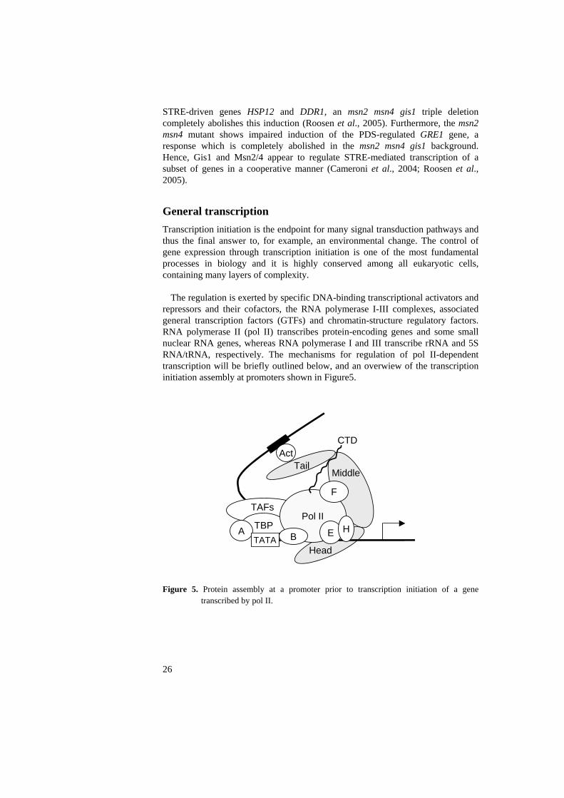
General transcription Transcription initiation is the endpoint for many signal transduction pathways and thus the final answer to, for example, an environmental change. The control of gene expression through transcription initiation is one of the most fundamental processes in biology and it is highly conserved among all eukaryotic cells, containing many layers of complexity.
The regulation is exerted by specific DNA-binding transcriptional activators and repressors and their cofactors, the RNA polymerase I-III complexes, associated general transcription factors (GTFs) and chromatin-structure regulatory factors. RNA polymerase II (pol II) transcribes protein-encoding genes and some small nuclear RNA genes, whereas RNA polymerase I and III transcribe rRNA and 5S RNA/tRNA, respectively. The mechanisms for regulation of pol II-dependent transcription will be briefly outlined below, and an overwiew of the transcription initiation assembly at promoters shown in Figure5.
TBPAPol II
F
E HBTATA
TAFs
Head
MiddleTail
CTDAct
Figure 5. Protein assembly at a promoter prior to transcription initiation of a gene
transcribed by pol II.
26
27
Promoters and promoter sites The upstream region of a gene, the promoter, plays a key role in regulating its expression, being the target of binding of regulatory proteins. The architecture of the promoter, i.e. the position and number of activator and repressor sites, the regulation of activators and repressors, and the chromatin state, together determine the gene's accessibility for transcription initiation. Regulatory proteins bind to different sequence elements that can be divided into two larger groups based on their positions, the core promoter and the upstream regulatory regions.
The core promoter elements are found close to the transcription start site and direct the polymerase to the correct start site. Three kinds of sequence elements are included in this definition, the first one identified was the TATA box, which is bound by the TATA-Binding Protein TBP (see below). Many genes also have an initiator element, INR, which can function either independently or together with the TATA box (Hampsey, 1998 and references therein; Smale et al., 1990). Finally, there are also downstream promoter elements (DPEs), that have been shown to be important for the binding of TFIID (see below) to TATA-less promoters (Burke and Kadonaga, 1996).
More distal promoter sequence elements, referred to as regulatory regions or enhancers are also involved in the regulation of transcription initiation. In yeast, these elements are typically located within a few hundred base pairs upstream of the core promoter, and they are called UASs for Upstream Activating Sequences and URSs for Upstream Repressing Sequences. These sequences interact with different types of DNA-binding transcription factors that can be either activators, in the case of UASs, or repressors, in the case of URSs. In higher eukaryotes, these regulatory sequences may be located both upstream and downstream of the core promoter, and sometimes very far away from it (thousands or even millions of base pairs). The RNA polymerase II enzyme The synthesis of eukaryotic mRNA from protein encoding genes is carried out by RNA polymerase II (pol II). Pol II in S. cerevisiae comprises 12 subunits that are highly conserved among the eukaryotes. The resolution of the three-dimensional structure of the 10-subunit core of the yeast pol II provided fundamental new insights into the basic mechanisms of transcription (Cramer et al., 2000; Cramer et al., 2001; Gnatt et al., 2001). Visualised in these studies were the active site, above which the melted DNA binds, and the wall and the clamp which restrict access to the active site. The Rpb4 and Rpb7 subunits form a distinct subcomplex which can dissociate from the core complex. This small subcomplex is not needed during elongation but is essential for initiation. It has further been suggested to be involved in the interaction of pol II with the newly synthesised RNA (Edwards et al., 1991).
The largest subunit of pol II, Rpb1, has a unique carboxy-terminal domain (CTD) consisting of multiple copies of a heptapeptide repeat (YSPTSPS) which
28
protrudes as a tail from the complex. This tail is conserved among all eukaryotic organisms, only differing in the number of repeats (Corden et al., 1985). The tail is the target of phosphorylations which regulate the assembly of the transcription complex and the start of transcription elongation (Payne et al., 1989). Interestingly, it has further been shown that the CTD plays a role in mRNA processing by the binding of enzymes that are involved in mRNA capping, polyadenylation and splicing (Hirose and Manley, 2000; Proudfoot, 2000).
During the mRNA transcription cycle, pol II is dependent on its association with several different GTFs and other proteins for its activity. Indeed, mild purification of the yeast pol II led to the identification of a complex containing the Mediator (see below) together with the pol II core enzyme, a complex referred to as the RNA pol II holoenzyme. The Mediator is necessary for transcription initiation at nearly all pol II transcribed promoters (Kim et al., 1994). During initiation, pol II also interacts with GTFs that mediate promoter recognition and melting of the promoter DNA, with coactivators that transmit regulatory signals, with elongation factors that enable the efficient production of long RNAs, and with factors that are involved in 3' processing of the RNA and in transcription termination. Still, the limiting factor in the rate of transcription is the number of molecules of pol II that can enter the transcription cycle in a given amount of time. Assembly of the pre-initiation complex Transcription by pol II is a multistep process which requires the assembly of a complex of initiation factors on the promoter. This complex is referred to as the pre-initiation complex (PIC) and consists of six GTFs: TFIID, TFIIB, TFIIA, TFIIF, TFIIE and TFIIH. The first step is promoter recognition by TFIID, which consists of the TATA-box binding protein TBP and 14 tightly associated proteins called TBP-associated factors (TAFs). This binding results in a 90 degree bend in the DNA which creates a platform for the interactions of the remaining factors and also brings the DNA sequences located immediately upstream and downstream of the TATA box closer to each other (Roeder, 1996 and references therein). Once TFIID has bound to either the TATA box or an INR element, TFIIB and TFIIA are recruited to stabilise the TFIID binding to the core promoter in different ways. TFIIB, which consists of a single polypeptide, stabilises the TBP-TATA complex through contacts with both TBP and DNA sequences flanking the bent TATA element while TFIIA helps by binding to TBP and DNA upstream of the TATA element (Nikolov et al., 1995; Tsai and Sigler, 2000; Geiger et al., 1996; Tan et al., 1996; Bleichenbacher et al., 2003). The formation of a TFIID-IIA-IIB-DNA complex allows for the subsequent recruitment of TFIIF in association with pol II. The complex also helps to position the pol II active site close to the transcription start site which is approximately 30 bp downstream of the TATA box.
The assembly of the TFIID-TFIIA-TFIIB-pol II-TFIIF complex is sufficient to create a stable functional initiation complex (Tyree et al., 1993; Parvin et al., 1993; Parvin et al., 1994). However, promoter clearance and hence the synthesis of longer transcripts also requires the two GTFs TFIIH and TFIIE (Goodrich and Tijan, 1994). TFIIE helps to recruit TFIIH and also stimulates its enzymatic

29
activities (Maxon et al., 1994; Ohkuma et al., 1995). The TFIIH complex contains a helicase that catalyses the melting of the promoter at the transcriptional start site by local unwinding of the promoter DNA (Dvir, 2001 and references therein). TFIIH also contains a kinase that is responsible for phosphorylation of the CTD. There is no phosphorylation of the CTD tail as long as pol II is associated with the promoter bound initiation complex. Phosphorylation of the CTD tail by TFIIH is the signal that triggers the onset of elongation (Cadena and Dahmus, 1987; Payne et al., 1989).
After promoter clearance, the TFIIF-bound pol II elongates downstream of the gene, while TFIIA and TFIID remain bound to the core promoter and TFIIB, TFIIE and TFIIH are released (Zawel et al., 1995). Interestingly however, it has been shown that after pol II escape, Mediator subunits can remain associated with the core promoter together with TFIIA, TFIID, TFIIE and THIIH, forming a scaffold that is stabilised by promoter-bound activators. This suggests that the second and subsequent rounds of transcription initiation (re-initiation) may not require de novo recruitment of TFIIA, TFIID, TFIIE, TFIIH or the entire pol II-Mediator holoenzyme, but may just require re-incorporation of TFIIB, TFIIF and pol II (Yudkowski et al., 2000). After transcription termination, the CTD tail is dephosphorylated. In yeast, this is achieved by a single phosphatase which is encoded by the FCP1 gene, and pol II is thereafter able to enter a new pre-initiation complex for a second round of transcription (Kobor et al., 1999). DNA-binding transcription factors The PIC complex which is recruited to promoters is a general transcription machinery that is common to all genes transcribed by pol II. Gene-specific regulation of transcription is, however, mediated by gene specific transcription factors which bind to upstream promoter elements and regulate transcription in response to specific signals. These gene specific transcription factors can stimulate transcription by helping to recruit the general transcription machinery and/or by clearing the promoters from nucleosomes. However, they can also inhibit transcription, e.g. by stabilising nucleosome-promoter interactions, in which case they work as repressors. A transcriptional activator usually possesses two distinct domains that are important for its activity, a DNA binding domain and an activating domain that mediates its function as an activator. Negatively acting transcription factors instead possess a domain capable of repression, either by interfering with the recruitment of pol II or by interacting with cofactors (see below).
The DNA-binding activity of a transcription factor is usually mediated by a highly conserved DNA-binding domain recognising specific promoter sequences. The DNA-binding domain is frequently, but not always, the most conserved part of a transcription factor. Therefore, transcription factors are usually classified into families depending on which DNA-binding domain(s) they possess.
The most common DNA-binding domain in eukaryotic transcription factors is the zinc finger motif, which consists of an α-helix and a β-sheet held together by a

30
zinc ion in a structure where the α-helix can interact with the major groove of the DNA helix, making specific interactions with a triplet of bases. The zinc finger motif is almost always repeated several times, thus creating a DNA-binding domain that can read six, nine or even twelve bases, thereby increasing the strength and specificity of the DNA-protein interaction (Struhl, 1997 and references therein). DNA binding can also be mediated by a basic-leucine zipper (bZIP) domain containing a basic DNA-binding region followed by a leucine zipper that mediates protein dimerization (Hurst, 1994 and references therein). The helix-turn-helix motif resembles the bZIP motif in that it has a basic DNA-binding region, but instead of a classical leucine zipper its dimerization region contains two α-helices connected by a short stretch of amino acids representing the turn (Struhl, 1997 and references therein). Cofactors In order to regulate pol II dependent transcription, other proteins than the gene specific transcription factors are also required. These additional proteins are termed cofactors and fall into two broad classes, those that function by regulating chromatin structure (as further discussed below), and those that interact with the general transcription machinery. The second class is often referred to as mediators or transcriptional adaptors since they serve as a bridge between the activator or repressor and the basal transcription machinery. A significant overlap of function is seen between these two classes. These cofactors are essential for the transduction of signals from promoter bound activator and repressor proteins to the pol II and the GTFs.
The first cofactors of the second class that were discovered were the TBP-associated factors or TAFs which together with the TBP protein form the TFIID complex mentioned above (Poon et al., 1995). The TBP protein itself is sufficient for promoter recognition and subsequent recruitment assembly of the PIC, but TBP is still seen at the promoter as part of the larger TFIID complex. This complex formation has been shown to be important for the response to transcriptional regulatory proteins, but not for basal transcription in in vitro studies (Tanese et al., 1991; Chen et al., 1994). This point has further been substantiated by the fact that most of the TAFs in yeast can be deleted without any drastic effects on the global regulation of transcription (Moqtaderi et al., 1996; Walker et al., 1996). However, TFIID complex formation seems to be important in gene specific regulation where specific TAFs can contact specific activators (Reese et al., 1994; Hampsey, 1998 and references therein). Also, certain TAFs have enzymatic activities, for example TAF250 possesses protein kinase and histone acyl transferase (HAT) activities (Dikstein et al., 1996; Mizzen et al., 1996) as well as an ubiquitin-activating and -conjugating activity that is required for full transcriptional activation by certain activators in Drosophila embryos (Pham and Sauer, 2000).
The TFIIA complex can also be said to function as a coactivator. When present in the PIC, it does not only interact with TBP but also with specific activators and TAFs (Ozer et al., 1994; Kobayashi et al., 1995; Burlay and Roeder, 1996;
31
Kraemer et al., 2001). Its recruitment to PIC is not required for basal transcription but it is required for optimal activator-induced transcription, where it functions both as an antirepressor and as a coactivator (Kang et al., 1995; Ma et al., 1996).
Finally there is one adaptor complex of great importance for the initiation of transcription, the multiprotein complex termed the Mediator, which will be discussed below. The Mediator Another way by which activators and co-activators may recruit pol II to a promoter is by interacting with a multiprotein complex, the Mediator. The Mediator was first discovered in yeast after the finding that purified activators and components of the general transcription machinery were not sufficient for regulated in vitro transcription. The activator dependent transcription also required the addition of crude yeast extracts, and the unknown component mediating the missing function was named the Mediator (Kelleher et al., 1990; Flanagan et al., 1991). This complex is required for regulated transcription of nearly all RNA polymerase II-dependent genes in S. cerevisiae and functions as a bridge between regulatory proteins and the basal pol II transcription machinery, as a regulator of the phosphorylation status of the CTD, and possibly as a modulator of the chromatin structure.
A combination of biochemical, genetic and structural data suggests that the Mediator comprises 21 subunits, and is found in both a free form and in a complex with the pol II core enzyme, a complex referred to as the pol II holoenzyme (Kim et al., 1994; Myers et al., 1998). There are therefore also two different conformational states of the Mediator. One is a compact arrangement which is seen in the absence of the pol II enzyme. The second state, which is seen within the pol II holoenzyme, is an elongated structure which consists of three functionally and physically distinct modules (Figure 6.). The three domains are named according to their position relative to the polymerase: the head domain, which makes contacts with pol II, the middle domain, and the tail domain which makes contacts with activators. Both the different subunits and the domain composition of the Mediator are strongly conserved between yeast and higher eukaryotes, and a unifying nomenclature has recently been proposed (Bourbon et al., 2004).
The head module comprising Med6, Med17, Med18, Med19, Med20 and Med22 plays a general role in transcription and interacts with the CTD of pol II (Lee and Kim, 1998). Next comes the middle module comprising Med1, Med4, Med7, Med8, Med9, Med10, Med11, Med14, Med21 and Med31 which has been shown to be in direct contact with the CTD and also with the TFIIE complex (Kang et al., 2001). It is debated whether Med14 is a part of the middle domain or of the tail because of its unique position, which connects these two domains. Finally, the tail module comprising Med2, Med3, Med5, Med15 and Med16 is the module presumed to be responsible for recognising and binding to activators (Bhoite et al., 2001). Electron microscopy observations suggest a high degree of

structural conservation of the head and the middle domains between different eukaryotes, which is consistent with the fact that they interact with the conserved pol II enzyme. In contrast, the tail domain shows structural differences between different species, which most likely reflect the fact that it is involved in interactions with less conserved or species specific activators and repressors (Asturias et al., 1999; Dotson et al., 2000).
There is also a fourth distinct subcomplex of the Mediator, the Srb8-11 complex, which is composed of the Med12, Med13, Cdk8 and CycC subunits (also known as Srb8, 9, 10 and 11). The cyclin-dependent kinase Cdk8 and its associated cyclin CycC have been proposed to phosphorylate the CTD prior to PIC assembly, hence negatively regulating transcription by causing premature dissociation of the Mediator from the pol II core enzyme. Accordingly, this subcomplex has been shown to be involved in the repression of a number of genes (Hengartner et al., 1998; Carlson, 1997).
Med31
Med19
Med17
Med11Med1
Med9Med14
Med2
Med5
Med7Med22
Med4
Med6
Med21
Med10Med18Med8 Med20
Srb8
Srb10
Srb9
Srb11
Middle
Tail Head
Med16
Med15Med3
Figure 6. Schematic overview of the Mediator in Saccharomyces cerevisiae.
Despite the great insights that have been obtained into the components and the structure of the Mediator it is still unclear how it works mechanistically. The first model that was proposed suggested that the function of the Mediator would be to act as a bridge between activators and the basal transcription machinery, thus helping to recruit pol II to the promoter. This model suggests that physical recruitment of pol II to a promoter should be sufficient for activation of transcription, and several experiments support this notion. For example, it is possible to activate transcription by fusing a DNA-binding domain to a subunit of the Mediator, which can then recruit the latter to a given promoter (Farrell et al., 1996; Ptashne and Gann, 1997; Keaveney and Struhl, 1998; Balciunas et al., 2003). The finding that some Mediator subunits have a negative effect on
32
33
transcription, does, however, complicate the picture (Sternberg et al., 1987; Jiang et al., 1992).
The Mediator has also been suggested to be involved in more than just forming bridges between activators and the pol II core enzyme. Thus, a role as a modifier of the chromatin structure is suggested by the fact that one of the Mediator subunits, Med5, has HAT activity (see below) (Lorch et al., 2000). Acetylation of histones is generally associated with a dissociation of nucleosomes from the DNA, thereby increasing the accessibility of the transcription machinery to the DNA.
Chromatin dynamics and gene regulation Chromatin organisation Eukaryotic DNA is organised within the nucleus through its interaction with histone proteins, forming the fundamental and repeating unit of chromatin, the nucleosome. This unit consists of 146 bp of DNA wrapped around a core histone octamer including a (H3-H4) tetramer associated with two H2A-H2B heterodimers. Histones are small proteins consisting of a globular domain which binds to other histones within the nucleosome to form the octamer. The positively charged N-terminus of the histone is flexible and protrudes from the nucleosome, and is therefore referred to as the histone tail. These tails facilitate the binding of the histones to the DNA by electrostatic interactions with the negatively charged DNA backbone, thus promoting nucleosome formation. However, the residues in the histone tails are subject to different enzymatic modifications which can affect the chromatin structure (as further described below). The arrays of nucleosomes also contain the linker histone H1, which is situated on the inside of the chromatin fibre, and which is responsible for twisting and folding the chromatin fibre into more compact filaments, thereby giving it a defined higher order structure. The chromatin organization decides whether the DNA is accessible to the transcription machinery or not. Tightly packed chromatin is inaccessible for transcription and is referred to as heterochromatin, whereas chromatin in a looser structure, which is more accessible to transcription, is referred to as euchromatin. The final chromatin structure determined by the totality of different modifications at the histone level dictates the particular biological outcome and is referred to as the histone code (Morales et al., 2001 and references therein). Covalent modifications of histones For proper regulation of DNA accessibility there is a need for flexibility of the chromatin structure. To this end, histones are subjected to covalent post-translational modifications such as actetylation, methylation, phosphorylation, sumoylation and ubiquitination. These alterations affect a number of processes including transcription, gene silencing, heterochromatin formation, replication of DNA, apoptosis and the response to DNA damage (Millar and Grunstein, 2006 and references therein). As histones can be modified on several different amino acid residues, an attractive hypothesis is that different combinations of histone modifications at a genetic loci lead to distinct biological outcomes. For example, a

34
combination of modified and unmodified histone residues at the promoter of a gene could lead to transcriptional activation.
The most studied histone modification so far is acetylation, where a single acetyl group is added to the ε-amino group of a lysine (K). This is a reversible modification which is catalyzed by histone acetyltransferases (HATs) that add the modification, and histone deacetylases (HDACs) that remove them. Acetylation neutralises the positive charges of the lysine residues and can thus alter the histone-DNA and nucleosome-nucleosome interactions. An acetylation can also function as a mark that serves as a signal for binding of a protein or, alternatively, the non-modified lysine can be the target of a protein interaction which is disrupted by the modification (Kurdistani and Grunstein, 2003 and references therein). The overall level of acetylation generally correlates positively with the level of transcription as increased acetylation of histones results in a more open chromatin structure. However, there is at least one exception, the acetylation of H4 at lysine 12 is found in regions of silent chromatin where histones are otherwise hypoacetylated (Braunstein et al., 1996). The regulation of the acetylation status of a histone can be either promoter targeted or global. Many HATs are components of large coactivator complexes that are recruited to promoters by interaction with DNA-bound activator proteins. The most well studied example is the SAGA complex, a HAT complex that functions in transcriptional initiation of many RNA polymerase II dependent genes (Thomas, and Chiang, 2006). However, some HATs also seem to have a more general role in establishing the global genomic balance of histone acetylation, and thus modulating basal transcription (Kurdistani and Grunstein, 2003 and references therein). In this context the HATs and HDACs function independently of any sequence-specific DNA-binding proteins.
Methylation of histones is catalyzed by histone methyltransferases (HMTases), and can occur in three different states on lysine (K) and arginine (R) residues; lysine can be mono- di- or trimethylated, whereas arginine can be monomethylated, symmetrically dimethylated or assymetrically dimethylated. These modifications are also involved in regulating a wide range of processes including transcription, chromatin structure, dosage compensation and epigenetic memory (Völkel and Angrand, 2006). In general, lysine methylation on H3K9, H3K27 and H4K20 is associated with regions of transcriptionally silenced chromatin, whereas methylation on H3K4, H3K36 and H3K79 is associated with transcriptionally active regions (Martin and Chang, 2005 and references therein). However, a more detailed analysis of histone methylation has revealed exceptions to these correlations, thus, H3K9 methylation has been found in transcriptionally active genes and H3K36 methylation is thought to represses transcription initiation (Vakoc et al., 2005; Carrozza et al., 2005).
One way that histone methylation can affect nucelosome assembly, and thus gene expression, is by preventing acetylation of the same lysine residue. When this occurs, the effect of the methylation on transcription is usually negative, since it prevents acetylation-dependent nucleosome alterations. However, as in the case of acetylation, the biological effects of histone methylation may also rely on proteins that can bind to specific methylated histone residues, and then mediate subsequent

35
effects on the surrounding chromatin. Furthermore, the methylation state of a lysine residue can dictate which effector proteins that will bind. Hence, both the state of methylation and the site of methylation within the histone tail is important for determining the functional outcome of the modification.
Unlike other histone modifications, methylation was for a long time thought to be a permanent modification, representing strictly epigenetic marks of chromatin structure. Recently, this dogma was challenged by the identification of several histone demethylases (HDMs). The first demethylase described was the human LSD1 protein which was shown to function as a transcriptional corepressor by demethylating H3K4 in cases where H3K4 methylation is linked to active transcription (Shi et al., 2004; Metzger et al., 2004). The LSD1-mediated demethylation involves several positive and negative regulatory factors including HDACs, CoREST and BHC80 (Shi et al., 2005). Interestingly, LSD1 can also activate transcription by demethylation of H3K9 (Metzger et al., 2005). A second group of demethylases is represented by enzymes that contain a jumonji C (jmjC) domain, a highly conserved domain that is found in many eukaryotic transcription factors (Balciuans and Ronne, 2000; Clissold and Ponting, 2001). Unlike Lsd1, which can only demethylate mono- and dimethyl lysine modifications, the jmjC domain-containing histone demethylases (JHDMs) can remove all three methyl groups from trimethylated lysine residues (Klose et al., 2006a and references therein). Although the main part of these studies so far have been made in human cells, one functional jmjC-type demethylase, Jhd1, has been identified in S. cerevisiae and several additional jmjC domain-containing proteins with putative demethylase activities have been identified in yeast based on sequence similarity, among them Gis1 and Rph1 (Tsukada et al., 2006). It should be noted, however, that the methylation pattern in yeast differs from that in higher eukaryotes in that H3K9 and H3K27 methylation has not yet been described in S. cerevisiae.
Histone H2B has long been known to be ubiquitinated but only more recently has this modification been linked to a transcriptional function. The ubiquitin conjugating enzyme Rad6, together with the Bre1 ubiquitin ligase, is recruited to promoters to participate in the activation of several highly inducible genes, most notably the GAL genes (Henry et al., 2003; Daniel et al., 2004; Kao et al., 2004). Furthermore, it is known that ubiquitination of H2B is essential for both mitotic growth and meiosis in yeast and that H2B ubiquitination is particularly low in the telomeric regions (Robzyk et al., 2000; Emre et al., 2005).
One of the known functions of the conjugation of SUMO to proteins other than histones is to antagonise ubiquitination. Sumoylation has also been reported as a modification targeting histones although the consequences of this modification are not yet well understood. In 2003, it was thus reported that the human core histone H4 is modified by sumoylation when acetylated and that this modification is linked to transcriptional repression as a result of the subsequent recruitment of histone deactetylases and HP1 (Shiio and Eisenman, 2003). These data led to the suggestion that the histone acetylation at an active gene functions as a signal that recruits histone sumoylation enzymes which in turn recruit HDACs for the attenuation of transcription (Nathan et al., 2003). In yeast, histone sumoylation has
36
also been shown to correlate with repression, and it occurs at slightly higher levels in subtelomeric regions. It was therefore suggested that histone sumoylation serves to block activating histone modifications such as acetylation or ubiquitination (Nathan et al., 2006).
Histones can also be by phosphorylated. One example is phosphorylation of serine 10 on histone H3 which has been shown to be involved in both transcriptional activation and in chromosome condensation during mitosis (Cheung et al., 2000). Since transcriptional activation and chromosome condensation are expected to involve opposing physical alterations of the chromatin, the finding that the same modification is involved in both processes supports the notion that some modifications or rather combinations of modifications serve as marks for the recruitment of remodelling proteins, rather than causing direct alterations of the chromatin structure.
As more pieces are added to the puzzle of gene regulation by histone modification it has become obvious that the full picture is more complex than first imagined. The final biological outcome is dependent on combinations of modifications which themselves inhibit or promote additional modifications of other histone residues. For instance, H3K4 methylation is inversely correlated with the methylation of H3K9 (Litt et al., 2001; Wang et al., 2001). H3S10 phosphorylation inhibits H3K9 methylation, but is linked to K9 and K14 acetylation of H3 (Jenuwein and Allis, 2001). Hence, pinpointing what modifications target what residues at what timepoint is just a first step towards a fuller understanding of the histone code. Chromatin remodeling The organisation of DNA into chromatin must be compatible with access of DNA binding factors that regulate genome replication, the transcription of genes, recombination during meiosis and repair of damaged DNA. Chromatin remodelling is a dynamic mechanism for altering chromatin structure in order to enable these processes. It occurs through interactions between chromatin remodelling complexes and nucleosomes. In contrast to the histone modifications described above that alter the state of the chromatin through direct covalent alterations of the histones, chromatin remodeling complexes mechanically rearrange nucleosomes. Activation of many genes need a combination of both mechanisms of chromatin alterations, in fact histone modifications may be what recruits the remodeling complexes (Becker, 2002; Tsukiyama, 2002 and references therein). The outcome of chromatin remodelling can be relocalisation of the nucleosomes to adjacent DNA segments, a process referred to as nucleosome sliding, or it may even lead to displacement of a nucleosome to a completely different DNA segment (Lorch et al., 1999; Phelan et al., 2000; Jaskelioff et al., 2000; Becker, 2002). ChIP-on-ChIP experiments have shown that nucleosome density differs with gene activity, with the lowest density being found in the promoter regions and coding regions of actively transcribed genes (Lee et al., 2004; Bernstein et al., 2004).
37
Several chromatin remodelling complexes have been identified in S. cerevisiae, whereof three have been described in some detail, the SWI/SNF complex, the RSC complex and the ISWI complex. The mechanisms by which these complexes rearrange nucleosomes differ, enabling both general effects on transcription and promoter-specific regulation. However, in common for all three complexes is that they carry out their activity in an ATP-driven reaction.
SWI/SNF was the first identified complex that was shown to alter chromatin structure in an ATP dependent reaction (Côte et al., 1994; Kwon et al., 1994). SWI/SNF is a relatively rare complex in yeast and is required for the transcription of approximately 5% of all yeast genes. The complex is recruited to its target genes by gene-specific activators (Côté et al., 1994; Peterson and Workman, 2000 and references therein). Two different mechanisms have been described by which SWI/SNF can facilitate the formation of nucleosome-free regions in the chromatin. The first is by displacement of histone octamers from the DNA in trans by a reaction that is enhanced by the interactions of some DNA binding transcription factors which contribute to nucleosome destabilisation (Owen-Hughes et al., 1996; Lorch et al., 1999). Secondly, SWI/SNF can also facilitate the sliding of histone octamers along the DNA by cis-acting displacement (Whitehouse et al., 1999).
The RSC (remodels chromatin structure) complex was identified by sequence homology to the SWI/SNF complex, a homology that extends beyond the ATPase subunits, with six proteins being either similar or shared between the two complexes (Cairns et al., 1996; Cairns et al., 1999). RSC is an abundant 15 subunit complex which in contrast to SWI/SNF is essential for viability and cell cycle progression (Du et al., 1998; Laurent et al., 1992; Treich and Carlson, 1997; Tsuchiya et al., 1992; Angus-Hill et al., 2001). The mechanism by which RSC facilitates chromatin accessibility is through DNA translocation. Thus, the ATPase activity of RSC enables the complex to actively and reversibly generate large loops of DNA within which the DNA is underwound (Saha et al., 2002; Lia et al., 2006).
In S. cerevisiae two chromatin remodeling ATPases, Isw1 and Isw2, have been identified as being members of a third group of remodeling enzymes, the ISWI (imitation switch) class (Tsukiyama et al., 1999). These proteins are found in several different complexes that are presumed to help to target the ATPases to carry out specific functions in the cell. The ISWI proteins are involved in a broad variety of activities including gene silencing, gene repression, gene activation, controlled transcriptional elongation and transcriptional termination (Mellor and Morillon, 2004 and references therein). Several of these activities are dependent on the ATPase activity of the complex, which is used for sliding nuclesomes (Kassabov et al., 2002).
38
Regulation of biological processes by sumoylation Ubiquitin and ubiquitin-like modification systems are related pathways that covalently attach a protein modifier to a lysine residue of a target protein. The prototypic modifier ubiquitin classically marks proteins for proteasomal destruction when polymeric chains of ubiquitin are formed on the target protein (Weissman, 2001). The small ubiquitin-related modifier (SUMO) is another protein of ~100 amino acids that is conserved in all eukaryotes which also functions in this way, becoming attached to other proteins as a post-translational modification. This process is referred to as sumoylation and alters the destiny of the particular protein targeted. S. cerevisiae contains one single SUMO protein encoded by the SMT3 gene, which is essential for viability (Meluh and Koshland, 1995).
SUMO shares a similar three dimensional structure with ubiquitin and has a related but separate three step pathway of activation, conjugation and ligation resulting in its conjugation to a target protein (Figure 7.) (Bayer et al., 1998; Johnson, 2004 and references therein). The heterodimer Aos1/Uba2 representing the SUMO-activation enzyme (E1) in yeast, activates the C terminus of SUMO by the formation of a high-energy thioester bond (Johnson et al., 1997). The activated form is then transferred to Ubc9, the only known SUMO-conjugating enzyme (E2) in yeast and higher eukaryotes, forming a SUMO-E2 thiolester intermediate (Johnson and Blobel, 1997). While Ubc9 is capable of ligation of SUMO to its target protein, several SUMO ligases (E3s) have been characterised that confer specificity to this last step in the sumoylation process. In yeast, the known E3 ligases are Siz1, Siz2 (Nfi1) and Mms21 (Johnson and Gupta, 2001; Takahashi et al., 2001; Takahashi et al., 2003; Zhao and Blobel, 2005). The linkage between SUMO and its conjugation targets is an isopeptide bond between the C-terminal carboxyl group of SUMO and the ε-amino group of a lysine residue in the target protein. Many of the lysine residues that are targeted by sumoylation are found within a short consensus sequence, ψKXE, where ψ is a large hydrophobic amino acid, generally isoleucine, leucin or valine; K is the lysine residue that is modified; X is any residue; and E is a glutamic acid. This motif is directly bound by Ubc9 while E3 ligases probably enhance specificity by interacting with other residues in the target protein. It should be noted that SUMO conjugations to lysines within other motifs also have been described, indicating a more complex specificity of sumoylation targeting. Unlike ubiquitin, chain formation of SUMO proteins is not very common. It does occur, but the function of chain formation is unclear as it can be eliminated, at least in yeast, without notable effects on either SUMO function or the pattern of conjugates (Johnson and Gupta, 2001; Bylebyl et al., 2003).
The pattern of sumoylation is dynamic and changes in response to various stimuli (Li and Hochstrasser, 1999). The reversal of sumoylation, SUMO cleavage, is carried out by isopeptidases which have two functions in this process, to remove SUMO from proteins, thus making the modification reversible, and thereby also providing a source of free SUMO to be used for conjugation to other proteins. The same isopeptidases are also needed for the generation of mature

SUMO from newly synthesised SUMO precursor, which contains a short peptide that blocks its C-terminus (Johnson, 2004 and references therein). SUMO cleaving enzymes all contain an Ulp domain which has the SUMO cleaving activity (Li and Hochstrasser, 1999; Strunnikov et al., 2001). Two such desumoylation enzymes have been described in yeast and they have been shown to have distinct functions. Thus, Ulp1 localises to the nuclear pore complex and is required for cleaving both the SUMO precursor and some SUMO conjugates, whereas Ulp2 localises to the nucleus and only appears to desumoylate a distinct set of SUMO conjugates (Li and Hochstrasser. 1999; Li and Hochstrasser, 2000; Schwienhorst et al., 2000).
SUMO-S-Aos1-Uba1SUMO-Protein
SUMO
SUMOProtein
Aos1-Uba1-SH
Ubc9
Ubc9
Ulp1/2
Siz1/Siz2/Mms21
Protein
Activation: E1
Conjugation: E2
Ligation: E3
De-modificationATP
Aos1-Uba1
Ubc9
Figure 7. The SUMO pathway in Saccharomyces cerevisiae.
Recently, several global proteomic screens for sumoylated proteins have been
reported, revealing a variety of targets of which a substantial proportion are involved in transcription regulation. Such sumoylation targets include transcription factors, basal transcription machinery proteins, and proteins that modify chromatin structure (Panse et al., 2004; Wohlslegel et al., 2004; Zhou et al., 2004, Hannich et al., 2005). In most cases that have been studied so far, sumoylation of transcriptional activators results in inhibition of their activities (Gill, 2003; Hay, 2005 and references therein). Many of the known transcriptional effects of sumoylation involve sumoylation of proteins that are localised to PML nuclear bodies (PML NBs). The activities of many transcription factors are regulated by their reversible association with PML NBs, and assembly of PML NBs requires sumoylation of the PML protein (Muller and Dejean, 1999). Many of the PML associated proteins are also targeted for sumoylation, and it has been suggested that nuclear bodies can function as a nuclear repository where transcription factors are stored in a repressive environment (Girdwood et al., 2004). Though the correlation of sumoylation with PML NBs is a well studied
39
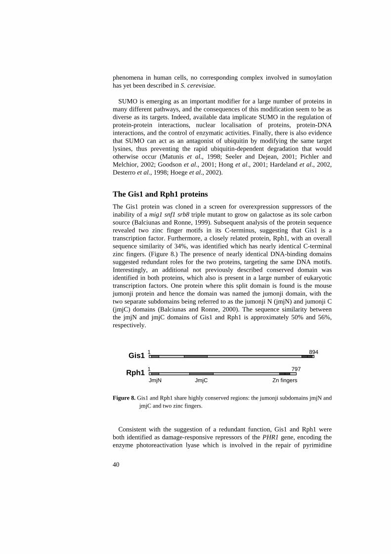
phenomena in human cells, no corresponding complex involved in sumoylation has yet been described in S. cerevisiae.
SUMO is emerging as an important modifier for a large number of proteins in many different pathways, and the consequences of this modification seem to be as diverse as its targets. Indeed, available data implicate SUMO in the regulation of protein-protein interactions, nuclear localisation of proteins, protein-DNA interactions, and the control of enzymatic activities. Finally, there is also evidence that SUMO can act as an antagonist of ubiquitin by modifying the same target lysines, thus preventing the rapid ubiquitin-dependent degradation that would otherwise occur (Matunis et al., 1998; Seeler and Dejean, 2001; Pichler and Melchior, 2002; Goodson et al., 2001; Hong et al., 2001; Hardeland et al., 2002, Desterro et al., 1998; Hoege et al., 2002).
The Gis1 and Rph1 proteins The Gis1 protein was cloned in a screen for overexpression suppressors of the inability of a mig1 snf1 srb8 triple mutant to grow on galactose as its sole carbon source (Balciunas and Ronne, 1999). Subsequent analysis of the protein sequence revealed two zinc finger motifs in its C-terminus, suggesting that Gis1 is a transcription factor. Furthermore, a closely related protein, Rph1, with an overall sequence similarity of 34%, was identified which has nearly identical C-terminal zinc fingers. (Figure 8.) The presence of nearly identical DNA-binding domains suggested redundant roles for the two proteins, targeting the same DNA motifs. Interestingly, an additional not previously described conserved domain was identified in both proteins, which also is present in a large number of eukaryotic transcription factors. One protein where this split domain is found is the mouse jumonji protein and hence the domain was named the jumonji domain, with the two separate subdomains being referred to as the jumonji N (jmjN) and jumonji C (jmjC) domains (Balciunas and Ronne, 2000). The sequence similarity between the jmjN and jmjC domains of Gis1 and Rph1 is approximately 50% and 56%, respectively.
JmjN
Gis1 1 894
Rph1 797
JmjC
1
Zn fingers
Figure 8. Gis1 and Rph1 share highly conserved regions: the jumonji subdomains jmjN and
jmjC and two zinc fingers.
Consistent with the suggestion of a redundant function, Gis1 and Rph1 were both identified as damage-responsive repressors of the PHR1 gene, encoding the enzyme photoreactivation lyase which is involved in the repair of pyrimidine
40
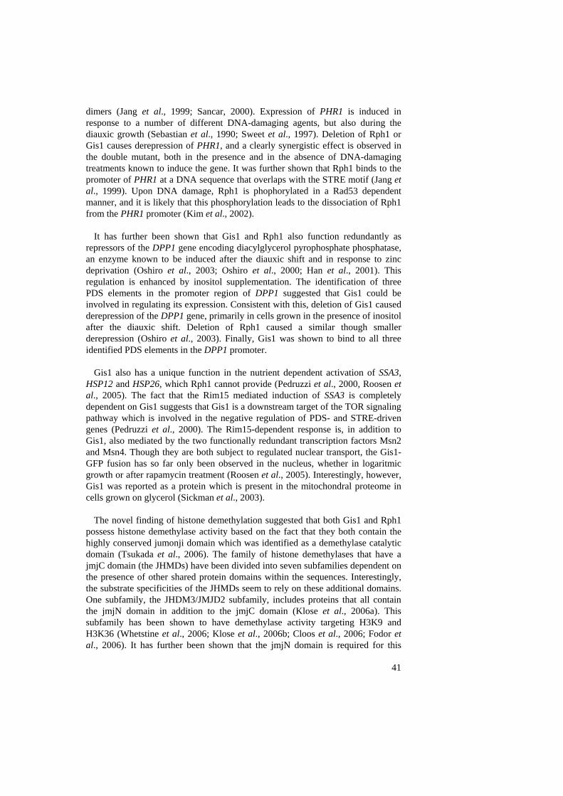
41
dimers (Jang et al., 1999; Sancar, 2000). Expression of PHR1 is induced in response to a number of different DNA-damaging agents, but also during the diauxic growth (Sebastian et al., 1990; Sweet et al., 1997). Deletion of Rph1 or Gis1 causes derepression of PHR1, and a clearly synergistic effect is observed in the double mutant, both in the presence and in the absence of DNA-damaging treatments known to induce the gene. It was further shown that Rph1 binds to the promoter of PHR1 at a DNA sequence that overlaps with the STRE motif (Jang et al., 1999). Upon DNA damage, Rph1 is phophorylated in a Rad53 dependent manner, and it is likely that this phosphorylation leads to the dissociation of Rph1 from the PHR1 promoter (Kim et al., 2002).
It has further been shown that Gis1 and Rph1 also function redundantly as repressors of the DPP1 gene encoding diacylglycerol pyrophosphate phosphatase, an enzyme known to be induced after the diauxic shift and in response to zinc deprivation (Oshiro et al., 2003; Oshiro et al., 2000; Han et al., 2001). This regulation is enhanced by inositol supplementation. The identification of three PDS elements in the promoter region of DPP1 suggested that Gis1 could be involved in regulating its expression. Consistent with this, deletion of Gis1 caused derepression of the DPP1 gene, primarily in cells grown in the presence of inositol after the diauxic shift. Deletion of Rph1 caused a similar though smaller derepression (Oshiro et al., 2003). Finally, Gis1 was shown to bind to all three identified PDS elements in the DPP1 promoter.
Gis1 also has a unique function in the nutrient dependent activation of SSA3, HSP12 and HSP26, which Rph1 cannot provide (Pedruzzi et al., 2000, Roosen et al., 2005). The fact that the Rim15 mediated induction of SSA3 is completely dependent on Gis1 suggests that Gis1 is a downstream target of the TOR signaling pathway which is involved in the negative regulation of PDS- and STRE-driven genes (Pedruzzi et al., 2000). The Rim15-dependent response is, in addition to Gis1, also mediated by the two functionally redundant transcription factors Msn2 and Msn4. Though they are both subject to regulated nuclear transport, the Gis1-GFP fusion has so far only been observed in the nucleus, whether in logaritmic growth or after rapamycin treatment (Roosen et al., 2005). Interestingly, however, Gis1 was reported as a protein which is present in the mitochondral proteome in cells grown on glycerol (Sickman et al., 2003).
The novel finding of histone demethylation suggested that both Gis1 and Rph1 possess histone demethylase activity based on the fact that they both contain the highly conserved jumonji domain which was identified as a demethylase catalytic domain (Tsukada et al., 2006). The family of histone demethylases that have a jmjC domain (the JHMDs) have been divided into seven subfamilies dependent on the presence of other shared protein domains within the sequences. Interestingly, the substrate specificities of the JHMDs seem to rely on these additional domains. One subfamily, the JHDM3/JMJD2 subfamily, includes proteins that all contain the jmjN domain in addition to the jmjC domain (Klose et al., 2006a). This subfamily has been shown to have demethylase activity targeting H3K9 and H3K36 (Whetstine et al., 2006; Klose et al., 2006b; Cloos et al., 2006; Fodor et al., 2006). It has further been shown that the jmjN domain is required for this
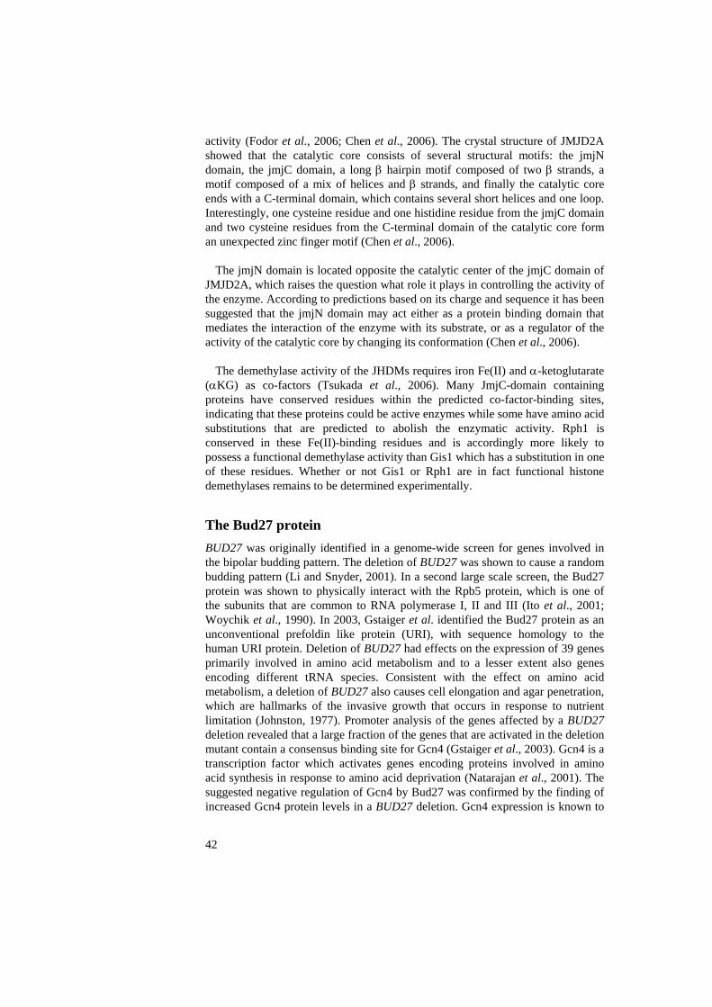
42
activity (Fodor et al., 2006; Chen et al., 2006). The crystal structure of JMJD2A showed that the catalytic core consists of several structural motifs: the jmjN domain, the jmjC domain, a long β hairpin motif composed of two β strands, a motif composed of a mix of helices and β strands, and finally the catalytic core ends with a C-terminal domain, which contains several short helices and one loop. Interestingly, one cysteine residue and one histidine residue from the jmjC domain and two cysteine residues from the C-terminal domain of the catalytic core form an unexpected zinc finger motif (Chen et al., 2006).
The jmjN domain is located opposite the catalytic center of the jmjC domain of JMJD2A, which raises the question what role it plays in controlling the activity of the enzyme. According to predictions based on its charge and sequence it has been suggested that the jmjN domain may act either as a protein binding domain that mediates the interaction of the enzyme with its substrate, or as a regulator of the activity of the catalytic core by changing its conformation (Chen et al., 2006).
The demethylase activity of the JHDMs requires iron Fe(II) and α-ketoglutarate (αKG) as co-factors (Tsukada et al., 2006). Many JmjC-domain containing proteins have conserved residues within the predicted co-factor-binding sites, indicating that these proteins could be active enzymes while some have amino acid substitutions that are predicted to abolish the enzymatic activity. Rph1 is conserved in these Fe(II)-binding residues and is accordingly more likely to possess a functional demethylase activity than Gis1 which has a substitution in one of these residues. Whether or not Gis1 or Rph1 are in fact functional histone demethylases remains to be determined experimentally.
The Bud27 protein BUD27 was originally identified in a genome-wide screen for genes involved in the bipolar budding pattern. The deletion of BUD27 was shown to cause a random budding pattern (Li and Snyder, 2001). In a second large scale screen, the Bud27 protein was shown to physically interact with the Rpb5 protein, which is one of the subunits that are common to RNA polymerase I, II and III (Ito et al., 2001; Woychik et al., 1990). In 2003, Gstaiger et al. identified the Bud27 protein as an unconventional prefoldin like protein (URI), with sequence homology to the human URI protein. Deletion of BUD27 had effects on the expression of 39 genes primarily involved in amino acid metabolism and to a lesser extent also genes encoding different tRNA species. Consistent with the effect on amino acid metabolism, a deletion of BUD27 also causes cell elongation and agar penetration, which are hallmarks of the invasive growth that occurs in response to nutrient limitation (Johnston, 1977). Promoter analysis of the genes affected by a BUD27 deletion revealed that a large fraction of the genes that are activated in the deletion mutant contain a consensus binding site for Gcn4 (Gstaiger et al., 2003). Gcn4 is a transcription factor which activates genes encoding proteins involved in amino acid synthesis in response to amino acid deprivation (Natarajan et al., 2001). The suggested negative regulation of Gcn4 by Bud27 was confirmed by the finding of increased Gcn4 protein levels in a BUD27 deletion. Gcn4 expression is known to

43
be controlled by the TOR pathway, hence the Bud27 activity is likely to be TOR regulated (Valenzuela et al., 2001). Considering all known data, Gstaiger et al. suggested that part of the function of Bud27 is to contribute directly or indirectly to the expression of both RNA pol II and pol III transcripts, specifically those genes that are involved in nutrient responses regulated by the TOR pathway.
Aims of the present investigation
The general aim of this work has been to contribute to a better understanding of general and gene-specific control of gene expression in eukaryotes. The first paper in this thesis is focused on general transcription initiation, specifically on contributing to the understanding of the internal interactions between the Mediator subunits. The jumonji domain is a highly conserved domain found in many transcription factors in different eukaryotes, including the yeast zinc finger protein Gis1. The second paper aims for a better understanding of the function of this domain by studying its physical interactions. The work on Gis1 was extended by genome wide analysis of gene expression in order to elucidate its function in different growth phases. In this paper we also addressed the question to what extent the Gis1 homologue Rph1 overlaps with Gis1 in function. The final paper aims at gaining more insight into the function of the Gis1 interacting protein Bud27 and its role in TOR signaling.
Results and discussion
Genetic and physical interactions of Med21 (I)
The Med21 protein is a small essential subunit of the middle domain of the Mediator and shows, together with Med7, the highest degree of sequence conservation among all core Mediator subunits (Myers et al., 1998; Hengartner et al., 1995). It has been proposed that Med21 together with Med3 are targets of Ssn6-Tup1 binding (Pasamichos-Chronakis et al., 2000; Gromöller and Lehming, 2000a). It has further been shown that Med21 interacts with Med4 and Med8, also located in the middle domain (Balciunas et al., 2003). A temperature sensitive allele of MED21 negatively effects transcription of some but not all genes to a moderate degree, while impairing the activation of a subset of genes more severely
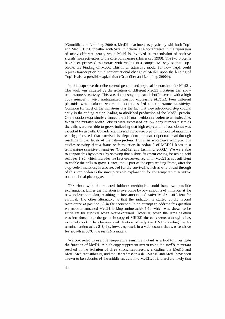
44
(Gromöller and Lehming, 2000b). Med21 also interacts physically with both Tup1 and Med6. Tup1, together with Ssn6, functions as a co-repressor in the repression of many different genes, while Med6 is involved in transmission of positive signals from activators to the core polymerase (Han et al., 1999). The two proteins have been proposed to interact with Med21 in a competitive way so that Tup1 blocks the binding of Med6. This is an attractive model for how Tup1 could repress transcription but a conformational change of Med21 upon the binding of Tup1 is also a possible explanation (Gromöller and Lehming, 2000b).
In this paper we describe several genetic and physical interactions for Med21. The work was initiated by the isolation of different Med21 mutations that show temperature sensitivity. This was done using a plasmid shuffle screen with a high copy number in vitro mutagenized plasmid expressing MED21. Four different plasmids were isolated where the mutations led to temperature sensitivity. Common for most of the mutations was the fact that they introduced stop codons early in the coding region leading to abolished production of the Med21 protein. One mutation suprisingly changed the initiator methionine codon to an isoleucine. When the mutated Med21 clones were expressed on low copy number plasmids the cells were not able to grow, indicating that high expression of our clones was essential for growth. Considering this and the severe type of the isolated mutations we hypothesised that survival is dependent on transcriptional read-through resulting in low levels of the native protein. This is in accordance with previous studies showing that a frame shift mutation in codon 3 of MED21 leads to a temperature sensitive phenotype (Gromöller and Lehming, 2000b). We were able to support this hypothesis by showing that a short fragment coding for amino acid residues 1-30, which includes the first conserved region in Med21 is not sufficient to enable the cells to grow. Hence, the 3' part of the open reading frame, after the stop codon mutation, is also needed for the survival, which is why a read-through of this stop codon is the most plausible explanation for the temperature sensitive but non-lethal phenotype.
The clone with the mutated initiator methionine could have two possible explanations. Either the mutation is overcome by low amounts of initiation at the new isoleucine codon, resulting in low amounts of native Med21 sufficient for survival. The other alternative is that the initiation is started at the second methionine at position 15 in the sequence. In an attempt to address this question we made a truncated Med21 lacking amino acids 1-14 which was shown to be sufficient for survival when over-expressed. However, when the same deletion was introduced into the genomic copy of MED21 the cells were, although alive, extremely sick. The chromosomal deletion of only the DNA encoding the N-terminal amino acids 2-8, did, however, result in a viable strain that was sensitive for growth at 38°C, the med21-ts mutant.
We proceeded to use this temperature sensitive mutant as a tool to investigate the function of Med21. A high copy suppressor screen using the med21-ts mutant resulted in the isolation of three strong suppressors, encoding the Med10 and Med7 Mediator subunits, and the HO repressor Ash1. Med10 and Med7 have been shown to be subunits of the middle module like Med21. It is therefore likely that

45
the genetic interactions in this case reflect physical interactions. This was verified by testing Med21 for 2-hybrid interactions with a β-galactosidase reporter, resulting in high levels of activity far above the background for both Med7 and Med10 preys. Both interactions were also confirmed by co-immunoprecipitations. It was further found that the amino acids 2-8 of Med21 are important for both the interaction with Med7 and with Med10. Additional 2-hybrid experiments also revealed an interaction between Med10 and Med7.
To determine what parts of Med21 that are involved in the interaction with Med10, several different constructs expressing different parts of Med21 were made. These were then tested for 2-hybrid interactions with Med10. None of the deletions tested abolished the interaction. Interestingly, these deletions included a construct where both of the two most strongly conserved regions in Med21 were deleted, showing that the Med21-Med10 interaction is not mediated by these regions.
Furthermore, in 2-hybrid experiments using Med21 both as bait and as prey, we suprisingly found a strong self-interaction. This self-interaction was also seen with the same deletion constructs that were tested against Med10. Hence, we concluded that the two strongly conserved regions of Med21 are not involved in mediating either the interaction with Med10 or with Med21 itself.
Identification of Gis1 jumonji domain interacting proteins (II) The jumonji domain was identified as a bipartite protein domain which is highly conserved and present in many eukaryotic transcription factors (Balciunas et al., 2000). This bipartite domain consists of the jmjN and the jmjC subdomains, although proteins can also be found that only contain the larger jmjC subdomain. In S. cerevisiae there are four proteins that belongs to the family of proteins that contains both subdomains: Gis1, Rph1, Ecm5 and Yjr119c (Balciunas et al., 2000).
As a first step towards elucidating the function of this domain we performed a 2-hybrid screen with a bait expressing the jumonji domain of the yeast transcription factor Gis1. The screen resulted in the identification of 19 different interactors, among them the RecQ helicase Sgs1, the silencing factors Esc1 and Sir4, the URI-type prefoldin Bud27 and the PIAS type SUMO ligase Nfi1.
We next asked the question whether both parts of the domain are needed for these interactions. The finding of protein families containing only the larger jmjC domain suggest that this domain can have a distinct function from the jmjN domain. This question was addressed by making three different constructs expressing the jmjN domain and the intervening region (amino acid residues 1-183), the intervening region and the jmjC domain (residues 58-333) and the intervening region with adjacent parts of both domains (residues 39-223), respectively as baits. These constructs were tested for 2-hybrid interaction against all proteins identified in our screen. No interactions were detected with any of the

baits. Furthermore, in a repression assay, we found that while the entire jumonji domain of Gis1 can repress transcription when recruited to the GAL1 promoter, none of the partial constructs were sufficient to perform the same repression. The facts that the partial constructs failed to interact with our 19 interactors and that only the full domain could mediate repression suggest that the function of the domain is dependent on both the jmjN and the jmjC subdomains. This is consistent with the recent finding that the jmjN domain was required for the jmjC-associated histone demethylase activity of a protein containing both subdomains (Chen et al., 2006; Fodor et al., 2006).
Zrg8
Bud27
Jip5
Pan1
Pig2
Esc1
Taf8
Jip3Csm3
Mft1 Bms1
Sgs1Gis1Gis1
Jip4
Mga2
Gds1 Sir4
Fir1
Nis1 Nfi1
Figure 9. Possible organisation of the Gis1 interacting proteins.
The large amount of identified interactors in the 2-hybrid screen posed the question whether they all interact directly with the Gis1 jumonji domain, or if some of the interactions are indirect interactions which depend on one or several of the other identified proteins. To address this question, we disrupted all non-essential genes encoding the interactors and then tested each interaction in these deletion strains. This resulted in a complex pattern of dependencies, which suggests the presence of a larger complex in which only a subset of the proteins interact directly with the jumonji domain of Gis1. Only four proteins Bud27, Sgs1, Nfi1 and Fir1 were not dependent on any other proteins for their interaction with Gis1. Consistent with the dependency studies, a quantification of all Gis1 interactions in a β-galactosidase assay showed that the four strongest interactors were Nis1, Fir1, Nfi1 and Sgs1. Furthermore, three of the identified interactors, Bud27, Taf8 and Jip5, also interacted with Gis1 in a co-immunoprecipitation
46

47
experiment. The overall conclusions from the interaction studies are summerised in Figure 9.
Of key importance in the above described interactions is the Bud27 protein, since all interactors except for Fir1, Nfi1, Nis1, and Sgs1 depend on Bud27 for their binding to Gis1. To further test the interpretation that these interaction dependencies reflect the formation of a complex, Bud27 was used as a 2-hybrid bait and tested for interaction against all other Gis1 interactors. We found that all interactors except Nfi1, Nis1 and Fir1 also can interact with Bud27, which is consistent with the fact that these three proteins seem to interact with Gis1 in a Bud27 independent manner (Figure 9).
Finally we reasoned that if the fact that the jumonji domain is conserved in
sequence in different eukaryotes also represents a conservation of function, the Gis1 jumonji domain should be able to interact with human homologues of our interactors. Interestingly, the recQ helicase Sgs1 which was identified in our screen has a human homologue, the Werner syndrome protein (WRN). To test whether the Gis1 jumonji domain of Gis1 can interact with the WRN protein we tested for a 2-hybrid interaction using the Gis1 jumonji domain as bait and the WRN protein as a prey. The positive result suggests that this interaction is indeed conserved between yeast and human.
Global expression analysis of Gis1 and Rph1 function in different growth phases (III) Gis1 and Rph1 are two closely related proteins with an overall sequence similarity of 34%. They also share three regions that are particularly well conserved, the jmjN subdomain, the jmjC subdomain and two nearly identical zinc fingers (Balciunas and Ronne, 2000). The highly conserved zinc fingers suggest that Gis1 and Rph1 bind to similar or identical DNA motifs. Indeed, previous studies have identified shared functions for these two transcription factors which are mediated through similar STRE and PDS elements (Oshiro et al., 2003; Jang et al., 1999). However, Gis1 also has a unique role as an activator in the diauxic shift response where no overlap in function of Rph1 has yet been described (Pedruzzi et al., 2000). Both Gis1 and Rph1 expression is continuously increased after diauxic shift and in the post diauxic growth phase suggesting a role for both proteins after glucose deprivation (Gasch et al., 2000).
Using microarrays, we studied the impact of rph1 single, gis1 single and rph1 gis1 double deletions in logaritmic grow phase, in cells immediately after the diauxic shift, and in cells grown for 3 days. Our data suggest a complex pattern of regulation where some genes are activated and some are repressed by either Gis1 alone, Rph1 alone, redundantly by both proteins, or synergistically by both proteins.
Genes that differed at least two-fold in expression levels between two of the strains were considered as being significantly affected. Among those genes we
48
saw an enrichment of genes with PDS or STRE elements in their 5' region. The PDS and STRE elements further appeared to have different roles in the different growth phases. Interestingly, also the orientation of the elements showed clear differences between genes that were differently regulated.
Our results further show that Gis1 and Rph1 can act as both repressors and activators, and that they have distinct roles in gene regulation after the diauxic shift. A similar pattern is seen after three days although the effect of gis1 deletion stands out from the other strains having a pronounced effect. Furthermore this pronounced effect after three days is reduced in the rph1 gis1 double deletion, one interpretation being that Gis1 negatively regulates Rph1.
Among the genes affected by our deletions were genes related to ribosome function, which were especially affected during the diauxic shift. Significant differences were, however, seen in between the regulation of cytoplasmic and mitochondrial ribosomal proteins. Another cluster of interest included cell cycle regulated genes that were negatively regulated by both Gis1 and Rph1 with a synergistic effect in the double deletion.
The roles of Bud27 and Gis1 in TOR-dependent regulation (IV) The Bud27 protein is an unconventional prefoldin-like protein (URI) which has been suggested to participate in TOR-dependent gene regulation in response to nutrients. This suggestion was based on the fact that cells deleted for BUD27 show similar changes in gene expression as are seen upon rapamycin treatment. Furthermore, it has been shown that a bud27 deletion leads to decreased protein levels of Gcn4, a protein that activates genes involved in amino acid biosynthesis (Gstaiger et al., 2003). Considering the fact that Bud27 interacts with the RNA polymerase subunit, Rpb5, it was suggested that Bud27 mediates its effect on regulation at the transcriptional level (Ito et al., 2001; Gstaiger et al., 2003). We identified Bud27 as an interactor of Gis1 (paper II). Furthermore, our data showed that most of the other identified interactors depend on Bud27 for their interaction with Gis1, suggesting a key role for the Bud27 protein among our Gis1 interactors. This prompted us to further study Bud27 and its functional link to Gis1.
As a first step towards elucidating the function of Bud27, we performed a screen for genes that when over-expressed could suppress the temperature sensitivity of a bud27 deletion mutant. We obtained 21 different suppressors, of which the strongest mainly included genes encoding proteins involved in ribogenesis and genes encoding RNA polymerase subunits. As the deletion of Bud27 also results in rapamycin sensitivity, all suppressors were also tested for their ability to suppress this phenotype. Three suppressors, showed strong suppression of the rapamycin sensitivity; MKT1, RPO26 and RBP5, while a subset of the other suppressors showed weak but distinct suppression. Furthermore, we found that the over-expression of Gis1 partly suppresses the Bud27 rapamycin sensitivity, suggesting that Gis1 may function together with or downstream of Bud27. Though
49
the genetic interactions with RNA polymerse subunits further support a model in which Bud27 is involved in the regulation of gene expression at the transcriptional level, the Bud27 protein has never been visualised in the nucleus, but instead shows a predominantly cytoplasmic localisation. This cytoplasmic localisation was not effected by rapamycin treatment. The localisation of Bud27 is a question that must be resolved before we can fully understand the function of Bud27.
To further investigate the possible connections between the cloned suppressors and TOR signaling, all suppressors were tested for their ability to suppress the rapamycin sensitive phenotypes of tor1, tco89 and rim15 deletion mutants. Three bud27 suppressors were able to suppress all three mutations; the two close homologues ZDS1 and ZDS2 encoding proteins involved in the stress and starvation induced relocalisation of Bcy1 (Griffioen et al., 2001; Griffioen et al., 2003) and MKT1, encoding an RNA-binding protein involved in the regulation of HO expression (Tadauchi et al., 2004). Their ability to suppress all three TOR pathway mutants suggest that their functions may be linked to the TOR regulated function of Bud27.
The Rim15 kinase is known to be a negatively regulated target of TOR signaling and is also known to partly mediate its function in a Gis1 dependent manner (Reinders et al., 1998; Pedruzzi et al., 2000). Therefore, we asked if Bud27, as a Gis1 interactor, is also regulated by Rim15. The deletion of RIM15 had no effect on the mRNA levels of Bud27. However, a Western blot showed that though the Bud27 protein level in a wild type strain decreases drastically after the diauxic shift, the level remain high after the diauxic shift in the rim15 strain. This suggests that Rim15 negatively regulates Bud27 at the post-transcriptional level.
The Gcn4 protein has been shown to be negatively regulated by Bud27 (Gstaiger et al., 2003). We could show that this regulation was mediated at the transcriptional level as seen by increased mRNA levels using reverse transcriptase analysis. Also the deletion of GIS1 had a positive effect on the GCN4 level, suggesting that Bud27 and Gis1 may function together as negative regulators of GCN4.
Conclusions • The Med21 subunit of the Mediator physically and genetically interacts
with Med7 and Med10.
• Med21 shows a 2-hybrid self-interaction, which suggest a possible Med21-Med21 dimerisation within the Mediator complex.
• The jumonji domain of Gis1 physically interacts directly or indirectly
with 19 proteins, and all those interactions are dependent on both the jmjN and the jmjC subdomains.
50
• The jumonji domain of Gis1 can interact with WRN, a human homologue of one of the identified interactors (Sgs1) suggesting a conserved role for the domain from yeast to man.
• Rph1 and Gis1 regulate unique as well as overlapping target genes, many
of which have STRE or PDS elements in their promoters. The resulting gene expression is dependent on both the growth phase and the orientation of the elements.
• Several genetic interactions emphasise the possible role for Bud27 as a
downstream effector in TOR-dependent gene regulation.
• Bud27 is post-transcriptionaly down-regulated by the Rim15 protein kinase, and is involved in negative regulation of GCN4 mRNA transcripts, a function it partly shares with Gis1.
Future Perspectives We intend to further investigate the functions of the three proteins of main focus in this thesis, Gis1, Rph1 and Bud27. Below, some of the questions we intend to target in the near future are briefly discussed.
The complex interaction pattern of Gis1 and its 19 interactors remains to be further investigated in order to better understand the nature of these interactions i. e. if it is one or several subcomplexes. The most straight forward method to address this question is by protein complex-purification using TAP-tagged Gis1 and Bud27. In this context it is important to consider under what conditions we might be able to purify a complex. Our data suggest that the main role of Gis1 is after glucose depletion with a pronounced effect being seen after 3 days. The question is whether the occurance of our interactions is correlating with the function of Gis1 under these conditions.
Finding out the functions of the Gis1 jumonji domain interactions will also be a main target for our investigations. As discussed in paper II, the interaction with the SUMO ligase Siz2 (Nfi1) opens for two possible interpretations. The first interpretation is that Gis1 itself is subjected to sumoylation as step in the regulation of its activity. A second more interesting interpretation that is supported by the fact that the many of the Gis1 interactors have been shown to be sumoylated, is that their sumoylation is stimulated by their interactions with Gis1. This would suggest the existence of a "sumoylation factory" similar to the PML NBs identified in mammalian cells. Whether Gis1 is sumoylated can be investigated by Gis1 purification and subsequently using Western blots targeting the yeast SUMO protein, in wild type cells as well as in cells deleted for SIZ2. Furthermore, the sumoylation of the Gis1 interactors would also be monitored in the abscence and in the presence of Siz2 and also, to test for Gis1 dependent sumoylation, in cells deleted for GIS1.
51
Of main importance for understanding the interaction patterns and the possible existence of a complex are further investigations of the localisation of the different proteins involved. Present knowledge about the localisation of our interactors presents a scattered picture of proteins localised in both the nucleus, the cytoplasm and even double localisations to the nucleus and to the mitochondria. A challenge will be to identify a method for localising also very small amounts of proteins, and to find the appropriate conditions for undertaking these studies.
Our results in paper III on Bud27 and its genetic interactions and regulations provide a starting point for a fuller understanding of the Bud27 function. Question about the mechanism behind the Rim15-dependent down-regulation of Bud27 and the effect on GCN4 expression will be targeted using further protein studies and reverse transcriptase-PCRs. Further studies of the correlation of Bud27 and Gis1 functions will involve analysis of the Bud27 effect on Gis1-targeted genes.
In paper III the result in the samples taken after three days suggest that Gis1 might act as a repressor of Rph1 under these conditions. To investigate this possibility we will make more careful analysis of the effect of the gis1 deletion on RPH1 mRNA levels using real time PCR analysis. Furthermore, genes specifically targeted for dual regulation by Gis1 and Rph1 will be analysed in wild type cells as well as in gis1 single, rph1 single and gis1 rph1 double deletion mutants, also using real time PCR.
The broad information about the effects of Gis1 and Rph1 that was obtained in our array analysis lacks one piece of information of great interest, as it does not separate direct effects from secondary effects. To obtain information about directly targeted genes, we will use ChIP-on-CHIP analysis.
Finally, with the novel finding that the jumonji domain has demethylase activity in several mammalian proteins, the question that automatically follows is whether the jumonji domains of Rph1 or Gis1 also have demethylase activity. Purified Rph1 and Gis1 as well as constructs with mutations targeting conserved amino acid residues important for the substrate binding will be used for demethylase assays specific for different lysine residues in the histone. Furthermore, the general effects of the gis1 and rph1 deletions on the over all chromatin methylation status will be investigated.

52
References
Ahuatzi, D., Herrero, P. de la Cera, T. and Moreno, F. (2004) The glucose-regulated nuclear localization of hexokinase 2 in Saccharomyces cerevisiae is Mig1-dependent. J Biol Chem. 279:14440-14446
Allen, C., Buttner, S., Aragon, A. D., Thomas, J. A., Meirelles, O., Jaetao, J., Benn, D., Ruby, S. W., Veenhuis, M., Madeo, F. and Werner-Washburn, M. (2006) Isolation of quiescent and nonquiescent cells from yeast stationary-phase cultures. J Cell Biol. 174:89-100
Angus-Hill, M. L., Schlichter, A., Roberts, D., Erdjument-Bromage, H., Tempst, P. and Cairns, B. R. (2001) A Rsc3/Rsc30 zinc cluster dimer reveals novel roles for the chromatin remodeler RSC in gene expression and cell cycle control. Mol Cell. 7:741-751
Ashrafi, K., Sinclari, D., Gordon, J. I. and Guarente, L. (1999) Passage through stationary phase advances replicative aging in Saccharomyces cerevisiae. Proc Natl Acad Sci USA 96:9100-9105
Asturias, F. J., Jiang, Y. W., Myers, L. C., Gustafsson, C. M. and Kornberg, R. D. (1999) Conserved structures of mediator and RNA polymerase II holoenzyme. Science. 283:985-987
Balciunas, D. and Ronne, H. (1999) Yeast genes GIS1-4: multicopy suppressors of the Gal- phenotype of snf1 mig1 srb8/10/11 cells. Mol Gen Genet. 262:589-599
Balciunas, D. and Ronne, H. (2000) Evidence of domain swapping within the jumonji family of transcription factors. Trends Biochem Sci. 25:274-276
Balciunas, D., Hallberg, M., Björklund, S. and Ronne, H. (2003) Functional interactions within yeast mediator and evidence of differential subunit modifications. J Biol Chem. 278:3831-3839
Bayer, P. Arndt, A., Metzger, S., Mahajan, R., Melcior, F., Jaenicke, R. and Becker, J. (1998) Structure determination of the small ubiquitin-related modifier SUMO-1. J Mol Biol. 280:275-286
Beck, T. and Hall, M. N. (1999) The TOR signaling pathway controls nuclear localization of nutrient-regulated transcription factors. Nature. 402:689-692
Becker, P. B. (2002) Nucleosome sliding: facts and fiction. EMBO J. 21:4749-4753 Bernstein, B. E., Liu, C. L., Humphrey, E. L., Perlstein, E. O. and Schreiber, S. L. (2004)
Global nucleosome occupancy in yeast. Genome Biol. 5:R62 Bhoite, L. T., Yu, Y. and Stillman, D. J. (2001) The Swi5 activator recruits the Mediator
complex to the HO promoter without RNA polymerase II. Genes Dev. 15:2457-2469 Bitterman, K. J., Medvedik, O. and Sinclair, D. A. (2003) Longevity regulation in
Saccharomyces cerevisiae: linking metabolism, genome stability, and heterochromatin. Microbiol Mol Biol Rev. 67:376-399
Bleichenbacher, M., Tan, S. ad Richmond, T. J. (2003) Novel interactions between the components of human and yeast TFIIA/TBP/DNA complexes. J Mol Biol. 332:783-793
Boles, E., Heinisch, J. and Zimmerman, F. K. (1993) Different signal control the activation of glycolysis in the yeast Saccharomyces cerevisiae. Yeast. 9:761-770
Boorstein, W. R. and Craig, E. C. (1990) Regulation of a yeast HSP70 gene by a cAMP responsive transcriptional control. EMBO J. 9:2543-2553
Boucheri, H. (1985) Protein synthesis during transition and stationary phases under glucose limitation in Saccharomyces cerevisiae. J Baceriol. 161: 385-392
Boy-Marcotte, E., Perrot, M., Bussereau, F., Boucherie, H. and Jaquet, M. (1998) Msn2 and Msn4 control a large number of genes induced at the diauxic transition which are repressed by cyclic AMP in Saccharomyces cerevisiae. J Bacteriol. 180:1044-1052
Braunstein, M., Sobel, R. E., Allis, C. D., Turner, B. M. and Broach, J. R. (1996) Efficient transcriptional silencing in Saccharomyces cerevisiae requires a heterochromatin histone acetylation pattern. Mol Cell Biol.16:4349-4356
Broek, D., Toda, T., Michaeli, T., Levin, L., Birchmeier, C., Zoller, M., Powers, S. and Wigler, M. (1987) The S. cerevisiae CDC25 gene product regulates the RAS/adenylate cyclase pathway. Cell. 48:789-799
53
Burke, T. W. and Kadonaga, J. T. (1996) Drosophila TFIID binds to a conserved downstream basal promoter element that is present in many TATA-box-deficient promoters. Genes Dev. 10:711-724
Butow, R. A. and Avadhani, N. G. (2004) Mitochondrial signaling: the retrograde response. Mol Cell. 14:1-15
Bylebyl, G. R., Belichenko, I. and Johnson, E. S. (2003) The SUMO isopeptidase Ulp2 prevents accumulation of SUMO chains in yeast. J Biol Chem. 278:44113-44120
Cadena, D. L. and Dahmus, M. E. (1987) Messenger RNA synthesis in mammalian cells is catalyzed by the phosphorylated form of polymerase II. J Biol Chem. 262:12468-12474
Cairns, B. R., Lorch, Y., Li, Y, Zhang, M., Lacomis, L., Erdjument-Bromoge, H., Tempst, P., Du, J., Laurent, B. and Kornberg, R. D. (1996) RSC, an essential, abundant chromatin-remodeling complex. Cell. 87:1249-1260
Cairns, B. R., Schlichter, A., Erdjument-Bromage, H., Tempst, P., Kornberg, R. D. and Winston, F. (1999) Two functionally distinct forms of the RSC nucleosome-remodeling complex, containing essential AT hook, BAH, and bromodomains. Mol Cell. 4:715-723.
Cairns, B., Erdjument-Bromage, H., Tempst, P., Winston, F. and Kornberg, R. D. (1998) Two actin-related proteins are shared functional components of the chromatin remodeling complexes RSC and SWI/SNF. Mol Cell. 2:639-651
Cameroni, E., Hulo, N., Roosen, J., Winderickx, J. and De Virgilio, C. (2005) The novel PAS kinase Rim15 orchestrates G0-associated antioxidant defence mechanisms. Cell Cycle. 3:462-468
Carlson, M. (1997) Genetics of transcriptional regulation in yeast: connections to the RNA polymerase II CTD. Annu Rev Cell Dev Biol. 13:1-23
Carrol, A. S., Bishop, A. C., DeRisi, J. L., Shokat, K. M. and O'Shea, E. K. (2001) Chemical inhibition of the Pho85 cyclin-dependent kinase reveals a role in the environmental stress response. Proc Natl Acad Sci U S A. 98:12578-12583
Carrozza, M. J., Li, B., Florens, L., Suganuma, T., Swanson, S. K., Lee, K. K., Shia, W. J., Andersson, S., Yates, J., Washburn, M. P. and Workman, J. L. (2005) Histone H3 methylation by Set2 directs deacetylation of coding regions by Rpd3S to suppress spurious intragenic transcription. Cell. 123:581-592
Causton H. C., Ren, B., Koh, S. S., Harbison, C. T., Kanin, E., Jennings, E. G., Lee, T. I., True, H. L., Landers, E. S. and Young, R. A. (2001) Remodelling of yeast genome expression in response to environmental changes. Mol Cell Biol. 12:323-337
Chang, E., Yang, J. W., Nagavarapu, U. and Herron, G. S. (2002) Aging and survival of cutaneous microvasculature. J Invest Dermatol. 118:752-758
Chappell, T. G., Welch, W. J., Schlossman, D. M., Palter, K. B., Schlesinger, M. J. and Rothman, J. E. (1986) Uncoating ATPase is a member of the 70 kilodalton family of stress proteins. Cell. 45:3-13
Chen, J. L., Attardi, L. D., Verrijzer, C. P., Yokomori, K. and Tjian, R. (1994) Assembly of recombinant TFIID reveals differential coactivator requirements for distinct transcriptional activators. Cell. 79:93-105
Chen, Z., Zang, J., Whetstine, J., Hong, X., Davrazou, F., Kutateladze, T. G., Simpson, M., Mao, Q., Pan, C. H., Dai, S., Hagman, J., Hansen, K., Shi, Y. and Zhang, G. (2006) Structural insights into histone demethylation by JMJD2 family members. Cell. 125:691-702
Cheung, P., Allis, C. D. and Sassone-Corsi, P. (2000) Signaling to chromatin through histone modifications. Cell. 103:263-271
Choder, M. (1991) A general topoisomerase I-dependent transcriptional repression in the stationary phase in yeast. Genes Dev. 5:2315-2326
Clayton, A. R., White, O., Ketchum, K. A. and Venter, J. C. (1997) The first genome from the third domain of life. Nature. 387:459-462
Clissold, P. M. and Ponting, C. P. (2001) JmjC: cupin metalloenzyme-like domains in jumonji, hairless and phospholipase A2beta. Trends Biochem Sci. 26:7-9
Cloos, P. A. (2006) The putative oncogene GASC1 demethylates tri- and dimethylated lysine 9 on histone H3. Nature. 442:307-311

54
Corden, J. L., Cadena, D. L., Ahearn, L. M. Jr. and Dahmus, M. E. (1985) A unique structure at the carboxyl terminus of the largest subunit of eukaryotic RNA polymerase II. Proc Natl Acad Sci U S A. 82:7934-7938
Côte, J., Quinn, J., Workman, J. L. and Peterson, C. L. (1994) Stimulation of GAL4 deriative binding to nucleosomal DNA by the yeast SWI/SNF protein complex. Science. 265:53-60
Cramer, P., Bushnell, D. A. and Kornberg, R. D. (2001) Structural basis of transcription: RNA polymerase II at 2.8 angstrom resolution. Science. 292:1863-1876
Cramer, P., Bushnell, D. A., Fu, J., Gnatt, A. L., Mavier-Davies, B., Thompson, N. E., Burgess, R. R., Edwards, A. M., David, P. R. and Kornberg, R. D. (2000) Architecture of RNA polymerase II and implications for the transcription mechanism. Science. 288:640-649
Crauwels, M, Donaton, M. C. V., Pernambuco, M. B., Winderickx, J., de Winde, J. H. and Thevelein, J. M. (1997) The Sch9 protein kinase in the yeast Saccharomyces cerevisiae controls cAPK activity and is required for nitrogen activation of the fermentable-growth-medium-induced (FGM) pathway. Microbiology. 143:2627-2637
Crespo, J. L. and Hall, M. N. (2002) Elucidating TOR signaling and rapamycin action: Lessons from Saccharomyces cerevisiae. Microbiol Mol Biol Rev. 66:579-591
Crespo, J. L., Powers, T., Fowler, B. and Hall, M. N. (2002) The TOR controlled transcription activators GLN3, RTG1 and RTG3 are regulated in response to intracellular levels of glutamine. Proc Natl Acad Sci USA. 99:6784-6789
Damak, F., Boy-Marcotte, E., Le-Roscouet, D., Guilbaud, R. and Jacquet, M. (1991) SDC25, a CDC25-like gene which contains a RAS-activating domain and is a dispensable gene of Saccharomyces cerevisiae. Mol Cell Biol. 11:202-212
Daniel, J. A., Torok, M. S., Sun, Z. W., Scieltz, D., Allis, C. D., Yates, J. R. and Grant, P. A. (2004) Deubiquitination of histone H2B by a yeast acetyltransferase complex regulates transcription. J Biol Chem. 279:1867-1871
De Vit, M. J., Waddle, J. A. and Johnston, M. (1997) Regulated nuclear translocation of the Mig1 glucose repressor. Mol Biol Cell. 8:1603-1618
DeNobel, H., Ruiz, C., Martin, H., Morris, W., Brul, S., Molina, M. and Klis, F. M. (2000) Cell wall perturbations in yeast result in dual phosphorylation of the Slt2/Mpk1 MAP kinases and the Slt2-mediated increase in FKS-lacZ expression, glucanase resistance and thermotolerance. Microbiology 146:2121-2123
DeNobel, J. M., Klis, F. M., Munnik, T., Priem, J., and van den Ende, H. (1990) An assay of relative cell wall porosity in Saccharomyces cerevisiae, Klyveromyces lactis and Schizo-saccharomyces pombe. Yeast 6: 483-490(b)
DeNobel, J.G., Klis, F. M., Priem, J., Munnik, T. and van den Ende, H. (1990) The glucanase-soluble mannoproteins limit cell wall porosity in Saccharomyces cerevisiae. Yeast 6:491-499(a)
DeRisi, J. L., Iyer, V. R. and Brown, P. O. (1997) Exploring the metabolic and genetic control of gene expression on a genomic scale. Science. 278:680-686
Deshaies, R. J., Koch, B. D., Werner-Washburn, M., Craig, E. A. and Schekman, R. (1988) A subfamily of stress proteins facilitates translocation of secretory and mitochondrial precursor polypeptides. Nature. 332:800-805
Desterro, J. M., Rodrigues, M. S. and Hay, R. T. (1998) SUMO-1 modification of IκBα inhibits NF-κB activation. Mol Cell. 2:233-239
Dikstein, R., Ruppert, S. and Tjian, R. (1996) TAFII250 is a bipartite protein kinase that phosphorylates the base transcription factor RAP74. Cell. 84:781-790
Dotson, M. R., Yuan, C. X., Roeder, R. G., Myers, L. C., Gustafsson, C. M., Jiang, Y. W., Li, Y., Kornberg, R. D. and Asturias, F. J. (2000) Structural organisation of yeast and mammalian mediator complexes. Proc Natl Acad Sci U S A. 97:14307-14310
Drebot, M. A., Johnston, G. C. and Singer, R. A. (1987) A yeast mutant conditionally defective for reentry into the mitotic cell cycle from stationary phase. Proc Natl Acad Sci USA, 84:7948-7952
Du, J., Nasir, I., Benton, B. K., Kladde, M. P. and Laurent, B. C. (1998) Sth1p, a Saccharomyces cerevisiae Snf2p/Swi2p homolog, is an essential ATPase in RSC and
55
differs from Snf/Swi in its interactions with histones and chromatin-associated proteins. Genetics. 150:987-1005
Düvel, K., Santhanam, A., Garrett, S., Schneper, L. and Broach, J R. (2003) Multiple roles of Tap42 in mediating rapamycin-induced transcriptional changes in yeast. Mol Cell. 11:1467-1478
Dvir, A., Conaway, J. and Conaway, R. C. (2001) Mechanism of transcription initiation and promoter escape by RNA polymerase II. Curr Opin Genet Dev. 11:209-214
Edwards, A. M., Kane, C. M., Young, R. A. and Kornberg, R. D. (1991) Two dissociable subunits of yeast RNA polymerase II stimulate the initiation of transcription at a promoter in vitro. J Biol Chem. 266:71-75
Emre, N. C., Ingvardottir, K., Wyce, A., Wood, A., Krogan, N. J., Li, K., Marmorstein, R., Greenblatt, J. F., Shilatifard, A. and Berger, S. L. (2005) Maintenance of low histone ubiquitylation by Ubp10 correlates with telomere-proximal Sir2 association and gene silencing. Mol Cell. 17:585-594
Ephrussi, B., Slonimsky, P. P., Yotsuyanagi, Y. and Tavlitzki, J. (1956) Variations physiologiques et cytologiques de la levure au cours du cycle de la croissance aèrobe. C R Lab Carlsberg Sér Physiol. 26:87-102
Estruch, F. (2000) Stress-controlled transcription factors, stress-induced genes and stress tolerance in budding yeast. FEMS Microbiol Rev. 24:469-486
Farrel, S., Simkovich, N., Wu, Y., Barberis, A. and Ptashne, M. (1996) Gene activation by recruitment of the RNA polymerase II holoenzyme. Genes Dev. 10:2359-2367
Flanagan, P. M., Kelleher, R. J. 3rd, Sayre, M. H., Tschochner, H., and Kornberg, R. D. (1991) A Mediator required for activation of RNA polymerase II transcription in vitro. Nature. 350:436-438
Fodor, B. D. (2006) Jmjd2b antagonizes H3K9 trimethylation at pericentric heterochromatin in mammalian cells. Genes Dev. 20:1557-1562
Gasch, A. P., Spellman, P. T., Kao, C. M., Carmel-Harel, O., Eisen, M. B., Storz, G., Botstein, D. and Brown, P. O. (2000) Genomic expression programs in the response of yeast to environmental changes. Mol Biol Cell. 11:4241-4257
Geiger, J. H., Hahn, S., Lee, S. and Siegler, P. B. (1996) Crystal structure of the yeast TFIIA/TBP/DNA complex. Science. 272:830-836
Gill, G. (2003) Post-translational modification by the small ubiquitin-related modifier SUMO has big effects on transcription factor activity. Curr Opin Genet Dev. 13:108-113
Girdwood, D. W. H., Tatham, M. H. and Hay, R. T. (2004) SUMO and transcriptional regulation. Semin Cell Dev Biol. 15:201-210
Gnatt, A. L., Cramer, P., Fu, J., Bushnell, D. A. and Kornberg, R. D. (2001) Structural basis of transcription: an RNA polymerase II elongation complex at 3.3A resolution. Science. 292:1876-1882
Goodrich, J. A. and Tjian, R. (1994) Transcription factors IIE and IIH and ATP hydrolysis direct promoter clearance by RNA polymerase II. Cell. 77:145-156
Goodson, M. L., Hong, Y., Rogers, R., Matunis, M. J., Park-Sarge, O. K. and Sarge, K. D. (2001) SUMO-1 modifications regulates the DNA binding activity of heat shock transcription factor 2, a promyelocytic leukemia nuclear body associated transcription factor. J Biol Chem. 276:18513-18518
Görner, W., Durchschlag, E., Martinez-Pator, M. T., Estruch, F., Ammerer, G., Hamilton, B., Ruis, H. and Shüller, C. (1998) Nuclear localization of the zinc finger protein Msn2p is regulated by stress and protein kinase A activity. Genes Dev. 12:586-597
Granot, D. and Snyder, M. (1991) Glucose induces cAMP-independent growth-related changes in stationary phase cells of Saccharomyces cerevisiae. Proc Natl Acad Sci USA, 88:5724-5728
Grey, J. V., Petsko, G. A., Johnston, G. C., Ringe, D., Singer, R. A. and Werner-Washburn, M. (2004) "Sleeping beauty": Quiescence in Saccharomyces cerevisiae. Microbiol. Mol Biol Rev 15:5295-5305
Griffioen, G., Anghileri, P., Imre, E., Baroni, M. D. and Ruis, H. (2000) Nutritional control of nucleocytoplasmic localization of cAMP-dependent protein kinase catalytic and regulatory subunits in Saccharomyces cerevisiae. J Biol Chem. 275:1449-1456
56
Griffioen, G., Branduardi, P., Ballarini, A., Anghileri, P., Norbeck, J., Baroni, M. D., Ruis, H. (2001) Nucleocytoplasmic distribution of budding yeast protein kinase A regulatory subunit Bcy1 requires Zds1 and is regulated by Yak1-dependent phosphorylation of its targeting domain. Mol Cell Biol. 21:511-523
Griffioen, G., Swinnen, S., Thevelein, J. M. (2003) Feedback inhibition on cell wall integrity signaling by Zds1 involves Gsk3 phosphorylation of a cAMP-dependent protein kinase regulatory subunit. J Biol Chem. 278:23460-23471
Gromöller, A. and Lehming, N. (2000a) Srb7p is a physical and physiological target of Tup1p.EMBO J. 19:6845-6852
Gromöller, A. and Lehming, N. (2000b) Srb7p is essential for the activation of a subset of genes. FEBS Lett. 484:48-54.
Gstaiger, M., Luke, B., Hess, D., Oakeley, E. J., Wirbelauer, C., Blondel, M., Vigneron, M., Peter, M. and Krek, W. (2003) Control of nutrient-sensitive transcription programs by the unconventional prefoldin URI. Science. 302:1208-1212
Hampsey, M. (1998) Molecular genetics of the RNA polymerase II general transcriptional machinery. Microbiol Mol Biol Rev. 62:465-503
Han, G. S., Johnston, C. N., Chen, X., Athenstaedt, K. and Carman, G. M. (2001) Regulation of the Saccharomyces cerevisiae DPP1-encoded diacylglycerol pyrophosphate phosphatase by zinc. J Biol Chem. 276:10126-10133
Han, S. J., Lee, Y. C., Gim, B. S., Ryu, G. H., Park, S. J., Lane, W. S. and Kim, Y. J. (1999) Activator-specific requirement of yeast mediator proteins for RNA polymerase II transcriptional activation. Mol Cell Biol. 19:979-988
Hannich, J. T., Lewis, A., Kroetz, M. B., Li, S. J., Heide, H., Emili, A., and Hochstrasser, M. (2005) Defining the SUMO-modified proteome by multiple approaches in Saccharomyces cerevisiae. J Biol Chem. 250:4102-4110
Hardeland, U., Steinhacher, R., Jiricny, J. and Schar, P. (2002) Modification of the human thymine-DNA glycosylase by ubiquitin-like proteins facilitates enzymatic turnover. EMBO J. 21:1456-1464
Hasan, R., Leroy, C., Isnard, A-D, Labarre, J., Boy-Marcotte, E. and Toledano, M. B. (2002) The control of the yeast H2O2 response by the Msn2/4 transcription factors. Mol Microbiol. 45:233-241
Hay, R. T. (2005) SUMO: A history of modification. Mol Cell. 18:1-12 Heitman, J., Movva, N. R. and Hall, M. N. (1992) Targets for cell cycle arrest by the
immunosuppressant rapamycin in yeast. Science. 233:113-121 Hengartner, C. J., Myer, V. E., Liao, S. M., Wilson, C. J., Koh, S. S. and Young, R. A.
(1998) Temporal regulation of RNA polymerase II by SRB10 and Kin28 cyclin-dependent kinases. Mol Cell. 2:48-53
Hengartner, C. J., Thompson, C. M., Zhang, J., Chao, D. M., Liao, S. M., Koleske, A. J., Okamura, S. and Young, R. A. (1995) Association of an activator with an RNA polymerase II holoenzyme. Genes Dev. 9:897-910
Henry, K. W., Wyce, A., Lo, W. S., Duggan, L. J., Emre, N. C., Kao, C. F., Pillus, L., Shilatifard, A., Osley, M. A. and Berger, S. L. (2003) Transcriptional activation via sequential histone H2B ubiquitylation and deubiquitylation, mediated by SAGA-associated Ubp8.Genes Dev. 17:2648-2663
Herman, P. K., (2002) Stationary phase in yeast. Curr Opin Microbiol. 5:602-607 Herrero, P., Galíndez, J., Ruiz, N., Martínez-Campa, C. and Moreno, F. (1995)
Transcriptional regulation of the Saccharomyces cerevisiae HXK1, HXK2 and GLK1 genes. Yeast. 11:137-144
Hirata, Y, Andoh, T., Ashara, T. and Kikuchi, A. (2003) Yeast glycogen synthase kinase-3 activates Msn2p-dependent transcription of stress responsive genes. Mol Biol Cell. 14:302-312
Hirimburegama, K., Durnez, P., Keleman, J., Oris, E., Vergauwen, R., Mergelsberg, H. and Thevelein, J. M. (1992) Nutrient-induced activation of trehalase in nutrient-starved cells of the yeast Saccharomyces cerevisiae: CAMP is not involved as second messenger. J Gen Microbiol. 138:2035-2043
Hirose, Y and Manley, J. L (2000) RNA polymerase II and the integration of nuclear events. Genes Dev. 14:1415-1429
57
Hoege,C., Pfander, B., Moldovan, G. L., Pyrowolakis, G. and Jentsch, S. (2002) Rad6 dependent DNA-repair is linked to modification of PCNA by ubiquitin and SUMO. Nature. 419:135-141
Hong, Y., Rogers, R., Matunis, M. J., Mayhew, C. N., Goodson, M. L. Park-Sarge, O. K. and Sarge, K. D. (2001) Regulation of heat shock transcription factor 1 by stress-induced SUMO-1 modification. J Biol Chem. 276:40263-40267
Hurst, H. C. (1995) Transcription factors 1: bZIP proteins. Protein profile. 2:101-168 Ingolia, T. D, Slater, M. R. and Craig, E. A. (1982) Saccharomyces cerevisiae contains a
complex multigene family related to the major heat shock-inducible gene of Drosophila. Mol Cell Biol. 2:1388-1398
Ito, T., Chiba, T., Ozawa, R., Yoshida, M. and Sakaki, Y. (2001) A comprehensive two-hybrid analysis to explore the yeast protein interactome. Proc Natl Acad Sci U S A. 98:4569-4574
Jacinto, E., Guo, B., Arndt, K. T., Schmelzle, T. and Hall, M. N. (2001) TIP41 interacts with TAP42 and negatively regulates the TOR signaling pathway. Mol Cell. 8:1017-1026
Jang, Y. K., Wang, L. and Sancar, G. B. (1999) RPH1 and GIS1 are damage-responsive repressors of PHR1. Mol Cell Biol. 19:7630-7638
Jaquet, M., Renault, G., Lallet, S., Mey, J. D. and Goldbeter, A. (2003) Oscillatory nucleocytoplasmic shuttling of the general stress response transcriptional activators Msn2 and Msn4 in Saccharomyces cerevisiae. J Cell Biol. 161:497-505
Jaskelioff, M., Gavin, I. M., Peterson, C. L. and Logie, C. (2000) SWI/SNF-mediated nucleosome remodeling: role of histone octamer mobility in the persistence of the remodeling state. Mol Cell Biol. 20:3058-3068
Jenuwein, T. and Allis, C. D. (2001) Translating the histone code. Science. 293:1074-1080 Jiang, Y. and Broach, J R. (1999) Tor proteins and protein phosphatase 2A reciprocally
regulate Tap42 in controlling cell growth. EMBO J. 18:2782-2792 Jiang, Y. W. and Stillman, D. J. (1992) Involvement of the SIN4 global transcriptional
regulator in the chromatin structure of Saccharomyces cerevisiae. Mol Cell Biol. 12:4503-4514
Johnson, E. S. (2004) Protein modification by SUMO. Annu Rev Biochem. 73:355-382 Johnson, E. S. and Blobel, G. (1997) Ubc9 is the conjugating enzyme for the ubiquitin-like
protein Smt3p. J Biol Chem. 272:26799-26802 Johnson, E. S. and Gupta, A. A. (2001) An E3-like factor that promotes SUMO conjugation
to the yeast septins. Cell. 106:735-744 Johnson, E. S., Schwienhorst, I., Dohmne, R. J. and Blobel, G. (1997) The ubiqutin-like
protein Smt3 is activated for conjugation to other proteins by an AOS1p/Uba2p heterodimer. EMBO J. 16:5509-5519
Johnston, G. C. (1977) Cell size and budding during starvation of the yeast Saccharomyces cerevisiae. J Bacteriol. 132:738-739
Kaffman, A., Herskowitz, I., Tjian, R. and O'Shea, E. K. (1994) Phosphorylation of the transcription factor PHO4 by a cyclin-CDK complex, PHO80-PHO85. Science. 263:1153-1156
Kaffman, A., Rank, N.M. and O'Shea, E. K. (1998) Phosphorylation regulates association of the transcription factor Pho4 with its import receptor Pse1/Kap121. Genes Dev. 1:2673-2683
Kang, J. J., Auble, D. T., Ranish, J. A. and Hahn, S. (1995) Analysis of the yeast transcription factor TFIIA: distinct functional regions and a polymerase II-specific role in basal and activated transcription. Mol Cell Biol. 15:1234-1243
Kang, J. S., Kim, S. H., Hwang, M. S., Han, S. J., Lee, Y. C. and Kim, Y. J. (2001) The structural and functional organization of the yeast mediator complex. J Biol Chem. 276:42003-42010
Kao, C. F., Hillyer, C, Tsukada, T., Henry, K., Berger, S. and Osley, M. A. (2004) Rad6 plays a role in transcriptional activation through ubiquitylation of histone H2B. Genes Dev. 18:184-195
Kassabov, S. R., Henry, N. M., Zofall, M., Tsukiyama, T. and Bartholomew, B. (2002) High-resolution mapping of changes in histone-DNA contacts of nucleosomes remodeled by ISW2. Mol Cell Biol. 22:7524-7534
58
Keaveney, M. and Struhl, K. (1998) Activator-mediated recruitment of the RNA polymerase II machinery is the predominant mechanism for transcriptional activation in yeast. Mol Cell. 1:917-924
Keith, A. D., Pollard, E. C. and Snipes, W. (1977) Inositol-less death in yeast results in a simultaneous increase in intracellular viscosity. Biophys J. 17:205-212
Keleher, C. A., Redd, M. J., Schultz, J., Carlson, M. and Johnson, A. D. (1992) Ssn6-Tup1 is a general repressor of transcription in yeast. Cell. 68:709-719
Kelleher, R. J. 3rd., Flanagan, P. M. and Kornberg, R. D. (1990) A novel Mediator between activator proteins and the RNA polymerase II transcription apparatus. Cell. 61:1209-1215
Kim, E. M., Jang, Y. K. and Park, S. D. (2002) Phosphorylation of Rph1, a damage-responsive repressor of PHR1 in Saccharomyces cerevisiae, is dependent upon Rad53 kinase. Nucleic Acid Res. 30:643-648
Kim, J. H., Polish, J. and Johnston, M. (2003) Specificity and regulation of DNA binding by the yeast glucose transporter gene repressor Rtg1. Mol Cell Biol. 23:5208-5216
Kim, Y. J., Björklund, S., Li, Y., Sayre, M. H. and Kornberg, R. D. (1994) A multiprotein mediator of transcriptional activation and its interaction with the C-terminal repeat domain of RNA polymerase II. Cell. 77:599-608
Klose, R. J., Kallin, A. M. and Zhang, Y. (2006) JmjC-domain-containing proteins and histone demethylation. Nat Rev Genet. 7:715-727(a)
Klose, R., Yamane, K., Bae, Y., Zhang, D., Erdjument-Bromage, H., Tempst, P., Wong, J. and Zhang, Y. (2006) The transcriptional repressor JHDM3A demethylases trimethyl histone H3 lysine 9 and 36. Nature. 442:312-316(b)
Kobor, M. S., Archambault, J., Lester, W, Holstege, F. C., Gileadi, O., Jansma, D. B., Jennings, E. G., Kouyoumdjian, F., Davidson, A. R., Young, R. A. and Greenblatt, J. (1999) An unusual eukaryotic protein phosphatase required for transcription by RNA polymerase II and CTD dephosphorylation in S. cerevisiae. Mol Cell. 5:55-62
Komeili, A., Wedaman, K. P., O'Shea, E. K. and Powers, T. (2000) Mechanism of metabolic control. Target of rapamycin signaling links nitrogen quality to the activity of the Rtg1 and Rtg3 transcription factors. J Cell Biol. 151:863-878
Kraakman, L., Lemaire, K., Ma, P., Teunissen, A. W., Donaton, M. C., Van Dijk, P., Winderickx, J., de Winde, J. H. and Thevelain, J. M. (1999) A Saccharomyces cerevisiae G-protein coupled receptor, Gpr1, is specially required for glucose activation of the cAMP pathway during the transition to growth on glucose. Mol Microbiol. 32:1002-1012
Kraemer, S. M., Ranallo, R. T., Ogg, R. C. and Stargell, L. A. (2001) TFIIA interacts with TFIID via association with TATA-binding protein and TAF40. Mol Cell Biol. 21:1737-1746
Kurdistani, S. K. and Grunstein, M. (2003) Histone acetylation and deacetylation in yeast. Nat Rev Mol Cell Biol. 4:276-284
Kuroda, Y., Suzuki, N. and Kataoka, T. (1993) The effect of posttranslational modifications on the interactions of Ras2 with adenylyl cyclase. Science. 259:683-686
Kwon, H., Imbalzano, A. N., Khavari, P. A., Kingston, R. E. and Green, M. R. (1994) Nucleosome disruption and enhancement of activation of activator binding by a human SWI/SNF complex. Nature. 370:477-481
Laurent, B. C., Yang, X. and Carlson, M. (1992) An essential Saccharomyces cerevisiae gene homologous to SNF2 encodes a helicase-related protein in a new family. Mol Cell Biol. 12:1893-1902
Lee, C. K., Shibata, Y., Rao, B., Strahl, B. D. and Lieb, J. D. (2004) Evidence for nucleosome depletion at active regulatory regions genome-wide. Nature Genet. 36:900-905
Lee, Y. C. and Kim, Y. J. (1998) Requirement for a functional interaction between mediator components Med6 and Srb4 in RNA polymerase II transcription. Mol Cell Biol. 18:5364-5370
Lewis, D. L. and Gattie, D. K. (1980) The ecology of quiescent microbes. ASM News. 57:27-32
Li, N. and Snyder, M. (2001) A genomic study of the bipolar bud site selection pattern in Saccharomyces cerevisiae. Mol Biol Cell. 12:2147-2170
59
Li, S. J. and Hochstrasser, M. (1999) A new protease required for cell-cycle progression in yeast. Nature. 398:246-251
Li, S. J. and Hochstrasser, M. (2000) The yeast ULP2 (SMT4) gene encodes a novel protease specific for the ubiquitin-like Smt3 protein. Mol Cell Biol. 20:2367-2377
Lia, G., Praly, E., Ferreira, H., Stockdale, C., Tse-Dinh, Y. C., Dunlap, D., Croquette, V., Bensimon, D. and Owen-Hughes, T. (2006) Direct observation of DNA distortion by the RSC complex. Mol Cell. 21:417-425
Lillie, S. H. and Pringle, J. R. (1980) Reserve carbohydrate metabolism in Saccharomyces cerevisiae: responses to nutrient limitation. J Bacteriol. 143:1384-1394
Litt, M. D., Simpson, M., Gaszner, M., Allis, C. D. and Felsenfeld, G. (2001) Correlation between histone lysine methylation and developmental changes at the chicken beta-globin locus. Science. 293:2453-2455
Loewith, R., Jacinto, E., Wullschleger, S., Lorberg, A., Crespo, J. L., Bonenfant, D., Oppliger, W., Jenoe, P. and Hall, M. N. (2002) Two TOR complexes, only one of which is rapamycin sensitive, have distinct roles in cell growth control. Mol Cell. 10:457-468
Longo, V. D. (1999) Mutations in signal transduction proteins increase stress resistance and longevity in yeast, nematodes, fruit flies and mammalian neuronal cells. Neurobiol Aging. 20:479-486
Lorch, Y., Beve, J., Gustafsson, C. M., Myers, L. C. and Kornberg, R. D. (2000) Mediator-Nucleosome Interaction. Mol Cell. 6:197-201
Lorch, Y., Zhang, M. and Kornberg, R. D. (1999) Histone octamer transfer by a chromatin-remodeling complex. Cell. 96:389-392
Ma, D. M., Olave, I., Merino, A. and Reinberg, D. (1996) Separation of the transcriptional coactivator and antirepression functions of transcription factor IIA. Proc Natl Acad Sci U S A. 93:6583-6588
Martin, C. and Zhang, Y. (2005) The diverse functions of histone lysine methylation. Nature Rev Mol Cell Biol. 6:838-849
Martinez-Pastor, M. T., Marchler, G., Schüller, C., Marchler-Bauer, A., Ruis, H. and Estruch, F. (1996) The Saccharomyces cerevisiae Zinc finger proteins Msn2p and Msn4p are required for transcriptional induction through the stress-response element (STRE). EMBO J. 15:2227-2235
Matile, P., Moor, H. and Robinow, C. F. (1969) Yeast cytology, p 219-302. In Rose A.H. and Harrison J. S. (ed.), The yeasts. Biology of yeasts, vol. 1. Academic Press inc., New York.
Matunis, M. J., Wu, J. and Blobek, G. (1998) SUMO-1 modification and its role in targeting the Ran GTPase-activating protein, RanGAP1, to the nuclear pore complex. J Cell Biol. 140:499-509
Maxon, M. E., Goodrich, J. A. and Tjian, R. (1994) Transcription factor IIE binds preferentially to RNA polymerase IIa and recruits TFIIH: a model for promoter clearance. Genes Dev. 8:515-524
Mellor, J. and Morillon, A. (2004) ISWI complexes in Saccharomyces cerevisiae. Biochim Biophys Acta. 1677:100-112
Meluh, P. B. and Koshland, D. (1995) Evidence that the MIF2 gene of Saccharomyces cerevisiae encodes a centromeric protein CENP-C. Mol Biol Cell. 6:793-807
Metzger, E., Wissman, M., Yin, N., Muller, J. M., Schneider, R., Peters, A. H., Gunther, T., Buettner, R. and Schule, R. (2005) LSD1 demethylates repressive histone marks to promote androgen-receptor-dependent transcription. Nature.437:436-439
Millar, C. B. and Grunstein, M. (2006) Genome-wide patterns of histone modifications in yeast. Nat Rev Mol Cell Biol. 7:657-666
Mizzen, C. A., Yang, X. J., Kokubo, T., Brownell, J. E., Bannister, A. J., Owen-Hughes, T., Workman, J., Wang, L., Berger, S. L., Kouzarides, T., Nakatani, Y. and Allis, C. D. (1996) The TAF(II)250 subunit of TFIID has histone acetyltransferase activity. Cell. 87:1261-1270
Moqtaderi, Z., Bai, Y., Poon, D., Weil, P. A. and Struhl, K. (1996) TBP-associated factors are not generally required for transcriptional activation in yeast. Nature. 383:188-191
60
Morales, V., Giamarchi, C., Chailleux, C., Moro, F., Marsaud, V., Le Ricousse, S. and Richard-Foy, H. (2001) Chromatin structure and dynamics: Functional implications. Biochimie. 83:1029-1039
Moreno, F. and Herrero, P. (2002) The hexokinase 2-dependent glucose signal transduction pathway of Saccharomyces cerevisiae. FEMS Microbiol Rev. 26:83-90
Mosley, A. L., Lakshamanan, J., Aryal, B. K. and Özcan, S. (2003) Glucose-mediated phosphorylation converts the transcription factor Rtg1 from a repressor to an activator. J Biol Chem. 278:10322-10327
Mouillon, J. M. and Persson, B. L. (2006) New aspects on phosphate sensing and signalling in Saccharomyces cerevisiae. FEMS Yeast Rev. 6:171-176
Mues, G. I., Munn, T. Z. and Raese, J. D. (1986) A human gene family with sequence homology to Drosophila melangoaster Hsp70 heat shock gene. J Biol Chem. 261:874-877
Muller, S. and Dejean, A. (1999) Viral immediate-early proteins abrogate the modification of SUMO-1 of PML and SP100 proteins, correlating with nuclear body disruption. J Virol. 73:5137-5143
Myers, L. C., Gustafsson, C. M., Bushnell, D. A., Lui, M., Erdjument-Bromage, H., Tempst, P. and Kornberg, R. D. (1998) The Med proteins of yeast and their function through the RNA polymerase II carboxy-terminal domain. Genes Dev. 12:45-54
Natarajan, K., Meyer, M. R., Jackson, B. M., Slade, D., Roberts, C., Hinnebusch, A. G. and Marton, M. J. (2001) Transcriptional profiling shows that Gcn4p is a master regulator of gene expression during amino acid starvation in yeast. Mol Cell Biol. 21:4347-4368
Nathan, D., Ingvarsdottir, K., Sterner, D. E., Bylebyl, G. R., Dokmanovic, M., Dorsey, J. A., Whelan, K. A., Krsmanovic, M., Lane, W. S., Meluh, P. B., Johnson, E. S. and Berger, S. L. (2006) Histone sumoylation is a negative regulator in Saccharomyces cerevisiae and shows dynamic interplay with positive-acting histone modifications. Genes Dev. 20:966-976
Nathan, D., Sterner, D. E. and Berger, S. L. (2003) Histone modifications: Now summoning sumoylation. Proc Natl Acad Sci U S A. 100:13118-13120.
Nehlin, J. O. and Ronne, H. (1990) Yeast MIG1 repressor is related to the mammalian early growth response and Wilm's tumour finger proteins. EMBO J. 9:2891-2898
Nikolov, D. B., Chen, H., Halay, E. D., Usheva, A. A., Hisatake, K., Lee, D. K., Roeder, R. G. and Burley, S. K. (1995) Crystal structure of a TFIIB-TBP-TATA-element ternary complex. Nature. 377:119-128
Nishizawa, M., Katou, Y., Shirahige, K. and Toh-e, A. (2004) Yeast Pho85 kinase is required for proper gene expression during the diauxic shift. Yeast. 21:903-918
Noda, T. and Oshumi, Y. (1998) Tor, a phophatidylinositol kinase homologue, controls autophagy in yeast. J Biol Chem. 273:3963-3966
Ogawa, N., DeRisi, J. and Brown, P. O. (2000) New components of a system for phosphate accumulation and polyphosphate metabolism in Saccharomyces cerevisiae revealed by genomic expression analysis. Mol Biol Cell. 11:4309-4321
Ohkuma, Y., Hashimoto, S., Wang, C. K., Horikoshi, M and Roeder, R. G. (1995) Analysis of the role of TFIIE in basal transcription and TFIIH-mediated carboxy-terminal domain phosphorylation through structure-function studies of TFIIE-alpha. Mol Cell Biol. 15:4856-4866
O'Neill, E. M., Kaffman, A., Jolly, E. R. and O'Shea, E. K. (1996) Regulation of Pho4 nuclear localisation by the Pho80-Pho85 cyclin-CDK complex. Science. 12:209-212
Oshima, Y. (1997) The phosphatase system in Saccharomyces cerevisiae. Genes Genet Syst. 72:323-334
Oshiro, J., Han, G. S., Iwanyshyn, W. M., Conover, K. and Carman, G. M. (2003) Regulation of the DPP1-encoded diacylglycerol pyrophosphate phosphatase by transcription factor Gis1p. J Biol Chem. 278:31495-31503
Oshiro, J., Rangaswamy, S., Chen, X., Han, G-S., Quinn, J. E. and Carman, G. M. (2000) Regulation of the DPP1-encoded diacylglycerol pyrophosphate (DGPP) phophatase by inositol and growth phase. J Biol Chem. 275:40887-40896
61
Owen-Hughes, T. A., Utley, R. T., Côté, J., Peterson, C. L. and Workman, J. L. (1996) Persistent site-specific remodeling of a nucleosome array by transient action of the SWI/SNF complex. Science. 273:513-516
Panek, A. D. and Panek, A. C. (1990) Metabolism and thermotolerance function of trehalose in Saccharomyces cerevisiae: a current perspective. J Biotechnol. 14:229-238
Panse, V. G., Hardeland, U., Werner, T., Kuster, B. and Hurt, E. (2004) A proteome-wide approach identifies sumoylated substrate proteins in yeast. J Biol Chem. 279:41346-41351
Papamichos-Chronakis, M., Conlan, R. R., Gounalaki, N., Copf, T. and Tzamarias, D. (2000) Hrs1/Med3 is a Cyc8-Tup1 corepressor target in the RNA polymerase II holoenzyme. J Biol Chem. 275:8397-8403
Parvin, J D., Shykind, B. M., Meyers, R. E., Kim, J. and Sharp, P. A. (1994) Multiple sets of basal factors initiate transcription by polymerase II. J Biol Chem. 269:18414-18421
Parvin, J. D. and Sharp, P. A. (1993) DNA topology and a minimal set of basal factors for transcription by RNA polymerase II. Cell. 73:533-540
Payne, J. M., Laybourn, P. J. and Dahmus, M. E. (1989) The transition of polymerase II from initiation to elongation is associated with phosphorylation of the carboxy-terminal domain of subunit IIa. J Biol Chem. 264:19621-19629
Pedruzzi, I., Bûrckert, N., Egger, P. and De Virgilio, C. (2000) Saccharomyces cerevisiae Ras/cAMP pathway controls post-diauxic shift element-dependent transcription through the zinc finger protein Gis1. EMBO J. 19:2569-2579
Pedruzzi, I., Dubouloz, F., Cameroni, E., Wanke, V., Roosen, J., Winderickx, J. and De Virgilio, C. (2003) TOR and PKA signaling pathways converge on the protein kinase Rim15 to control entry into G0. Mol Cell. 12:1607-1613
Peng, T., Golub, T. R. and Sabatini, D. M. (2002) The immunosuppressant rapamycin mimics a starvation-like signal distinct from amino acid and glucose deprivation. Mol Cell Biol. 22:5575-5584
Persson, B. L., Lagerstedt, J. O., Pratt, J. R., Pattison-Granberg, J., Lundh, K., Shokrollahzadeh, S. and Lundh, F. (2003) Regulation of phosphate acquisition in Saccharomyces cerevisiae. Curr Genet. 43:225-244
Peterson, C. L. and Workman, J. L. (2000) Promoter targeting and chromatin remodeling by the SWI/SNF complex. Curr Opin Genet Dev. 10:187-192
Petko, L. and Lindquist, S. (1986) Hsp26 is not required for growth at high temperatures, nor for thermotolerance, spore development, or germination. Cell. 45:885-894
Pham, A. D. and Sauer, F. (2000) Ubiquitin-activating/conjugating activity of TAFII250, a mediator of activation of gene expression in Drosophila. Science. 289:2357-2360
Phelan, M. L., Schnitzler, G. R. and Kingston, R. E. (2000) Octamer transfer and creation of stably remodeled nucleosomes by human SWI/SNF and its isolated ATPases. Mol Cell Biol. 20:6380-6389
Pichler, A. and Melchior, F. (2002) Ubiquitin-related modifier SUMO1 and nucleocytoplasmic transport. Traffic. 3:381-387
Pillar, T. M. and Bradshaw, R. E.(1991) Heat shock and stationary phase induce transcription of the Saccharomyces cerevisiae isi-2 cytochrome c gene. Curr Genet. 20:185-188
Plesset, J. J., Ludwig, R., Cox, B. S. and McLaughlin, C. S. (1987) Effect of cell cycle position in on thermotolerance in Saccharomyces cerevisiae. J Bacteriol. 169:779-784
Polakis, E. S., Bartley, W. and Meek, G. A. (1964) Changes in the structure and enzyme activity of Saccharomyces cerevisiae in response to changes in the environment. Biochem. J. 90:369-374
Poon, D., Bai, Y., Campbell, A. M., Björklund, S., Kim, Y. J., Zhou, S., Kornberg, R. D. and Weil, P. A. (1995) Identification and characterization of a TFIID-like multiprotein complex from Saccharomyces cerevisiae. Proc Natl Acad Sci U S A. 92:8224-8228
Praekelt, U. M. and Meacock, P. A. (1990) HSP12, a new small heat shock gene of Saccharomyces cerevisiae: analysis of structure, regulation and function. Mol Gen Genet. 223:97-106
Proudfoot, N. (2000) Connecting transcription to messenger RNA processing. Trend Biochem Sci. 25290-25293
62
Pruyvne, D. and Bretscher, A. (2000) Polarization of cell growth in yeast. J Cell Sci. 113:571-585
Ptashne, M. and Gann, A. (1997) Transcriptional activation by recruitment. Nature. 386:569-577
Reese, J. C., Apone, L., Walker, S. S., Griffin, L. A. and Green, M. R. (1994) Yeast TAFIIS in a multisubunit complex required for activated transcription. Nature. 371:523-527
Reifenberger, E., Boles. E. and Ciriacy, M. (1997) Kinetic characterization of individual hexose transporters of Saccharomyces cerevisiae and their relation to the triggering mechanisms of glucose repression. Eur J Biochem. 245:324-333
Reinders, A., Bürckert, N., Boller, T., Wiemken, A. and De Virgilio, C. (1998) Saccharomyces cerevisiae cAMP-dependent protein kinase controls entry into stationary phase through the Rim15p protein kinase. Genes Dev. 12:2943-2955
Reinke, A., Anderson, S., McCaffery, J. M., Yates III, J., Aronova, S., Chu, S., Fairclough, S., Iverson, C., Wedaman, K. P. and Powers, T. (2004) TOR complex 1 includes a novel component, Tco89 (YPL180w), and cooperates with Ssd1 to maintain cellular integrity in Saccharomyces cerevisiae. J Biol Chem. 279:14752-14762
Robzyk, K., Recht, J. and Osley, M. A. (2000) Rad6-dependent ubiquitination of histone H2B in yeast. Science. 287:501-504
Roeder, R. G. (1996) The role of general initiation factors in transcription by RNA polymerase II. Trends Biochem Sci. 21:327-335
Rolland, F., Winderickx, J. and Thevelein, J. M. (2002) Glucose-sensing and signaling mechanisms in yeast. FEM Yeast Res. 2:183-201
Ronne, H. (1995) Glucose repression in fungi. Trends Genet. 11:12-17 Roosen, J., Engelen, K., Marchal, K., Mathys, J., Griffioen, G., Cameroni, E., Thevelein, J.
M., De Virgilio, C., De Moor, B. and Winderickx, J. (2005) PKA and Sch9 control a molecular switch important for the proper adaptation to nutrient availability. Mol Microbiol. 55:862-880
Saha, A., Wittmeyer, J. and Cairns, B. R (2002) Chromatin remodeling by RSC involves ATP-dependent DNA translocation. Genes Dev. 16:2120-2134
Sancar, G. B. (2000) Enzymatic photoreactivation: 50 years and counting. Mutant Res. 451:25-37
Santangelo, G. M. (2006) Glucose signaling in Saccharomyces cerevisiae. Microbiol Mol Biol Rev. 70:253-282
Schmidt, A., Beck, T., Koller, A., Kunz, J. and Hall, M. N. (1998) The TOR nutrient signalling pathway phosphorylates NPR1 and inhibits turnover of the tryptophan permease. EMBO J. 17:6924-6931
Schmitt, A. P. and McEntee, K. (1996) Msn2p, a zinc finger DNA-binding protein, is the transcriptional activator of the multistress response in Saccharomyces cerevisiae. Proc Natl Acad Sci U S A. 93:5777-5782
Schwienhorst, I., Johnson, E. S. and Dohmen, R. J. (2000) SUMO conjugation and deconjugation. Mol Gen Genet. 263:771-786
Sebastian, J., Kraus, B. and Sancar, G. B. (1990) Expression of the yeast PHR1 gene is induced by DNA-damaging agents. Mol Cell Biol. 10:4630-4637
Seeler and Dejean, (2001) SUMO: of branched proteins and nuclear bodies. Oncogene. 20:7243-7249
Shi, Y. J., Matson, C., Iwase, S., Baba, T. and Shi, Y. (2005) Regulation of LSD1 histone demethylase activity by its associated factors. Mol Cell. 19:857-864
Shi, Y., Lan, F., Matson, C., Mulligan, P., Whetstine, J. R., Cole, P. A., Casero, R. A. and Shi, Y. (2004) Histone demethylation mediated by the nuclear amine oxidase homolog LSD1. Cell. 119:941-953
Shiio, Y and Eisenman, R. N. (2003) Histone sumoylation is associated with transcriptional repression. Proc Natl Acad Sci U S A. 100:13225-13230
Sickman, A., Reinders, J., Wagner, Y., Joppich, C., Zahedi, R., Meyer, H. E., Schönfisch, B., Perschil, I., Chacinska, A., Guiard, B., Rehling, P., Pfanner, N. and Meisinger, C. (2003) The proteome of Saccharomyces cerevisiae mitochondria. Proc Natl Acad Sci U S A. 100:13207-13212
63
Smale, S. T., Schmidt, M. C., Berk, A. J. and Baltimore, D. (1990) Transcriptional activation by Sp1 as directed through TATA or initiator: specific requirement for mammalian transcription factor IID. Proc Natl Acad Sci U S A. 87:4509-4513
Sneddon, A. A., Cohen P. T. and Stark, M. J. (1990) Saccharomyces cerevisiae protein phosphatase 2A performs an essential cellular function and is encoded by two genes. EMBO J. 9:4339-4346
Sternberg, P. W., Stern, M. J., Clark, I. and Herskowitz, I. (1987) Activation of the yeast HO gene by release from multiple negative controls. Cell. 48:567-577
Struhl, K. (1989) Helix-turn-helix, zinc-finger, and leucine-zipper motifs for eukaryotic transcriptional regulatory proteins. Trends Biochem Sci. 14:137-140
Strunnikov, A. V., Aravind, L. and Koonin, E. V. (2001) Saccharomyces cerevisiae SMT4 encodes an evolutionary conserved protease with a role in chromosome condensation regulation. Genetics. 158:95-107
Su, S. S. and Mitchell, A. P. (1993) Identification of functionally related genes that stimulate early meiotic gene expression in yeast. Genetics. 133:67-77
Suda, T., Arai, F. and Hirao, A. (2005) Hematopoetic stem cells and their niche. Trends Immunol. 26:426-433
Sweet, D. H., Jang, Y. K. and Sancar, G. B. (1997) Role of Ume6 in transcriptional regulation of a DNA repair gene in Saccharomyces cerevisae. Mol Cell Biol. 17:6223-6335
Swinnen, E., Wanke, V., Roosen, J., Smets, B., Dubouloz, F., Pedruzzi, I., Cameroni, E., De Virgilio, C. and Winderickx, J. (2006) Rim15 and the crossroads of nutrient signalling pathways in Saccharomyces cerevisiae. Cell Div. 1:3
Takahashi Y, Toh-E, A. and Kikuchi, Y. (2001) A novel factor required for the SUMO/Smt3 conjugation of yeast septins. Gene. 275:223-231
Takahashi, Y, Toh-E, A. and Kikuchi, Y. (2003) Comparative analysis of yeast PIAS-type SUMO ligases in vivo and in vitro. J Biochem (Tokyo). 133:415-422
Tan, S., Hunziker, Y., Sargent, D. F. and Richmond, T. J. (1996) Crystal structure of a yeast TFIIA/TBP/DNA complex. Nature. 381:127-151
Tanaka, K., Lin, B. K., Wood, D. R. and Tamanoi, F. (1991) IRA2, an upstream negative regulator of RAS in yeast, is a RAS GTPase-activation protein. Proc Natl Acad Sci USA. 88:468-472
Tanaka, K., Nakafuku, M., Satoh, T., Marshall, M. S., Gibbs, J. B., Matsumoto, K., Kaziro, Y. and Toh-e, A. (1990) S. cerevisiae genes IRA1 and IRA2 encode proteins that may be functionally equivalent to mammalian ras GTPase activating protein. Cell. 60:803-807
Tanese, N., Pugh, B. F. and Tjian, R. (1991) Coactivators for a proline-rich activator purified from the multisubunit human TFIID complex. Genes Dev. 5:2212-2224
Tayler, F. R. and Parks, L. W. (1979) Triaglycerol metabolism in Saccharomyces cerevisiae. Relation to phospholipid synthesis. Biochem Biophys Acta. 575:204-214
Thevelein, J. M. and de Winde, J. H. (1999) Novel sensing mechanisms and targets for the cAMP-protein kinase A pathway in the yeast Saccharomyces cerevisiae. Mol Microbiol. 33:904-918
Thomas, M. C. and Chiang, C. M. (2006) The general transcription machinery and general cofactors. Crit Rev Biochem Mol Biol. 41:105-178
Timblin, B. K. and Bergman, L. W. (1997) Elevated expression of stress response genes resulting from deletion of the PHO85 gene. Mol Microbiol. 26:981-990
Toda, T., Cameron, S., Sass, P. and Wigler, M. (1988) SCH9, a gene of Saccharomyces cerevisiae that encodes a protein distinct from, but functionally and structurally related to, cAMP-dependent protein kinase catalytic subunits. Genes Dev. 2:517-527
Treich, I. and Carlson, M. (1997) Interaction of a Swi3 homolog with Sth1 provides evidence for a Swi/Snf-related complex with an essential function in Saccharomyces cerevisiae. Mol Cell Biol. 17:1768-1775
Treitel, M. A. and Carlson, M. (1995) Repression by SSN6-TUP1 is directed by MIG1, a repressor/activator protein. Proc Natl Acad Sci USA. 92:3132-3136
Tsai, F. S. and Sigler, P. B. (2000) Structural basis of preinitiation complex assembly on human pol II promoters. EMBO J. 19:25-36

64
Tsuchiya, E., Uno, M., Kiguchi, A., Masuoka, K., Kanemori, Y., Okabe, S. and Mikayawa, T. (1992) The Saccharomyces cerevisiae NPS1 gene, a novel CDC gene which encodes a 160 kDa nuclear protein involved in G2 phase control. EMBO J. 11:4017-4026
Tsukada, Y., Fang, J., Erdjument-Bromage, H., Warren, M. E., Borcers, C. H., Tempst, P. and Zhang, Y. (2006) Histone demethylation by a family of JmjC domain-containing proteins. Nature. 439:811-816
Tsukiyama, T. (2002) The in vivo functions of ATP-dependent chromatin-remodelling factors. Nat Rev Mol Cell Biol. 3:422-429
Tsukiyama, T., Palmer, J., Landel. C, C., Shiloach, J. and Wu, C. (1999) Characterization of the imitation switch subfamily of ATP-dependent chromatin-remodeling factors in Saccharomyces cerevisiae. Genes Dev. 13:686-697
Tyree, C. M., George, C. P., Lira-DeVito, L. M., Wampler, S. L., Dahmus, M. E., Zawel, L. and Kadonaga, J. T. (1993) Genes Dev. 7:1254-1265
Uemura, H., Koshio, M., Inoue, Y., Lopez, M. C. and Baker, H. V. (1997) The role of Gcr1p in the transcriptional activation of glycolytic genes in yeast Saccharomyces cerevisiae. Genetics. 147:521-532
Ungewickel, E. (1985) The 70-kd mammalian heat shock proteins are structurally and functionally related to the uncoating protein that releases clathrin from coated vesicles. EMBO J. 4:3385-33391
Uno, I., Mitsuzawa, H., Tanaka, K., Oshima, T. and Ishikawa, T. (1987) Identification of the domain of Saccharomyces cerevisiae adenylate cyclase associated with the regulatory function of RAS product. Mol Gen Genet. 210:187-194
Vakoc, C. R., Mandat, S. A., Olenchock, B. A., Blobel, G. A. (2005) Histone H3 lysine 9 methylation and HP1gamma are associated with transcription elongation through mammalian chromatin. Mol Cell. 19:381-391
Valenzuela, L., Aranda, C., Gonzalez, A. (2001) TOR modulates GCN4-dependent expression of genes turned on by nitrogen limitation. J Bacteriol. 183:2331-2334
van der Plaat, J. B. (1974) Cyclic 3',5'-adenosine monophosphate stimulates trehalose degradation in baker's yeast. Biochem Biophys Res Commun. 56:580-587
Versele, M. J., de Winde, J. H. and Thevelein, J. M. (1999) A novel regulator of G protein signaling in yeast, Rgs2, downregulates glucose-activation of the cAMP pathway through direct inhibition of Gpa2. EMBO J. 18:5577-5591
Völkel, P. and Angrand, P-O. (2006) The control of histone lysine methylation in epigenetic regulation. Biochimie. [Epub ahead of print]
Walker, S. S., Reese, J. C., Apone, L. M. and Green, M. R. (1996) Transcription activation in cells lacking TAFIIS. Nature. 383:185-188
Wang, H., Cao, R., Xia, L., Erdjument-Bromage, H., Borchers, C., Tempst, P., Zhang, Y. (2001) Purification and functional characterization of a histone H3-lysine 4-specific methyltransferase. Mol Cell. 8:1207-1217
Wang, Y., Pierce, M., Schneper, L., Gildal, C. G., Zhang, X., Tavazoie, S. and Broach, J. R. (2004) Ras and Gpa2 mediate one branch of a redundant glucose signaling pathway in yeast. PLoS Biol. 2:0610-0622
Wanke, V., Pedruzzi, I., Cameroni, E., Dubouloz, F. and De Virgilio, C. (2005) Regulation of G0 entry by the Pho80-Pho85 cyclin-CDK complex. EMBO J. 24:4271-4278
Weissman, A. M. (2001) Themes and variations on ubiquitylation. Nat Rev Mol Cell Biol. 2:169-178
Werner-Washburn, M., Becker, J., Kosic-Smithers, J. and Craig, E. A. (1989) Yeast Hsp70 RNA levels vary in response to the physiological status of the cell. J. Bacteriol. 171:2680-2688
Werner-Washburn, M., Braun, E. L. Crawford, M. E. and Peck, V. M. (1996) Stationary phase in Saccharomyces cerevisiae. Mol Microbiol. 19:1159-1166
Werner-Washburn, M., Stone, D. E. and Craig, E. A. (1987) Complex interaction among members of an essential subfamily of Hsp70 genes in Saccharomyces cerevisiae. Mol cell Biol. 7:2568-77
Whetstine, J. R., Nottke, A., Lan, F., Huarte, M., Smolikov, S., Chen, Z., Spooner, E., Li, E., Zhang, G., Colaiacovo, M. and Shi, Y. (2006) Reversal of histone lysine trimethylation by the JMJD2 family of histone demethylases. Cell. 125:467-481
65
Whitehouse, I., Flaus, A., Cairns, B. R., White, M. F., Workman, J. L. and Owen-Hughes, T. (1999) Nucleosome mobilization catalysed by the yeast SWI/SNF complex. Nature. 400:784-787
Wiemken, A. (1990) Trehalose in yeast, stress protectant rather than reserve carbohydrate. Antonie Leeuwenhoek. 58:209-492
Wohlslegel, J. A., Johnson, E. S., Reed, S. I. and Yates, J. A. (2004) Global analysis of protein sumoylation in Saccharomyces cerevisiae. J Biol Chem. 279:45662-45668
Woychik, N. A., Liao, S. M., Kolodziej, P. A. and Yopung, R. A. (1990) Subunits shared by eukaryotic nuclear RNA polymerases. Genes Dev. 4:313-323
Xue, X., Wightman, J. D., Geller, B. L., Avram, D. and Bakalinsky, A. T. (1994) Isolation and characterization of sulfite mutants of Saccharomyces cerevisiae. Curr Genet. 25:488-496
Zhao, X. and Blobel, G. (2005) A SUMO ligase is part of a nuclear multiprotein complex that affects DNA repair and chromosomal organization. Proc Natl Acad Sci U S A. 102:4777-4782
Zhou, W., Ryan, J. J. and Zhou, H. (2004) Global analyses of sumoylated proteins in Saccharomyces cerevisiae. J Biol Chem. 279:32262-32268
Zlotnik, H., Fernandez, M. P., Bowers, B. and Cabib, E. (1984) Saccharomyces cerevisiae mannoproteins form an external cell wall layer that determines wall porosity. J Bacteriol. 159:1018-1026
Östling, J. and Ronne, H. (1998) Negative control of the Mig1p repressor by Snf1p-dependent phosphorylation in the absence of glucose. Eur J Biochem. 252:162-168
Özcan, S. and Johnston, M. (1999) Function and regulation of yeast hexose transporters. Microbiol Mol Biol Rev. 63:554-569

66
Acknowledgement
Over these last few years I have come to owe so much to so many people. Therefore I would like to express my sincere gratitude to all the people who have supported me in my work. It is impossible to mention everyone here, but I would like to give special thanks to the following people: Hans, my supervisor, for showing me that there is actually someone in the world more stubborn than I am and for always beaming at the thought of a new wild scientific challenge! As exhausting as it can be to work with someone with great visions, I am grateful for the great source of inspiration it has been. Thank you also for the friendly support during these years. The heads of the department during this time, Per Bergman and Christer Jansson for great support when needed. All GC-people for creating a friendly and constructive working environment. Special thanks to Marie Nyman for making teaching pure joy, Natalie and Cihan and rest of the "LG-group" for great friendships and shared laughter, the "virus" group, especially Anna and Elin for marevelous cakes and great company during "fika", all the people from pub night: Anders Falk, Jens Staahl, Ove Lindgren, Kjell Ståhlberg and all others, for wild discussions, theories and many laughs. Great thanks to all of my fellow lab members and friends who are so important to me: Gunilla, for being my room mate all these years, sharing pranks, despair and giving good back rubs! Eva, for also having ups and downs and for being an important counterweight and friend. Niklas, for being so level-headed when I tend to go down crazy scientific side roads on the project and for also singing in the lab. Zhen, for teaching me so much about yeast and life. You are one of the most generous and kind persons I have ever met. Micke, for your twisted sense of humor and all the kindness and wonderful support you have given me during the tough last months. You will always be my Fabio and I will always envy you your great talent for science! "Mattias and Mattias", for introducing me to the world of yeast during lab class and for great companionship over the years. Special thanks to Mattias T for staying in the lab for as long as you did, being a great source of inspiration for me to strive further in the field of science. Tina, for all giggles we have shared and for the girl power you instilled in ”the first girls in the lab". Anders, for always being so incredibly helpful and for making me happy just by opening your mouth, thanks to your wonderful accent. Marie for trying to bring some order into the lab. Mattias C. for filling the vacant "Mattias position" in Hans's lab and for being such a nice person. Darius, for teaching me so much in such a short time and for discouraging me from taking any shortcuts. Ulf for all your support and all the nice conversations we had and for being the one who made me feel that I belonged in the lab from the time I first arrived. Eva W, Annelie, Maria, Kristina and Inez for your great contributions to the crowded and friendly atmosphere in the lab. The "Pombe group", Jenny (for our shared love to cabbage), Carolina, Frida, Sara and Pernilla, for fruitful discussions

67
(scientific and personal) and for giving the lab an energy injection on your arrival. Karin Stensjö for insights into both life and science and for always brightening up the lab with your visits. Jacke, for all invaluable support and knowledge, and for your unforgettable interpretation of a “rundgång”. Anne-Sophie van Laere for not laughing at my attempts to speak some French and for trying to love yeast. The AgriFunGen program for important scientific and social events. Special thanks to Jenny Carlsson, Anne-Marie Somberg, and Mats Borén, for scientific discussions, nice dinners and shared bottles of wine. People outside the lab (whom I met either before I started this work or during the few times I left it ; ): Britt Phalén, my first chemistry teacher, for opening the world of chemistry to me and giving me the inspiration to continue on that road. Tomas Parling for giving me the tools to help myself back to real life. Andrea, for being a constant reminder of all the fun there is in life, for all the laughters, all the tears, all the shopping, all the nice food….I could go on and on! Susanne, for also trying to remind me of the existence of a life outside the lab. You are such a generous person, and I feel greatful having you as a friend. Both Johan and I are grateful for all opportunities you given us to spend time with Higgins and Fröja! Linnéa, for your great inner beauty and for always beeing a great source of inspiration to me. Uffe, the best friend I could ever imagine, thanks for spoiling me and singing duetts with me, but most of all for always being just your wonderful self! Philstöm for being someone who can always make me laugh but also for being someone to turn to in joy and despair, miss you immensly! Georg, for all the shared Barbra Streisand and Josh Groban moments. But most of all for being my guiding star. You make me a better person. Christine, for your fabulous sense of humor and for having an enormously warm heart. I will always look up to you. To the both of you for never ever letting me stagnate, but constantly challenging me to grow as a person. Min älskade familj: Arne och Märta för att ni varit som en extra uppsättning föräldrar för mig de senaste tretton åren. Tack för att jag får vara en del av er familj, för att ni är så goda grannar och för att ni står ut med vårt eviga lån av er dusch! Maja, för din fina vänskap och för att du lärt mig så mycket om livet, till exempel att man fortfarande kan ha saker att se fram emot när man är 97 år. AB, för att du är min hjältinna och förebild och för att våra samtalsämnen aldrig sinar. Farmor, för att du är min farmor och för att du har en underbar humor som du givit i arv till hela släkten. Mormor för att du gav mig mina rötter och mitt fotfäste. Min älskade syster Erika som jag inte kan tänka mig en värld utan. Tack till dig och Pelle för er vänskap och för att dörren till ert hem alltid står öppen för mig och
68
Johan. Mina älskade syskonbarn Alma och Ruben för att ni är en ständig källa till glädje och obegränsad kärlek. Mamma och pappa, för att ert stöd för inte ser några gränser, inte ens när jag ger mig in i saker jag inser ni inte kan förstå varför jag ger mig in i. Jag har alltid vågat ta risker i livet i vetskap om att ni tar emot mig om jag faller. Tack mamma för den kvinnliga förebild du alltid varit för mig med all din styrka och kärlek. Johan, min Johan, för att det fortfarande är vi, mot alla odds. Tack för att du inte vingklipper mig, utan låter min frihetstörstande själ flyga fritt. Av så många anledningar älskar och respekerar jag dig, för vår vänskap, för din godhet, för den stora människa som ryms i din själ och allt det romantiska trams jag kan komma på…(och inte minst för att du kan skilja på samtliga grannars traktorer bara genom att höra motorljudet!) And finally, to all of you close to me that saw me through the darkest period of my life and carried me through it. For all that you did, I will always be grateful. Thank you for giving me back my future. I'm fighting everyday not to waste it. I've conquered my past The future is here at last I stand at the entrance to a new world I can see The ruins to the right of me Will soon have lost sight of me Love, rescued me (Bono Vox and Bob Dylan) Sanna