functional instability of the ankle and the role of neuromuscular control- a comprehensive review
TRANSCRIPT

REVIEW
Functional Instability of the Ankle andthe Role of Neuromuscular Control: AComprehensive Review
Douglas H. Richie, Jr., DPM, FACFAS
A review of current knowledge of the clinical syndrome of functional ankle instability is presented.Recent evidence has demonstrated that the majority of patients with functional instability of the ankledo not have mechanical hypermobility of the ankle joint. Functional instability of the ankle results froma loss of neuromuscular contro l. Components of neuromuscular control include proprioception , musclestrength, muscle reaction time, and postural control. Proprioceptive deficits lead to a delay in peronealreaction time, which appears to be a peripheral reflex. Proprioception and eversion muscle strengthimprove with the use ofpassive supportive devices. Balance and postural control of the ankle appear tobe diminished after a lateral ankle sprain and can be restored through training that is mediated throughcentral nervous mechanisms. Methods of detecting defic its in neuromuscular control are presentedalong with rehabilitation techniques to treat functional instability of the ankle. (The Journal of Foot &Ankle Surgery 40(4):240-251 ,2001)
Key words: ankle instability , ankle proprioception, ankle sprains, ankle sprain rehabilitation
Ankle sprains are the most common injury affectingathletes, accounting for up to 25% of all lost time fromparticipation in sport s (1, 2) . Lateral ankle ligament sprainscomprise 85% of all ankle sprains while eversion sprainsof the deltoid ligament comprise 5% of sprains andsyndesmosis sprains comprise 10% of these injuries (3- 5).
Despite ongoing research and newer technologies fortrea tment interventions, patients suffering a lateral anklesprai n have a signi ficant chance of developing longterm sequela. After a single lateral ankle sprain, ithas been estimated that 70 .:... 80% of athletes will suffera recurrent sprain (6. 7). Sympt oms of residual instability develop in 20-40% of patients after a lateralankle sprain (8- 10). Long-standing lateral instability canpredispose to osteoarthriti s ( I I) .
Studies have show n that ove r half of patients with chroniclateral ankle instability do not have clini cal or radiographicev idence of mechanical instabil ity with standard anterior
Address correspondence to: Douglas H. Richie. Jr. , DPM. FACFAS,550 Pacific Coas t Hwy., Suite 209, Seal Beach, CA 90740.
Received for publication August 17, 2000 ; accepted in revised formfor publication April 15, 2001.The Journal of Foot & Ankle Surgery 1067-2516/01/4004-0240$4.00/0Copyright © 200 I by the America n College of Foot and Ankle Surgeons
240 THE JOURNAL OF FOOT & ANKLE SURGERY
drawer stress or inversion stress (12, 13). At the sametime, mechanical instability can be demonstrated in patientsshortly after a first-time ankle sprain, and this instabilitywill disappear in ju st 12 weeks ofrehabilitation (14).
Surgical procedures for the chronic unstable ankle aredesigned to repair, augmen t, or replace injured ligamentous structures (15 -24). Yet, less than half of patients withfunctional ankle instability do not have anatomic deficiencies in these ligaments. Thu s, surgical procedures cannotbe offered as a remedy to patients with functional ankleinstability.
Thi s article reviews current knowledge about funct ionalankle instabilit y beginnin g with an analysis of the pathomechanics of the lateral ankle sprain. Next, the components of neuromuscular control over stability are examined,including an analysis of the role of passive supportivedevices to augment dynamic defense mech anisms. Finally,a clinical assessment program is proposed that should bean essential component of the presurgical evaluation of anypatient with chronic instability of the ankle.
Mechanical Versus Functional Instability
Mech anical instabil ity involves an anatomic abnor mality such as disruption of one or more lateral collateral

ligaments of the ankle (25). Biomechanical deformitiessuch as tibial varum, rearfoot varus, or forefoot valguswill create mechanical instability by setting up compensation mechanisms that induce supination moment to thetalocrural joint (26, 27). Congenital ligamentous laxitycan also contribute to mechanical joint hypermobility (13).
Freeman first described functional instability in 1965 asa condition in which a patient has "recurrent sprains and/ora feeling of giving way of the ankle" (28). Konradsenattributed the cause of functional instability to bothmechanical and functional causes in stating that functional instability results from "damage to mechanoreceptors in the lateral ligaments or muscle/tendons withsubsequent partial de-afferentiation of the proprioceptive reflex" (29). Mechanical instability can cause functional instability (14). Damage to the sensorimotor controlsystem of the ankle has been implicated as a primary causeof recurrent inversion injuries (25, 28, 30, 31).
Not all ankles that are mechanically unstable actuallyfunction in an unstable fashion (14). In a study of 444soccer players, Tropp found that 128 players had functional instability of one or both ankle joints (12). Of the159 functionally unstable ankles, 66 (42%) were foundto be mechanically unstable (Fig. 1). In their study of117 functionally unstable ankles, Vaes et al. demonstratedmechanical instability in only 41 ankles (supine stresstalar tilt greater than 7°) (13). Staples has also questionedcause-effect relationship between functional and mechanical instability (32). A summary of the contributory factorsof functional ankle instability is provided in Figure 2.
Pathomechanics of the Lateral Ankle Sprain
Most inversion ankle sprains occur when the foot isplantarflexed at the ankle and a supination moment forceis applied to the foot while an external rotation force isapplied to the leg (33, 34). The plantarflexed ankle is mostvulnerable to injury because: 1) The plantarflexed talus
FIGURE 1 The association between functional and mechanicalinstability of the ankle joints in 444 soccer players. (Reprinted, withpermission, from Tropp, H., et al. lnt. J. Sports Med. 6:180, 1985.)
with its narrow posterior body is thrust forward betweenthe malleoli and has less stability than in its dorsi flexedposition, and 2) the anterior talofibular ligament and thecalcaneofibular ligament are under greatest mechanicalstrain in a plantarflexed position (35).
Clinical situations commonly attributed to precipitatingan ankle sprain include landing from a fall, landing froma jump on an opponent's shoe, stepping off a curb, andstepping down stairs (1-5, 33, 34). In each of these situations, forefoot contact would precede rearfoot (calcaneal)contact, and the forefoot through a gearing mechanism andmovement coupling sequence would transmit a supinationmoment to the talus and calcaneus.
The fact that the pedal joints are primarily responsible for transmitting supination moment to the ankle jointraises concern about simultaneous injury to the ligamentsof the foot when a patient suffers a lateral ankle sprain.Indeed, it has been documented that there is a subpopulation of patients with a history of lateral ankle sprains thatdemonstrate mechanical instability of both the ankle jointand subtalar joints (36-40). In their study of 12 subjectswith a history of unilateral ankle sprains compared to eighthealthy controls, Hertel and co-workers demonstrated that78% of patients with excessive talar tilt documented withfluoroscopy also demonstrated laxity of the subtalar jointwith manual testing, and 67% demonstrated talocalcaneallaxity of the subtalar joint under stress fluoroscopy (41).
The rotational force applied via the subtalar and midtarsal joints to the talocrural joint has much greater potential for creating ligamentous injury of the ankle thandoes pure inversion and anterior drawer stress commonlyemployed with clinical testing and experimental models. Aclear indication of this overlooked fact is found in studiesof the inherent osseous stability of the talocrural joint. Inone study by Fraser and Ahmed (42) and another studyby McCullough and Burge (43), cadaver models demonstrated that, with increased axial load, rotation in thetalocrural joint decreased. With intact ligaments and afterligament release, increased axial load was associated withdecreased axial rotation of the talus within the mortise.However, both of these studies did not permit normalsubtalar motion to occur when axial load was applied.Stormont et a1. allowed subtalar joint motion in their studyof 21 cadaver specimens with application of an axial loadof 150 pounds (44). After serial sectioning of the ankleligaments, Stormont and co-workers concluded that thearticular surfaces of the talocrural joint account for 30%of the stability in rotation and 100% of the stability withinversion force resistance (44).
Cass and Settles noted that Stormont's study would notallow rotation of the foot and ankle along with inversionand eversion (45). Either rotation or inversion -eversionwas constrained. Cass and Settles constructed an experimental model that would allow both ankle and subta1ar
VOLUME 40, NUMBER 4, JULY/AUGUST 2001 241

Muscle -+Weakness
Functional Instability
t..- Mechanical
Tnstabilitv
L Neurolnuscular Control
tBalance - Posture I
i
t
tFIGURE 2 A summary of the contributory factors of functional ankle instability.
jo int motion about all three refere nce axes of rotation(inversion!eversion, dorsiflexion!plantarflex ion, adduct ion!abduction). They repeated Stormont's study utilizing 19cadaver specimens where axia l load was applied whileaxial rotation was unconstrained. In every specimen,significant talar tilt did not occur until after both ligaments were released, averaging 20.6°. External rotation ofthe leg increased by 4.9° afte r anterio r talofibular ligamentsectioning and 12.8° after sectioning of both the anteriortalofibular and calcaneofibu lar ligaments. These inves tigators concluded that the anterio r talofibular ligament andthe calcaneofibular ligament work in tandem to stabilizethe talocrural joint while the articular surfaces appear toprovid e minimal restraint in preventing tilting of the talusin the ankle mortise.
The se cadaver studies were all performed on axialloaded cadaver models where the foot was in a neutral(90°) position to the leg. Anatomically, the ankle is inits most stable osseou s position when it is dorsiflexedand in its most unstable osseo us configuration when itis plantarflexed (34) . While the plantar flexed ankle lacksosseous stability, this joi nt posi tion actually improvesneuromu scular stabilization of the ankle.
Neuromuscular Control: The Role ofProprioception
Considerabl e resea rch has been conducted on thecompensatory response to humans to perturbationsoccurring during gait (46 -48). The rapid response oflower extremity musculature to such perturbations hasbeen measured on tilting platforms and treadmills duringboth static stance and gait (14, 29, 46, 49, 50). The reflexmechanism of the body as a whole attempts to fulfill
242 THE JOURNAL OF FOOT & ANKLE SURGERY
one primary requ irement: to maintain the body' s centerof mass over the feet.
Sudd en displacement of the foot or feet activa tesa sequence of muscle firing that is dependent uponcentral generators and programs interacting with peripheral reflexes (5 1). Affe rent inform ation is provided byproprioceptive, visual, vestibular, and auditory systems.The prog rammed leg muscle response varies accordingto the direction of pertu rbation and the phase of gaitor alignme nt of the foot on the ground. Unilateral footand leg displacement evoke a bilateral response patternwith a similar latency of onset on both sides (approxi mately 55 ms) (52). Both proximal and distal activationof muscle s occur in the legs with activation of agonis tand antagonist groups. In cases of unilateral displacement of the foot (spra in), a rapid contraction of themuscles of the contral ateral occurs to provide a stablebase of support (53). The med iation of afferent input andcoordination of bilateral response is controlled by spinalinterneuronal circuits, which themselves are under controlof the cen tral nervous system (51).
Propriocepti ve afferen t input is provided by receptorslocated in muscles, tendons, joi nts, and other tissues.The monosynaptic stretch reflex involves muscle spindlereceptors connecting I-a nerve fibers as well as Golgitendon organs connec ting to I-b fibers (54). During rapidperturbation such as tripping or falling, monosynapticreflexes are abse nt and compensation occurs as a resultof transmission along group II and III afferent fibers fromsecondary muscle spindles (52) . These connect througha polysynaptic reflex system to generate an appropriaterespo nse. A centra l program as well as supraspinal influences interacts in a comp lex manner which is poorlyunderstood. The contribution of vestibular and visual input
to these reflexes is minimal (51). Gravity and pressure onthe joints and on the plantar skin surface of the feet may becritical to these reflexes (55). Experiments during weightlessness in space and during emersion in water show acompromise in peripheral afferent input and increasedreliance on visual and vestibular cues (51). Otherwise, thevestibular system is primarily involved during falls withstabilizing the head and, along with v.isual input, compensating body sway (49). At the same time, leg muscleEMG activity occurring after a fall is predominantlyinduced by proprioceptive reflex rather than vestibularinput (51).
Several studies have shown that ankle ligamentousinjury will lead to distortion of joint position sense. Glencross and Thornton found significant differences betweensprained and nonsprained ankles in an active positioningtask (31). Konradsen studied 44 patients with clinicalgrade II to III first-time ankle inversion sprains (14).A significant loss of ankle joint position sense wasfound in the sprained ankle that persisted 12 weeksafter injury. Interestingly, Konradsen found no increasedperoneal reaction time on the injured side compared tothe noninjured side at 3, 6, and 12 weeks after injury. Incomparing to previous work performed on subjects withchronic ankle instability, Konradsen concluded that a firsttime ankle sprain does not compromise peroneal reactiontime.
There is some debate about the true location of theproprioceptor organs necessary to maintain ankle jointstability. Freeman et al. have demonstrated that numerousarticular nerve fibers can be found in mechanoreceptorsof the capsules and ligaments of the ankle joint (56).They speculated that capsular and ligamentous injurieslead to partial or permanent joint de-afferentiation. Lackof sensory input from these joint mechanoreceptors wouldtheoretically lead to functional ankle instability. However,several investigations have demonstrated that ankle jointproprioception may not depend on ligament or capsulemechanoreceptors. DiCarlo and Talbot evaluated anklejoint proprioception following anesthetic injection of theanterior talofibular ligament (57). They found that bilateral balancing ability actually improved after anesthesia.Feuerbach et al. examined 12 noninjured subjects beforeand after anesthesia applied to the anterior talofibularand calcaneofibular ligaments (58). No significant differences could be found between accuracy of ankle positioning tasks between the nonanesthetized and anesthetized conditions.
Afferent feedback for ankle joint proprioception maybe provided from the skin, muscle, and other joint receptors (59-61). Robbins has demonstrated that cutaneousand pressure receptors located on the plantar surface of thefoot have significant influence over protective activationof the lower leg musculature (55).
Neuromuscular Control: Muscle Reaction Time
In terms of lateral foot and ankle perturbations, severalstudies have evaluated the timing and power of neuromuscular response in the lower leg muscles. Konradsen tested10 subjects with mechanically stable ankles walking andstanding on a trap door apparatus capable of suddenlyinverting the foot 30° in the frontal plane (62). Peroneallatency (time for initial peroneal EMG activity) was54 ms. This reflex latency was significantly faster whenthe foot was placed in an inverted position rather than aneverted position prior to sudden inversion on the trapdoor.
A significant faster reflex time was found in theperoneal muscles compared with the quadriceps andhamstring, leading Konradsen to conclude that these latermuscle reflexes rely on a centrally coordinated program.The peroneal reflex appeared more reliant on peripheralreceptors in the tendons or muscles because of the shortened reaction time occurring when the foot was placed inan inverted position rather than everted position.
Konradsen also studied electromechanical delay, whichis the time necessary to generate eversion moment afterinitial EMG activity is noted in the peroneal musculature.This delay of 72 ms must be added to the peroneal reaction time of 54 ms before sufficient muscle tension canbe developed to prevent inversion. At least 126 ms willthus pass before true protective muscle activity develops,which is too long of a delay before the ankle can invertto the point of ligament failure. In this study, it took only80 ms for subjects to invert 30° on a trapdoor. Isakovand Nawoczenski presented this concern about peronealreaction delay (30, 50).
Protective activation of the peroneal muscles (peronealreaction time) has shown significant delay in patientswith chronic lateral ankle instability. Konradsen and Ravnfound a significant mean delay of 17 ms in peronealreaction time in patients with unstable ankles comparedto patients with stable ankles (29). Brunt and co-workersfound a l3-m difference in patients with previous grade IIankle sprains compared to healthy subjects (46).
Karlsson studied peroneal reaction times in subjectswith unstable chronic lateral ankle instability (63). Thereaction time was significantly shorter in stable anklescompared to unstable ankles (68.8 ms vs. 84.5 ms, p <.001). Interestingly, when the unstable ankles were taped,the reaction time improved significantly (p < .05). Notall ankles responded the same to tape; the most unstableankles responded best. This interaction between passiveexternal supports augmenting the dynamic defense mechanism has been extensively studied (62-66).
A contradiction to these findings can be found intwo separate studies published by Isakov and Nawoczenski (30, 50). Both studies failed to show a significant decrease in peroneal reaction time in patients with
VOLUME 40, NUMBER 4, JULY/AUGUST 2001 243

sprained ankles versus uninjured patients. Howe ver, thesestudies used unilateral inversion stress cond itions, whilestudies by Karlsson and Brunt used bilateral perturbationon tilt platforms.
Experimental research suggests that the peroneallatency added to electromechanical delay in theperoneal musculature does not appear to be capableof protecting the ankle from sudden inversion forcewhen tripping, falling, or landing on uneven terrain. Inevaluating previous research, this delay would exceed150 rns (30, 62). However, these tests were conductedon subjects in a standing position with lower extremitymuscles at rest. In the walking, running, or jumpingconditions, human s demonstrate preact ivation of lowerextremity muscles prior to foot touchdown (67, 68).Preactivation of lower leg muscles prior to groundcontact increases segmental reflex activity and stretchvelocity (69-71 ). Preactivated peroneal muscles, withfully activated cros s-bridges of contractile units priorto foot touchdown, would provide significantly greatermuscle force upon touch down without significant timedelay (72) . A higher rate of tension rise will occur duringstretching as the foot inverted due to eccentric lengtheningcontractions that occur in the peroneal musculature .The force per active fiber ratio is greater duringeccentric muscular contractures than during concentricconditions (73).
Plyometric contractions involve a stretch-shorteningsequence which combines eccentric and concentric contractions (74). The force developed from stretch-shortening isgreater than in an isometric contraction (75). In normalrunning, stretch-shortening determines muscle stiffness andaccounts for the spring-like elastic properties of muscleduring landing, push-off, and acceleration of the body (72).Higher brain centers may regulate muscle stiffness prior totouchdown when perceiving changes of terrain or surfacehardness (68, 76).
Vestibular and auditory cues may be linked toprecontraction of lower leg muscles during tripping andfallin g (77). Melvill-Jones and Watt demonstrated thathuman subjects deprived of visual input when droppedfrom a height required a minimum of 74 ms to activatelower leg muscles and prepare for impact (78) . Effecti vebuildup of muscl e tension could not occur until at least102 ms. Falls from heights under 5 em, occurring inless than 100 ms, resulted in insuffic ient activation oflower leg musculature in human subjects. Thus, MelvillJones actuall y measured sharper impact force on the feetof human subjects falling less than 10 cm compared tothose falling from heights greater than 17 em. Falls above18 em, taking 190 ms, were required to fully activate aprotective shock-absorbing reaction. Some prote ction isformed from reflex occurring in the otolith apparatus, butrequires a height of at least 7.5 em to activate this reflex.
244 THE JOURNAL OF FOOT & ANKLE SURGERY
Winter has shown that the foot passes as close as 5 mm tothe ground during the swing phase of walking gait (79).
Higher impact force applied to the ankle has greaterpotential to cause an ankle sprain. A force of one bodyweight (BW) applied more than 3.4 em medial to themidline will cause an ankle sprain (80). A four-BW forceneed only be applied to 0.85 em medial to the midline fora sprain to occur.
From their data on muscle eversion power in anisometric condition, Ashton-Miller and co-workers calculated the potential effect of precontracted muscle actionprior to ground contact on a 1SO inverted surface (80).The resulting eccentric contraction would increase muscleforce from 35.8 to 68.0 N/m. Total equivalent muscle forcebased on the lever arm of the peroneal longus and breviswas calculated at 2533 N, enough to rupture or tear theperoneal tendons or avulse the styloid process of the fifthmetatarsal base .
Effects of Passive Support
Augmentation of the dynamic defen se system to preventlateral ankle sprains can be accomplished in two ways:passive supportive devices and specialized balancingtraining to augment neuromuscular control. Many studieshave been conducted on the effects of tape and bracesto prevent and treat ankle sprain s. Athletic tape hasbeen reported to effectively prevent ankle sprains inathletes (64, 81-84). Other studies have shown eitherno reduction of injury (85) or a hindrance of athleticperformance with taping of the ankle s (86-88). Untilrecently, most studies on the effecti veness of tape to resistankle inversion were carried out in nonweightbearingconditions (82, 89-93).
Manfroy and co-workers stud ied the effects of tape andprewrap on healthy subjects under weightbearing conditions and found that tape significantly improved ankleeversion moment (resistance to inversion) (65). However,after 40 minutes of exercise, this protective benefit of tapewas lost. Other studies have verified that tape can lose itsmechanical strength as soon as 10 minutes after exerci se(64,93 - 96).
The effect of footwear in providing passive supportfor the ankle has been studied. Garrick not only foundprotective benefit from taping but also a lowering of riskwhen athletes combined tape with a high-top shoe (5).Rovere found that a lace-up brace was more effectivethan tape in preventing ankle sprain s in collegiate footballplayers and that combining a brace with a low-top shoewas better than a high-top shoe (97).
The effecti veness of tape may not be dependent onits mechanical effect on the ankle. Karlsson studied20 patients with mechanically unstable ankle s verifiedby stress radiography utilizing the Telos device (63).
When these ankles were taped, no reduction of instabilitycould be measured with stress radiography. However,peroneal reaction time was significantly shortened whenthe unstable ankles were taped. Overall, peroneal reactiontime was significantly shorter in stable versus unstableankles. Karlsson concluded that tape helps patients withunstable ankles by facilitating proprioceptive and skinsensory input to the central nervous system. Glick alsofound that tape improved peroneal reaction time in patientswith significant talar tilt (64). In patients with stableankles, tape caused no improvement of peroneal reaction time.
As with previous studies on tape, studies on theuse of ankle braces have been primarily performed onnonweightbearing subjects or on cadaver models that hadthe foot disarticulated. Greene studied a semirigid anklebrace in nonweightbearing human subjects to determinepassive restriction of ankle joint range of motion (90).In this study, the semirigid orthosis was more effective than the tape in limiting inversion, both duringand after exercise. Gross has published three studiestesting various ankle braces utilizing The Biodex StabilitySystemf" device to measure passive inversion or eversionin nonweightbearing human subjects (91, 92, 98). Shapirostudied five cadaver ankles to determine the effects oftaping and bracing (60). A Materials Test Systems devicedetermined passive inversion force, moment, and stiffness. The cadaver specimens were mounted on a platformwith only the calcaneus contacting and with the forefootremoved at the tarsometatarsal joints. The braces and tapeprovided a more than two times improvement of resistanceto inversion.
Ashton-Miller and Manfroy designed a special testingapparatus to measure functional eversion ankle strength ofhuman subjects under full weightbearing conditions in aneutral position and at 32° plantarftexion (65, 80). AshtonMiller utilized this unipedal strength test to measuremaximal voluntary resistance to inversion moment developed by 20 healthy adult men in various conditions,including with ankle taping in place, or anyone of threedifferent ankle braces (80). A 3/4 high-top shoe increasedeversion strength significantly at 0° plantarftexion (5.9 Nm increase) and at 32° plantarflexion (3.3 N-m increase).Ankle taping or bracing increased inversion resistancesignificantly, although no differences were found betweentaping and any of the three braces. In any shoe, ankletaping or bracing improved inversion resistance by 7.8%at 0° plantarftexion and by 4.6% at 32° plantarflexion.
Of interest in this study was the finding that increasedmuscular eversion moment developed as the ankle wasmore plantarflexed, yet effectiveness of passive support
I Biodex, Shirley, NY.
provided by ankle braces decreased as the ankle was plantarftexed. Ashton-Miller calculated that at 15° inversion,the ankle evertor muscles isometrically developed an eversion moment up to six times larger than that developed bya 3/4 high athletic shoe alone (80). This eversion musclestrength was also three times larger than that developedpassively by tape or anyone of three popular anklebraces. In a plantarflexed, inverted ankle, the activatedand powerful contraction of the peroneal muscles providesa dynamic defense mechanism that is far more effectivethan any combination of footwear, taping, or bracing.
This dynamic defense mechanism appears most effective when the ankle is in its most vulnerable position: plantarflexed and inverted. Peroneal latency is significantlyshorter when the ankle is placed in inversion compared toeversion (62). Active tension in the peroneal musculatureis more than 63% greater in an inverted ankle comparedto a neutral ankle (80). When the ankle is plantarftexed32°, the peroneal muscles generate 73% more power thanin a neutral ankle position (80). These findings suggest alength-tension phenomenon as well as an augmentation ofthe stretch reflex.
Coordination, Balance, and Postural Control
Loss of proprioception may lead to lengthened peronealreaction time as well as impaired balance on the supportivelimb (99, 100). Loss of balance and postural controlcan be measured objectively with stabilometry, involvingcalculation of center of pressure on a force platform. Withstabilometry evaluation, subjects are required to stand onone leg with the arms folded across the chest in a modifiedRobert position. The force platform detects anteroposterior and medial lateral shifts in the center of mass as it ismaintained over the supportive foot.
Single-leg balance, as measured by stabilometry, ismediated by joint proprioception. Since balance assessment and training is performed in a weightbearing position, it may be more representative of lower extremityproprioception ability than other nonweightbearing assessments (10 I, 102). Cornwall used stabilometry to measurepostural sway during single-leg stance in 20 subjects witha history of inversion ankle sprain (99). These subjectsshowed a significant impairment of single-leg stancecompared to a control group.
Tropp et al. studied 47 male soccer players andfound that those players with functional ankle instabilityhad significantly higher stabilometry scores, indicatingdecreased balance control, than players without functionalinstability (12). This same study showed no difference inpostural balance, as measured by stabilometry, betweenplayers with mechanically unstable ankles and thosewithout. Therefore, loss of postural control and balanceappears to be more closely associated with functional
VOLUME 40, NUMBER 4, JULY/AUGUST 2001 245
instability of the ankle than mechanical instability .Previously, Tropp had shown a predilection for anklesprains among soccer players with abnormal stabilometryfindings (103).
Restoration of balance and proprioception is the corne rstone of rehabilitation programs for the functionallyunstab le ankle. These programs often employ unstablebalancing platform s or disks that require trainin g on aweightbeari ng limb (12, 104, 105). Several studies haveshown that disk or wobble board training will improvecoordination, balance, strength, and proprioception, whilereducing overall functional instability of the ankle.
Tropp et a1. placed 65 male soccer players with ahistory of ankle instability into a 10-week ankle diskcoordination program ( 106) . This group was followed andcomp ared to a control group for 6 months of soccercomp etition and practice. Ankle disk training reduced therisk of inju ry in previously injured players to a levelof those without any previous injury. Also, ankle disktraining prevented ankle injury at the same level as did anankle orthosis.
Tropp and Gillqu ist have shown that ankle disk trainingcan improve stabilometry scores and reduce symptoms offunctional instability (105, 107). Gauffin et a!. studied 10male socce r players with functional instability of the ankle,before and after 8 weeks of ankle disk training (108) .Stabilometry record ings were made with a force platform and optoelectronic movement recording system . Asignificant decrease in postural sway occurred after ankledisk training. The training program achieved supranormalvalues and even improved scores on the untrained limb.The authors concluded that these findings validated thetheory that balance relies on central motor programs.Pathologic stabilometric values reflecting functional ankleinstability do not appear to be the result of peripheralproprioceptor deficits (99, 108).
Rozzi and co-workers offered further challenge to theproprioceptive deficit theory (109) . Twenty-six subjec tswere divided into two groups: one experimental groupwith self-reported functional ankle instability and onegroup without functional instability ( 13 subjects in eachgro up). The Biodex Stability System" was utilized asan assessment and training. The experi mental group hadsignificantly impaired balance ability at the beginning ofthe study. Howe ver, there was no difference between thegroups after trainin g three times a week for 4 weeks. Adeficit imbalance ability was also found in the uninvolvedlimb. Also, a balance score improvement was found in theuntrained limb. These findings also suggest a centrallymediated neuromu scular mechanism responsible for themaintenance of balance and posture.
Muscle weakness has been cited as a causative factorof chronic ankle instability. However, data ju stifying thisconcern have been misleading. Bosien found peroneal
246 THE JOURNAL OF FOOT & ANKLE SURGERY
muscle weakness in 23 of 35 ankles (66%) using manualmuscle testing techniqu es (1 10). In a long-term clinical study by Staples, 9 of 21 (43%) patients demonstrated residual weakness of the peroneal muscles withmanual muscle testing (Ill). This subjective assessmentof peroneal muscle weakness has not been verified withobjective measurements taken of subjects with chron icallyunstable ankles after grade II or grade III ankle sprains.
Lentell and co-workers measured peak torque of inversion and eversion on 33 subjects with chronic lateralankle instability utilizing objective isometric and isokenetic techniqu es (104). No significant differences werefound in muscle streng th between the involved and uninvolved ankles. However, balance testing of these samesubjec ts with a modified Romberg test showed abnormalities in 55% of the subjects.
Konradsen et a!. measured isometric eccentric ankleeversion strength in 44 patients at 3 weeks and 12 weeksafter a first-time grade II or grade HI ankle sprai n (14).At 3 weeks postinjury, eversion strength was significantlyless on the injured side. However, strength had normalizedby 12 weeks postinjury with no significant differencebetween the injured and uninjured sides.
Both of these studies measured eversion strength in anopen-chain environment. Furth er research on functionalankle instability in a closed-chain situation is needed todetermine the role of muscle weakness in the overallsyndrome of functional ankle instability. In terms ofneuromuscular control of the ankle, proprioception, coordination, balance, and posture control have consistentlydemonstrated a more crucial role than peroneal muscleweakness.
Clinical Recommendations for Assessment
Although many exper imental models have been presented to evaluate functional instability of the ankle,severa l simple techn iques can be utilized in the clinicalsetting to detect the presence of functional instabilityand allow monitoring of progression of rehabilitationprograms. Clin ical assessment of function al ankle instability falls into two areas: subjecti ve historical informationand clinical testing of the patient.
A patient history of recurrent sprains and/or a feelingof "giving way" of the ankle is a consistent finding amongpeople with functional ankle instability (28). The abilityto recove r from these sprains can allow a further subclas sification of this instability. Vaes proposed a classificationsystem of people with functional ankle instability as beingeither compensated or noncompensated (13). Noncompensated ankles remain swollen for a minimum of 5 daysafter each sprain with maintenance of feeling of instabilityafterward.
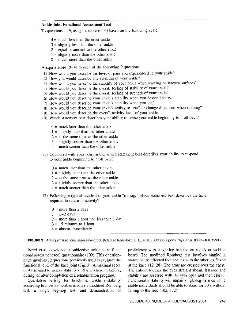
AnkleJointFunctional Assessment ToolTo questions 1-9, assign a score (0-4) based on the following scale:
4 = much less than the other ankle3 = slightly less than the other ankle2 = equal in amount to the other ankle1 = slightly more than the other ankleo= much more than the other ankle
Assign a score (0-4) to each of the following 9 questions:
1) How would you describe the level of pain you experienced in your ankle?2) How you would describe any swelling of your ankle?3) How would you describe the stability of your ankle when walking on uneven surfaces?4) How would you describe the overall feeling of stability of your ankle?5) How would you describe the overall feeling of strength of your ankle?6) How would you describe your ankle's stability when you descend stairs?7) How would you describe your ankle's stability when you jog?8) How would you describe your ankle's ability to "cut" or change directions when running?9) How would you describe the overall activity level of your ankle?
10) Which statement best describes your ability to sense your ankle beginning to "roll over?"
o= much later than the other ankle1 = slightly later than the other ankle2 = at the same time as the other ankle3 = slightly sooner than the other ankle4 = much sooner than the other ankle
11) Compared with your other ankle, which statement best describes your ability to respondto your ankle beginning to "roll over?"
o= much later than the other ankle1 = slightly later than the other ankle2 = at the same time as the other ankle3 = slightly sooner than the other ankle4 = much sooner than the other ankle
12) Following a typical incident of your ankle "rolling," which statement best describes the timerequired to return to activity?
o= more than 2 days1 = 1-2 days2 = more than 1 hour and less than 1 day3 = 15 minutes to 1 hour4 = almost immediately
FIGURE 3 Ankle joint functional assessment tool. (Adapted from Rozzi, S. L., et al. J. Orthop. Sports Phys. Ther. 8:478-486, 1999.)
Rozzi et al. developed a subjective ankle joint functional assessment tool questionnaire (109). This questionnaire involves 12questions previously used to evaluate thefunctional level of the knee joint (Fig. 3). A maximal scoreof 48 is used to assess stability of the ankle joint before,during, or after completion of a rehabilitation program.
Qualitative testing for functional ankle instabilityaccording to most authorities involve a modified Rombergtest, a single leg-hop test, and demonstration of
proficiency with single-leg balance on a disk or wobbleboard. The modified Romberg test involves single-legstance on the affected foot and leg with the other leg flexedat the knee (12, 28). The arms are crossed over the chest.The patient focuses the eyes straight ahead. Balance andstability are assessed with the eyes open and then closed.Functional instability will impair single-leg balance whilestable individuals should be able to stand for 30 s withoutfalling to the side (103, 112).
VOLUME 40, NUMBER 4, JULY/AUGUST 2001 247
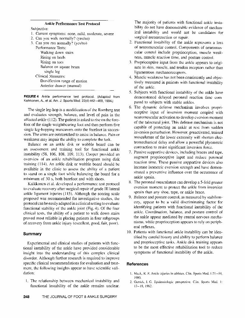
Ankle Performance Test ProtocolSubjective:I. Current symptoms: none, mild, moderate, severe2. Can you walk normally? (yes/no)3. Can you run normally? (yes/no)
Performance Tests:Walking down stairsRising on heelsRising on toesBalance on square beam
single legClinical Measures:
Dorsiflexion range of motionAnterior drawer (manual)
FIGURE 4 Ankle performance test protocol. (Adapted fromKaikkonen, A., et al. Am. J. Sports Med. 22(4):462-469, 1994.)
The single leg-hop is a modification of the Romberg testand eva luates strength, balance, and level of pain in theaffec ted ankle (112). The patient is asked to rise on the forefoot of the single weightbearing foot and then perform fivesingle leg-hopping maneuvers onto the forefoot in succession. The arms are outstretched to assis t in balance. Pain orweakness may impair the ability to complete the task.
Balance on an ankle disk or wobble board can bean assess ment and training tool for functional ankleinstability (58, 106, 108, 109, 113). Cooper provided anoverview of an ankle rehabilitation program using disktraining (1 14). An ankle disk or wobble board should beavai lable in the clin ic to assess the abilit y of a patientto stand on a single foot while balancing the board for aminimum of 30 s, both barefoot and with shoes .
Kaikkonen et a1. developed a performance test protocolto evaluate recovery after surgical repair of grade III lateralankle ligament injuri es (1 15) . Although the scoring scaleproposed was recommended for investigative studies, theprotocol can be easily adapted in a clinical setting to evalu atefunctional stability of the ankle joint (Fig. 4). Of the fourclinical tests, the abilit y of a patient to walk down stairsproved most reliable in placing patients in four subgro upsof recovery from ankle injury (exce llent, good, fair, poor).
Summary
Experimental and clinical studies of patient s with functional instability of the ankle have provided considerabl einsight into the understanding of this complex clinicaldisorder. Although furthe r research is required to impro vespecific clinical recommendations for eva luation and treatment , the following insights appear to have scientific validation:
I. The relationship between mechanical instabilit y andfunctional instability of the ankle remains unclear.
248 THE JOURNAL OF FOOT & ANKLE SURGERY
The majority of patients with functional ankle instability do not have demonstrable evidence of mechanical instability and would not be candidates forsurgical reconstruction or repair.
2. Functional instability of the ankle repre sents a lossof neuromuscular control. Components of neuromu scular control include proprioception, muscle weak ness, muscle reaction time, and posture control.
3. Propriocepti ve input from the ankle appears to orig inate in skin, muscle, and tendon receptors rather thanligamentous mechanoreceptors.
4. Muscle weakness has not been consistentl y and objectively measured in patients with functional instabilityof the ankle.
5. Subjects with functional instability of the ankle havedemonstrated delayed peroneal reaction time compared to subjects with stable ankles.
6. The dynamic defense mechanism involves proprioceptive input of inversion moment coupled withneuromu scula r activation to develop eversion momentof the talocrural joi nt. Th is defense mechanism is notcapable of protecting an ankle at rest from suddeninversion perturbation. However, preactivated, trainedmusculature of the lower extremity will shorten elec tromechanical delay and allow a powerful plyometriccontraction to resist significant inversion force.
7. Passive supportive devices, including braces and tape,augment propri ocepti ve input and reduce peronealreaction time. These passive supportive devices alsoincrease isometric eversion strength and have demonstrated a prevent ive influence over the occurrence ofankle sprain.
8. The peroneal musculature can develop a 5-fold greatereversion mom ent to protect the ankle from inversionsprain than any shoe, tape, or ankle brace.
9. Balance and posture control, as measured by stabilometry, appear to be a valid discriminating factor foridentifying pat ients with functional instability of theankle. Coordination, balance, and posture cont rol ofthe ankle appear mediated by central nervous mechanisms, while proprioception appears to rely on peripheral reflexes.
10. Patients with functional ankle instabil ity can be identified by careful history and ability to perform balanceand proprioceptive tasks. Ankle disk trainin g appearsto be the most effect ive rehabilit ation tool to reducesymptoms of functional instability of the ankle.
References
I. Mack, R. P. Ankle injuries in athletes. Clin. Sports Med. 1:7 1- 84,
1980.2. Garrick, J. G. Epidemiologic perspective . Clin. Sports Med. 1:
13-18,1982.

3. Baldwin, F. C; Tetzlaff, 1. Historical perspectives on injuries ofthe ligaments of the ankle. Clin. Sports Med. 1:3-13, 1982.
4. Jackson, D.W., Ashley, R. L., Powell, J. W. Ankle sprains inyoung athletes. Relation of severity and disability. Clin. Orthop.101:201-215,1974.
5. Garrick, 1. G., Requa, R. F. Role of external support in the prevention of ankle sprains. Med. Sci. Sports 5(3):200- 203, 1973.
6. Smith, R W., Reischl, S. F. Treatment of ankle sprains in youngathletes. Am. J. Sports Med. 14:465-471, 1986.
7. Yeung, M. S., Chan, C. H., Yuan, W.Y. An epidemiological surveyon ankle sprain. Br. J. Sports Med. 28:112-116, 1994.
8. Itay, S., Ganel, A., Horoszowski, H., Farine, 1. Clinical and functional status following lateral ankle sprains. Orthop. Rev. 11:73- 76,1982.
9. Bosien, W. R, Staples, O. S., Russell, S. W. Residual disabilityfollowing acute ankle sprain. J. Bone Joint Surg. 37-A: 1237-1243,1955.
10. Brand, R L., Black, H. M., Cox, J. S. The natural history of theinadequately treated ankle sprain. Am. J. Sports Med. 5:248-249,1977.
II. Harrington, D. C. Degenerative arthritis of the ankle secondaryto long-standing lateral ligament instability. J. Bone Joint Surg.61-A:354-461,1979.
12. Tropp, H., Odenrick, P., Gillquist, 1. Stabilometry recordings infunctional and mechanical instability of the ankle joint. Int. J.Sports Med. 6:180-182,1985.
13. Vaes, P.H., Duquet, W., Pierre-Powel, C., et al. Static and dynamicroentgenographic analysis of stability in braced and non-bracedstable and functionally unstable ankles. Am. 1. Sports Med. 26:692,1998.
14. Konradsen, L., Olesen, S., Hansen, H. Ankle sensorimotor controland eversion strength after acute ankle inversion injuries. Am. J.Sports Med. 26:72-77, 1998.
15. Ahlgren, O. H., Larsson, S. Reconstruction for lateral ligamentinjuries of the ankle. J. Bone Joint Surg. 71-B(2):300-303, 1989.
16. Brostrom, L. Sprained ankles: VI. Surgical treatment of "chronic"ligament ruptures. Acta Chir. Scand. 132:551-565, 1966.
17. Gould, N., Seligson, D., Gassman, 1. Early and late repair oflateral ligament of the ankle. Foot Ankle 1(2):84-89, 1980.
18. Karlsson, J., Bergsten, T., Lansinger, 0., Peterson, L. Reconstruction of the lateral ligaments of the ankle for chronic lateral instability. J. Bone Joint Surg. 70-A(4):581-588, 1988.
19. Karlsson.T, Bergsten, T., Lansinger,O., Peterson, L. Surgicaltreatment of chronic lateral instability of the ankle joint: a newprocedure. Am. J. Sports Med. 17(2):268-273, 1989.
20. Chrisman, O.D., Snook, G. A. Reconstruction of lateral ligamenttears of the ankle: an experimental study and clinical evaluationof seven patients treated by a new modification of the Elmslieprocedure. J. Bone Joint Surg. 51-A:904-912, 1969.
21. Elmslie, R. C. Recurrent subluxation of the ankle joint. Ann. Surg.110:364-367,1934.
22. Evans, D. L. Recurrent instability of the ankle: a method ofsurgical treatment. Proc. R. Soc. Med. 46:343-344, 1953.
23. Lee, H. G. Surgical repair in recurrent dislocation of the anklejoint. J. Bone Joint Surg. 39-A:828-834, 1957.
24. Nilsonne, H. Making a new ligament in ankle sprain. J. Bone JointSurg. 31-A:380-381, 1949.
25. Karlsson, 1., Bergsten, T., Lansinger, 0., et al. Surgical treatmentof chronic lateral instability of the ankle joint: a new procedure.Am. J. Sports Med. 17:268-273, 1989.
26. Inman, V. T. In The Joints of the Ankle, pp. 1-117, Williams &Wilkins, Baltimore, 1976.
27. Root, M. C., Weed, J. H., Orien, W. P. In Normal and AbnormalFunction ofthe Foot, pp. 1-161,295-346, Clinical BiomechanicsCorporation, Los Angeles, 1977.
28. Freeman, M. A Instabilities of the foot after lateral ligamentinjuries of the ankle. 1. Bone Joint Surg. 47-B(4):669-677, 1965.
29. Konradsen, L., Raun, J. B. Ankle instability caused by delayedperoneal reaction time. Acta Orthop. Scand. 61:388- 390, 1990.
30. Isakov, E., Mizrahi, 1., Solzi, P., et al. Response of the peronealmuscles to sudden inversion of the ankle during standing. lnt. J.Sport Biomech. 2:100-109, 1986.
31. Glencross, D., Thornton, E. Position sense following joint injury.1. Sports Med. Phys. Fitness 21:23 -27, 1981.
32. Staples, O. S. Ruptures of the fibular collateral ligaments of theankle. J. Bone Joint Surg. 57-A:IOI-107, 1975.
33. Dufek, J. S., Bates, B. T. Biomechanical factors associated withinjury during landing during sports. Sports Med. 12:326-337,
1991.34. Garrick, J. G. The frequency of injury, mechanism of injury, and
epidemiology of ankle sprains. Am. J. Sports Med. 5:241-242,
1977.35. Colville, M. R., Marder, R. A., Boyle, J. 1., et al. Strain measure
ment in lateral ankle ligaments. Am. J. Sports Med. 18:196-200,
1990.36. Ishii, T. S., Miyagawa, Fukubayashi, T., Hayashi, K. Subtalar
stress radiography using dorsiflexion and supination. 1. Bone JointSurg. 78-B:56-60, 1996.
37. Louwerens, J. W., Ginai, A. Z., Van Linge, B., Snuders, C. 1.Stress radiography of the talocrural and subtalar joints. Foot AnkleInt. 16:148-155, 1995.
38. Zell, B., Shereff, M. J., Greenspan, A., Liebowitz, S. Combinedankle and subtalar instability. Bull Hosp. J. Dis. Orthop. lnst.46:37-46,1986.
39. Brantigan, 1. W., Pedcgana, L. R, Lippert, F. G. Instability of thesubtalar joint: diagnosis by stress tomography in three cases. 1.
Bone Joint Surg. 59-A:321-324, 1977.40. Meyer, J. M., Garcia, J., Hoffmeyer, P., Fritschy, D. The subtalar
sprain: a roentgenographic study. Clin. Orthop. 226:169-173,1986.
41. Hertel, J., Dcnegar, C. R, Monroe, M. M., Stokes, W. L. Talocrural and subtalar joint instability after lateral ankle sprain. Med.Sc. Sports Exerc. 31:1501-1508,1999.
42. Fraser, G. H., Ahmed, A. M. Passive rotational stability of theweight-bearing talocrural joint: an in-vitro biomechanical study(abstract). Orthop. Trans. 7:248, 1983.
43. McCullough, C. J., Burge, P. D. Rotary stability of the loadbearing ankle: an experimental study. J. Bone Joint Surg. 62B:460-464, 1980.
44. Stormont, D. M., Morrey, B. F., Kai-Nan, A., et al. Stability ofthe loaded ankle: relation between articular restraint and primaryand secondary static restraint. Am. J. Sports Med. 13:295-300,1985.
45. Cass, J. R., Settles, H. Ankle instability: in vitro kinematics inresponse to axial load. Foot Ankle 15:134-140, 1994.
46. Brunt, D., Anderson, J. C, Huntsman, B., et al. Postural responsesto lateral perturbation in healthy subjects and ankle sprain patients.Med. Sci. Sports Exerc. 24: 171-176, 1992.
47. Dietz, V., Berger, W. Spinal coordination of bilateral leg muscleactivity during balancing. Exp. Brain Res. 47:172-176, 1982.
48. Nashner, L. M. Balance adjustments of humans perturbed whilewalking. 1. Neurophysiol. 44:650-664, 1980.
49. Nashner, L. M., Black, F. 0., Wall, C. Adaption to altered supportand visual conditions during stance: patients with vestibulardeficits. J. Neurosci. 2:536-544, 1982.
50. Nawoczenski, D. A, Owen, M. G., Ecker, M. L., et al. Objectiveevaluation of peroneal response to sudden inversion stress. J.Orthop. Sports Phys. Ther. 7:107-109,1985.
VOLUME 40, NUMBER 4, JULY/AUGUST 2001 249
51. Dietz, V. Human neuronal control of automatic functional move
ments: interaction between central programs and afferent input.Physiol. Rev. 72:33-42, 1992.
52. Dietz, V., Quintern, J., Sillem, M. Stumbling reactions in man:significance of proprioceptive and pre-programmed mechanisms.1. Physiol. 386:149-163,1987.
53. Nashner, L. M. Adapting reflexes controlling the human posture.Exp. Brain Res. 26:59-72, 1976.
54. Mathews, P. B., Stein, R B. The regularity of primary andsecondary muscle spindle afferent discharges. J. Physiol. Lond.202:59-82, 1969.
55. Robbins, S. E., Hanna, A. M., Gouw, G. J. Overload protection:avoidance response to heavy plantar surface loading. Med. Sci.Sports Exerc. 20:85-92, 1988.
56. Freeman, M. A. R, Dean, M. R E., Hanham, 1. W. F. The etiology and prevention of functional instability of the foot. J. BoneJoint Surg. 47-B:678-685, 1965.
57. DiCarlo, M. S., Talbot, R W. Evaluation of ankle joint proprioception following injection of the anterior talofibular ligament. J.Orthop. Sports Phys. Ther. 8:70-76, 1986.
58. Feuerbach, M. A., Brabiner, M. 0., Koh, T. J., Weiker, G. G.Effect of an ankle orthosis and ankle ligament anesthesia on anklejoint proprioception. Am. J. Sports Med. 20:223-229, 1994.
59. Barrack, R. L., Skinner, H. B., Brunet, M. E. Functional performance of the knee after Intra-articular anesthesia. Am. 1. SportsMed. 11:258-261, 1983.
60. McCluster, G. M., Blackburn, T. A., Lewis, T. Prevention ofankle sprains. Am. 1. Sports Med. 4: 151-157, 1976.
61. Skinner, H. B., Wyatt, M. P., Hodgdon, D. W., Connerd, R C.Effect of fatigue on joint position sense of the knee. 1. Orthop.Res. 4: 112-118, 1986.
62. Konradsen, L., Voight, M., Hojsgaard, C. Ankle inversion injuries:the role of the dynamic defense mechanism. Am. J. Sports Med.25:54-58, 1997.
63. Karlsson, J., Andreasson, G. O. The effect of external anklesupport in chronic lateral ankle joint instability: an electromyographic study. Am. J. Sports Med. 20:257-261, 1992.
64. Glick, J. M., Gordon, R. B., Nishimoto, D. The prevention andtreatment of ankle injuries. Am. 1. Sports Med. 4:136-141,1976.
65. Manfroy, P. P., Ashton-Miller, 1. A., Wojtys, E. M. The effect ofexercise, prewrap and athletic tape on the maximal active andpassive ankle resistance to ankle inversion. Am. J. Sports Med.25:156-163, 1997.
66. Shapiro, M. S., Kabo, J. M., Mitchell, P. W., et al. Ankle sprainprophylaxis: an analysis of the stabilizing effects of bracing andtape. Am. 1. Sports Med. 22:78-82, 1994.
67. Dietz, V., Schmidtbleicher, D., Noth, J. Neuronal mechanisms ofhuman locomotion. J. Neurophysiol. 42:1212-1222, 1979.
68. Berger, W. G., Dietz, V., Quintern, 1. Corrective reactions tostumbling reactions in man: neuronal coordination of bilateral legmuscle activity during gait. J. Physiol. Lond. 357: 109-125, 1984.
69. Dietz, V., Schmidtbleicher, D. Interaction between pre-activityand stretch reflex in human triceps brachii during landing fromforward falls. J. Physiol. 311:113-125, 1981.
70. Gottlieb, G., Agarwal, G. Response to sudden torques about theankle in man. J. Neurophysiol. 42:91-106,1979.
71. Nichols, T., Houk, J. Improvements in linearity and regulation of
stiffness that results from action of stretch reflex. J. Neurophysiol.39:119-142,1976.
72. Gollhofer, A., Schmidtbleicher, D., Dietz, V. Regulation of musclestiffness in human locomotion. Int. J. Sports Med. 5:19-22,1984.
73. Armstrong, R B. Initial events in exercise induced muscularinjury. Med. Sci. Sports Exerc. 22:429-436, 1990.
250 THE JOURNAL OF FOOT & ANKLE SURGERY
74. Norman, R W., Komi, P. V. Electromechanical delay in skeletalmuscle under normal movement conditions. Acta. Physiol, Scand.100:241-252,1979.
75. Cavagna, G., Dunsman, B., Margaria, R Positive work done by apreviously stretched muscle. 1. Appl. Physiol, 65:11-19, 1988.
76. Richie, D. H., Endo, C. K., DeVries, H. Shin muscle activity andsport surfaces: an electromyographic study. J. Am. Podiatr. Med.Assoc. 83:181-187, 1993.
77. Linge, B. V. Activity of peroneal muscles, the maintenanceand balance, and the prevention of injury of the ankle: aneJectromyographic and kinematic study. Acta. Orthop. Scand.(suppl. 227):67 - 72, 1988.
78. Melvill-Jones, G., Watt, D. G. D. Muscular control of landingfrom unexpected falls in man. 1. Physiol. 219:729-737, 1971.
79. Winter, D. A. The Biomechanics and Motor Control of HumanGait: Normal, Elderly, and Pathological, 2nd ed., p. 21, Universityof Waterloo Press, Waterloo, Ontario, Canada, 1991.
80. Ashton-Miller, 1. A., Ottavian, R A., Hutchinson, C., Wojtysue, M. What best protects the inverted ankle against further inversion? Am. J. Sports Med. 24:800-809, 1996.
81. Bullard, R H., Dawson, J., Arenson, D. J. Taping the "athleticankle." 1. Am. Podiatr. Assoc. 69:727, 1979.
82. Fumich, R M., Ellis, A. E., Guerin, G. 1., et al. The measuredeffect of taping on combined foot and ankle motion before andafter exercise. Am. 1. Sports Med. 9:165-170, 1981.
83. Larsen, E. Taping the ankle for chronic instability. Acta. Orthop.Scand. 55:551-553, 1984.
84. Quigley, T. B., Cox, J., Murphy, 1. Protective wrapping for theankle. JAMA 132:924, 1946.
85. Barrett, J. R, Tangi, J. L., Drake, c., et al. High versus low topshoes for the prevention of ankle sprains in basketball players: aprospective, randomized study. Am. 1. Sports Med. 21:582-585,1993.
86. Burks, R T., Bean, B. G., Marcus, R, et al. Analysis of athleticperformance with prophylactic ankle devices. Am. J. Sports Med.19:104-106, 1991.
87. Journal, J. P. The effects of taping on vertical jumping ability. J.Athletic Train. 7:146-152,1972.
88. Mayhew, J. L. Effects of ankle taping on motor performance. 1.Athletic Train. 7:10- 18, 1972.
89. Delacerda, F. G. Effect of underwrap conditions on the supportiveeffectiveness of ankle strapping with tape. J. Sports Med. Phys.Fitness 18:77-84, 1978.
90. Greene, T. A., Hillman, S. K. Comparison of support provided bya semirigid orthosis and adhesive ankle taping before, during andafter exercise. Am. 1. Sports Med. 18:498-506, 1990.
91. Gross, M. T., Bradshaw, M. K., Ventry, L. C., et al. Comparisonof support provided by ankle taping and semirigid orthosis. J.Orthop. Sports Phys. Ther. 9:33-39, 1987.
92. Gross, M. T., Lapp, A. K., Davis, J. M. Comparison of Sweedo universal ankle support and Aircast sport stirrup orthoses andankle tape in restricting ankle eversion-inversion before and afterexercise. J. Orthop. Sports Phys. Ther. 13:11- 19, 1991.
93. Laughman, R K., Carr, T. A., Chao, E. Y., et al. Three-dimensional kinematics of the taped ankle before and after exercise.Am. 1. Sports Med. 8:425-431, 1980.
94. Bunch, R P., Bednarski, K., Holland, D., et al. Ankle joint support: a comparison of reusable lace-on braces with taping andwrapping. Physician Sports Med. 13(5):59-62, 1985.
95. Malina, R M., Plagenz, L. B., Rarick, G. L. Effect of exerciseupon the measurable supporting strength of cloth and tape anklewraps. Res. Q. 34:158-165,1963.
96. Rarick, G. L., Bigley, G., Karst, R, et al. The measurable supportof the ankle joint by conventional methods of taping. 1. Bone JointSurg. 44-A:1l83-1190, 1962.

97. Rovere, G. D., Clarke, T. 1., Yates, S. c. et al. Retrospectivecomparison of taping and ankle stabilizers in preventing ankleinjuries. Am. 1. Sports Med. 16:228~233, 1988.
98. Gross, M. T., Ballard, C. L., Mears, H. C., Watkins, E. J. Comparisons of Don-Joy ankle ligament protector and Aircast sportstirrup orthoses in restricting foot and ankle motion beforeand after exercise. J. Orthop. Sports Phys. Ther. 16:60-67,
1992.99. Cornwall, M. W., Murrell, P. Postural sway following inversion
sprain of the ankle. 1. Am. Podiatr. Med. Assoc. 81:243-247.
1991.100. Lofvengerg, R., Karrholm, J., Sudelin, G., Ahigren, O. Prolonged
reaction time in patients with chronic lateral instability of theankle. Am. J. Sports Med. 23:414~417, 1995.
101. Irrgang, J. J., Whitney, S. L., Cox, E. D. Balance and proprioception training for rehabilitation of the lower extremity. J. SportsRehab. 3:68-83, 1994.
102. Pinciuro, D., Lephart, S. M., Henry, T. Learning effects and reliability of the Biodex Stability System". J. Athletic Train. 30:535-541,1995.
103. Tropp, H., Ekstrind, 1., Gillquist, 1. Stabilometry in functionalinstability of the ankle and its value in predicting injury. Med.Sci. Sports 16:64-66, 1984.
104. Lentell, G. L., Katzman, L. L., Walters, M. K. The relationshipbetween muscle function and ankle stability. J. Orthop. SportsPhys. Ther. 11:605-611, 1990.
lOS. Hoffman, M., Payne, V. G. The effects of proprioceptive ankledisk training n healthy subjects. 1. Orthop. Sports Phys. Ther.21:90-93,1995.
106. Tropp, H., Askling, C., Gillquist, 1. Prevention of ankle sprains.Am. 1. Sports Med. 13:259-262, 1985.
107. Tropp, H., Gillquist, J. Factors affecting stabilometry recordingsof single limb stance. Am. J. Sports Med. 12:185-188, 1985.
108. Gauffin, H., Tropp, H., Oderrick, P. Effect of ankle disk trainingon postural control in patients with functional instability of theanklejoint.lnt. J. Sports Med. 9:141~144, 1988.
109. Rozzi, S. L., Lephart, S. M., Sterne, R., Kuligowski, L. Balancetraining for persons with functionally unstable ankles. 1. Orthop.Sports Phys. Ther. 8:478-486, 1999.
110. Bosien, W. R., Staples, O. S., Russell, S. W. Residual disabilityfollowing acute ankle sprains. J. Bone Joint Surg. 37-A: 1237 -1243,1955.
III. Staples, S. Ruptures of the fibular collateral ligaments of the ankle.J. Bone Joint Surg. 57:101-107,1975.
112. Glasoe, W. M., Allen, M. K., Awtry, B. F., Yack, H. J. Weightbearing immobilization and early exercise treatment followinga grade II lateral ankle sprain. Orthop. Sports Phys. Ther.29:394-399, 1999.
113. Gauffin, H., Tropp, H., Odenrick, P. Effect of ankle disk trainingon postural control in patients with functional instability of theankle joint. lnt. 1. Sports Med. 9: 141-144, 1988.
114. Cooper, D., Farr, J. Ankle rehabilitation using the ankle disk.
Physician Sports Med. 6:141, 1978.115. Kaikkonen, A., Kannus, P., Jarvinen, M. A performance test proto
col and scoring scale for the evaluation of ankle injuries. Am. J.Sports Med. 22(4):462-469, 1994.
Suggested Reading
Bahr, R., Lian, 0., Bahr, I. A. A twofold reduction in the incidenceof ankle sprains in volleyball after the introduction of an injuryprevention program: a prospective cohort study. Scand. 1. Med. Sci.Sports 7(3):172, 1997.
Brand, R. L., Black, H. M., Cox, J. S. The natural history of the inadequately treated ankle sprain. Am. 1. Sports Med, 5:248-249, 1977.
Brantigan, J. W., Pedegana, L. R., Lippert, F. G. Instability of thesubtalar joint: diagnosis by stress tomography in three cases. J. BoneJoint Surg. 59-A:321-324, 1977.
Bruns, J., Staerk, H. Mechanical ankle stabilization due to the useof orthotic devices and peroneal muscle strength an experimentalinvestigation. lnt. J. Sports Med. 13:611 ~615, 1992.
Cawley, P. W., France, E. P. Biomechanics of the lateral ligaments ofthe ankle: an evaluation of the effects of axial load and single planemotions on ligament strain patterns. Foot Ankle, 12:92-99, 1991.
Fraser, G. A., Ahmed, A. M. Passive rotational stability of the weightbearing talocrural joint: an in vitro biomechanical study (Abstract).Orthop. Trans. 7:248, 1983.
Friden, T., Zatterstrom, R., Lindstrand, A., Moritz, U. A stabilometrictechnique for evaluation of lower limb instability. Am. 1. Sports Med.17:118~ 122, 1989.
Guskiewicz, K. M., Perrin, D. H. Effect of orthotics on postural swayfollowing inversion ankle sprain. J. Orthop. Sports Phys. Ther.23:326-331,1996.
Kelikian, H., Kelikian, A. S. Disorders of the Ankle, pp. 58~59, W.B.Saunders, Philadelphia, 1985.
Lephart, S. M., Henry, T. J. Functional rehabilitation for the upper andlower extremity. Orthop. Clin. North Am. 26:579~592, 1995.
Ramot, Y, Susak, Z. The dynamics of the subtalar joint in suddeninversion of the foot. 1. Biomech. Eng. 112:9, 1990.
Sheth, P., Yu, B., Laskowski, E. R., et al. Ankle disk training influencesreaction times of selected muscles in a simulated ankle sprain. Am.J. Sports Med. 25:538, 1997.
Verhagen, R. A., Kclzer, G., Van Dijk, C. N. Long-term follow-upof inversion trauma of the ankle. Arch. Orthop. Trauma Surg.114:92-96, 1995.
VOLUME 40, NUMBER 4, JULY/AUGUST 2001 251