functional imaging of the auditory processing applied to...
TRANSCRIPT
Phil. Trans. R. Soc. B (2008) 363, 1023–1035
doi:10.1098/rstb.2007.2157
on June 2, 2018http://rstb.royalsocietypublishing.org/Downloaded from
Functional imaging of the auditory processingapplied to speech sounds
Published online 7 September 2007
Roy D. Patterson1,* and Ingrid S. Johnsrude2
One confrom sou
*Autho
1Centre for the Neural Basis of Hearing, Department of Physiology, Development and Neuroscience,University of Cambridge, Downing Street, Cambridge CB2 3EG, UK
2Department of Psychology, Queen’s University, 62 Arch Street, Kingston, Ontario, Canada K7L 3N6
In this paper, we describe domain-general auditory processes that we believe are prerequisite to thelinguistic analysis of speech. We discuss biological evidence for these processes and how they mightrelate to processes that are specific to human speech and language. We begin with a brief review of (i)the anatomy of the auditory system and (ii) the essential properties of speech sounds. Section 4describes the general auditory mechanisms that we believe are applied to all communication sounds,and how functional neuroimaging is being used to map the brain networks associated with domain-general auditory processing. Section 5 discusses recent neuroimaging studies that explore where suchgeneral processes give way to those that are specific to human speech and language.
Keywords: auditory anatomy; speech sounds; auditory processing; neuroimaging of pitch;neuroimaging of speech sounds
1. INTRODUCTIONSpeech is a rich social signal that conveys a wealth of
information. Not only is it a linguistic signal, used tocommunicate information and ideas, but it alsocontains non-linguistic information about the size,sex, background, social status and emotional state ofthe speaker. Finally, it is usually experienced as amultisensory and interactive signal; these are importantaspects that also do not fall within the traditional realmof linguistic analysis. These non-linguistic aspects ofcommunication are a reminder that speech sharescharacteristics with communication in other animals,including other primates. The initial stages of auditoryprocessing, which rely on a neural organization that isevolutionarily conserved among many primate species,are probably general and apply to all communicationsounds, not just to speech. Accordingly, we begin witha brief overview of primate anatomy. At the same time,the complexity of human communication indicates thatit engages additional neural apparatus subservinglinguistic and social cognition. The point in the systemwhere the processing radiates out into divergentfunctions is the topic of §5.
2. A BRIEF OVERVIEW OF AUDITORY ANATOMY(a) The subcortical auditory system in humans
In humans, the principal components of the subcorticalauditory system lie in a frontal plane that extends fromthe ear canal to the upper surface of the central portionof the temporal lobe. Between the cochlea and theauditory cortex, there are four major centres of neuralprocessing: the cochlear nucleus (CN); the superior
tribution of 13 to a Theme Issue ‘The perception of speech:nd to meaning’.
r for correspondence ([email protected]).
1023
olivary complex (SOC); the inferior colliculus (IC);and the medial geniculate body (MGB) of thethalamus. Work in other primates suggests that thereare mandatory synapses for auditory processing inthree of the four nuclei (CN, IC and MGB), whichsupports the view that these nuclei perform transfor-mations that are applied to all sounds as they proceedup the pathway, much as the cochlea performs amandatory frequency analysis on all sounds enteringthe auditory system. In the visual system, there is onlyone synapse between the retina and visual cortex in thelateral geniculate nucleus.
Information from the two ears is probably integratedin several nuclei in the subcortical auditory system. TheCN projects to both the contralateral and the ipsilateralSOC, where minute differences in the timing of theversions of a sound at the two ears are correlated,permitting estimation of source location. The CN alsoprojects to both contralateral and ipsilateral IC, and thetwo ICs are themselves densely interconnected. Thus,the subcortical auditory system does not maintain aclear segregation of information by the ear of entry. Incontrast, in the visual system, there is no binocularprocessing prior to visual cortex. The complexity of thesubcortical auditory system is probably due, at least inpart, to the temporal precision of the neural represen-tation of sound (Patterson et al. 1999). Auditory nervefibres between the cochlea and the CN fire in phasewith basilar membrane motion up to approximately5000 Hz, and the nuclei that process this sub-millisecond information must be close to the sourceto minimize temporal distortion. The maximum rate ofphase locking drops to approximately 500 Hz in the IC,and to approximately 50 Hz in the MGB and primaryauditory cortex (PAC), which suggests that the form ofthe neural code changes at least twice as theinformation progresses from cochlea to cortex, onceat the level of the IC and once at the level of the MGB.
q 2007 The Royal Society
PS
AS
CSIPS
LS
STS
STGLF
RPB
CPB
cut
insulaAI
RRT
(a)
CLCM
MLRM
AL
RTLRTM
TptAF
PaAlt
Ts2
Ts 3
Ts1
Un Bd
Extrn Cap
SLF
AS
PS
CS IPS
LS
IOS
STS
Pro
LF
(b)
PS
AS
CSIPS
LS
STG
LF
(c)
Pro
STS
PS
AS
CSIPS
LS
STS
STG
LFRPB
CPBTPt
(d )
TPO
TPO
TPO
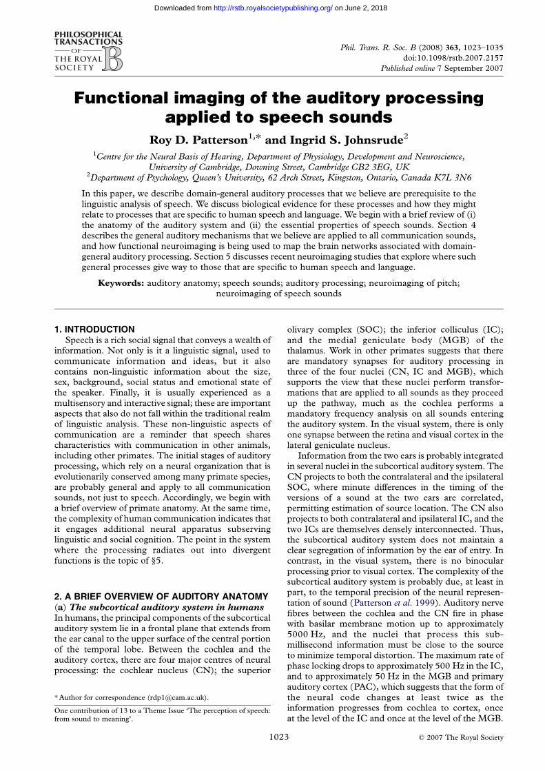
Figure 1. Four representations of the anatomical connections of the temporal lobe in the macaque brain. (a) The anatomicalorganization of the auditory cortex is consistent with at least four levels of processing, including core regions (darkest shading) beltregions (lighter shading), parabelt regions (stripes) and temporal and frontal regions that interconnect with belt and parabelt (lightershading). (Adapted from Kaas et al. (1999) and Hackett & Kaas (2004)). Dotted lines indicate sulci that have been opened to showauditory regions. Regions along the length of (b) superior temporal gyrus and (c) dorsal bank of the superior temporal sulcus connectwith prefrontal regions in a topographically organized anterior-to-posterior fashion. (b) Adapted from Petrides & Pandya (1988,p. 64); (c) adapted from Seltzer & Pandya (1989a). (d ) Connectivity of auditory belt and parabelt; adapted from Hackett & Kaas(2004). AF, arcuate fasciculus; AS, arcuate sulcus; CS, central sulcus; Extm Cap, extreme capsule; IOS, inferior occipital sulcus;IPS, intraparietal sulcus; LF, lateral fissure; LS, lunate sulcus; PS, principal sulcus; SLF, superior longitudinal fasciculus; STG,superior temporal gyrus; STS, superior temporal sulcus; UnBd, uncinatebundle. (Note. Abbreviations are not spelt out if they are theconventional label for a microanatomically or physiologically defined area).
1024 R. D. Patterson & I. S. Johnsrude Imaging of speech-sound processing
on June 2, 2018http://rstb.royalsocietypublishing.org/Downloaded from
(b) The anatomy of auditory cortex and its
projections, in the macaque
In humans, the principal components of the cortical
auditory system are not well understood. Microelectrode
recordings, the cornerstone of non-human neurophy-
siology, can only be undertaken in rare circumstances
(e.g. during neurosurgery; Howard et al. 2000; Brugge
et al. 2003). Post-mortem histological material is scarce
and of relatively poor quality (Hackett et al. 2001;
Wallace et al. 2002), and in vivo tracer studies in humans
are currently not possible. The rhesus macaque monkey
(Macaca mulatta) provides an animal model for the
organization of auditory cortex (Rauschecker et al. 1997;
Rauschecker 1998; Kaas et al. 1999; Kaas & Hackett
2000), and this can be supplemented by the (relatively
few) anatomical and neurophysiological studies that
have been conducted in humans (Liegeois-Chauvel et al.1991; Rivier & Clarke 1997; Howard et al. 2000; Hackett
et al. 2001; Morosan et al. 2001; Rademacher et al. 2001;
Wallace et al. 2002; see Hall et al. (2003) and Scott &
Johnsrude (2003), for reviews).
A note of caution must be sounded in assuming
anatomical and functional homologies between maca-
ques and humans. Most obviously, functional
Phil. Trans. R. Soc. B (2008)
specialization must diverge in the two species at, or
before, the point where speech-specific processing
begins in humans. Furthermore, unlike our own
species, vocalization is not an important form of
communication in macaques. Also, auditory research
in the macaque has been largely restricted to experi-
ments with very simple sounds such as clicks and pure
tones, which may not require extensive cortical
processing. As a result, the functional specialization
of the core, belt and parabelt regions is simply not
known, and macaque research provides only the most
general indication of where to look for specific forms of
processing in humans.
The organization in the macaque is shown in
figure 1a. Cortical afferents from the ventral division
of the MGB project to three tonotopically organized
fields on the superior temporal gyrus (STG;
Rauschecker et al. 1997; Kaas et al. 1999; Kaas &
Hackett 2000). This ‘core’ of primary areas projects to
a surrounding ‘belt’ of anatomically distinguishable
cortical fields which exhibit interconnections among
adjacent regions (Merzenich & Brugge 1973; Pandya &
Sanides 1973; Jones et al. 1995; Pandya 1995; Hackett
et al. 1998; Rauschecker 1998; Kaas & Hackett 2000;

Imaging of speech-sound processing R. D. Patterson & I. S. Johnsrude 1025
on June 2, 2018http://rstb.royalsocietypublishing.org/Downloaded from
Rauschecker & Tian 2000). Belt areas connect withlateral ‘parabelt’ fields, again through connectionsbetween physically adjacent regions. The hierarchicalconnections of the core, belt and parabelt areas suggestat least three discrete levels of processing in themacaque (Pandya 1995; Hackett et al. 1998;Rauschecker 1998; Kaas et al. 1999; Kaas & Hackett2000; Rauschecker & Tian 2000).
Recent neuroimaging studies (reviewed in §5)indicate that the superior temporal sulcus (STS) regionin humans is important for speech-sound perception.Drawing inferences from macaque cortical organiz-ation is problematic in the STS, since humans have amiddle temporal gyrus (including the ventral bank ofthe STS) and macaques do not. Human homologiesof the ventral bank regions that have been mapped inthe macaque are particularly uncertain. Nevertheless,the anatomical organization of the upper bank of themacaque STS may be somewhat conserved in humans,and it is currently the best evidence we have as to whatto expect in human STS.
The STS in the macaque is anatomically hetero-geneous, but much of its upper bank, running the lengthof the STS, comprises a region (area TAa) that receivesits input mainly from auditory cortex (Seltzer & Pandya1978, 1989b). This region projects into adjacentpolysensory cortex in the depth of the STS, as well asto the inferior parietal lobule and prefrontal cortex(Seltzer & Pandya 1989a,b). Furthermore, anteriorSTS regions project to ventral and anterior frontalregions, and more posterior STS regions project to moreposterior and dorsal frontal regions (and to parietalcortex; figure 1b). Similarly, as shown in figures 1c,danterior belt and parabelt also interconnect directly, andin a topographically organized way, with multiple siteswithin orbitofrontal, ventrolateral and dorsolateralfrontal cortex including Brodmann areas 46, 12 and45 (Petrides & Pandya 1984; Hackett et al. 1998, 1999;Romanski et al. 1999a,b). Importantly, area 45 inhumans, located in the inferior frontal gyrus (IFG;pars triangularis), is considered as one of the architec-tonic constituents of Broca’s area (Amunts et al. 1999).This distributed set of fields in STG, STS, parietal andprefrontal cortex constitutes a potential fourth stage ofprocessing (Kaas et al. 1999; figure 1a).
(c) Links between perception and production in
humans
At the level of cortex, anatomical connectivity suggeststhat auditory perception and vocal production may bequite intimately linked. Auditory core, belt andparabelt regions all project into the dorsal caudateand putamen—components of the basal ganglia—which are traditionally considered to serve a primarilymotor function (Yeterian & Pandya 1998). STSregions that receive projections from auditory cortices,in turn project to regions of the inferior parietal lobulethat interconnect with motor cortex via premotorcortex (Pandya & Seltzer 1982; Seltzer & Pandya1991; Petrides & Pandya 2002). Finally, Brodmannareas 45 and 46 in frontal cortex, which receiveauditory projections, interconnect with motor regionsvia area 44 and premotor cortex.
Phil. Trans. R. Soc. B (2008)
Physiological data are consistent with a link betweenauditory perception and vocal production, and theyindicate that the coupling is quite rapid. Matt Howard,John Brugge and colleagues have used depth electrodestimulation and electrophysiological recording inneurosurgical patients to explore the evoked responsesand connectivity in a circuit involving PAC, a poster-olateral region of the STG which they call posteriorlateral superior temporal (PLST), IFG (pars triangu-laris and opercularis) and orofacial motor cortex(Garell et al. 1998; Howard et al. 2000; Brugge et al.2003; Greenlee et al. 2004). Evoked responses in PACof Heschl’s gyrus (HG) had response latencies rangingfrom 15 to 25 ms, which are compatible with themagnetoencephalography (MEG) data on click latencyin PAC reported by Lutkenhoner et al. (2003). Then,when this region of HG was electrically stimulated, itresulted in an evoked potential in PLST (Howard et al.2000; Brugge et al. 2003). The average onset latencyfor this evoked response was only 2.0 ms, consistentwith an ipsilateral corticocortical connection betweenHG and PLST. PLST appears to make a functionalconnection with the IFG (Garell et al. 1998) with onsetlatencies of approximately 10 ms, and cortical stimu-lation of posterior IFG elicits responses in orofacialmotor cortex with onset latencies of approximately6.0 ms (Greenlee et al. 2004). Taken together, theseresults suggest that a sound in the environment could,in principle, have an impact on neural activity inorofacial motor cortex within 35 ms of stimulus onset,and most of that time is spent in the pathway from thecochlea to PAC.
In summary, this overview of the anatomy of auditorycortex suggests that, following the succession of nuclei inthe subcortical pathway, the information in auditorycortex radiates out in parallel paths from core areas, andcascades into at least three spatially distributed sets ofregions, comprising at least three further processingstages. Other sense information is integrated withauditory information early on in cortical processing,and prominent feedback routes connect adjacent regionsat all levels. Perceptual processes must depend on thisanatomical organization.
Now we turn to the characteristics of speech soundsand describe a model of the processes that we believeare applied to all communication sounds beforespeech-specific processing begins in cortex.
3. GENERAL AUDITORY PROCESSES INVOLVEDIN SPEECH PERCEPTIONWhen a child and an adult utter the ‘same’ syllable, it isonly the linguistic message of the syllable that is the same.The child has a shorter vocal tract and lighter vocal cords,and as a result, the waveforms carrying the message arequite different for the child and the adult. Althoughhumans have no difficulty in understanding that a childand an adult have said the same word, evaluating theequivalence is far from trivial, as indicated by the fact thatspeech-recognition machines find this task difficult.Indeed, when trained on the speech of a man, recognitionmachines are notoriously bad at understanding thespeech of a woman, let alone a child. The robustness ofauditory perception has led Irino & Patterson (2002) to

low
high
tall
short
(a)
(b)
(c)
(d )
8ms12ms
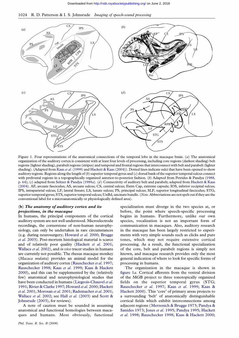
Figure 2. Internal structure of voiced sounds illustrating thesize factors: pulse rate and resonance rate. (a,b) Glottal pulserate and (c,d ) vocal-tract length have a major effect on boththe waveform and the spectrum of the sound, but humanperception is extremely robust to changes in both of thesefactors.
8 12 16 20 24 28 32 36 400.1
0.3
0.5
0.9
1.5
2.5
3.9
6.0
freq
uenc
y (k
Hz)
time (ms)
(a)
081632 240.1
0.3
0.5
0.9
1.5
2.5
3.9
6.0
freq
uenc
y (k
Hz)
time interval (ms)
(b)
1026 R. D. Patterson & I. S. Johnsrude Imaging of speech-sound processing
on June 2, 2018http://rstb.royalsocietypublishing.org/Downloaded from
hypothesize that the auditory system possesses
mechanisms that automatically assess the vocal-tract
length (VTL)and glottal pulse rate (GPR) of the speaker.
Moreover, since humansproduce speech sounds inmuch
the same way as all other mammals, it is assumed that
such mechanisms are part of the processing applied to all
sounds. The value of this analysis is that it helps to
produce a size-invariant representation of the timbral
cues that identify a species, and this greatly facilitates
communication. In speech communication, such pro-
cesses may be responsible for what is referred to as vowel
normalization (e.g. Miller 1989).
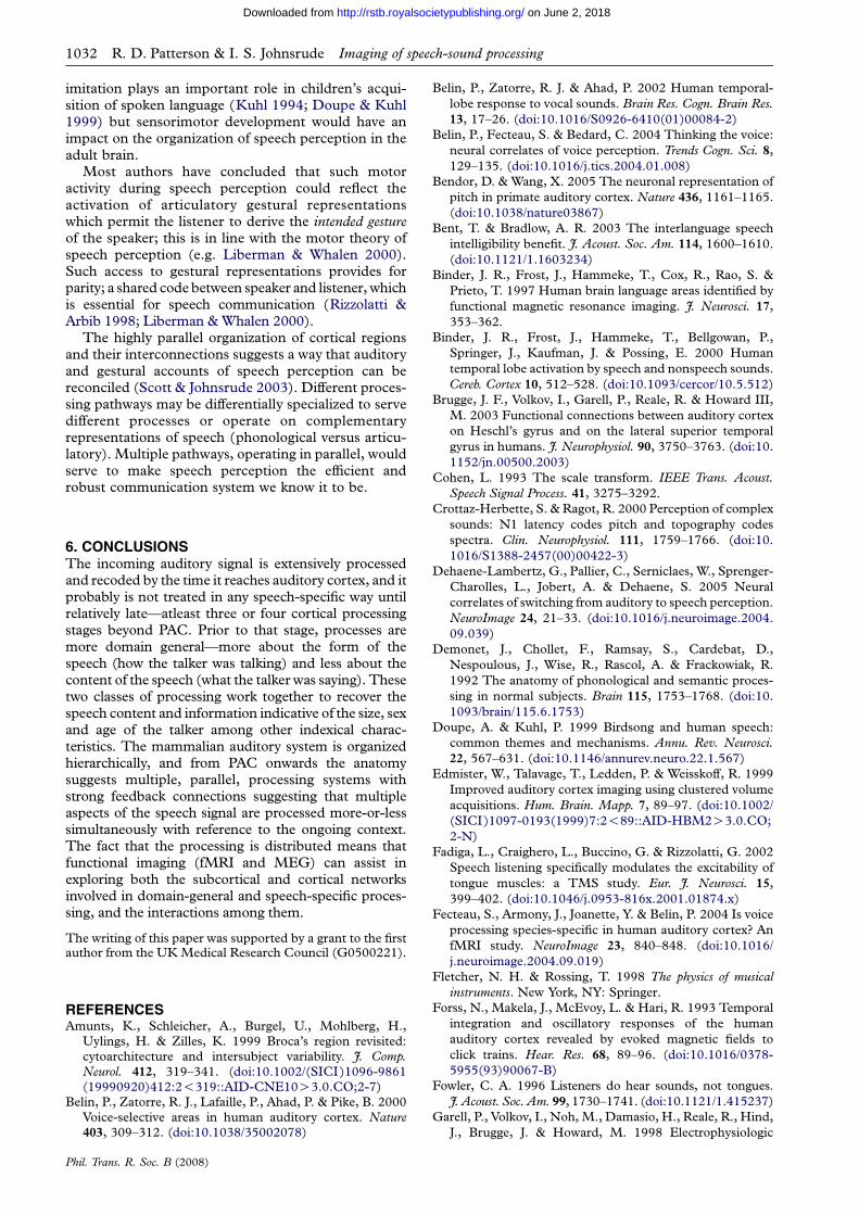
Figure 3. (a) The neural activity pattern and (b) the auditoryimage produced by the /a/ of ‘hat’. Note that the abscissa ofthe auditory image (b) is ‘time interval’ rather than time itself. (a) Communication soundsAt the heart of each syllable of speech is a vowel.
Figure 2 shows four versions of the vowel /a/ as in ‘hall’.
From the auditory perspective, a vowel is a ‘pulse-
resonance’ sound, that is, a stream of glottal pulses each
with a resonance showing how the vocal tract
responded to that pulse. From the speech perspective,
the vowel contains three important components of the
information in the larger communication (Irino &
Patterson 2002). The first is the phonological
‘message’; for the vowels in figure 2, the message is
that the vocal tract is currently in the shape that the
brain associates with the phoneme /a/. This message is
contained in the shape of the resonance which is the
same in every cycle of all four waves. In figure 2a,b one
person has spoken two versions of /a/ using a high and a
low GPR, respectively; the pulse rate determines the
pitch of the voice. The resonances have the same form
since it is the same person speaking the same vowel. In
figure 2c,d a short person and a tall person, respectively,
have spoken versions of /a/ on the same pitch. The pulse
rate and the shape of the resonance are the same, but
the rate at which the resonance proceeds within the
glottal cycle is slower in figure 2d. This person has the
longer vocal tract and so their resonances ring longer.
Since the vocal tract connects the mouth and nose to
the lungs, VTL is highly correlated with the height of
the speaker. In summary, it is the shape of the
resonance that corresponds to the message or content
of the speech sound. The GPR, which corresponds to
the pitch, and the resonance rate, which corresponds to
VTL, are derived from the ‘form’ of the message.
Phil. Trans. R. Soc. B (2008)
(b) The auditory image model and auditory
adaptation to GPR and VTL
The general transforms involved in analysing GPR and
VTL will be presented in the context of the auditory
image model (AIM; Patterson et al. 1992, 1995), a
model that focuses on the internal ‘auditory images’
produced by communication sounds and how these
images can be produced with an ordered set of three
transforms. The cochlea performs a spectral analysis of
all incoming sounds. In AIM, this is simulated with an
auditory filterbank in which the bandwidths of the
filters are proportional to filter centre frequency. The
filterbank converts an incoming sound wave into a
multi-channel representation of basilar membrane
motion. The most recent version of the auditory filter
includes the fast-acting compression and two-tone
suppression observed in the cochlea (Irino & Patterson
2006; Unoki et al. 2006). A ‘transduction’ mechanism
involving half-wave rectification and low-pass filtering
converts each channel of membrane motion into a
simulation of the neural activity produced in the
auditory nerve at that point on the basilar membrane.
The result is a multi-channel, neural activity pattern
(NAP) like that shown in figure 3a; the dimensions of
the NAP are time (the abscissa) and auditory-filter
centre frequency on a quasi-logarithmic axis (the
ordinate). The surface defined by the set of lines is
AIM’s simulation of the NAP produced in response to a
short segment of this vowel. The channels cover the
frequency range from 100 to 6000 Hz. The glottal

Imaging of speech-sound processing R. D. Patterson & I. S. Johnsrude 1027
on June 2, 2018http://rstb.royalsocietypublishing.org/Downloaded from
pulses initiate activity in most of the channels everytime they occur. The concentrations of energy in themid-frequency region reveal the formants. Thus, theNAP of a vowel is a repeating pattern consisting of awarped vertical structure with triangular resonances onone side, which provide information about the shape ofthe vocal tract. The pattern repeats at the GPR which isheard as the voice pitch.
Whereas the activity in the NAP of a periodic soundoscillates on and off over the course of the glottal cycle,the percept evoked by such a stationary vowel does notflutter or wobble; indeed, periodic sounds produce themost stable of auditory perceptions. The contrastbetween the form of the NAP, which summarizes ourunderstanding of the representation of sound in theearly stages of the auditory pathway (CN and SOC),and the auditory image we hear indicate that there issome form of temporal integration between the NAPand the representation that is the basis of our initialperception of the sound. One process that couldproduce this stabilization is ‘strobed’ temporal inte-gration (STI). It is assumed that there is a neural unitassociated with each NAP channel that monitors itsactivity, to locate peaks like those produced by glottalpulses. The peaks cause the unit to ‘strobe’ thetemporal integration process which (i) measures thetime intervals from the strobe time to succeeding peaksin the decaying resonance and (ii) enters the timeintervals into an interval histogram as they aregenerated (Patterson 1994). The histogram isdynamic; the information in it decays with a half-lifeof approximately 30 ms. The array of dynamic intervalhistograms across NAP channels is AIM’s represen-tation of the stabilized auditory image (SAI) that wehear in response to this kind of sound. The SAI of theNAP in figure 3a is presented in figure 3b. The rate ofglottal cycles in speech is high relative to the rate ofsyllables; so, even for men who have GPRs in the rangeof 125 Hz, there are about four glottal cycles per 30 mshalf-life. As a result, the level of the auditory imagewould be incremented four times during the time that itwould otherwise take the image to decay to half its level,and so, a stable pattern builds up in the auditory imageand remains there as long as the sound is on.
The process of stabilizing repeating patterns bycalculating time intervals from NAP peaks applies toany periodic sound with amplitude modulation, that is,for sounds where one pulse of the period is somewhatlarger than the others. This condition is common in thecommunication calls of animals and the soundsproduced by most musical instruments (Fletcher &Rossing 1998; van Dinther & Patterson 2006). Thenormalization happens with the analysis; there is noneed for a central pitch mechanism as in spectralmodels of perception (e.g. Terhardt 1974), and no needfor a central neural net to learn that tokens of a vowelwith different pitches should all be mapped to the samevowel type. The STI does provide information aboutpitch and pitch strength (Yost et al. 1996; Pattersonet al. 2000), but the pitch information arises as aby-product of image stabilization and adaptation to thesound’s pulse rate.
Adaptation to resonance rate, and thereby to VTL,involves a mathematically straightforward affine-scaling
Phil. Trans. R. Soc. B (2008)
transform (Cohen 1993; Irino & Patterson 2002). Whenthe VTL becomes shorter, the formants move up infrequency and they shrink in time. If the time-intervaldimension in each channel of the auditory image isstretched, by multiplying time interval by the centrefrequency of the channel, then the upper formants arestretched horizontally relative to the lower formants, andthe mathematics tells us that the image becomes scalecovariant. That is, changes in VTL just cause theformants to move up or down, as a group, withoutchanging shape, and the vertical position of the pattern isthe size information.
In summary, temporal models of auditory perceptionlike AIM suggest that, following the initial frequencyanalysis performed in the cochlea, two relatively simpletransforms are applied to the internal representation ofthe sound,whichextract the pulse rateand resonance rateof the sound, and produce a largely invariant represen-tation of the linguistic message. The result is that threekinds of information relating directly to GPR, VTL andto formant structure are available for processing. Themodel is helpful because it specifies a set of physio-logically plausible processes and the order in which theyoccur. That is, the GPR and VTL adaptationmechanisms must occur, like frequency analysis andthe binaural analysis of interaural phase and intensitycues, before the analysis of the sound’s spectral structure(its timbre or formants). If the signature of one of theprocesses can be identified at a specific site in the auditorypathway, it places constraints on where the remainingprocesses are instantiated. This is the approach adoptedby a loose consortium of auditory scientists to searchinitially for a ‘hierarchy of pitch and melody processing’in the auditory system, and more recently to beginsearching for the site of acoustic scaling in the auditorysystem. Neuroimaging research aimed at identifying andlocalizing the putative adaptation mechanisms isreviewed in §4.
4. IMAGING METHODS AND GENERAL AUDITORYPROCESSINGWe began by searching for evidence of processing ofperiodicity, such as would be used to extract GPR,within the auditory pathway using functional magneticresonance imaging (fMRI). fMRI is a non-invasivetechnique that indirectly measures regional neuralactivity, by measuring regional changes in bloodoxygenation level. Griffiths et al. (2001) conducted astudy with regular-interval (RI) sounds (Pattersonet al. 1996; Yost et al. 1996) that are spectrally matchedstimuli with and without temporal regularity, whichgive rise to a noisy percept with and without a buzzypitch, respectively (see Patterson et al. 2002; figure 1).RI noise with pitch produces an auditory image with avertical ridge at the pitch period, similar to the ridgeproduced by the glottal period in steady-statevowels, but without the formant structure. The studyemployed a 2-Tesla MR system, cardiac gating(Vlaardingerbroek & den Boer 1996; Guimaraes et al.1998), sparse imaging (Edmister et al. 1999; Hall et al.1999) and a magnet-compatible, high-fidelity soundsystem (Palmer et al. 1998). There were also 48repetitions of each condition, which provided sufficient

left hemisphere
saggital saggitalaxial axial
right hemisphere
structural structural coronalcoronal
noise–silencefixed–noisetonic–fixedrandom–fixed 34.4° 34.4°
–78 10 78x
group analysis –10
Figure 4. A summary of the results of Patterson et al. (2002). Group activation for four contrasts from Patterson et al. (2002),using a fixed-effects model, rendered onto the average structural image of the group (threshold p!0.05, corrected for multiplecomparisons across the whole brain). The position and orientation of the sections are shown in the bottom panels of the figure.The axial sections show the activity in a plane parallel to the surface of the temporal lobe and just below it. The highlightedregions in the structural sections show the average position of HG in the two hemispheres; they are replotted under thefunctional activation in the axial sections above. The functional activation shows that, as a sequence of noise bursts acquires theproperties of melody (first pitch and then changing pitch), the region sensitive to the added complexity changes from a large areaon HG and planum temporale (blue), to a relatively focused area in the lateral half of HG (red), and then on out intosurrounding regions of the planum polare (PP) and STG (green and cyan mixed). The orderly progression is consistent with thehypothesis that the hierarchy of melody processing that begins in the brainstem continues in auditory cortex and subsequentregions of the temporal lobe. The activation is largely symmetric in auditory cortex and becomes asymmetric abruptly as itmoves on to PP and STG with relatively more activity in the right hemisphere.
1028 R. D. Patterson & I. S. Johnsrude Imaging of speech-sound processing
on June 2, 2018http://rstb.royalsocietypublishing.org/Downloaded from
sensitivity to reveal activation in the four majorsubcortical nuclei of the auditory pathway. Thecontrast between the activation produced by soundswith and without temporal regularity revealed that theprocessing of temporal regularity begins in subcorticalstructures (CN and IC). A contrast with the samepower, between sounds with a fixed pitch and soundswhere the pitch was varied to produce a melody,revealed that changing pitch does not produce moreactivation than fixed pitch in these nuclei.
The cortical activation from this fMRI study wasreported in Patterson et al. (2002); the main results arepresented in figure 4 (fig. 3, Patterson et al. 2002). Themorphological landmark for PAC in humans is HG(Rademacher et al. 1993; Rivier & Clarke 1997;Morosan et al. 2001; Rademacher et al. 2001). Thelocation of HG was identified in each of the subjects;the conjoint volume is shown in white in the centralpanels of figure 4 and its location is in good agreementwith the locations reported in other studies (Penhuneet al. 1996; Leonard et al. 1998). The figure shows thatnoise on its own (the blue regions in figure 4) producedmore activation than silence, bilaterally, in a largecluster of voxels centred on HG and planum temporale(PT) behind it. The same region is activated by thestimuli with temporal regularity, whether the regularityis fixed (so that pitch is fixed) or varying (as in themelodies). The activation peaks in this region are
Phil. Trans. R. Soc. B (2008)
highly significant and they appear with remarkableconsistency in all of the contrasts between sound andsilence, and in all subjects.
The region of the noise—silence contrast (blue) atthe medial end of HG was particularly consistent. Inthis region, despite strong activation to all stimuli, therewas no differential activity when activation to onesound condition was contrasted with that of any other.For example, figure 4 shows that when the fixed-pitchcondition was contrasted with noise (red), or when thechanging-pitch conditions (melody) were contrastedwith noise (cyan and green), there was no differentialactivation in medial HG. The obvious interpretation isthat this is core auditory cortex (PAC) which is fullyengaged by the processing of any complex sound, so thelevel of activation is the same for sounds with thesame loudness.
When the fixed-pitch condition was contrasted withnoise (red), it revealed differential activation, bilater-ally, in anterolateral HG (al-HG), a region that couldbe auditory belt (Wallace et al. 2002) or core (Morosanet al. 2001). When activity in the melody conditionswas contrasted with that in the fixed-pitch condition, itrevealed differential activation in planum polare (PP)and in STG just below HG (cyan and green in figure 4),and the activation was stronger in the right hemisphere.In bilateral al-HG, melody conditions producedroughly the same level of activity as fixed-pitch

Imaging of speech-sound processing R. D. Patterson & I. S. Johnsrude 1029
on June 2, 2018http://rstb.royalsocietypublishing.org/Downloaded from
conditions. Taken together, these results suggest thatneurons in the al-HG region are involved in the cross-frequency evaluation of pitch value and pitch strength,and subsequent temporal regions, particularly in theright hemisphere, evaluate changes in pitch over time.
Penagos et al. (2004) extended the results ofGriffiths et al. (2001) and Patterson et al. (2002)using harmonic complex tones with and withoutresolved harmonics. They used a 3-Tesla MR system,cardiac gating and sparse imaging which enabled themto measure activation in CN, IC and HG, and showthat the level of activation does not vary significantlywith pitch salience, except in a small region of al-HG,bilaterally. There were no changing-pitch conditions inthis study. Finally, Bendor & Wang (2005) haverecently reported finding cells in marmoset (Callithrixjacchus) cortex sensitive to the low pitch of harmoniccomplex tones. The cells were in an auditory core areaadjacent to A1 (area R) which, Bendor & Wang (2005)argue, is probably homologous to al-HG in humans.
The results of these experiments can be interpreted toindicate that there is a ‘hierarchy of processing’ in theauditory pathway. With regard to AIM and the transformthat adapts to GPR, the results suggest that (i) theextraction of time-interval information from the firingpattern in the auditory nerve probably occurs in thebrainstem, (ii) the construction of the time-intervalhistograms probably occurs in, or near, the thalamus(MGB), and the resulting SAI is in PAC, (iii) the cross-channel evaluation of pitch value and pitch strengthprobably occurs in al-HG, and (iv) assessment of pitchvariation for the perception of melody, and perhapsprosody appears to occur in regions beyond auditorycortex (anterior STG) particularly in the right hemi-sphere. It is this last process, requiring integration overlong time periods that gives rise to the hemisphericasymmetries observed in neuropsychological andfunctional neuroimaging studies of pitch perception,rather than pitch extraction per se (Patterson et al. 2002)
(a) Imaging the auditory system with MEG
MEG measures the strength and direction of themagnetic dipole produced by activation in nerve fibresrunning parallel to the scalp; it is largely insensitive toradial sources. This is an advantage for measuringactivity along the surface of the temporal lobe in theregion of lateral belt and parabelt auditory cortex, and adisadvantage for imaging of activity in higher-levelauditory areas like the STS. However, the mainadvantage of MEG for the investigation of auditoryfunction is that it has millisecond temporal resolutionwhich can be used to investigate the order of events inauditory cortex. It is also the case that recent MEGmachines with hundreds of sensors make it possible tolocalize sources sufficiently well to associate them withregions of activation observed with fMRI.
The auditory evoked field (AEF) is dominated by alarge negative deflection associated with stimulus onset;it appears in the interval between 80 and 130 ms post-stimulus onset, and when the source can be located, it isusually in PT just posterior to al-HG. It is referred to asN1m or N100m, and it is generally assumed to representthe aggregate activity of several sources involved ingeneral auditory processing. There are several techniques
Phil. Trans. R. Soc. B (2008)
for dissecting the components of this large, broad,negative deflection. One can simply gather sufficientMEG data to reveal smaller positive and negative peakson the flanks of the N100m by averaging. For example,Lutkenhoner et al. (2003) showed that the first corticalresponse in humans to transient sounds is a negativedeflection in PAC 19 ms post-stimulus onset (N19m),and Rupp et al. (2002) showed that short chirps producean N19m–P30m complex from a source on HG nearPAC. The P30 is the first of a set of positive fieldgenerators that appear in, or near, PAC between 30 and60 mspost-onset, and which are collectively referred toas‘P1m’. Gutschalk et al. (2004) have shown that the P1m isrelated to stimulus onset but not the processing oftemporal regularity.
Forss et al. (1993) have shown that the latency of theN100m elicited by a regular click train is inverselyrelated to the pitch of the sound, which ledCrottaz-Herbette & Ragot (2000) to propose that thegenerators of the N100m are involved in pitchprocessing. However, an earlier review of a widerange of studies (Naatanen & Picton 1987) concludedthat an N100m can be elicited by the onset of almostany kind of sound. So, while it is the case that thelatency of the N100m varies with pitch, the response isfundamentally confounded with the activation of othergenerators that reflect features like loudness and timbrerather than pitch. To isolate the pitch component of theN100m, Krumbholz et al. (2003) developed a continu-ous stimulation technique in which the sound beginswith a stationary noise and then, after a second or so,when the N100m has passed and the AEF has settledinto a sustained response, the fine structure of the noiseis regularized to produce a RI sound (RIS) withoutchanging the energy or spectral distribution of theenergy. There is a marked perceptual change at thetransition from noise to RIS, and it is accompanied by aprominent negative deflection in the magnetic field,referred to as the pitch onset response (POR). Theinverse transition, from RIS to noise, produces virtuallyno deflection. Krumbholz et al. (2003) showed that thelatency of the POR varies inversely with the pitch of theRIS, and the magnitude of the response increases withpitch strength. Gutschalk et al. (2004) constructed acontinuous stimulus of alternating regular and irregularclick trains, and used it to isolate the POR from anintensity-related response in PT. The source of thePOR was located in al-HG very near the pitch centreidentified by Patterson et al. (2002) and Penagos et al.(2004). The PORs were surprisingly late: approxi-mately 120 ms plus four times the period of the clicktrain. This is substantially longer than might beanticipated from temporal models of pitch perception(e.g. Patterson et al. 1995; Krumbholz et al. 2003). Theresults suggest that the POR reflects relatively latecortical processes involved in the cross-channel esti-mation of pitch and pitch strength. It stands in sharpcontrast to the initial extraction of periodicity infor-mation with STI which is thought to be in thebrainstem and thalamus.
The notes of music and the vowels of speechproduce sustained pitch perceptions, and there is asustained component of the AEF that appears to reflectthe sustained perception of pitch. The advent of MEG

1030 R. D. Patterson & I. S. Johnsrude Imaging of speech-sound processing
on June 2, 2018http://rstb.royalsocietypublishing.org/Downloaded from
systems with 125–250 sensors means that it is nowpossible to measure sustained fields. Gutschalk et al.(2002) contrasted the activity produced by regular andirregular click trains and performed the experiment atthree intensities. This enabled them to isolate twosources in each hemisphere adjacent to PAC. The moreanterior source was located in lateral HG in the pitchregion identified with fMRI by Patterson et al. (2002)and Penagos et al. (2004). This source was particularlysensitive to regularity and largely insensitive to soundlevel. The second source was located just posterior tothe first in PT; it was particularly sensitive to soundlevel and largely insensitive to regularity. This doubledissociation provided convincing evidence that thesource of the POR in al-HG also produces a sustainedfield that is related to the sustained perception of pitch.The posterior source in PT would appear to be moreinvolved with the perception of loudness.
The studies discussed to this point are all related to thetransforms that adapt auditory analysis to the GPR of thevowel and evaluate the pitch. There is only one veryrecent study (von Kriegstein et al. 2006) of the processesthat adapt auditory analysis to the resonance rate of thevowel, that is, the VTL of the speaker. Subjects listened tosequences of syllables in which GPR and VTL eitherremained fixed or varied randomly in a 2!2 factorialdesign. The results are compatible with the model ofhierarchical processing inasmuch they indicate that theadaptation begins in the MGB and is not completed untilregions of STG beyond auditory cortex. However, theseinitial results do not reveal one simple region for theprocessing of VTL information. The cortical activationarises from the interaction of GPR and VTL, whichoccurs naturally as children grow; however, it compli-cates the interpretation of the results.
5. THE BEGINNINGS OF VOICE-SPECIFIC ANDSPEECH-SPECIFIC PROCESSING IN THE BRAIN,AND THE IMPLICATIONS FOR MODELS OFSPEECH PERCEPTIONAs noted above, talker normalization is an importantpreliminary step to recovering the content of anutterance. Not until GPR and VTL have been takeninto account, and a transformed auditory imagereflecting the shape of the vocal apparatus achieved,can the linguistic content (phonemes/words/phrases)be analysed and interpreted. Until recently, research onspeech perception (as a linguistic signal) assumed thattalker-specific information, such as GPR and VTL, wassimply stripped away from the linguistic content of themessage relatively early in processing—it was thoughtthat this was simply noise that did not contributehelpfully to interpreting speech. A growing body ofresearch makes it clear, however, that form and contentmust, to some extent, be processed together (Goldinger1998). Detailed information about an individualtalker’s voice is encoded and retained and cansubsequently improve intelligibility of this familiarvoice (Pisoni 1997; Nygaard & Pisoni 1998; Sheffertet al. 2002; Bent & Bradlow 2003). Thus, thetransformations discussed in §3 must also permittalker-specific information such as GPR and VTL tocontact the linguistic information processed in cortex.
Phil. Trans. R. Soc. B (2008)
The brain networks underlying both voice-specificprocessing and the transformation from general audi-tory to speech-specific processing have been studiedusing imaging methods, particularly fMRI. Voice-specific processing has not been extensively investi-gated, and speech-specific processing—particularly thequestion of where the transformation from auditory tolinguistic processing occurs—has been neglected untilrecently. In fact, this is not one but many questions: Isthe transformation localizable? If localizable, do weobserve evidence for one neural locus or for several? Issuch a transformation dependent only upon theacoustics of the signal or also upon the cognitive stateof the individual? Is this a modular auditory process, orcan it be influenced by other sensory modalities (i.e.visual; somatomotor)? Recent imaging studies havebegun to answer these questions. First, however, we willbriefly review the evidence supporting localized voice-specific processing in cortex.
Thierry et al. (2003) compared auditory processingof speech and environmental sound sequences matchedfor duration, rhythm, content and interpretability, inaddition to identifying a network of areas activatedduring comprehension of both kinds of sound, severalareas in the left anterior superior and middle temporalgyri, straddling the STS, were activated more by speechthan by environmental sounds. As the authors point out(see Price et al. 2005), increased sensitivity to speechover environmental sounds could arise either becausethese STS areas are sensitive to speech qua linguisticsignal or qua vocal signal. It is difficult to separate thesetwo types of processing, but studies indicate that bothtypes of processing appear to recruit similar regions, inboth hemispheres. However, the processing of speechas a linguistic signal seems to recruit left-hemisphereSTS areas preferentially, whereas processing of speechas a voice signal seems to recruit right-hemisphere STSareas preferentially.
Pascal Belin and colleagues (Belin et al. 2000, 2002;Fecteau et al. 2004; see Belin et al. 2004 for a review)used non-speech vocalizations such as laughs and criesto distinguish between processing of speech as voiceand speech as linguistic content. They observed robustbilateral activity in the STS for human voices whencompared with non-human sounds, irrespective of thelinguistic content of the voice (Belin et al. 2000, 2002).In most listeners the peak of this activity appeared to bein the upper bank of the STS. Although anatomicalhomologies between humans and macaque monkeys inthe STS are not yet known, this region in the macaquecorresponds to a third or fourth stage of corticalauditory processing (Kaas et al. 1999, fig. 2a), and maysimilarly subserve late-stage auditory processing inhumans. In a subsequent study with different listeners,similar, but somewhat more inferior, regions exhibitedgreater sensitivity to human than to animal vocaliza-tions, suggesting that these STS regions are not justsensitive to voices, but are more sensitive to humanvoices. Von Kriegstein & Giraud (2004) used twodifferent tasks with the same set of sentences spoken bydifferent talkers to observe three regions in the rightSTS that are more active during recognition of targetvoices than during recognition of target sentencecontent, confirming a role for multiple right STS

Imaging of speech-sound processing R. D. Patterson & I. S. Johnsrude 1031
on June 2, 2018http://rstb.royalsocietypublishing.org/Downloaded from
regions in voice perception. Unlike the studies by Belinand colleagues, differential activity in left STS regionswas not observed in this contrast. Note however thatvoices were equally present in all conditions: if voiceprocessing is more obligatory in the left hemisphere(perhaps because it a necessary concomitant of theextraction of linguistic content), then it would be‘subtracted out’ in the contrast.
In general, the evidence suggests that the transfor-mation from an auditory signal to speech is localizableand is distributed across several neural loci, includingPT (probable belt or parabelt) and STS but not HG(probable core and belt). Uppenkamp et al. (2006)scanned volunteers while they listened to natural andsynthetic vowels, or to non-speech stimuli matched tothe vowel sounds in terms of their long-term energy andspectro-temporal profiles. Vowels produced moreactivation than non-speech sounds in several regionsalong the STS bilaterally, in anterolateral PT as well asin premotor cortex, but not in any anatomically earlierauditory region Other researchers observe multiple focithroughout the temporal lobe when speech and non-speech perceptions are compared, even despite the8–12 mm smoothing that is common in imagingstudies (e.g. Giraud et al. 2004; Rimol et al. 2005).Jacquemot et al. (2003) performed a study in whichthey examined the neural correlates of acousticdifferences within, or across, phonetic categories.They exploited cross-linguistic differences in phonol-ogy between French and Japanese, to achieve acounterbalanced design in which stimuli that wereperceived by one language group as belonging to thesame phonetic category were perceived by the othergroup as belonging to different phonetic categories.When across- versus within-category stimuli werecompared across groups, activation was observed inthe supramarginal gyrus and in a region of anterior PT.Since linguistic stimuli were present in both conditions,any activation that was due to these stimuli beingtreated as speech would be subtracted out, and theactivity could reflect some common, experience-dependent, magnification of acoustic differences acrossgroups. The results are generally compatible with thehierarchical model of primate auditory processing, andwith the idea that early cortical stages of processingrespond indiscriminately to speech and non-speechsounds, and only regions at a higher stage of processingare specialized for speech perception. However, eventhough speech and non-speech stimuli were acousti-cally closely matched in the study by Uppenkamp et al.(2006), the synthetic speech stimuli had a perceptualcoherence, eliciting a voice-like percept that the non-speech stimuli lacked. Thus, we cannot determinewhether the activation foci observed in this study, andin many other studies comparing speech and non-speech stimuli (e.g. Demonet et al. 1992; Binder et al.1997, 2000; Jancke et al. 2002), reflect voice orlinguistic perception, or both.
Activity in speech-sensitive regions can be con-tingent upon the cognitive state of the individual. Inseveral fMRI experiments (Liebenthal et al. 2003;Giraud et al. 2004; Dehaene-Lambertz et al. 2005;Mottonen et al. 2006) the physical identity of auditorystimuli was held constant between two conditions, but
Phil. Trans. R. Soc. B (2008)
the cognitive state of the listeners was systematicallymanipulated so that they heard the stimuli as non-speech in one condition, but as speech in another. Forexample, in the study by Giraud et al. (2004), listenersheard sentences that had been noise-vocoded (dividedinto four frequency bands and then resynthesized ontoa noise carrier) both before and after a period oftraining. These sentences were initially not heard asspeech, but after they had been presented pairwise withtheir natural-speech homologues (training), listenerscould understand them. A control stimulus setconsisted of vocoded sentences that were degradedacoustically so that training did not increase compre-hension. The post- versus pre-training contrast wasessentially tested as an interaction with post–precontrol stimuli, to remove systematic order-of-testingeffects, and revealed areas that are selectively activeduring comprehension. Interestingly, this contrast didnot reveal activity in the left STS, instead activity wasobserved in right STS and in left and right middle andinferior temporal areas. Dehaene-Lambertz et al.(2005) used sine-wave consonant–vowel syllableswhich can be perceived either as non-speech whistlesor, following instructions and training, as syllables.Posterior left STS/STG was the only region that wasmore active when listeners heard the stimuli as speechcompared to when they heard them as non-speech.Mottonen et al. (2006) also demonstrated left posteriorSTS activity when perceiving sine-wave processed non-word bisyllables as speech compared to non-speech.Again, whether such activation arises as a result of voiceor linguistic perception is an open question.
Researchers have observed activity in premotorcortex or motor cortex during the perception of speech(words: Fadiga et al. 2002; monosyllables: Wilson et al.2004, Uppenkamp et al. 2006; connected speech:Watkins et al. 2003, Watkins & Paus 2004). It istherefore possible that the acoustic-to-speech transfor-mation relies on multiple regions in a distributednetwork including both temporal-lobe and motor–premotor regions, although the stimuli used in thesestudies may have engaged lexical, semantic andsyntactic processes in addition to speech-sound proces-sing, and further studies are required to determinewhether premotor/motor activity reflects processes thatare prerequisite to speech perception, or insteadreflects some late process that is merely correlatedwith speech perception (e.g. semantically relevantimagery; cf. Hauk et al. 2004).
The anatomical connections between the auditorysystem and motor structures are highly compatible witha wealth of information attesting to speech perceptionas a sensorimotor phenomenon. Several models ofspeech perception, positing a basis in articulatory orgestural representations, have been formulated (Fowler1996; Rizzolatti & Arbib 1998; Liberman & Whalen2000; MacNeilage & Davis 2001). The motor theory ofspeech perception is unlikely to hold in its orthodoxform (e.g. Liberman & Whalen 2000), but moremoderate positions that acknowledge at least pre-liminary domain-general auditory processing of thespeech signal also propose that speech perception andproduction are linked (Kluender & Lotto 1999). Thisis seen most clearly during development where
1032 R. D. Patterson & I. S. Johnsrude Imaging of speech-sound processing
on June 2, 2018http://rstb.royalsocietypublishing.org/Downloaded from
imitation plays an important role in children’s acqui-sition of spoken language (Kuhl 1994; Doupe & Kuhl1999) but sensorimotor development would have animpact on the organization of speech perception in theadult brain.
Most authors have concluded that such motoractivity during speech perception could reflect theactivation of articulatory gestural representationswhich permit the listener to derive the intended gestureof the speaker; this is in line with the motor theory ofspeech perception (e.g. Liberman & Whalen 2000).Such access to gestural representations provides forparity; a shared code between speaker and listener, whichis essential for speech communication (Rizzolatti &Arbib 1998; Liberman & Whalen 2000).
The highly parallel organization of cortical regionsand their interconnections suggests a way that auditoryand gestural accounts of speech perception can bereconciled (Scott & Johnsrude 2003). Different proces-sing pathways may be differentially specialized to servedifferent processes or operate on complementaryrepresentations of speech (phonological versus articu-latory). Multiple pathways, operating in parallel, wouldserve to make speech perception the efficient androbust communication system we know it to be.
6. CONCLUSIONSThe incoming auditory signal is extensively processedand recoded by the time it reaches auditory cortex, and itprobably is not treated in any speech-specific way untilrelatively late—atleast three or four cortical processingstages beyond PAC. Prior to that stage, processes aremore domain general—more about the form of thespeech (how the talker was talking) and less about thecontent of the speech (what the talker was saying). Thesetwo classes of processing work together to recover thespeech content and information indicative of the size, sexand age of the talker among other indexical charac-teristics. The mammalian auditory system is organizedhierarchically, and from PAC onwards the anatomysuggests multiple, parallel, processing systems withstrong feedback connections suggesting that multipleaspects of the speech signal are processed more-or-lesssimultaneously with reference to the ongoing context.The fact that the processing is distributed means thatfunctional imaging (fMRI and MEG) can assist inexploring both the subcortical and cortical networksinvolved in domain-general and speech-specific proces-sing, and the interactions among them.
The writing of this paper was supported by a grant to the firstauthor from the UK Medical Research Council (G0500221).
REFERENCESAmunts, K., Schleicher, A., Burgel, U., Mohlberg, H.,
Uylings, H. & Zilles, K. 1999 Broca’s region revisited:
cytoarchitecture and intersubject variability. J. Comp.Neurol. 412, 319–341. (doi:10.1002/(SICI)1096-9861
(19990920)412:2!319::AID-CNE10O3.0.CO;2-7)Belin, P., Zatorre, R. J., Lafaille, P., Ahad, P. & Pike, B. 2000
Voice-selective areas in human auditory cortex. Nature403, 309–312. (doi:10.1038/35002078)
Phil. Trans. R. Soc. B (2008)
Belin, P., Zatorre, R. J. & Ahad, P. 2002 Human temporal-
lobe response to vocal sounds. Brain Res. Cogn. Brain Res.
13, 17–26. (doi:10.1016/S0926-6410(01)00084-2)
Belin, P., Fecteau, S. & Bedard, C. 2004 Thinking the voice:
neural correlates of voice perception. Trends Cogn. Sci. 8,
129–135. (doi:10.1016/j.tics.2004.01.008)
Bendor, D. & Wang, X. 2005 The neuronal representation of
pitch in primate auditory cortex. Nature 436, 1161–1165.
(doi:10.1038/nature03867)
Bent, T. & Bradlow, A. R. 2003 The interlanguage speech
intelligibility benefit. J. Acoust. Soc. Am. 114, 1600–1610.
(doi:10.1121/1.1603234)
Binder, J. R., Frost, J., Hammeke, T., Cox, R., Rao, S. &
Prieto, T. 1997 Human brain language areas identified by
functional magnetic resonance imaging. J. Neurosci. 17,
353–362.
Binder, J. R., Frost, J., Hammeke, T., Bellgowan, P.,
Springer, J., Kaufman, J. & Possing, E. 2000 Human
temporal lobe activation by speech and nonspeech sounds.
Cereb. Cortex 10, 512–528. (doi:10.1093/cercor/10.5.512)
Brugge, J. F., Volkov, I., Garell, P., Reale, R. & Howard III,
M. 2003 Functional connections between auditory cortex
on Heschl’s gyrus and on the lateral superior temporal
gyrus in humans. J. Neurophysiol. 90, 3750–3763. (doi:10.
1152/jn.00500.2003)
Cohen, L. 1993 The scale transform. IEEE Trans. Acoust.
Speech Signal Process. 41, 3275–3292.
Crottaz-Herbette, S. & Ragot, R. 2000 Perception of complex
sounds: N1 latency codes pitch and topography codes
spectra. Clin. Neurophysiol. 111, 1759–1766. (doi:10.
1016/S1388-2457(00)00422-3)
Dehaene-Lambertz, G., Pallier, C., Serniclaes, W., Sprenger-
Charolles, L., Jobert, A. & Dehaene, S. 2005 Neural
correlates of switching from auditory to speech perception.
NeuroImage 24, 21–33. (doi:10.1016/j.neuroimage.2004.
09.039)
Demonet, J., Chollet, F., Ramsay, S., Cardebat, D.,
Nespoulous, J., Wise, R., Rascol, A. & Frackowiak, R.
1992 The anatomy of phonological and semantic proces-
sing in normal subjects. Brain 115, 1753–1768. (doi:10.
1093/brain/115.6.1753)
Doupe, A. & Kuhl, P. 1999 Birdsong and human speech:
common themes and mechanisms. Annu. Rev. Neurosci.
22, 567–631. (doi:10.1146/annurev.neuro.22.1.567)
Edmister, W., Talavage, T., Ledden, P. & Weisskoff, R. 1999
Improved auditory cortex imaging using clustered volume
acquisitions. Hum. Brain. Mapp. 7, 89–97. (doi:10.1002/
(SICI)1097-0193(1999)7:2!89::AID-HBM2O3.0.CO;
2-N)
Fadiga, L., Craighero, L., Buccino, G. & Rizzolatti, G. 2002
Speech listening specifically modulates the excitability of
tongue muscles: a TMS study. Eur. J. Neurosci. 15,
399–402. (doi:10.1046/j.0953-816x.2001.01874.x)
Fecteau, S., Armony, J., Joanette, Y. & Belin, P. 2004 Is voice
processing species-specific in human auditory cortex? An
fMRI study. NeuroImage 23, 840–848. (doi:10.1016/
j.neuroimage.2004.09.019)
Fletcher, N. H. & Rossing, T. 1998 The physics of musical
instruments. New York, NY: Springer.
Forss, N., Makela, J., McEvoy, L. & Hari, R. 1993 Temporal
integration and oscillatory responses of the human
auditory cortex revealed by evoked magnetic fields to
click trains. Hear. Res. 68, 89–96. (doi:10.1016/0378-
5955(93)90067-B)
Fowler, C. A. 1996 Listeners do hear sounds, not tongues.
J. Acoust. Soc. Am. 99, 1730–1741. (doi:10.1121/1.415237)
Garell, P., Volkov, I., Noh, M., Damasio, H., Reale, R., Hind,
J., Brugge, J. & Howard, M. 1998 Electrophysiologic

Imaging of speech-sound processing R. D. Patterson & I. S. Johnsrude 1033
on June 2, 2018http://rstb.royalsocietypublishing.org/Downloaded from
connections between the posterior superior temporalgyrus and lateral frontal lobe in humans. Soc. Neurosci.Abstr. 24, 1877.
Giraud, A. L., Kell, C., Thierfelder, C., Sterzer, P., Russ, M.,Preibisch, C. & Kleinschmidt, A. 2004 Contributions ofsensory input, auditory search and verbal comprehensionto cortical activity during speech processing. Cereb. Cortex14, 247–255. (doi:10.1093/cercor/bhg124)
Goldinger, S. D. 1998 Echoes of echoes? An episodic theoryof lexical access. Psychol. Rev. 105, 251–279. (doi:10.1037/0033-295X.105.2.251)
Greenlee, J. D., Oya, H., Kawasaki, H., Volkov, I., Kaufman,O., Kovach, C., Howard, M. & Brugge, J. 2004 Afunctional connection between inferior frontal gyrus andorofacial motor cortex in human. J. Neurophysiol. 92,1153–1164. (doi:10.1152/jn.00609.2003)
Griffiths, T. D., Uppenkamp, S., Johnsrude, I., Josephs, O. &Patterson, R. D. 2001 Encoding of the temporal regularityof sound in the human brainstem. Nat. Neurosci. 4,633–637. (doi:10.1038/88459)
Guimaraes, A. R., Melcher, J., Talavage, T., Baker, J.,Ledden, P., Rosen, B., Kiang, N., Fullerton, B. &Weisskoff, R. 1998 Imaging subcortical auditory activityin humans. Hum. Brain Mapp. 6, 33–41. (doi:10.1002/(SICI)1097-0193(1998)6:1!33::AID-HBM3O3.0.CO;2-M)
Gutschalk, A., Patterson, R. D., Rupp, A., Uppenkamp, S. &Scherg, M. 2002 Sustained magnetic fields reveal separatesites for sound level and temporal regularity in humanauditory cortex. NeuroImage 15, 207–216. (doi:10.1006/nimg.2001.0949)
Gutschalk, A., Patterson, R. D., Scherg, M., Uppenkamp, S.& Rupp, A. 2004 Temporal dynamics of pitch in humanauditory cortex. NeuroImage 22, 755–766. (doi:10.1016/j.neuroimage.2004.01.025)
Hackett, T. A., & Kaas, J. H. 2004 Auditory cortex inprimates: functional subdivisions and processing streams.In The cognitive neurosciences III (ed. M. Gazzaniga),ch. 16, pp. 215–232. Cambridge, MA: MIT Press.
Hackett, T. A., Stepniewska, I. & Kaas, J. 1998 Subdivisionsof auditory cortex and ipsilateral cortical connections ofthe parabelt auditory cortex in macaque monkeys. J. Comp.Neurol. 394, 475–495. (doi:10.1002/(SICI)1096-9861(19980518)394:4!475::AID-CNE6O3.0.CO;2-Z)
Hackett, T. A., Stepniewska, I. & Kaas, J. 1999 Prefrontalconnections of the parabelt auditory cortex in macaquemonkeys. Brain Res. 817, 45–58. (doi:10.1016/S0006-8993(98)01182-2)
Hackett, T. A., Preuss, T. & Kaas, J. 2001 Architectonicidentification of the core region in auditory cortex ofmacaques, chimpanzees, and humans. J. Comp. Neurol.441, 197–222. (doi:10.1002/cne.1407)
Hall, D. A., Haggard, M., Akeroyd, M., Palmer, A.,Summerfield, A. Q., Elliott, M., Gurney, E. & Bowtell,R. 1999 “Sparse” temporal sampling in auditory fMRI.Hum. BrainMapp. 7, 213–223. (doi:10.1002/(SICI)1097-0193(1999)7:3!213::AID-HBM5O3.0.CO;2-N)
Hall, D. A., Hart, H. & Johnsrude, I. 2003 Relationshipsbetween human auditory cortical structure and function.Audiol. Neurootol. 8, 1–18. (doi:10.1159/000067894)
Hauk, O., Johnsrude, I. & Pulvermuller, F. 2004 Somatotopicrepresentation of action words in human motor andpremotor cortex. Neuron 41, 301–307. (doi:10.1016/S0896-6273(03)00838-9)
Howard, M. et al. 2000 Auditory cortex on the humanposterior superior temporal gyrus. J. Comp. Neurol. 416,79–92. (doi:10.1002/(SICI)1096-9861(20000103)416:1!79::AID-CNE6O3.0.CO;2-2)
Irino, T. & Patterson, R. D. 2002 Segregating informationabout the size and shape of the vocal tract using a time-
Phil. Trans. R. Soc. B (2008)
domain auditory model: the stabilised wavelet-Mellin
transform. Speech Commun. 36, 181–203. (doi:10.1016/
S0167-6393(00)00085-6)
Irino, T. & Patterson, R. D. 2006 A dynamic, compressive
gammachirp auditory filterbank. IEEE Audio, Speech
Lang. Process (ASLP) 14, 2222–2232. (doi:10.1109/
TASL.2006.874669)
Jacquemot, C., Pallier, C., LeBihan, D., Dehaene, S. &
Dupoux, E. 2003 Phonological grammar shapes the
auditory cortex: a functional magnetic resonance imaging
study. J. Neurosci. 23, 9541–9546.
Jancke, L., Wustenberg, T., Scheich, H. & Heinze, H. 2002
Phonetic perception and the temporal cortex. NeuroImage
15, 733–746. (doi:10.1006/nimg.2001.1027)
Jones, E. G., Dell’Anna, M., Molinari, M., Rausell, E. &
Hashikawa, T. 1995 Subdivisions of macaque monkey
auditory cortex revealed by calcium-binding protein
immunoreactivity. J. Comp. Neurol. 362, 153–170.
(doi:10.1002/cne.903620202)
Kaas, J. & Hackett, T. 2000 Subdivisions of auditory cortex
and processing streams in primates. Proc. Natl Acad. Sci.
USA 97, 11 793–11 799. (doi:10.1073/pnas.97.22.11793)
Kaas, J., Hackett, T. & Tramo, M. 1999 Auditory processing
in primate cerebral cortex. Curr. Opin. Neurobiol. 9,
164–170. (doi:10.1016/S0959-4388(99)80022-1)
Kluender, K. R. & Lotto, A. 1999 Virtues and perils of an
empiricist approach to speech perception. J. Acoust. Soc.
Am. 105, 503–511. (doi:10.1121/1.424587)
Krumbholz, K., Patterson, R. D., Seither-Preisler, A.,
Lammertmann, C. & Lutkenhoner, B. 2003 Neuromag-
netic evidence for a pitch processing center in Heschl’s
gyrus. Cereb. Cortex 13, 765–772. (doi:10.1093/cercor/13.
7.765)
Kuhl, P. 1994 Learning and representation in speech and
language. Curr. Opin. Neurobiol. 4, 812–822. (doi:10.
1016/0959-4388(94)90128-7)
Leonard, C., Puranik, C., Kuldau, J. & Lombardino, L. 1998
Normal variation in the frequency and location of human
auditory cortex landmarks. Heschl’s gyrus: where is it?
Cereb. Cortex 8, 397–406. (doi:10.1093/cercor/8.5.397)
Liberman, A. & Whalen, D. 2000 On the relation of speech to
language. Trends Cogn. Sci. 4, 187–196. (doi:10.1016/
S1364-6613(00)01471-6)
Liebenthal, E., Binder, J., Piorkowski, R. & Remez, R. 2003
Short-term reorganization of auditory analysis induced by
phonetic experience. J. Cogn. Neurosci. 15, 549–558.
(doi:10.1162/089892903321662930)
Liegeois-Chauvel, C., Musolino, A. & Chauvel, P. 1991
Localization of the primary auditory area in man. Brain
114, 139–151.
Lutkenhoner, B., Krumbholz, K., Lammertmann, C.,
Seither-Preisler, A., Steinstrater, O. & Patterson, R. D.
2003 Localization of primary auditory cortex in humans
by magnetoencephalography. NeuroImage 18, 58–66.
(doi:10.1006/nimg.2002.1325)
MacNeilage, P. F. & Davis, B. L. 2001 Motor mechanisms in
speech ontogeny: phylogenetic, neurobiological and
linguistic implications. Curr. Opin. Neurobiol. 11,
696–700. (doi:10.1016/S0959-4388(01)00271-9)
Merzenich, M. & Brugge, J. 1973 Representation of the
cochlear partition of the superior temporal plane of the
macaque monkey. Brain Res. 50, 275–296. (doi:10.1016/
0006-8993(73)90731-2)
Miller, J. D. 1989 Auditory–perceptual interpretation of the
vowel. J. Acoust. Soc. Am. 85, 2114–2134. (doi:10.1121/
1.397862)
Morosan, P., Rademacher, J., Schleicher, A., Amunts, K.,
Schormann, T. & Zilles, K. 2001 Human primary

1034 R. D. Patterson & I. S. Johnsrude Imaging of speech-sound processing
on June 2, 2018http://rstb.royalsocietypublishing.org/Downloaded from
auditory cortex: cytoarchitectonic subdivisions and map-ping into a spatial reference system. NeuroImage 13,684–701. (doi:10.1006/nimg.2000.0715)
Mottonen, R., Calvert, G., Jaaskelainen, I., Matthews, P.,Thesen, T., Tuomainen, J. & Sams, M. 2006 Perceivingidentical sounds as speech or non-speech modulatesactivity in the left posterior superior temporal sulcus.NeuroImage 30, 563–569. (doi:10.1016/j.neuroimage.2005.10.002)
Naatanen, R. & Picton, T. 1987 The N1 wave of the humanelectric and magnetic response to sound: a review and ananalysis of the component structure. Psychophysiology 24,375–425. (doi:10.1111/j.1469-8986.1987.tb00311.x)
Nygaard, L. & Pisoni, D. 1998 Talker-specific learning inspeech perception. Percept. Psychophys. 60, 355–376.
Palmer, A. R., Bullock, D. & Chambers, J. 1998 A high-output, high-quality sound system for use in auditoryfMRI. NeuroImage 7, S359.
Pandya, D. N. 1995 Anatomy of the auditory cortex. Rev.Neurol. (Paris) 151, 486–494.
Pandya, D. N. & Sanides, F. 1973 Architectonic parcellationof the temporal operculum in rhesus monkey and itsprojection pattern. Z. Anat. Entwicklungsgesch. 139,127–161. (doi:10.1007/BF00523634)
Pandya, D. N. & Seltzer, B. 1982 Intrinsic connections andarchitectonics of posterior parietal cortex in the rhesusmonkey. J. Comp. Neurol. 204, 196–210. (doi:10.1002/cne.902040208)
Patterson, R. D. 1994 The sound of a sinusoid: time-intervalmodels. J. Acoust. Soc. Am. 96, 1419–1428. (doi:10.1121/1.410286)
Patterson, R. D., Robinson, K., Holdsworth, J., McKeown,D., Zhang, C. & Allerhand, M. 1992 Complex sounds andauditory images. In Auditory physiology and perception,Proc. 9th Int. Symp. Hear. (eds Y. Cazals, L. Demany & K.Horner), pp. 429–446. Oxford, UK: Pergamon.
Patterson, R. D., Allerhand, M. & Giguere, C. 1995 Time-domain modelling of peripheral auditory processing: amodular architecture and a software platform. J. Acoust.Soc. Am. 98, 1890–1894. (doi:10.1121/1.414456)
Patterson, R. D., Handel, S., Yost, W. & Datta, A. 1996 Therelative strength of the tone and noise components initerated rippled noise. J. Acoust. Soc. Am. 100, 3286–3294.(doi:10.1121/1.417212)
Patterson, R. D., Hackney, C. M. & Iversen, I. D. 1999Interdisciplinary auditory neuroscience. Trends Cognit.Neurosci. 3, 245–247. (doi:10.1016/S1364-6613(99)01347-9)
Patterson, R. D., Yost, W., Handel, S. & Datta, A. 2000 Theperceptual tone/noise ratio of merged iterated ripplednoises. J. Acoust. Soc. Am. 107, 1578–1588. (doi:10.1121/1.428442)
Patterson, R. D., Uppenkamp, S., Johnsrude, I. & Griffiths,T. 2002 The processing of temporal pitch and melodyinformation in auditory cortex. Neuron 36, 767–776.(doi:10.1016/S0896-6273(02)01060-7)
Penagos, H., Melcher, J. & Oxenham, A. 2004 A neuralrepresentation of pitch salience in nonprimary humanauditory cortex revealed with functional magnetic reso-nance imaging. J. Neurosci. 24, 6810–6815. (doi:10.1523/JNEUROSCI.0383-04.2004)
Penhune, V. B., Zatorre, R. J., MacDonald, J. & Evans, A.1996 Interhemispheric anatomical differences in humanprimary auditory cortex: probabilistic mapping andvolume measurement from magnetic resonance scans.Cereb. Cortex 6, 661–672. (doi:10.1093/cercor/6.5.661)
Petrides, M. & Pandya, D. N. 1984 Projections to the frontalcortex from the posterior parietal region in the rhesusmonkey. J. Comp. Neurol. 228, 105–116. (doi:10.1002/cne.902280110)
Phil. Trans. R. Soc. B (2008)
Petrides, M. & Pandya, D. N. 1988 Association fiberpathways to the frontal cortex from the superior temporalregion in the rhesus monkey. J. Comp. Neurol. 273, 52–66.(doi:10.1002/cne.902730106)
Petrides, M. & Pandya, D. N. 2002 Comparative cytoarch-itectonic analysis of the human and the macaqueventrolateral prefrontal cortex and corticocortical connec-tion patterns in the monkey. Eur. J. Neurosci. 16, 291–310.(doi:10.1046/j.1460-9568.2001.02090.x)
Pisoni, D. 1997 Some thoughts on ‘normalization’ in speechperception. In Talker variability in speech processing (edsK. Johnson & J. W. Mullennix), pp. 9–32. San Diego, CA:Academic Press.
Price, C., Thierry, G. & Griffiths, T. 2005 Speech-specificauditory processing: where is it? Trends Cogn. Sci. 9,271–276. (doi:10.1016/j.tics.2005.03.009)
Rademacher, J., Caviness Jr, V., Steinmetz, H. & Galaburda,A. 1993 Topographical variation of the human primarycortices: implications for neuroimaging, brain mapping,and neurobiology. Cereb. Cortex 3, 313–329. (doi:10.1093/cercor/3.4.313)
Rademacher, J., Morosan, P., Schormann, T., Schleicher, A.,Werner, C., Freund, H. & Zilles, K. 2001 Probabilisticmapping and volume measurement of human primaryauditory cortex. NeuroImage 13, 669–683. (doi:10.1006/nimg.2000.0714)
Rauschecker, J. P. 1998 Parallel processing in the auditorycortex of primates. Audiol. Neurootol. 3, 86–103. (doi:10.1159/000013784)
Rauschecker, J. P. & Tian, B. 2000 Mechanisms and streamsfor processing of “what” and “where” in auditory cortex.Proc. Natl Acad. Sci. USA 97, 11800–11806. (doi:10.1073/pnas.97.22.11800)
Rauschecker, J. P., Tian, B., Pons, T. & Mishkin, M. 1997Serial and parallel processing in rhesus monkey auditorycortex. J. Comp. Neurol. 382, 89–103. (doi:10.1002/(SICI)1096-9861(19970526)382:1!89::AID-CNE6O3.0.CO;2-G)
Rimol, L. M., Specht, K., Weis, S., Savoy, R. & Hugdahl, K.2005 Processing of sub-syllabic speech units in theposterior temporal lobe: an fMRI study. NeuroImage 26,1059–1067. (doi:10.1016/j.neuroimage.2005.03.028)
Rivier, F. & Clarke, S. 1997 Cytochrome oxidase, acetyl-cholinesterase, and NADPH-diaphorase staining inhuman supratemporal and insular cortex. NeuroImage 6,288–304. (doi:10.1006/nimg.1997.0304)
Rizzolatti, G. & Arbib, M. 1998 Language within our grasp.Trends Neurosci. 21, 188–194. (doi:10.1016/S0166-2236(98)01260-0)
Romanski, L., Bates, J. & Goldman-Rakic, P. 1999a Auditorybelt and parabelt projections to the prefrontal cortex inthe rhesus monkey. J. Comp. Neurol. 403, 141–157.(doi:10.1002/(SICI)1096-9861(19990111)403:2!141::AID-CNE1O3.0.CO;2-V)
Romanski, L., Tian, B., Fritz, J., Mishkin, M., Goldman-Rakic, P. & Rauschecker, J. 1999b Dual streams ofauditory afferents target multiple domains in the primateprefrontal cortex. Nat. Neurosci. 2, 1131–1136. (doi:10.1038/16056)
Rupp, A., Uppenkamp, S., Gutschalk, A., Beucker, R.,Patterson, R. D., Dau, T. & Scherg, M. 2002 Therepresentation of peripheral neural activity in themiddle-latency evoked field of primary auditory cortex inhumans(1). Hear. Res. 174, 19–31. (doi:10.1016/S0378-5955(02)00614-7)
Scott, S. K. & Johnsrude, I. 2003 The neuroanatomical andfunctional organization of speech perception.TrendsNeurosci.26, 100–107. (doi:10.1016/S0166-2236(02)00037-1)
Seltzer, B. & Pandya, D. N. 1978 Afferent corticalconnections and architectonics of the superior temporal

Imaging of speech-sound processing R. D. Patterson & I. S. Johnsrude 1035
on June 2, 2018http://rstb.royalsocietypublishing.org/Downloaded from
sulcus and surrounding cortex in the rhesus monkey.
Brain Res. 149, 1–24. (doi:10.1016/0006-8993(78)
90584-X)
Seltzer, B. & Pandya, D. N. 1989a Frontal lobe connections
of the superior temporal sulcus in the rhesus monkey.
J. Comp. Neurol. 281, 97–113. (doi:10.1002/cne.
902810108)
Seltzer, B. & Pandya, D. N. 1989b Intrinsic connections and
architectonics of the superior temporal sulcus in the rhesus
monkey. J. Comp. Neurol. 290, 451–471. (doi:10.1002/
cne.902900402)
Seltzer, B. & Pandya, D. N. 1991 Post-rolandic cortical
projections of the superior temporal sulcus in the rhesus
monkey. J. Comp. Neurol. 312, 625–640. (doi:10.1002/
cne.903120412)
Sheffert, S. M., Pisoni, D., Fellowes, J. & Remez, R. 2002
Learning to recognize talkers from natural, sinewave, and
reversed speech samples. J. Exp. Psychol. Hum. Percept.
Perform. 28, 1447–1469. (doi:10.1037/0096-1523.28.6.
1447)
Terhardt, E. 1974 Pitch, consonance, and harmony. J. Acoust.
Soc. Am. 55, 1061–1069. (doi:10.1121/1.1914648)
Thierry, G., Giraud, A. L. & Price, C. 2003 Hemispheric
dissociation in access to the human semantic system.
Neuron 38, 499–506. (doi:10.1016/S0896-6273(03)
00199-5)
Unoki, M., Irino, T., Glasberg, B., Moore, B. C. J. & Patterson,
R. D. 2006 Comparison of the roex and gammachirp filters
as representations of the auditory filter. J. Acoust. Soc. Am.
120, 1474–1492. (doi:10.1121/1.2228539)
Uppenkamp, S., Johnsrude, I., Patterson, R. D., Norris, D. &
Marslen-Wilson, W. 2006 Locating the initial stages of
speech-sound processing in human temporal cortex.
NeuroImage 31, 1284–1296. (doi:10.1016/j.neuroimage.
2006.01.004)
Phil. Trans. R. Soc. B (2008)
van Dinther, R. & Patterson, R. D. 2006 Perception of acousticscale and size in musical instrument sounds. J. Acoust. Soc.Am. 120, 2158–2176. (doi:10.1121/1.2338295)
Vlaardingerbroek, M. & den Boer, J. A. 1996. Magneticresonance imaging. New York, NY: Springer.
von Kriegstein, K. & Giraud, A. L. 2004 Distinct functionalsubstrates along the right superior temporal sulcus for theprocessing of voices. NeuroImage 22, 948–955. (doi:10.1016/j.neuroimage.2004.02.020)
von Kriegstein, K., Warren, J. D., Ives, D. T., Patterson, R. D.& Griffiths, T. D. 2006 Processing the acoustic effect ofsize in speech sounds. NeuroImage 32, 368–375. (doi:10.1016/j.neuroimage.2006.02.045)
Wallace, M. N., Johnston, P. W. & Palmer, A. R. 2002Histochemical identification of cortical areas in theauditory region of the human brain. Exp. Brain Res. 143,499–508. (doi:10.1007/s00221-002-1014-z)
Watkins, K. & Paus, T. 2004 Modulation of motor excitabilityduring speech perception: the role of Broca’s area. J. Cogn.Neurosci. 16, 978–987. (doi:10.1162/0898929041502616)
Watkins, K. E., Strafella, A. P. & Paus, T. 2003 Seeing andhearing speech excites the motor system involved inspeech production. Neuropsychologia 41, 989–994.(doi:10.1016/S0028-3932(02)00316-0)
Wilson, S. M., Saygin, A. P., Sereno, M. I. & Iacoboni, M.2004 Listening to speech activates motor areas involved inspeech production. Nat. Neurosci. 7, 701–702. (doi:10.1038/nn1263)
Yeterian, E. H. & Pandya, D. N. 1998 Corticostriatalconnections of the superior temporal region in rhesusmonkeys. J. Comp. Neurol. 399, 384–402. (doi:10.1002/(SICI)1096-9861(19980928)399:3!384::AID-CNE7O3.0.CO;2-X)
Yost, W. A., Patterson, R. & Sheft, S. 1996 A time domaindescription for the pitch strength of iterated rippled noise.J. Acoust. Soc. Am. 99, 1066–1078. (doi:10.1121/1.414593)