functional eye capacity and fitness for work
TRANSCRIPT

Functional Eye Capacity
and Fitness for WorkStephen J Best
Ophthalmologist, Auckland

The Eye
• Very small but very important sensory organs
• Highly organised neuro-physiology
• Occipital lobes primary visual cortex but ~ 51% of entire brain associated with
visual processing!
• Primate evolution upright posture
• Binocular perception….2 eyes!!!…Stereopsis
• Colour perception mediated via cone photoreceptors
• Extensive visual fields ~ 210 degrees….movement detection mediated via rod
photoreceptors
• Dark adaptation cf daylight ( rod photoreceptors )

Anatomy
Optics

Boney Orbit
• Mechanical protection but orbital walls with thin
bones cf orbital rim bones
• Ocular adenexa in particular extra ocular
muscles
• Vascular supply and innervation

Boney Anatomy
Orbital Contents

Visual Acuity
• Fine vision subserved by foveal anatomy
• Normal visual acuity 6/6 ( 20/20 ) = Emmetropia with Snellen
Fraction “EVOTZ” ie the bottom line …cf 6/60 being the large “E”
at the top of the chart
• Pinhole vison corrects ametropia!
• Refractive errors…myopia,hyperopia,astigmatism corrected by
glasses/contact lenses/surgery
• Near acuity N5…( Newsprint N10 ), and expected deterioration
with age ..presbyopia…around age 45 yrs need +1.00 reading
glasses and at 65 yrs need + 3.00

Ocular Coherence
Tomography OCT


Glasses and Occupation
• Appropriate correction of refractive error
• Reading glasses
• Computer screen working distance
• Safety glasses with Poly-carbonate in workplace
• Special indications eg Welding visors and specific safety
standards
• NB: sports injuries eg badminton shuttle cocks cf squash
ball protection by wrap around safety glasses

Standard Sussex Near
Chart

Corneal Refractive Surgery
• Radial Keratotomy…..regression
• Photorefractive Keratectomy (PRK)
• Automated lamellar keratoplasty (ALK)
• Laser-assisted in situ Keratomileusis (LASIK)
• Small Incision Lenticule Extraction (SMILE) It is blade
free and uses a femtosecond laser to create a lens-
shaped disc of tissue within the cornea which is removed
via a 4.5 mm incision with forceps perhaps creating the
most robust mechanical outcome

Refractive Laser Surgery

Pupillary Function
• An objective measure of visual function
comparing one eye to the other
• PERLA
• Relative Afferent Pupillary
• Defect (RAPD)


Colour Vision
• Colour vision mediated via three cone receptor system
• Variability in perception (~8% males with red/green
deficiency )
• Testing may be important in certain occupations
• Red/Green Lantern test for railway workers 1800’s
• Ishihara Pseudo-Isochromatic Plates
• Farnsworth Munsell 100 Hue Testing - advanced colour
matching used in paint and textile industries


Electricians
• Mostly male dominated occupation
• ~8% males “colour blind”
• Colour perception obviously a safety issue given
the risk of colour perception variables!

NZ Standard Wiring

Stereopsis
• There are two distinct aspects to stereopsis: coarse
stereopsis and fine stereopsis, and provide depth information
of different degree of spatial and temporal precision.
• The ability of stereopsis can be tested by, for example, the
Lang stereo-test, which consists of a random-dot stereogram
• Specific employment situations that require testing eg
surgeons!
• Note patients with long standing amblyopia often adapt
surprising well using other clues to depth perception
Peripheral Vision• Each eye has a horizontal field @ 160 degrees and a
vertical field @ 100 degrees
• Binocular visual field
• Testing visual fields in clinical situation best with
confrontation to a red target/counting fingers in quadrants
• Formal visual field testing with Automated Perimetry (
Zeiss Humphrey/ Medmont…testing central 30 degrees
• Binocular visual field testing = Esterman Perimetry with
both eyes open ie usual functional situation

Visual Fields

Vision and Driving
• The standard of visual acuity required is 6/12 using both
eyes together, with or without correcting lenses for a
standard Class 1 License
• A person with two functional eyes must have a field vision
of 140 degrees.
• There should be no significant pathological field defect in
the binocular field that encroaches within 20 degrees of
fixation either above or below the horizontal meridian. This
includes homonymous hemianopic, homonymous
quadrantanopic and bitemporal hemianopic defects within
20 degrees of fixation.

6/12
0.5
20/40

Right Homonymous Hemianopia
(Left sided brain injury)

Right Optic Atrophy

Esterman
Binocular
Visual
Field
Both eyes open
Represents reality of vision
Same patient as previous
slides with marked vision
loss OD (CF), OS (6/6)

Bi-Temporal Hemianopia = Chiasmal Lesion

Special Licenses
• Heavy Traffic : 6/9 OU
• Commercial Drivers : 6/9 OU
• Train Drivers : 6/9 better eye and 6/12 fellow eye
• Airline Pilots : Distant visual acuity, with or without correction,
shall be 6/9 or better monocularly, and 6/6 or better binocularly.
• Police / Fire services with specific requirements related to
occupational hazards!
• Maritime NZ : Visual acuity and colour vision!

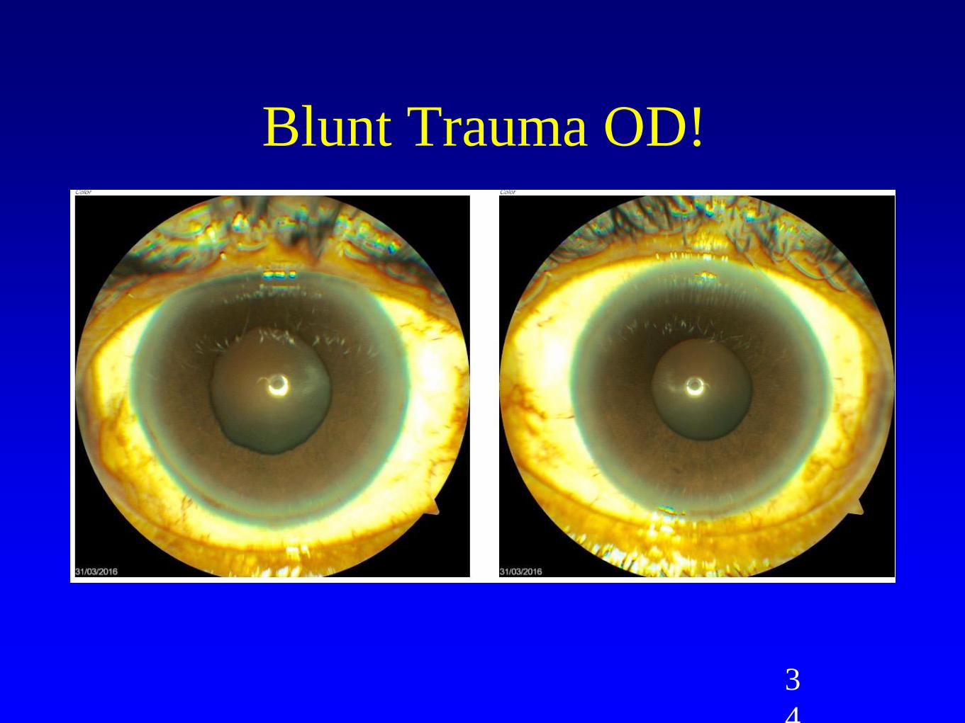
Eye Injuries
• Blunt trauma to the eye causing traumatic mydriasis - irido-
dialysis - angle recession resulting in secondary glaucoma -
cyclo-dialysis cleft resulting in hypotony
• Hyphaema - blood in anterior chamber and if prolonged risk of
corneal blood staining. ( Risk of recurrent haemorrhage )
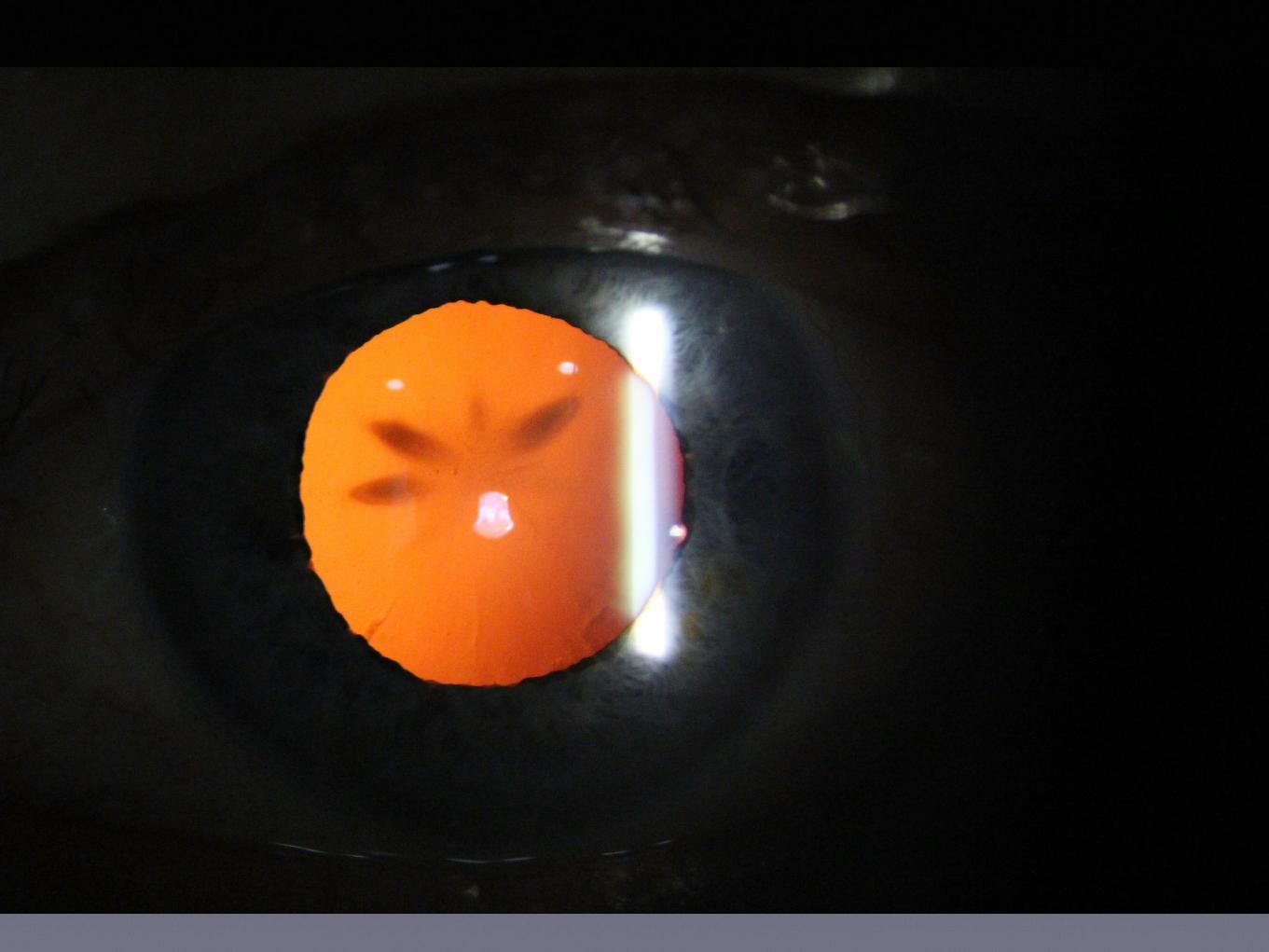
• Traumatic cataract with anterior petalloid pattern
• Globe rupture without open wound - Note of risk from
previous eye surgery
• Lacerations to cornea and sclera with hallmark irregular pupil

Left optic disc examination
3
4
Blunt Trauma OD!

3
5
Blunt Trauma OD(Nail gun!)
Iridodialysis
Polycoria

Corneal Injuries

Eye Injuries
• Commotio retinae = “contra coup”equivalent with
energy transferred to posterior segment and
specific damage to the macula. Acutely
haemorrhage may be present.
• Retinal detachment when trauma results in a
retinal tear/hole with fluid under the
retina…symptoms of flashes and floaters with a
shadow across the vision…early detection and
surgical treatment with vitrectomy results in better
outcomes

Eye Injuries• Surface foreign bodies especially on the cornea symptomatic and
easily seen but should be removed under slit lamp control
• Rust rings
• History of injury paramount to determine required
evaluation…grinding injury more likely to be low velocity rather than
hitting metal on metal with high velocity and risk of penetration
• Plain X Ray orbits no longer adequate and best imaging via CT
Scan
• Removal depending on size and nature of FB…magnet for iron
• Missed diagnosis = risk of siderosis and loss of vision!

CT Scan showing three
intra-ocular FB’s Corneal FB
Head Injuries
• Extremely variable deficits associated with severity of head
injury
• Recovery prolonged and often more rapid in younger patients
• Visual field defects often homonymous
• Parietal lobe injuries resulting in hemi-field neglect
• Convergence/accommodative insufficiency commonly occurs
but usually slowly resolves with time
• Beware of reading difficulties in presbyopic age group which
appear to be potentiated by the injury (coincidental)

Thank you