functional anatomy of the spine
TRANSCRIPT

Chapter 32
Functional anatomy of the spine
NIKOLAI BOGDUK*Newcastle Bone and Joint Institute, University of Newcastle, Newcastle, Australia
Abstract
Among other important features of the functional anatomy of the spine, described in this chapter, is theremarkable difference between the design and function of the cervical spine and that of the lumbar spine.In the cervical spine, the atlas serves to transmit the load of the head to the typical cervical vertebrae. Theaxis adapts the suboccipital region to the typical cervical spine. In cervical intervertebrtal discs the anulusfibrosus is not circumferential but is crescentic, and serves as an interosseous ligament in the saddle jointbetween vertebral bodies. Cervical vertebrae rotate and translate in the sagittal plane, and rotate in themanner of an inverted cone, across an oblique coronal plane. The cervical zygapophysial joints are themost common source of chronic neck pain. By contrast, lumbar discs are well designed to sustain com-pression loads, but rely on posterior elements to limit axial rotation. Internal disc disruption is the mostcommon basis for chronic low-back pain. Spinal muscles are arranged systematically in prevertebral andpostvertebral groups. The intrinsic elements of the spine are innervated by the dorsal rami of the spinalnerves, and by the sinuvertebral nerves. Little modern research has been conducted into the structure ofthe thoracic spine, or the causes of thoracic spinal pain.
INTRODUCTION
In writing a chapter on anatomy for neurologists the riskarises of being arcane or banal. Neurologists will alreadybe familiar with the precepts of classic anatomy, andwould not be inclined to read a chapter that repeats bor-ing, undergraduate material. For these reasons, the pre-sent chapter has been cast in a different manner.Although conventional elements of anatomy arereprised, they are permeated by several themes. Newfacts are provided, stemming frommodern research intothe structure of the spine, along with new perceptionsabout design and function. Throughout, the focus ison clinical relevance, particularly with respect to themechanisms of spinal injury and spinal pain. In thatregard, certain structures – ignored in conventionalundergraduate curricula – are promoted to epidemiolog-ically significant, clinical importance.
CERVICAL SPINE
The cervical spine serves as a mobile support for the sen-sory platform of the head. It allows the sensory apparatusfor vision, hearing, and smell to be elevated or depressedin the sagittal plane, and to scan the environment in thehorizontal plane. In order to subserve these functions,the cervical spine has to be mobile, yet sufficiently strongto support the weight of the head. Its vulnerability, toeither minor or major injuries, lies in being long, slender,and carrying the large mass of the head at its summit.
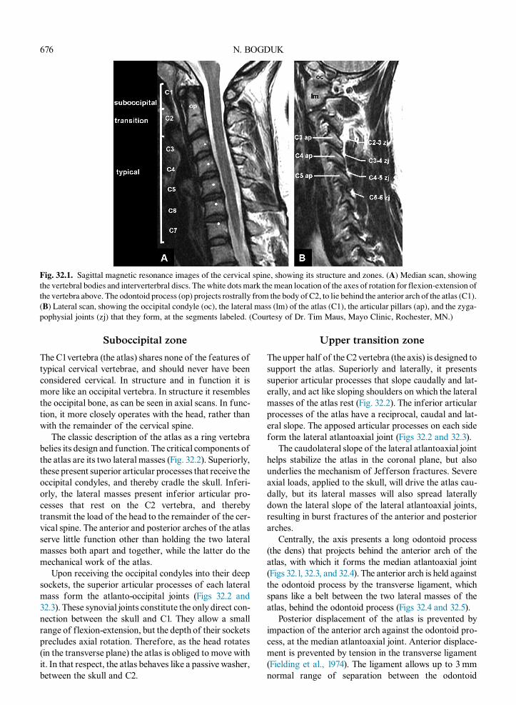
Both for descriptive purposes and functionally, thecervical spine can be divided into three zones: the suboc-cipital zone, centered on the C1 vertebra; a transitionalzone formed by the C2 vertebra; and the typical zone,encompassing the C–7 vertebrae (Bogduk and Mercer,2000) (Fig. 32.1). These zones differ both in structureand in function.
*Correspondence to: Nikolai Bogduk, PO Box 128, The Junction, New South Wales 2291, Australia. E-mail: nbogduk@bigpond.
net.au
Handbook of Clinical Neurology, Vol. 136 (3rd series)Neuroimaging, Part IIJ.C. Masdeu and R.G. Gonzalez, Editors© 2016 Elsevier B.V. All rights reserved
Suboccipital zone
The C1 vertebra (the atlas) shares none of the features oftypical cervical vertebrae, and should never have beenconsidered cervical. In structure and in function it ismore like an occipital vertebra. In structure it resemblesthe occipital bone, as can be seen in axial scans. In func-tion, it more closely operates with the head, rather thanwith the remainder of the cervical spine.
The classic description of the atlas as a ring vertebrabelies its design and function. The critical components ofthe atlas are its two lateral masses (Fig. 32.2). Superiorly,these present superior articular processes that receive theoccipital condyles, and thereby cradle the skull. Inferi-orly, the lateral masses present inferior articular pro-cesses that rest on the C2 vertebra, and therebytransmit the load of the head to the remainder of the cer-vical spine. The anterior and posterior arches of the atlasserve little function other than holding the two lateralmasses both apart and together, while the latter do themechanical work of the atlas.
Upon receiving the occipital condyles into their deepsockets, the superior articular processes of each lateralmass form the atlanto-occipital joints (Figs 32.2 and32.3). These synovial joints constitute the only direct con-nection between the skull and C1. They allow a smallrange of flexion-extension, but the depth of their socketsprecludes axial rotation. Therefore, as the head rotates(in the transverse plane) the atlas is obliged to move withit. In that respect, the atlas behaves like a passive washer,between the skull and C2.
Upper transition zone
The upper half of the C2 vertebra (the axis) is designed tosupport the atlas. Superiorly and laterally, it presentssuperior articular processes that slope caudally and lat-erally, and act like sloping shoulders on which the lateralmasses of the atlas rest (Fig. 32.2). The inferior articularprocesses of the atlas have a reciprocal, caudal and lat-eral slope. The apposed articular processes on each sideform the lateral atlantoaxial joint (Figs 32.2 and 32.3).
The caudolateral slope of the lateral atlantoaxial jointhelps stabilize the atlas in the coronal plane, but alsounderlies the mechanism of Jefferson fractures. Severeaxial loads, applied to the skull, will drive the atlas cau-dally, but its lateral masses will also spread laterallydown the lateral slope of the lateral atlantoaxial joints,resulting in burst fractures of the anterior and posteriorarches.
Centrally, the axis presents a long odontoid process(the dens) that projects behind the anterior arch of theatlas, with which it forms the median atlantoaxial joint(Figs 32.1, 32.3, and 32.4). The anterior arch is held againstthe odontoid process by the transverse ligament, whichspans like a belt between the two lateral masses of theatlas, behind the odontoid process (Figs 32.4 and 32.5).
Posterior displacement of the atlas is prevented byimpaction of the anterior arch against the odontoid pro-cess, at the median atlantoaxial joint. Anterior displace-ment is prevented by tension in the transverse ligament(Fielding et al., 1974). The ligament allows up to 3 mmnormal range of separation between the odontoid
Fig. 32.1. Sagittal magnetic resonance images of the cervical spine, showing its structure and zones. (A) Median scan, showing
the vertebral bodies and interverterbral discs. The white dotsmark themean location of the axes of rotation for flexion-extension of
the vertebra above. The odontoid process (op) projects rostrally from the body of C2, to lie behind the anterior arch of the atlas (C1).
(B) Lateral scan, showing the occipital condyle (oc), the lateral mass (lm) of the atlas (C1), the articular pillars (ap), and the zyga-
pophysial joints (zj) that they form, at the segments labeled. (Courtesy of Dr. Tim Maus, Mayo Clinic, Rochester, MN.)
676 N. BOGDUK

process and the anterior arch in adults, and 5 mm in chil-dren. In the past, the magnitude of the interval betweenthe anterior arch and the odontoid process has been usedas ameasure of atlantoaxial instability, but as a predictorof neurologic compromise the posterior atlantodentalinterval (Fig. 32.3) has greater sensitivity and specificity(Wasserman et al., 2011).
Severe forces delivered anteriorly to the head can frac-ture the odontoid process. Such fractures threaten the sag-ittal stability of the atlas. In turn, anterior or posteriordisplacement of the atlas can threaten the spinal cord.Rheumatoid arthritis of the atlantoaxial joints canweakenthe transverse ligament of the atlas, resulting in anteriorsubluxation of the atlas (Wasserman et al., 2011).
Fig. 32.2. Coronal magnetic resonance images of the cervical spine, showing the structure of its components. (A) Anterior scan,
showing the occipital condyles (oc) resting in the sockets of the lateral masses (lm) of the atlas, and forming the atlanto-occipital
joints (aoj); and the lateral masses bracketing the odontoid process (op), and resting on the “shoulders” of the axis (C2), where they
form the lateral atlantoaxial joints (laaj). The vertebral bodies of C2–7 form the anterior column of the cervical spine. (B) Posterior
scan, through the synovial joints of the cervical spine. Note the wedge shape of the lateral mass (lm) between the atlanto-occipital
joint (aoj) and the lateral atlantoaxial joint (laaj). The zygapophysial joint at C2–3 slopes caudally and medially, but those at suc-
cessive levels are essentially horizontal. The dotted line illustrates the ellipsoid shape depicted by the C2–3 zygapophysial joints
and the C2–3 disc, into which the atlas (C2) nestles on to the typical cervical spine.
Fig. 32.3. Close-up views of sagittal magnetic resonance images of the suboccipital joints. (A) Median scan through the odon-
toid process (op) and vertebral body of C2. With the front of the odontoid process, the anterior arch (aa) of the atlas forms the
median atlantoaxial joint (maaj). The transverse arrow marks the posterior atlantodental (pa) interval. (B) Lateral scan through
the lateral mass (lm) of the atlas. With the superior sockets of the lateral mass of the atlas, the occipital condyle (oc) forms the
atlanto-occipital joint (aoj). With C2, the lateral mass forms the lateral atlantoaxial joint (laaj). Note the bi-convex shape of
the lateral atlantoaxial joint. The triangular, white signals anteriorly and posteriorlywithin the joint are the fibroadiposemeniscoids
that it contains. (Courtesy of Dr. Tim Maus, Mayo Clinic, Rochester MN.)
FUNCTIONAL ANATOMY OF THE SPINE 677

Although the osseous articular processes of the lat-eral atlantoaxial joint are flat, their articular cartilagesare convex (along the sagittal plane) (Koebke andBrade, 1982) (Fig. 32.3). As a result, the atlas perchessomewhat precariously on the axis, with its convex infe-rior articular cartilages balancing on the convexities ofthe superior articular cartilages of the axis. The spacesanteriorly and posteriorly between the convex cartilagesare filled by wedge-shaped fibrocartilaginous menis-coids (Mercer and Bogduk, 1993).
Although a small degree of flexion and extension ispossible between the atlas and the axis (Bogduk andMercer, 2000), the cardinal movement between thesetwo vertebrae is axial rotation. During this movementthe atlas pivots at the median atlantoaxial joint, whileits lateral masses slide backwards or forwards, circum-ferentially, at the lateral atlantoaxial joints. However,because of the convexity of the articular cartilages inthese joints, the lateral masses also descend, down theposterior or anterior slope of the cartilages, as theymovebackwards or forwards, respectively. As a result, theatlas settles (lowers or screws down) during axial rota-tion, and rises when the movement is reversed(Bogduk and Mercer, 2000). During these displace-ments, the meniscoids of the joints cover the exposedsurfaces of the subluxating articular cartilages.
The total range of axial rotation of the atlas is consid-erably large. It has been measured as 43�5.5° whicheffectively amounts to 50%of the range of axial rotationof the head and neck (Bogduk andMercer, 2000). At theextremes of this range, very little of the articular
cartilages of the lateral atlantoaxial joints remainopposed; the joint is almost dislocated.
In order to accommodate this large range of motion,the capsules of the lateral atlantoaxial joint are loose,and serve little to hold the atlas on the axis. That serviceis provided by the alar ligaments. On each side, these lig-aments pass essentially transversely from the upper endof the odontoid process to the margin of the foramenmagnum (Fig. 32.5). In doing so, they bypass the atlas,and lock the head into place on the axis, effectively clamp-ing the atlas between the skull and C2. The alar ligamentsare the cardinal restraint to axial rotation of the head(Dvorak et al., 1987). They are sufficiently strong to pre-vent anterior dislocation of the head even if the transverseligament is completely severed (Fielding et al., 1974). Dis-ruption of an alar ligament can result in rotatory instabil-ity of the head and atlas (Dvorak et al., 1987).
Excessive axial rotation of the atlas can result in a lat-eral mass dislocating at the lateral atlantoaxial joint,causing fixed atlantoaxial deformity (Wortzman andDewar, 1968). A less dramatic form of torticollis canarise if, after rotation of the head and atlas, a meniscoidof the lateral atlantoaxial joint fails to re-enter the jointspace, catches under the capsule of the joint, and acts likea loose body to prevent derotation of the joint (Mercerand Bogduk, 1993).
Fig. 32.4. A sketch of an axial (top) view of the atlas sitting on
the axis. The lateral masses of the atlas bracket the odontoid
process (op) of the axis, and are themselves joined by the ante-
rior (aa) and posterior (pa) arches of the atlas.With the anterior
arch, the odontoid process forms the median atlantoaxial joint
(maaj). The transverse ligament (tl) spans like a belt behind the
odontoid processes, between the two lateral masses.
Fig. 32.5. A sketch of the suboccipital joints and ligaments, as
view from behind with the posterior arch of the atlas resected.
The lateral mass (lm) of the atlas supports the occipital bone,
and rests of the axis, forming the atlanto-occipital joint (aoj)
above, and the lateral atlantoaxial joint (laaj) below. The trans-
verse ligament (tl) holds the atlas (al) against the odontoid pro-
cess (op). The alar ligaments bind the odontoid process to the
margins of the foramen magnum, thereby connecting the skull
to C2 but bypassing C1.
678 N. BOGDUK

Increasing interest has been focused on the lateralatlantoaxial joints as a possible source of cervicogenicheadache. This contention can be tested by controlled,intra-articular, diagnostic blocks of the putatively pain-ful joint (Bogduk and Bartsch, 2008; Bogduk andGovind, 2009; Bogduk, 2014).
Lower transition zone
The lower half of the C2 vertebra has the structure of atypical cervical vertebra (Figs 32.1 and 32.2). Centrally itpresents a vertebral body, and laterally it presents pairedinferior articular processes. Having received the lateralmasses of the atlas, the axis transmits the load of thehead along an anterior channel, through its vertebralbody to the vertebral bodies below, and along paired pos-terior channels, through the zygapophysial joints(Fig. 32.6). Approximately half of the axial load is trans-mitted through the anterior channel, and half through thetwo posterior channels.
Typical cervical vertebrae
The cardinal elements of a typical cervical vertebra areits vertebral body and two articular pillars (Figs 32.1
and 32.2). Secondarily, transverse processes project lat-erally from the articular pillars, and posteriorly the twopillars are united by a pair of laminae, which support amidline spinous process at their junction. The transverseprocesses and spinous processes serve as levers uponwhich act the muscles that control the position of the cer-vical vertebrae. Along its superior, posterolateral marginon each side, each vertebral body bears uncinate pro-cesses. Previously enigmatic, the uncinate processesunderlie the nature of the joints between the cervical ver-tebral bodies and how they operate.
Consecutive articular pillars are united by the zy-gapophysial joints (Figs 32.1 and 32.2), which are synovialjoints formed by the inferior articular process of thevertebra above and the superior articular process ofthe vertebra below. Fibroadipose meniscoids intervenebetween the articular cartilages of these joints (Mercerand Bogduk, 1993). The zygapophysial joints are planar,and at typical cervical levels are oriented at about 40° tothe coronal and transverse planes, so that they face back-wards and upwards (Nowitzke et al., 1994). At the C2–3level, however, the joints also facemedially, such that thepair of joints depict an ellipsoid socket into which nestlesthe weight of the axis, and the load that it carries fromthe head (Figs 32.2 and 32.6).
Consecutive vertebral bodies are united by interverteb-ral discs, and by the anterior and posterior longitudinal lig-aments (Mercer andBogduk, 1999). The anterior ligamentconnects only the typical cervical vertebrae, from C2 cau-dally. The posterior longitudinal ligament forms acarpet along the floor of the vertebral canal at typical cer-vical levels, but expands into the membrana tectoria tocover the back of the atlantoaxial region. In doing so,the ligament separates the dural sac and spinal cord fromthe mechanics of the median atlantoaxial joint.
Posterior ligaments are lacking in the cervical spine.Interspinous ligaments are represented by only a sagittallayer of fascia (Mercer and Bogduk, 2003). The ligamen-tum nuchae lacks the structure of a ligament. It consistslargely of a narrow, coronal raphe, anchored to the C7spinous process, and formed by interlacing tendons ofthe splenius muscles and trapezius (Mercer andBogduk, 2003).
The intrinsic structure of the cervical intervertebraldiscs is unlike that of lumbar discs, and differs withage (Oda et al., 1988; Mercer and Bogduk, 1999). Thenucleus pulposus of cervical discs is gelatinous only inchildren and young adults. By the age of 30 it driesout to form a fibrocartilaginous plate (Oda et al.,1988). Moreover, the nucleus is not surrounded by con-centric lamellae of the anulus fibrosus (Mercer andBogduk, 1999). The anulus fibrosus is largely deficientposteriorly, and consists of a thin, paramedian band ofcollagen fibers that run longitudinally between the
Fig. 32.6. A sketch of a coronal view of how forces from the
head are transmitted into the cervical spine. On each side, the
weight of the head passes through the occipital condyle (oc),
into the lateral mass (lm) of the atlas, and into the axis
(C2) through the lateral atlantoaxial joint. From there, the forces
diverge, partly into the posterior column of zygapophysial
joints, and partly into the anterior column of vertebral bodies
and discs. Half the load passes anteriorly and half posteriorly.
FUNCTIONAL ANATOMY OF THE SPINE 679

vertebral bodies (Fig. 32.7). Posterolaterally, the nucleusis covered by the posterior longitudinal ligament, ratherthan by anulus fibrosus. Anteriorly, the anulus fibrosusis crescentic in shape, thin posteriorly near the uncinateprocesses, but thicker anteriorly towards the midline. Allof its collagen fibers pass in a similar direction, effec-tively aiming to a median point on the lower anterior sur-face of the vertebral body above. This configurationendows the anulus fibrosus with the structure of a thickinterosseous ligament that binds the anterior edges ofconsecutive vertebral bodies.
The superior surface of each cervical vertebral bodypresents two curvatures: a slight convex curvature alongthe sagittal plane, and a deep concave curvature trans-versely between the uncinate processes (Fig. 32.8). Thesecurvatures endow the vertebral body with the configura-tion of a saddle joint (Bogduk and Mercer, 2000). Con-sequently, the cervical interbody joints operate like asaddle joint, with motion restricted to two planes: thesagittal plane and an oblique coronal plane.
In the sagittal plane, the vertebral bodies can rock andslide (rotate and translate), to provide for flexion andextension of the neck. From above downwards, the typ-ical cervical vertebrae exhibit progressively less transla-tion for each degree of rotation, during flexion orextension. This is reflected by the different locationsof their axes of movement. At higher levels the axes
lie in the vertebral body below the moving vertebra,but are progressively closer to their intervertebral discat lower levels (Amevo et al., 1991) (Fig. 32.1). These dif-ferences correlate strongly with the height of the articu-lar pillar at each segment (Nowitzke et al., 1994). Tallerpillars provide less space into which the vertebra cantranslate once it has commenced sagittal rotation. Con-versely, at segments with shorter pillars, sagittal rotationlifts the inferior articular processes of the moving verte-bra off the supporting articular pillar, and provides agreater gap into which it can translate (Nowitzkeet al., 1994).
The second plane of movement of typical cervicalvertebrae is set at 40° forwards of the coronal plane,and lies parallel to the plane of the zygapophysial joints
Fig. 32.7. Sketches of various views of the internal structure
of the cervical disc. In a front view, all fibers of the anterior
anulus fibrosus pass towards a point on the inferior anterior
surface of the vertebral body above. In a top view, the anulus
fibrosus (af) is crescentic in shape, thick anteriorly but tapering
at the uncinate processes (u). The nucleus pulposus (np) is a
fibrocartilaginous plate. Posteriorly the anulus is restricted
to a small bundle of paramedian, longitudinal fibers. A side
view shows the fibers of the anulus fibrosus passing upwards
and forwards. A transverse cleft runs from one uncinate pro-
cess to the other.
Fig. 32.8. Sketches of a posterolateral view of a typical cervi-
cal vertebral body, showing the two curvatures of its superior
surface: a downward concavity along the sagittal plane, and a
second concavity, facing upwards and forwards, between the
uncinate processes (u). These two concavities endow the inter-
vertebral disc with the features of a saddle joint.
680 N. BOGDUK
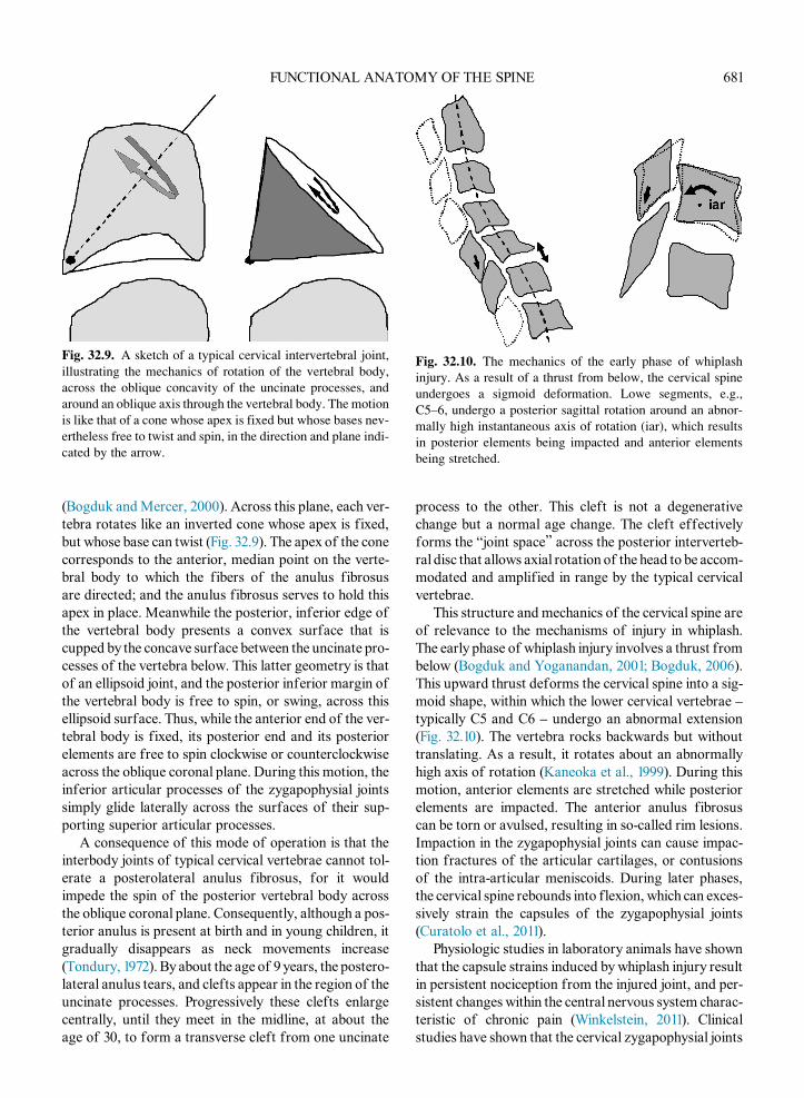
(Bogduk andMercer, 2000). Across this plane, each ver-tebra rotates like an inverted cone whose apex is fixed,but whose base can twist (Fig. 32.9). The apex of the conecorresponds to the anterior, median point on the verte-bral body to which the fibers of the anulus fibrosusare directed; and the anulus fibrosus serves to hold thisapex in place. Meanwhile the posterior, inferior edge ofthe vertebral body presents a convex surface that iscupped by the concave surface between the uncinate pro-cesses of the vertebra below. This latter geometry is thatof an ellipsoid joint, and the posterior inferior margin ofthe vertebral body is free to spin, or swing, across thisellipsoid surface. Thus, while the anterior end of the ver-tebral body is fixed, its posterior end and its posteriorelements are free to spin clockwise or counterclockwiseacross the oblique coronal plane. During this motion, theinferior articular processes of the zygapophysial jointssimply glide laterally across the surfaces of their sup-porting superior articular processes.
A consequence of this mode of operation is that theinterbody joints of typical cervical vertebrae cannot tol-erate a posterolateral anulus fibrosus, for it wouldimpede the spin of the posterior vertebral body acrossthe oblique coronal plane. Consequently, although a pos-terior anulus is present at birth and in young children, itgradually disappears as neck movements increase(Tondury, 1972). By about the age of 9 years, the postero-lateral anulus tears, and clefts appear in the region of theuncinate processes. Progressively these clefts enlargecentrally, until they meet in the midline, at about theage of 30, to form a transverse cleft from one uncinate
process to the other. This cleft is not a degenerativechange but a normal age change. The cleft effectivelyforms the “joint space” across the posterior interverteb-ral disc that allows axial rotation of the head to be accom-modated and amplified in range by the typical cervicalvertebrae.
This structure and mechanics of the cervical spine areof relevance to the mechanisms of injury in whiplash.The early phase of whiplash injury involves a thrust frombelow (Bogduk and Yoganandan, 2001; Bogduk, 2006).This upward thrust deforms the cervical spine into a sig-moid shape, within which the lower cervical vertebrae –typically C5 and C6 – undergo an abnormal extension(Fig. 32.10). The vertebra rocks backwards but withouttranslating. As a result, it rotates about an abnormallyhigh axis of rotation (Kaneoka et al., 1999). During thismotion, anterior elements are stretched while posteriorelements are impacted. The anterior anulus fibrosuscan be torn or avulsed, resulting in so-called rim lesions.Impaction in the zygapophysial joints can cause impac-tion fractures of the articular cartilages, or contusionsof the intra-articular meniscoids. During later phases,the cervical spine rebounds into flexion, which can exces-sively strain the capsules of the zygapophysial joints(Curatolo et al., 2011).
Physiologic studies in laboratory animals have shownthat the capsule strains induced by whiplash injury resultin persistent nociception from the injured joint, and per-sistent changes within the central nervous system charac-teristic of chronic pain (Winkelstein, 2011). Clinicalstudies have shown that the cervical zygapophysial joints
Fig. 32.9. A sketch of a typical cervical intervertebral joint,
illustrating the mechanics of rotation of the vertebral body,
across the oblique concavity of the uncinate processes, and
around an oblique axis through the vertebral body. The motion
is like that of a cone whose apex is fixed but whose bases nev-
ertheless free to twist and spin, in the direction and plane indi-
cated by the arrow.
Fig. 32.10. The mechanics of the early phase of whiplash
injury. As a result of a thrust from below, the cervical spine
undergoes a sigmoid deformation. Lowe segments, e.g.,
C5–6, undergo a posterior sagittal rotation around an abnor-
mally high instantaneous axis of rotation (iar), which results
in posterior elements being impacted and anterior elements
being stretched.
FUNCTIONAL ANATOMY OF THE SPINE 681

are the single most common source of chronic neckpain after whiplash, accounting for between 50% and60% of cases (Bogduk, 2011). Most commonly, neckpain – with referred pain to the shoulder girdle – stemsfrom the C5–6 joint, while headache stems from theC2–3 zygapophysial joint.
Lesswell understood is pain from the cervical interver-tebral discs. Conspicuously, degenerative disc disease isnot associated with neck pain. Furthermore, discogenicpain appears to be uncommon, once zygapophysial jointpain is taken into account (Yin and Bogduk, 2008). Per-haps discogenic pain is caused by strains of the inteross-eous ligament formed by the anterior anulus fibrosus, butdiagnostic techniques by which to test this propositionhave not been developed.
LUMBARSPINE
The cardinal role of the lumbar spine is to support thethorax and upper limbs – and any loads that theycarry – and to transmit those loads to the pelvis and lowerlimbs (Bogduk, 2012a). Secondarily, the lumbar spineaccommodates a modest range of movement betweenthe thorax and pelvis.
In order to subserve these functions, the essential ele-ments of the lumbar spine are the vertebral bodies of thefive lumbar vertebrae (Fig. 32.11). These are stacked intoa strong column, and are united by intervertebral discs
and by the anterior and posterior longitudinal ligaments(Bogduk, 2012a). Bowing the column into a lordosisendows the lumbar spine with the ability to absorbdynamic axial loads (bouncing). Axial impulses deformthe lordotic curve; the energy is absorbed by the elasticdiscs and longitudinal ligaments; and is returned torestore the more upright curve, once the axial impulsehas passed (Bogduk, 2012a).
The lumbar intervertebral discs are well designed toaccommodate compression loads (Hickey and Hukins,1980). Each consists of hydrated nucleus pulposus, sur-rounded by an anulus fibrosus, and capped superiorlyand inferiorly by a vertebral endplate that joins the discto the adjacent vertebral body (Fig. 32.12). The anulusfibrosus is formed by concentric layers of collagenfibers, in which the fibers in any one layer run in parallel,at about 60° to the long axis of the spine, but in succes-sive layers that orientation alternates.
Axial compression is resisted primarily by the concen-tric layers of the anulus fibrosus (Markolf and Morris,1974) (Fig. 32.12). However, the tendency of the anulusunder load is to buckle, both outwards and inwards. Thisbuckling is resisted by the hydrostatic nucleus pulposus.When the nucleus is compressed it exerts a radial pressurethat braces, and stiffens, the anulus, thereby preventing itfrom buckling. A small range of flexion-extension isaccommodated by the discs (about 13° per segment), dur-ing which the anulus fibrosus on the side to which
Fig. 32.11. Sagittal magnetic resonance images of the lumbar spine. (A) Median scan showing the vertebral bodies and spinous
processes (sp). The white dots mark the location of the axes of rotation of the vertebra above. (B) Lateral scan through the inter-
vertebral foramina and the L3–4 to L5–S1 zygapophysial joints. ped, pedicle of L3; sap, superior articular process of L4; iap,
inferior articular process of L5.
682 N. BOGDUK

movement occurs is compressed slightly, while the anuluson the opposite side is stretched (Bogduk, 2012a).
While strongly designed to resist compression, thelumbar discs are poorly designed to resist axial rotation.Because the collagen fibers of the anulus fibrosus alter-nate in direction in successive layers, only half are avail-able to resist axial rotation in one direction or the other.For stability in axial rotation, the lumbar vertebral bodiesand intervertebral discs rely on the posterior elements ofthe lumbar vertebrae (Bogduk, 2012a).
The posterior elements are based on an arch (Bogduk,2012a) (Fig. 32.13). The arch is supported by stout pedi-cles that emanate from the upper posterior surface ofeach vertebral body. The pedicles serve to transmitforces from the succeeding posterior elements to the ver-tebral bodies, which control the position or movementsof the vertebral bodies. The arch is completed by leftand right laminae that join in the midline. From the junc-tion of the two laminae springs a large spinous process,and from the junction between the pedicle and lamina oneach side arises a long transverse process. These pro-cesses serve as levers to which attach the muscles thatcontrol the movements of the lumbar vertebrae.
At its superior and inferior lateral corners respec-tively, each lamina bears a superior and inferior articularprocess. Like large mittens, the paired superior articularprocesses reach cranially to grasp the inferior articularprocesses of the vertebra above, and form the zygapo-physial joints. The plane of these joints is parallel tothe longitudinal axis of the lumbar spine. Consequently,during flexion of the vertebral bodies, the inferior artic-ular processes glide freely out of the sockets formed bythe superior articular processes, until movement isarrested by tension in the joint capsules (Bogduk,2012a). The axis of this movement typically lies in thedisc below the moving vertebra (Pearcy and Bogduk,1988) (Fig. 32.11A), which indicates only a small amountof translation for every degree of rotation of the moving
vertebra. As the inferior articular processes move, theylift away from the superior articular process, tanta-mount to partially subluxating the joint. Fibroadiposemeniscoids protect the exposed surfaces of the articularcartilages during this displacement (Engel and Bogduk,1982; Bogduk and Engel, 1984).
In axial views, the lumbar zygapophysial joints vari-ously present flat, C-shaped, or J-shaped appearances,which correspond to the primary functions of thesejoints (Horwitz and Smith, 1940). Flat joints essentiallyface medially and posteriorly. C-shaped joints have ananterior end that faces posteriorly, and a posterior endthat faces medially. J-shaped joints have a small anteriorlip facing posteriorly, and a larger surface facing medi-ally. The medially facing surfaces serve to resist axialrotation of the vertebrae. Attempted axial rotationswings the inferior articular process laterally, but thismovement is arrested by the opposing superior articularprocess. The range ofmotion is limited to about 2° or lessper segment (Pearcy and Tibrewal, 1984), and is accom-modated only by compression of the articular cartilage.The surfaces that face posteriorly serve to resist forwarddisplacement (listhesis) of the vertebra.
Impaction of an inferior articular process against itssuperior articular process tends to force the inferior pro-cess backwards, and lift the lamina from which it arises(like opening a hatchback). In turn this tendency stressesthe junction between the lamina and its pedicle. Repeatedimpactions – particularly during repeated axialrotation – can cause stress fractures at this point, result-ing in pars interarticularis defects.
The lumbar zygapophysial joints can be a source oflow-back pain, but its prevalence is uncertain. It appearsto be uncommon or rare in injured workers, but is com-mon in elderly patients (Bogduk, 2008, 2012b).
The most common cause of chronic low-back pain isinternal disc disruption (Bogduk et al., 2013). This condi-tion is characterized by degradation of the nucleus
Fig. 32.12. Close-up views of a sagittal magnetic resonance image of an L3–4 intervertebral disc. (A) The components of the disc.
np, nucleus pulposus; af, anulus fibrosus; vep, vertebral endplate. (B) The mechanics of the disc. Axial compression loads are
primarily borne by the lamellae of collagen in the anulus fibrosus. When compressed, the nucleus pulposus exerts radial pressure
to brace the anulus, and prevent it from buckling under load.
FUNCTIONAL ANATOMY OF THE SPINE 683

pulposus of the affected disc and the development ofradial fissures into the posterior or posterolateral anulus.The condition has been produced in laboratory animals,and pursued in numerous clinical studies. Its cause iscompression injuries that produce small fractures ofthe vertebral endplate. These result in degradation ofthe matrix of the nucleus pulposus. As the nucleusbecomes less able to retain water, it is no longer ableto pressurize and brace the anulus. Pressures in thenucleus drop, but rise in the posterior anulus. Theunbraced anulus progressively delaminates, particularlyin regions of high stress where the laminae are curved: atthe posterolateral corners or the posterior paramediansector. Pain arises as a result of chemical irritation ofnociceptors in the anulus by degradation products fromthe nucleus, and as a result of the increased mechanicalstresses on the surviving, intact laminae of anulus(Bogduk et al., 2013). To various degrees of certaintythe condition can be diagnosed by characteristic featuresonmagnetic resonance imaging, such asModic lesions inthe vertebral body or high-intensity zones in the anulusfibrosus, and by provocation discography (Bogduket al., 2013). No treatment has been vindicated, but sev-eral minimally invasive interventions are being pursued,which encompass ablating nociceptors in the disc, inject-ing restorative agents such as stem cells, or injectingantagonists of inflammation.
MUSCLES
The anatomy ofmuscles of the cervical and lumbar spineis made complex by the diversity of their numerousattachments. If those specifics are ignored, the anatomybecomes simpler.
Small muscles connect consecutive spinous processesand transverse processes. Too small to move their verte-brae effectively, these muscles serve as proprioceptorsfor the spine (Bastide et al., 1989).
Prevertebral muscles are represented only in the cer-vical spine (Standring, 2008). The longus cervicis con-nects the vertebral bodies and transverse processes ofthe cervical vertebrae. It is covered by the longus capitiswhich anchors the skull to the cervical vertebrae. Thesemuscles are weak flexors of the head and neck.
Various suboccipital muscles control movementsof the head in relation to the atlas and the axis. Theyare the rectus anterior and rectus lateralis anteriorly,and the rectus capitis posterior major and minor acc-ompanied by obliquus inferior and obliquus superior,posteriorly (Standring, 2008). Collectively these musclescontrol the orientation of the head on the atlas and axis.
The postvertebral muscles are aligned systematically,side by side and by layers (Standring, 2008). Multifidusis the deepest and most medial muscle. Its fascicles arisefrom a spinous process and descend to various insertionson articular processes and transverses processes one toseveral segments caudally. It is flanked by the longissi-mus system of muscles, which attach to transverse pro-cesses near their bases, and whose components are largeat lumbar levels, but virtuallyminiscule at cervical levels.Further laterally runs the iliocostalis system, whichattaches to transverse processes near their tips, andwhose components are, likewise, large at lumbar levelsbut miniscule at cervical levels. A semispinalis systemis vestigial at lumbar levels but well developed at cervicallevels. Semispinalis cervicis arises from the cervical spi-nous processes, and covers the multifidus with fascicleslonger than those of the latter muscle. Semispinalis capi-tis arises from the occiput, and is anchored to the cervical
Fig. 32.13. Sketches of the posterior elements of a lumbar vertebra. (A) Posterior view. The two laminae (la) form a quadrangular
plate, from whose corners project the superior (sap) and inferior (iap) processes. From the junction of the two laminae projects the
spinous process (sp). On each side, the inferior and superior articular processes of consecutive vertebrae form the zygapophysial
joint (zj) (B) Axial (top) view. The posterior elements are connected to the vertebral body (vb) by the pedicles (p). The transverse
process (tp) projects from the junction of the pedicle and lamina, on each side.
684 N. BOGDUK

transverse processes. It is the largest of the posteriorneck muscles. It is covered by the splenius muscle, whichpasses cranially and laterally from the raphe of the liga-mentum nuchae to wrap around all the other posteriormuscles of the neck. Splenius cervicis reaches the uppercervical transverse processes, while splenius capitisreaches the superior nuchal line. Variously and collec-tively, the cervical postvertebral muscles act to extendthe head and the cervical spine.
Other muscles use the vertebral column adventi-tiously, as a base from which to act on nonspinal struc-tures. In the neck, these include the scalene muscles,which act on the ribs; and levator scapulae and trapezius,which act on the shoulder girdle. Sternocleidomastoid isthe principal flexor and rotator of the head and neck, butpasses directly from the manubrium and clavicle to thehead, with no connection to the cervical spine. Beinglocked to the skull through the atlanto-occipital joints,the atlas is rotated when the sternocleidomastoid rotatesthe head.
In the lumbar spine, psoas major arises from the ver-tebral bodies, discs, and transverse processes to act onthe femur, but does not move the lumbar spine(Bogduk et al., 1992). Quadratus lumborum attachesto the lumbar transverse processes but acts principallyon the 12th rib; its actions on the lumbar vertebrae areeffectively trivial (Phillips et al., 2008). Transversusabdominis stems from the lumbar transverse processes,and has virtually no effect on the lumbar vertebrae(Macintosh et al., 1987). Likewise, latissimus dorsi gainssome anchorage to the lumbar spinous processes but hasa negligible action on the lumbar spine (Bogduket al., 1998).
INNERVATION
The C1 spinal nerve is unlike other spinal nerves, whichreinforces the atlas being suboccipital rather than cervi-cal in nature. This nerve lacks a typical dorsal root gan-glion, but ganglion cells can be found amongst therootlets of the spinal accessory nerve. The C1 dorsalramus appears amongst the posterior suboccipital mus-cles (Lazorthes and Gaubert, 1956). Sometimes it canhave a cutaneous branch. The C1 ventral ramus crossesthe posterior arch of the atlas, behind the superior artic-ular process. It innervates the atlanto-occipital jointbefore entering the cervical plexus (Lazorthes andGaubert, 1956).
The C2 spinal nerve lies behind the lateral atlantoaxialjoint, and forms a large dorsal ramus that supplies themore superficial posterior neck muscles, and becomescutaneous as the greater occipital nerve, over the occiput(Bogduk, 1982). The C2 ventral ramus supplies the
lateral atlantoaxial joint before joining the cervicalplexus (Lazorthes and Gaubert, 1956).
The C3–7 cervical spinal nerves lie above their like-numbered vertebrae, enclosed in their respective inter-vertebral foramina. They are joined by the C8 spinalnerve, which lies in the C7–T1 intervertebral foramen.The ventral rami of C3 and C4 join the cervical plexus,and the lower cervical ventral rami join the brachialplexus. The dorsal rami of the typical cervical spinalnerves form lateral branches that supply the splenius,longissimus, and iliocostalis; and medial branches thatsupply the deeper and medial posterior neck muscles,and the cervical zygapophysial joints (Bogduk, 1982).The cervical medial branches have constant locationson the cervical articular pillars, which allow them to betargeted for fluoroscopy-guided diagnostic blocks, bywhich pain from the zygapophysial joints can be diag-nosed (Bogduk, 1982, 2011).
Gray rami communicantes, from the stellate ganglionand from the cervical ventral rami, form a plexus –
called the vertebral nerve – that accompanies the verte-bral artery through the foramina transversaria ofthe neck, and into the posterior cranial fossa (Bogduket al., 1981a). Although migraine cervicale, or theBarre–Lieou syndrome, has been attributed to irritationof these nerves, and spasm of the vertebral artery, labo-ratory studies have shown the vertebrobasilar system tobe remarkable unresponsive to stimulation of the verte-bral nerve (Bogduk et al., 1981a; Lambert et al., 1984).
The cervical sinuvertebral nerves are formed bysomatic roots from the ventral rami and autonomic rootsfrom the rami communicantes in the vertebral nerve. Asrecurrent meningeal branches they innervate the cervicaldural sac, but also innervate the cervical discs and theposterior longitudinal ligament (Bogduk et al., 1988).The C1–3 sinuvertebral nerves innervate the ligamentsof the median atlantoaxial joint before passing throughforamen magnum to supply the dura mater over the cli-vus (Kimmel, 1960).
The lumbar spinal nerves lie obliquely in their inter-vertebral foramina, each below the like-numbered verte-bra. Their ventral rami enter the lumbar or lumbosacralplexus. Their dorsal rami form lateral and intermediatebranches that innervate the iliocostalis and longissimusmuscles respectively (Bogduk et al., 1982; Bogduk,1983). Medial branches innervate the lumbar zygapophy-sial joints and the multifidus (Bogduk et al., 1982;Bogduk, 1983). Where the medial branches cross the rootof the superior articular process they can be targeted forfluoroscopy-guided diagnostic blocks, by which painfrom the lumbar zygapophysial joints can be diagnosed(Bogduk, 1983, 2008, 2012b).
At each segmental level, the lumbar sinuvertebralnerves arise from the ventral ramus and gray ramus
FUNCTIONAL ANATOMY OF THE SPINE 685

communicans. Each passes back into the intervertebralforamen to supply the dural sac, the posterior longitudi-nal ligament, and the posterior anulus fibrosus (Bogduket al., 1981b; Bogduk, 1983). These nerves provide thesensory pathway for lumbar discogenic pain.
THORACIC SPINE
There have been no substantial advances in the descrip-tion of the anatomy of the thoracic spine since editionsof anatomy textbooks of the 19th and 18th century. Inparallel, there has been little advance in the understand-ing of thoracic spinal pain and its sources, let alonecauses. No diagnostic or treatment procedures have beenvalidated. Thoracic spinal pain essentially remains amystery.
Like cervical and lumbar vertebrae, the thoracic ver-tebrae have vertebral bodies that are connected by inter-vertebral discs and longitudinal ligaments, and posteriorelements that are connected by zygapophysial joints(Fig. 32.14). The distinction of the thoracic spine is thatit suspends the ribs. At typical thoracic levels, the headof the rib articulates with the intervertebral disc anddemifacets on the edges of the vertebrae that bind thatdisc, and the articular tubercle of the rib articulates withthe transverse process of the upper of the two vertebrae
(Fig. 32.15). Exceptions to this arrangement occur at T1and at T11 and T12, where the head of the rib fully artic-ulates with the like-numbered vertebrae.
Few studies have explored the innervation of the tho-racic spine (Bogduk, 2002). The thoracic sinuvertebralnerves are assumed to be homologous to those at cervicalor lumbar levels. The courses of the thoracic dorsal ramiappear to differ from those at cervical and lumbar levels,but are nevertheless homologous (Chua and Bogduk,1995). Whereas the medial branches at cervical and lum-bar levels wind around the base of the superior articularprocess at each segmental level, at thoracic levels thedorsal ramus stretches to the tip of the transverse pro-cess before dividing into medial and lateral branches.This difference is reconciled once it is realized that whatare called the transverse processes at cervical and lumbarlevels are embryologically costal elements (rudimentaryribs), whereas the embryologic transverse elements (ortrue transverse processes) are absorbed into the baseof the superior articular process. Consequently, at cervi-cal and lumbar levels, the medial branches cross thesuperior articular process because the true transverseprocesses also lie there. This distinction becomes perti-nent for minimally invasive, diagnostic, and treatmentprocedures that target thoracic medial branches. The tar-get lies on the transverse process, not on the superiorarticular process (Chua and Bogduk, 1995).
A persisting curiosity pertains to the structure of tho-racic intervertebral discs. Cervical discs differ greatlyfrom lumbar discs, but undiscovered is the transition
Fig. 32.14. Sagittal magnetic resonance images of the thoracic
spine. (A) Median section, through the vertebral bodies, spinal
cord (sc), and spinous processes (sp) cord. (B) Paramedian sec-
tion through the zygapophysial joints (zj). Intervertebral discs
(ivd) are evidence in both sections. (Courtesy of Dr. TimMaus,
Mayo Clinic, Rochester MN.)
Fig. 32.15. Axial magnetic resonance image of a typical tho-
racic spinal segment. vb, vertebral body; zj, zygapophysial
joint; sp, spinous process; cvj, costovertebral joint; ctj,
costotransverse joint. (Courtesy of Dr. Tim Maus, Mayo
Clinic, Rochester, MN.)
686 N. BOGDUK

zone. Are thoracic discs like cervical discs, or do they havethe structure of lumbar discs?Given that cervical uncinateprocesses are homologous to the heads of the ribs, unpub-lished observations suggest that discs change their struc-ture where uncinate processes or their rib equivalentcease. Thoracic discs become lumbar in nature at T11,where the rib no longer articulates with the disc.
REFERENCES
Amevo B, Worth D, Bogduk N (1991). Instantaneous axes of
rotation of the typical cervical motion segments: a study in
normal volunteers. Clin Biomech 6: 111–117.Bastide G, Zadeh J, Lefebvre D (1989). Are the ‘little muscles’
what we think they are? Surg Radiol Anat 11: 255–256.Bogduk N (1982). The clinical anatomy of the cervical dorsal
rami. Spine 7: 319–330.
Bogduk N (1983). The innervation of the lumbar spine. Spine
8: 286–293.BogdukN (2002). Innervation and pain patterns of the thoracic
spine. In: R Grant (Ed.), Physical therapy of the Cervical
and Thoracic Spine, 3rd edn. Churchill Livingstone, New
York, pp. 73–81.
Bogduk N (2006). Whiplash injury. In: F Cervero, TS Jensen
(Eds.), Handbook of Clinical Neurology Vol. 81: Pain,
Elsevier, Amsterdam, pp. 791–801.
Bogduk N (2008). Evidence-informedmanagement of chronic
back pain with facet injections and radiofrequency neurot-
omy. Spine J 8: 56–64.Bogduk N (2011). On cervical zygapophysial joint pain after
whiplash. Spine 36: S194–S199.BogdukN (2012a). Clinical Anatomy of the Lumbar Spine and
Sacrum, 5th edn. Elsevier, Edinburgh.
Bogduk N (2012b). Lumbar medial branch neurotomy. In:
S Dagenais, S Haldeman (Eds.), Evidence-Based
Management of Low Back Pain, Elsevier, St Louis,
pp. 351–363.
Bogduk N (2014). The neck and headaches. Neurol Clin 32:471–487.
Bogduk N, Bartsch T (2008). Cervicogenic headache. In:
SD Silberstein, RB Lipton, DW Dodick (Eds.), Wolff’s
Headache, 8th edn. Oxford University Press, New York,
pp. 551–570.
Bogduk N, Engel R (1984). The menisci of the lumbar zyga-
pophyseal joints. A review of their anatomy and clinical
significance. Spine 9: 454–460.
Bogduk N, Govind J (2009). Cervicogenic headache: an
assessment of the evidence on clinical diagnosis, invasive
tests, and treatment. Lancet Neurol 8: 959–968.Bogduk N, Mercer SR (2000). Biomechanics of the cer-
vical spine. I: Normal Kinematics. Clin Biomech 15:633–648.
Bogduk N, Yoganandan N (2001). Biomechanics of the cervi-
cal spine Part 3: minor injuries. Clin Biomech 16: 267–275.Bogduk N, Lambert G, Duckworth JW (1981a). The anatomy
and physiology of the vertebral nerve in relation to cervical
migraine. Cephalalgia 1: 11–24.Bogduk N, TynanW,Wilson AS (1981b). The nerve supply to
the human lumbar intervertebral discs. J Anat 132: 39–56.
Bogduk N, Wilson AS, Tynan W (1982). The human lumbar
dorsal rami. J Anat 134: 383–397.BogdukN,WindsorM, Inglis A (1988). The innervation of the
cervical intervertebral discs. Spine 13: 2–8.
BogdukN, PearcyM,Hadfield G (1992). Anatomy and biome-
chanics of psoas major. Clin Biomech 7: 109–119.Bogduk N, Johnson G, Spalding D (1998). The morphology
and biomechanics of latissimus dorsi. Clin Biomech 13:377–385.
Bogduk N, Aprill C, Derby R (2013). Lumbar discogenic pain:
state-of-the-art review. Pain Med 14: 813–836.ChuaWH, BogdukN (1995). The surgical anatomy of thoracic
facet denervation. Acta Neurochir 136: 140–144.Curatolo M, Bogduk N, Ivancic PC et al. (2011). The role of
tissue damage in whiplash-associated disorders. Spine 36:S309–S315.
Dvorak J, Hayek J, Zehnder R (1987). CT-functional diagnos-
tics of the rotatory instability of the upper cervical spine
part 2. An evaluation on healthy adults and patients with
suspected instability. Spine 12: 726–731.
Engel R, Bogduk N (1982). The menisci of the lumbar zyga-
pophysial joints. J Anat 135: 795–809.Fielding JW, Cochran G van B, Lawsing JF et al. (1974). Tears
of the transverse ligament of the atlas. J Bone Joint Surg
56A: 1683–1691.Hickey DS, Hukins DWL (1980). Relation between the struc-
ture of the anulus fibrosus and the function and failure of
the intervertebral disc. Spine 5: 100–116.Horwitz T, Smith RM (1940). An anatomical, pathological
and roentgenological study of the intervertebral joints
of the lumbar spine and of the sacroiliac joints. Am
J Roentgenol 43: 173–186.Kaneoka K, Ono K, Inami S et al. (1999). Motion analysis of
cervical vertebrae during whiplash loading. Spine 24:763–770.
Kimmel DL (1960). Innervation of the spinal dura mater and
dura mater of the posterior cranial fossa. Neurology 10:800–809.
Koebke J, Brade H (1982).Morphological and functional stud-
ies on the lateral joints of the first and second cervical ver-
tebrae in man. Anat Embryol 164: 265–275.Lambert GA, Duckworth JW, Bogduk N et al. (1984). Low
pharmacological responsiveness of the vertebro-basilar cir-
culation inMacaca nemestrina monkeys. Eur J Pharmacol
102: 451–458.Lazorthes G, Gaubert J (1956). L’innervation des articulations
interapophysaire vertebrales. Comptes Rendues de
l’Association des Anatomistes 43: 488–494.Macintosh JE, Bogduk N, Gracovetsky S (1987). The biome-
chanics of the thoracolumbar fascia. Clin Biomech 2:
78–83.Markolf KL, Morris JM (1974). The structural components
of the intervertebral disc. J Bone Joint Surg 56A:
675–687.Mercer S, Bogduk N (1993). Intra-articular inclusions of the
cervical synovial joints. Br J Rheumatol 32: 705–710.
Mercer S, Bogduk N (1999). The ligaments and anulus fibro-
sus of human adult cervical intervertebral discs. Spine 24:619–626.
FUNCTIONAL ANATOMY OF THE SPINE 687

Mercer SR, Bogduk N (2003). Clinical anatomy of ligamen-
tum nuchae. Clin Anat 16: 484–493.Nowitzke A, Westaway M, Bogduk N (1994). Cervical zyga-
pophyseal joints: geometrical parameters and relationship
to cervical kinematics. Clin Biomech 9: 342–348.Oda J, Tanaka H, Tsuzuki N (1988). Intervertebral disc
changes with aging of human cervical vertebra from the
neonate to the eighties. Spine 13: 1205–1211.PearcyMJ, BogdukN (1988). Instantaneous axes of rotation of
the lumbar intervertebral joints. Spine 13: 1033–1041.
Pearcy MJ, Tibrewal SB (1984). Axial rotation and lateral
bending in the normal lumbar spine measured by three-
dimensional radiography. Spine 9: 582–587.Phillips S, Mercer S, Bogduk N (2008). Anatomy and
biomechanics of quadratus lumborum. J Eng Med 222:151–159.
Standring S (Ed.), (2008). Gray’s Anatomy, 40th edn.
Churchill Livingstone, Edinburgh, pp. 736–743.
Tondury G (1972). The behaviour of the cervical discs during
life. In: C Hirsch, Y Zotterman (Eds.), Cervical pain,
Pergamon Press, Oxford, pp. 59–66.
Wasserman BR, Moskovitch R, Razi AE (2011). Rheumatoid
arthritis of the cervical spine. Clinical considerations. Bull
Hosp Joint Dis 68: 136–148.Winkelstein BA (2011). How can animal models inform on the
transition to chronic symptoms in whiplash? Spine 36:
S218–S225.Wortzman G, Dewar FP (1968). Rotary fixation of the atlan-
toaxial joint: rotational atlantoaxial subluxation.
Radiology 90: 479–487.
YinW, Bogduk N (2008). The nature of neck pain in a private
pain clinic in the United States. Pain Med 9: 196–203.
688 N. BOGDUK