functional analysis of the cis-regulatory elements …functional analysis of the cis-regulatory...
TRANSCRIPT

Functional analysis of the cis-regulatory elements I56i,
I56ii and I12b that control Dlx gene expression in the
developing forebrain of mouse and zebrafish
Man Yu
Thesis submitted to the Faculty of Graduate and Postdoctoral Studies
in partial fulfillment of the requirements for the degree of Doctor of
Philosophy in Cellular and Molecular Medicine
Department of Cellular and Molecular Medicine
Faculty of Medicine
University of Ottawa
© Man Yu, Ottawa, Canada, 2011
II
Statement of contribution
My PhD thesis entitled “Functional analysis of the cis-regulatory elements I56i,
I56ii and I12b that control Dlx expression in the developing forebrain of mouse and
zebrafish” is composed of three independent but highly relevant studies.
I have co-authored with Dr. Noël Ghanem on the first study entitled
“Characterization of a distinct subpopulation of striatal projection neurons expressing the
Dlx genes in the basal ganglia through the activity of the I56ii enhancer”. This study has
been published in Developmental Biology 2008; 322: 415-424. The following individuals
contributed to this work: Dr. Noël Ghanem prepared most of the transgenic constructs
and generated lines of transgenic mice reflecting the activity of different Dlx forebrain
enhancers, with the assistance of Gary Hatch, a senior research assistant in the laboratory.
Dr. Noël Ghanem also completed the characterization and comparison of different
enhancer activities at various time points. I completed the rest of the experiments shown
in this chapter, mainly including double immunohistochemistry, in situ hybridization,
sequence analysis of transcription factor binding sites within the I56ii enhancer, co-
transfection and chloramphenicol acetyltransferase (CAT) assays. Dr. Luc Poitras, a
former postdoctoral research associate in the laboratory, has provided important technical
advice and resources for the design and analysis of the co-transfection and CAT assays.
The creation and regular maintenance of transgenic mice were performed by Adrianna
Gamborotta and Gary Hatch, respectively. Dr. John Rubenstein (University of California
at San Francisco) reviewed this study. The article was collaboratively written by Dr. Noël
Ghanem and me, and was corrected by Dr. Rubenstein and Dr. Ekker.
III
The second study, “Roles of distinct cis-regulatory elements, I56i and I56ii, from
the dlx5a/dlx6a intergenic region, during zebrafish GABAergic neuron development and
their cross-regulatory interaction”, was wholly conducted and completed in Dr. Ekker’s
laboratory. This manuscript has been accepted for publication by International Journal of
Developmental Neuroscience. The transgenic zebrafish line, tg(1.4kbdlx5a/dlx6a:GFP),
was previously established by Dr. Qiaoming Long, a former postdoctoral fellow in Dr.
Ekker’s laboratory. The transgenic line, tg(1.1kbI56i:GFP), was originally established by
me, with a tremendous technical assistance from Yanwei Xi, a PhD student in Dr.
Ekker’s laboratory. Ryan MacDonald, a former PhD student in Dr. Ekker’s laboratory,
generated the 300bpI56ii:GFP transgene construct and tested its activity in primary
zebrafish embryos. The daily maintenance of these transgenic zebrafish lines was
performed by me, with the technical assistance from Vishal Saxena, a research assistant
in Dr. Ekker’s laboratory. I have wholly designed and performed all the comparative
analyses as well as the morpholino knockdown experiments described in this chapter and
wrote the article, which was reviewed and corrected by Dr. Ekker.
I have wholly completed a vast majority of the phenotypic analyses shown in the
third study entitled “Targeted deletion of a Dlx enhancer I12b reduces Dlx1/Dlx2
expression and inhibits cell proliferation in the developing mouse forebrain”. This
manuscript is under preparation for final submission. Dr. Luc Poitras and Lisa Tran, a
summer student in Dr. Ekker’s laboratory, collaboratively designed and completed the
real-time RT-PCR assays. The mutant mice carrying a targeted deletion of I12b enhancer
was generated and maintained by Dr. Luc Poitras and me, with technical assistance from
the colleagues at McGill Cancer Center on the work of genetic targeting in embryonic

IV
stem cells. Gary Hatch and Sylvie Emond also offered tremendous technical supports in
regular maintenance of I12b mutants. The article was written by me and corrected by Dr.
Poitras and Dr. Ekker.

V
Abstract
The vertebrate Dlx gene family generally consists of multiple convergently transcribed
bigene clusters and encodes a group of homeodomain-containing transcription factors
crucial for the development of forebrain, branchial arches, sensory organs and limbs.
During embryogenesis, the expression patterns of various Dlx genes display a high
degree of overlaps in many tissues and their expression is well conserved among distantly
related vertebrates, such as mammals and zebrafish, probably due to shared regulatory
mechanisms. At least four cis-regulatory elements (CREs) are responsible for Dlx
expression in the forebrain: URE2 and I12b in the Dlx1/Dlx2 (zebrafish dlx1a/dlx2a)
locus, and, I56i and I56ii in the Dlx5/Dlx6 (zebrafish dlx5a/dlx6a) locus. Here, we first
show that unlike the other three forebrain enhancers, mouse I56ii CRE does not label
interneuron progenitor cells in the subpallial telencephalon; instead, I56ii-positive cells
are a group of GABAergic projection neurons expressing striatal markers Meis2 and
Islet1. Meis2 and Islet1 proteins can bind and activate reporter gene transcription via the
I56ii CRE, suggesting that they may be potential upstream regulators of Dlx genes in
vivo. To determine whether there exists a dlx-mediated regulatory pathway during
zebrafish GABAergic neuron formation similar to that was previously found in mice, we
establish two independent lines of transgenic fish in which the GFP reporter gene is
controlled by a 1.4kb dlx5a/dlx6a intergenic sequence (encompassing zebrafish I56i and
I56ii) and a 1.1kb fragment containing only I56i CRE, respectively. Our observations
reveal that dlx5a/dlx6a regulatory elements exhibit a fairly specific activity in the
zebrafish forebrain and may be essential for GABAergic neuron generation, while I56i
and I56ii are likely to play distinct roles in modulating this process in different
VI
subpopulations of cells. Additionally, disruption of dlx1a/dlx2a or dlx5a/dlx6a function
leads to a marked decrease of enhancer activity in the diencephalon and midbrain as well
as a comparatively lesser extent of reduction in the telencephalon, indicating that a
possible functional divergence of dlx genes or their CREs may have occurred in the
telencephalon between zebrafish and mammals during evolution. In order to define the
specific contribution of various individual CREs to overall Dlx regulation as well as to
proper GABAergic neuron production, we also generate a mutant mouse model in which
I12b CRE is selectively deleted. Despite that mice homozygous for I12b loss develop
normally and harbor no overt morphological defects in the forebrain, targeted deletion of
this enhancer results in a statistically significant reduction of Dlx1/Dlx2 transcript levels
and seemingly perturbs cell proliferation in the subpallial telencephalon, particularly in
the ventricular and subventricular zones of ganglionic eminences. Taken together, these
data illustrate a complex and dynamic Dlx regulation in the early developing forebrain
through the implications of multiple Dlx CREs with overlapping and diverse functions.
Further study of dlx CRE activity in zebrafish or other related vertebrate species may
eventually contribute to our understanding of the evolution of the Dlx gene family.
VII
Table of contents
Statement of contribution .................................................................................................. II
Abstract ............................................................................................................................. V
Table of contents ............................................................................................................. VII
List of figures and tables ............................................................................................... XIII
List of abbreviations ................................................................................................... XVII
Acknowledgements .................................................................................................... XXIII
1. Introduction .................................................................................................................. 1
1.1. Overview of the mammalian central nervous system ........................................ 1
1.2. Origin and general structures of the subpallial telencephalon ......................... 2
1.3. Regional patterning and specification of the subpallial telencephalon. ........... 3
1.4. Cell migration in the developing telencephalon. ................................................ 9
1.4.1. Overview of cell migration in the developing telencephalon ......................... 9
1.4.2. Tangential migration in the developing telencephalon . ............................... 10
1.5. GABAergic interneurons in the telencephalon ................................................ 15
1.6. Development of the zebrafish telencephalon .................................................... 18
1.6.1. Advantages of using zebrafish as a model organism .................................... 18
1.6.2. Eversion versus evagination: a different mode of telencephalic development
in zebrafish (and other teleost) as compared to most other vertebrate
lineages ......................................................................................................... 19
1.6.3. Conserved genetic cascades in the developing zebrafish subpallium as
compared to mouse ....................................................................................... 23

VIII
1.7. The Dlx gene family ............................................................................................ 24
1.7.1. Origin, genomic organization and evolution of the Dlx genes ..................... 24
1.7.1.1. The Drosophila distal-less (Dll) gene ............................................... 24
1.7.1.2. Genomic organization of the Dlx genes ............................................ 27
1.7.1.3. Evolution of the vertebrate Dlx gene family ..................................... 28
1.7.2. Expression and function of Dlx genes in the forebrain ................................. 29
1.7.3. Regulation of Dlx gene expression ............................................................... 40
1.7.3.1. Upstream regulators of Dlx genes in the forebrain ........................... 40
1.7.3.2. Downstream targets of Dlx genes in the forebrain............................ 41
1.7.3.3. Cis-regulatory elements (CREs) of Dlx genes .................................. 42
1.7.3.4. Consensus Dlx-binding motifs present in various Dlx CREs ........... 49
Statement of inquiry ...................................................................................................... 52
2. Characterization of a distinct subpopulation of striatal projection neurons
expressing the Dlx genes in the basal ganglia through the activity of I56ii
enhancer ...................................................................................................................... 56
Abstract ....................................................................................................................... 57
2.1. Introduction ......................................................................................................... 58
2.2. Material and methods ......................................................................................... 61
2.2.1. Transgenic animals ....................................................................................... 61
2.2.2. Histology ....................................................................................................... 61
2.2.3. Double immunohistochemistry ..................................................................... 62
2.2.4. In situ RNA hybridization ............................................................................. 63

IX
2.2.5. Co-transfection and chloramphenicol acetyltransferase (CAT) assays ........ 63
2.3. Results .................................................................................................................. 66
2.3.1.Spatio-temporal comparisons of lacZ expression in four CRE lines during
the development of the telencephalon............................................................ 66
2.3.2. I56ii marks a subgroup of striatal projection neurons expressing Meis2 and
Islet1 .............................................................................................................. 76
2.3.3. Meis2 and Islet1 proteins can bind and activate reporter gene transcription
via the I56ii enhancer sequence .................................................................... 86
2.4. Discussion............................................................................................................. 95
2.4.1. Comparisons of the lacZ reporter gene expression driven by the four Dlx
CREs with endogenous Dlx gene expression ............................................... 96
2.4.2. I56ii CRE displays a differential activity in comparison with the I56i, I12b
and URE2 CREs ........................................................................................... 99
2.4.3. I56ii marks a subgroup of striatal projection neurons that are probably
derived from the ventral LGE that tangentially migrate into the pallium... 100
2.4.4. Meis2 and Islet1 are potential upstream regulators of Dlx genes ............... 101
Acknowledgements .................................................................................................. 102
3. Roles of distinct cis-regulatory elements, I56i and I56ii, from the dlx5a/dlx6a
intergenic region, during zebrafish GABAergic neuron development and their
cross-regulatory interaction ................................................................................... 103
Abstract ..................................................................................................................... 104
3.1. Introduction ....................................................................................................... 105

X
3.2. Material and methods ....................................................................................... 109
3.2.1. Animal maintenance ................................................................................... 109
3.2.2. Construction of dlx CRE transgene vectors ................................................ 109
3.2.3. Generation and visualization of transgenic zebrafish ................................. 110
3.2.4. Immunohistochemistry ............................................................................... 113
3.2.5. Morpholino knockdown of dlx mRNA expression ..................................... 115
3.2.6. Neuroanatomical terminology .................................................................... 116
3.3. Results ................................................................................................................ 116
3.3.1. Characterization and comparison of the activity of dlx5a/dlx6a intergenic
CREs in the zebrafish forebrain .................................................................. 116
3.3.2. dlx5a/dlx6a intergenic CREs do not extensively mark diverse GABAergic
interneuron subtypes .................................................................................. 123
3.3.3. Knockdown of dlx1a/dlx2a or dlx5a/dlx6a dramatically affects enhancer
activity in the zebrafish diencephalon ......................................................... 129
3.4. Discussion........................................................................................................... 133
3.4.1. Conserved activities of dlx regulatory elements bewteen zebrafish and
mouse .......................................................................................................... 133
3.4.2. Differential activity and contribution of zebrafish I56i and I56ii activity .. 135
3.4.3. Functional divergence of dlx forebrain CREs between zebrafish and
mouse .......................................................................................................... 137
3.4.4. Functional implication of dlx genes in zebrafish GABAergic neuron
differentiation .............................................................................................. 140
Acknowledgements .................................................................................................. 142
XI
4. Targeted deletion of a dlx intergenic enhancer I12b reduces Dlx1/Dlx2 expression
and inhibits cell proliferation in the developing mouse forebrain ...................... 144
Abstract ..................................................................................................................... 145
4.1. Introduction ....................................................................................................... 146
4.2. Material and methods ....................................................................................... 149
4.2.1. Animal maintenance ................................................................................... 149
4.2.2. Construction of I12b CRE targeting vector ................................................ 150
4.2.3. Production of I12b null mice ...................................................................... 150
4.2.4. Genotyping of embryos and mice ............................................................... 152
4.2.5. Tissue preparation and sectioning ............................................................... 152
4.2.6. Nissl staining ............................................................................................... 153
4.2.7. Immunohistochemistry ............................................................................... 154
4.2.8. In situ RNA hybridization on cryostat sections .......................................... 154
4.2.9. Quantitative real-time RT-PCR assays ....................................................... 156
4.3. Results ................................................................................................................ 158
4.3.1. Production of mutant mice carrying a targeted deletion of I12b CRE ....... 158
4.3.2. Morphological analyses of brain structures in I12b mutants ...................... 161
4.3.3. I12b CRE deletion impacts normal cell proliferation in the basal ganglia.. 161
4.3.4. Removal of I12b leads to a reduction of Dlx1/Dlx2 mRNA levels
in the forebrain ............................................................................................ 173
4.4. Discussion........................................................................................................... 174
4.4.1. Functional redundancy between Dlx forebrain CREs ................................. 174
4.4.2. Enhancer sharing between the Dlx genes .................................................... 180

XII
4.4.3. Contribution of I12b to the regulation of the genetic pathway involving
Mash1 and Dlx1/Dlx2 ................................................................................. 181
4.4.4. Possible implication of I12b in cell-fate determination in the developing
forebrain ...................................................................................................... 183
4.4.5. Potential importance of I12b to GABAergic interneuron differentiation ... 184
Acknowledgements .................................................................................................. 189
5. Conclusions ............................................................................................................... 190
5.1. Investigation of specific contribution of I56ii CRE to overall Dlx regulation ... 191
5.2. Identification of upstream activators regulating Dlx CRE activities .................. 195
5.3. Conserved activity of Dlx CREs in the forebrain of other vertebrate species .... 196
5.4. Investigation of molecular mechanisms underlying enhancer sharing ............... 198
5.5. Functional redundancy between different forebrain CREs and existence of
additional CREs in the Dlx locus ........................................................................ 200
5.6. Association of Dlx CRE aberration with human diseases .................................. 202
6. References ................................................................................................................. 205

XIII
List of figures and tables
Figure 1.1. Anatomical organization of the mouse developing forebrain ..................... 4
Figure 1.2. Neuronal migration in the mammalian telencephalon during early
embryogenesis....................................................................................... ….11
Figure 1.3. Two different modes of telencephalic development from the neural tube
in vertebrates. ....................................................................................... …..21
Figure 1.4. Schematic diagrams summarizing expression patterns of various key
regulatory genes in the developing zebrafish telencephalon . ............. …..25
Figure 1.5. A hypothesized model depicting the evolution of the Dlx gene family …..30
Figure 1.6. Expression domains of the Dlx genes in the mouse ventral telencephalon
............................................................................................................... …..34
Table 1.1. A summary of forebrain phenotypes of single and double Dlx knockout
mice ....................................................................................................... …..38
Figure 1.7. Genomic organization of the mouse Dlx genes and the activities of various
Dlx enhancers ........................................................................................ …..46
Supplementary Figure 2.1. Schematic diagram showing the conserved CREs (I56i and
I56ii) within the Dlx5/Dlx6 intergenic region and alignment of I56ii
sequences from human, mouse and zebrafish ....................................... …..64
Figure 2.1. Comparative activities of URE2, I12b, I56i and I56ii CREs in E10.5
transgenic mouse embryos and coronal sections as shown by the expression
of lacZ reporter gene .................................................................................... 69
Figure 2.2. Different enhancer activities of URE2, I12b, I56i and I56ii in the subpallial

XIV
telencephalon of transgenic mice ........................................................... …..71
Supplementary Figure 2.2. Activities of four different Dlx CREs in the rostral and
caudal levels of subpallial telencephalon at E11.5 ................................ …..73
Supplementary Figure 2.3. Comparisons of various activities of URE2, I12b, I56i and
I56ii CREs in rostral, medial and caudal levels of the subpallial
telencephalon at E12.5 ........................................................................... …..77
Supplementary Figure 2.4. Enhancer activitiy of URE2, I12b, I56i and I56ii in rostral
and caudal levels of the subpallial telencephalon at E13.5 .................... …..79
Supplementary Figure 2.5. Activities of four Dlx CREs in the subpallial telencephalon
as shown by coronal hemisections at E15.5........................................... …..81
Figure 2.3. The I56ii CRE is active in a number of Dlx5- and Dlx6-expressing cells in
the telencephalon ......................................................................................... 83
Figure 2.4. Characterization of the I56ii CRE activity in the LGE and MGE
at E13.5 .................................................................................................. …..87
Figure 2.5. I56ii-lacZ positive cells are immunoreactive for GABA at E13.5 ........ .…..89
Figure 2.6. The I56ii CRE is active in a subset of cell expressing Islet1 and Meis2 ...... 91
Supplementary Figure 2.6. Unlike the activity of the I56ii CRE, only a few weakly
labeled URE2-lacZ positive cells express either Islet1 or Meis2 in the
mantle of the MGE at E13.5 ........................................................................ 93
Figure 2.7. The Meis2 and Islet1 proteins can activate reporter gene transcription
via the I56ii CRE in vitro ............................................................................. 97
Figure 3.1. A schematic drawing of GFP transgene constructs used to establish
stable transgenic zebrafish lines ............................................................... 111
XV
Figure 3.2. Comparison of reporter gene expression patterns in the
1.1kbdlx5a/dlx6a:GFP and 1.1kbI56i:GFP live embryos at various
developmental stages(1, 2, 3 and 5 dpf) ................................................... 118
Figure 3.3. Double immunostaining on transverse sections showing colocalization
of GFP and GABA in the 1.4kbdlx5a/dlx6a:GFP transgenic embryos at
3 dpf .......................................................................................................... 120
Figure 3.4. Double immunohistochemistry analysis of GFP and GABA on transverse
sections of the 3 dpf 1.1kbI56i:GFP forebrain ......................................... 124
Figure 3.5. Immunolocalization of GFP and various GABAergic interneuron
markers in the 1.4kbdlx5a/dlx6a:GFP transgenic embryos at 3 dpf ........ 127
Table 3.1. A summary of forebrain phenotypes after knockdown of dlx1a/dlx2a and
dlx5a/dlx6a genes in the 3dpf 1.4kbdlx5a/dlx6a:GFP and 1.1kbI56i:GFP
embryos ..................................................................................................... 130
Figure 3.6. Double knockdown of dlx1a/dlx2a and dlx5a/dlx6a markedly reduces
GFP reporter expression in the diencephalon and the midbrain tectum
but not in the telencephalon in the 1.4kbdlx5a/dlx6a:GFP and
1.1kbI56:GFP morphants at 3 dpf ............................................................ 131
Figure 4.1. Experimental strategy for targeted deletion of I12b CRE in ES cells and
genotyping analysis of I12b mutant offsprings ......................................... 159
Figure 4.2. Histological analysis of I12b-/-
mutants at various developmental stages
(E13.5, E15.5 and P0) ............................................................................... 162
Figure 4.3. Coronal section through the brains of newborn I12b null and wild-type

XVI
mice stained with cresyl violet ................................................................. 164
Figure 4.4. The I12b-/-
telencephalon exhibits no obvious disruption of tangential
migration of GABA-expressing cells at E13.5 ........................................ 167
Figure 4.5. I12b enhancer deletion does not lead to an apparent defect of tangential
neuronal migration at E15.5 ..................................................................... 169
Figure 4.6. I12b CRE deletion may potentially affect cell proliferation in the basal
ganglia ...................................................................................................... 171
Figure 4.7. In situ hybridization analysis of Dlx expression in the ventral forebrain
of I12b-deficient mouse embryos at E13.5 ............................................... 175
Figure 4.8. Real-time RT-PCR assay displaying the relative expression levels of
Dlx1/Dlx2, Dlx5/Dlx6, Mash1, Nkx2.1 and Gsh2 transcripts in E13.5
I12b heterozygous and homozygous mutants ........................................... 177
Supplementary Figure 4.1. A proposed model showing the Dlx1&2/Mash1/Olig2-
mediated pathway that control the switch between neurogenesis and
oligodendrogliogenesis in the developing telencephalon .................. …..185
Figure 5.1. A model of the genetic network mediated by Dlx genes and various
forebrain-specific Dlx CREs ..................................................................... 192

XVII
List of abbreviations
AEP: anterior entopeduncular area
AER: apical ectodermal ridge
AH: anterior hypothalamus
AP: alkaline phosphatase
A/P: anterior/posterior
BAC: Bacterial Artificial Chromosome
BCIP: 5-bromo-4-chloro-3-indolyl-phosphate
BF: basal forebrain
bHLH: basic helix-loop-helix
bp: base pair
BT: basal telencephalon
CAT: chloramphenicol acetyltransferase
CB: calbindin
CCK: cholecystokinin
cDNA: complementary deoxyribonucleic acid
CGE: caudal ganglionic eminence
ChIP: chromatin immunoprecipitation
CNS: central nervous system
CR: calretinin
CRE: cis-regulatory element
cRNA: complementary RNA
DEPC: diethylpyrocarbonate
XVIII
Di: diencephalon
DIG: digoxigenin
Dl: lateral zones of the dorsal telencephalon;
dLGE: dorsal lateral ganglionic eminence
Dll: distal-less gene
Dlx: distal-less related mouse homologue
dlx: distal-less related zebrafish homologue
DLX: distal-less related human homologue
Dm: medial zones of the dorsal telencephalon
dMGE: dorsal medial ganglionic eminence
DP: dorsal pallium
Dp: posterior zones of the dorsal telencephalon;
dpf: day post fertilization
D/V: dorsal/ventral
E: embryonic day
EMSA: electrophoretic mobility shift assay
ES cells: embryonic stem cells
FGF: fibroblast growth factor
FN: frontonasal prominence
GABA: γ-aminobutyric acid
Gad: glutamic acid decarboxylase
GAPDH: glyceraldehyde-3-phosphate dehydrogenase
GE: ganglionic eminence

XIX
GFP: green fluorescent protein
GP: globus pallidus
h: hour
hb: hindbrain
HGF/SF: hepatocyte growth factor/scatter factor
Hy: the second branchial arch
hy: hypothalamus
IZ: intermediate zone
Kb: kilobase
L: limbs
LGE: lateral ganglionic eminence
LP: lateral pallium
LV: lateral ventricle
MAP2: microtubule-associated protein 2
Mb: midbrain
Md: mandibular component of the first branchial arch
MGE: medial ganglionic eminence
min: minutes
MOs: morpholino oligonucleotides
MP: medial pallium
μm: micron
MZ: mantle zone
NBT: nitroblue tetrazolium chloride

XX
NCX: neocortex
neo: neomycin
NPY: neuropeptide Y
nt: nucleotide
O/N: overnight
OP: optic eminences
OPCs: oligodendrocyte precursor cells
P: post-natal day
PB: phosphate buffered saline
PBS: phosphate buffered solution
PBST: phosphate buffered solution and Tween-20
PCNA: proliferating cell nuclear antigen
PCR: polymerase chain reaction
PCX: palliocortex
P/D: proximal-distal
PEP: posterior entopeduncular area
PFA: paraformaldhyde
PH3: phospho-histone H3
PLAP: placental-like alkaline phosphatase
POA: preoptic area
POP: posterior preoptic area
PT: pretectum
pTh: prethalamus (ventral thalamus)

XXI
PTU: 1-phenyl-2-thiourea
PV: parvalbumin
RA: retinoid acid
RT: room temperature/reverse transcription
s: second
SCH: suprachiasmatic nucleus
Sdd: dorsal subdivision of the dorsal subpallium
Sdv: ventral subdivision of the dorsal subpallium
Shh: Sonic hedgehog protein
SNP: single-nucleotide polymorphism
SOM: somatostatin
Sp/Sep: septum
ST: subpallial telencephalon
St: somites
Str: striatum
Sv: ventral subpallium
SVZ: subventricular zone
tec: tectum
tel: telencephalon
TBST: Tris buffered solution and Tween-20
TH: tyrosine hydroxylase
TU: tuberal region
URE2: upstream regulatory element 2

XXII
VIP: vaso-active intestinal peptide
vLGE: ventral lateral ganglionic eminence
vMGE: ventral medial ganglionic eminence
VP: ventral pallium
VZ: ventricular zone
III: third ventricle

XXIII
Acknowledgements
The work shown in this doctoral thesis was mostly conducted at the Centre for
Advanced Research in Environmental Genomics (CAREG), University of Ottawa. I
would like to first express my sincere gratitude to everyone who has encouraged me and
provided me with invaluable help during my graduate studies.
First and foremost, I would like to deeply thank my supervisor, Dr. Marc Ekker, for
providing me such an unforgettable opportunity and experience to work with you. I am
always grateful to you for sharing your extensive knowledge in developmental genetics
and neuroscience, and for your great support and consistent encouragement throughout
my research. I greatly appreciate my advisory committee members, Drs. Ruth Slack,
Marie-Andrée Akimenko and Micheline Paulin-Levasseur for their critical reviews and
constructive suggestions on my PhD projects. My sincere thanks also go to the members
of my examination committee: Drs. Vincent Tropepe, Rashmi Kothary, Barbara
Vanderhyden and Linda Bonen for your extraordinary efforts in reviewing my thesis.
I would like to express my gratitude to my colleagues and friends in Dr. Ekker and
Dr. Akimenko’s laboratories. My special acknowledgements go to: Dr. Luc Poitras for
providing me with an unmatchable francophone environment as well as for his kind help,
support, teaching and patient explanation throughout my entire PhD studies; Gary Hatch,
Sylvie Emond, Vishal Saxena and Jing Zhang for teaching me all basic techniques in
transgenic mice and zebrafish and for their constant willingness to answer my questions
and give me practical advices; Dr. Amanda Smith and Danielle Guay for a short but
wonderful time we worked together. I would like to also thank the rest of the past and

XXIV
present members of the Ekker laboratory for their discussion, advice, help and faithful
friendship. I truly enjoyed every minutes spent in your midst!
Many thanks go to the past and present CMM departmental assistants, Donna
Hooper and Sylvie Deblois for their constant help and guidance with academic matters. I
also thank my former supervisor, Dr. Ruifang Niu, for her continuous support,
encouragement and collaboration in many ways during the period of my PhD studies in
Canada. Thanks to Pastor Mingtao Chen’s family, Pengfei Wang & Lingzhen Dong
family, Qi Hong & Wenxia Zhao family, Yandong Wu & Hanqing Qiu family, Robert
Xiong & Maggie Wang family, Michael Liu & Rachel Yang family, and Yanwei Xi &
Jing Zhang family for all great support and kind help in my working and private life. The
financial supports provided by the OGS postgraduate scholarships from the Ministry of
Education, the Admission and Excellence Scholarships from the University of Ottawa,
and a Chinese Government Award are gratefully acknowledged.
Finally, I would like to extend my extreme appreciation to my parents, Keju Yu &
Jingxiang Gao and Daigui Wang & Yanlin Lu in Tianjin and Chengdu, China. You have
been a great inspiration to me, thank you for your unfailing love and mental support and
encouragement in showing me always that you are proud of me. My sincere and deepest
thanks go also to my lovely wife, Linming Wang, for her endless love, being with me all
the time, taking care of my daily life and bringing happiness into my life. Thanks to all
my family members and relatives in China for their motivation in my study and offering
tremendous practical help to my parents.
1
1. Introduction
1.1. Overview of the mammalian central nervous system
The central nervous system (CNS) has complex morphology and structural
organization and is composed of hundreds to thousands of different cell types, which
makes it arguably the most intricate organ system throughout the body of vertebrates
(Kandel et al., 2000). The CNS of mammals comprises two main subdivisions, the brain
at the anterior end and the spinal cord posteriorly. During embryonic development, the
process named neurulation by which the CNS is established as well as the diverse basic
structures of the CNS appear to be largely similar among different mammalian species
and thus are thought to be highly conserved over evolutionary time (Ghysen, 1992;
Reichert, 2002). The CNS is originally derived from the ectoderm of the early embryo as
a single uniform sheet of cells that subsequently evolve into the neural plate (Lumsden
and Krumlauf, 1996; Stern, 2005). During the process of neurulation, the lateral edges of
the neural plate edges fold up, roll bilaterally, and close at the dorsal midline along the
anterior-posterior (A/P) axis and then form the neural tube, which contains the primordia
of different structures of the CNS (Colas and Schoenwolf, 2001; Gilbert, 2006).
Specifically, the posterior parts of the neural tube will eventually develop into the spinal
cord, whereas the anterior end of the neural tube by itself will be temporarily transformed
into three consecutive enlargements (also called primary vesicles), namely the forebrain
(prosencephalon), the midbrain (mesencephalon) and the hindbrain (rhombencephalon)
(Moore, 1993; Puelles and Rubenstein, 1993; Gilbert 2006). These three primary vesicles
can be further regionalized during development and turn into a total of five subdivisions
(also as secondary vesicles). The prosencephalon is divided into two major domains, the

2
more anterior telencephalon and the diencephalon, while the rhombencephalon becomes
subdivided into the metencephalon and the myelencephalon (Moore, 1993; Gilbert,
2006). After a series of complex morphological transitions, each of these embryonic
structures will ultimately develop into their respective corresponding adult structures with
distinct cellular composition and physiological function (Moore, 1993; Gilbert, 2006).
For example, the telencephalon will develop into the cerebral hemispheres (cerebrum),
the hippocampus and the olfactory lobes, and, the diencephalon will give rise to the
thalamus and the hypothalamus (Gilbert, 2006).
1.2. Origin and general structures of the subpallial telencephalon
Despite the fact that the general anatomical morphologies of the adult
telencephalon across distinct mammalian species are highly variable, the overall
organization and morphological structures of the telencephalon appear to be well
conserved in the early developing forebrain (Puelles et al., 1999; Puelles et al., 2000).
The telencephalon is basically formed as two major subdivisions along the dorsal-ventral
(D/V) axis in the alar plate of the embryonic forebrain, namely the pallium (dorsal) and
the subpallium (ventral) (Figure 1.1A) (Kallen, 1951; Striedter, 1997; Nieuwenhuys,
1999). On the other hand, the basal plate (undersurface) of the early developing forebrain
contains parts of diencephalic structures, such as the mammillary bodies (Kallen, 1951;
Striedter, 1997; Nieuwenhuys, 1999). Hence, the pallial and subpallial compartments
have been respectively described as the roof and floor of the developing telencephalic
vesicle. Herein, I will briefly outline the various anatomical structures within the
mammalian subpallium that is of particular relevance to my research. As delineated in

3
Figure 1.1B-D, the subpallium is comprised of three principal subregions at different
rostral, medial and caudal levels: the striatal, pallidal, and telencephalic stalk domains
(Puelles et al., 1999; Puelles et al., 2000; Rossant and Tam, 2002; Marin and Rubenstein,
2003; Lindsay et al., 2005). The striatal domain lies lateral to the subdivision of the
ventral pallium (VP) and is populated and established by progenitor cells originally
generated from an area called the lateral ganglionic eminence (LGE) (Rossant and Tam,
2002; Marin and Rubenstein, 2003). As development progresses, the striatal domain can
produce several structures including the caudoputamen nucleus, nucleus accumbens,
several parts of the septum and amygdala (Rossant and Tam, 2002; Marin and
Rubenstein, 2003). The pallidal domain mainly derived from progenitors born in the
medial ganglionic eminence (MGE) is situated right below the striatal domain and its
derivatives include the globus pallidus, ventral pallidum, some central parts of the bed
nucleus of the stria terminalis and septum. (Rossant and Tam, 2002; Marin and
Rubenstein, 2003). Moreover, the telencephalic stalk domain, primarily consisting of the
anterior entopeduncular area (AEP) and preoptic area (POA), is located further
underneath the pallidal domain (Rossant and Tam, 2002). Apart from its implication in
regulating neuron generation, it is also an important subpallial area through which a set of
major fiber tracts, such as the medial and lateral forebrain bundles and the anterior
commissure, travel when moving in or away from their different source regions (Marin et
al., 2000; Rossant and Tam, 2002; Marin and Rubenstein, 2003).
1.3. Regional patterning and specification in the subpallial telencephalon
4
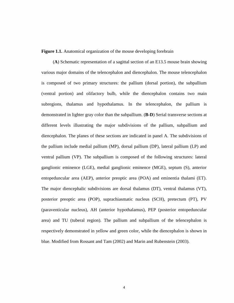
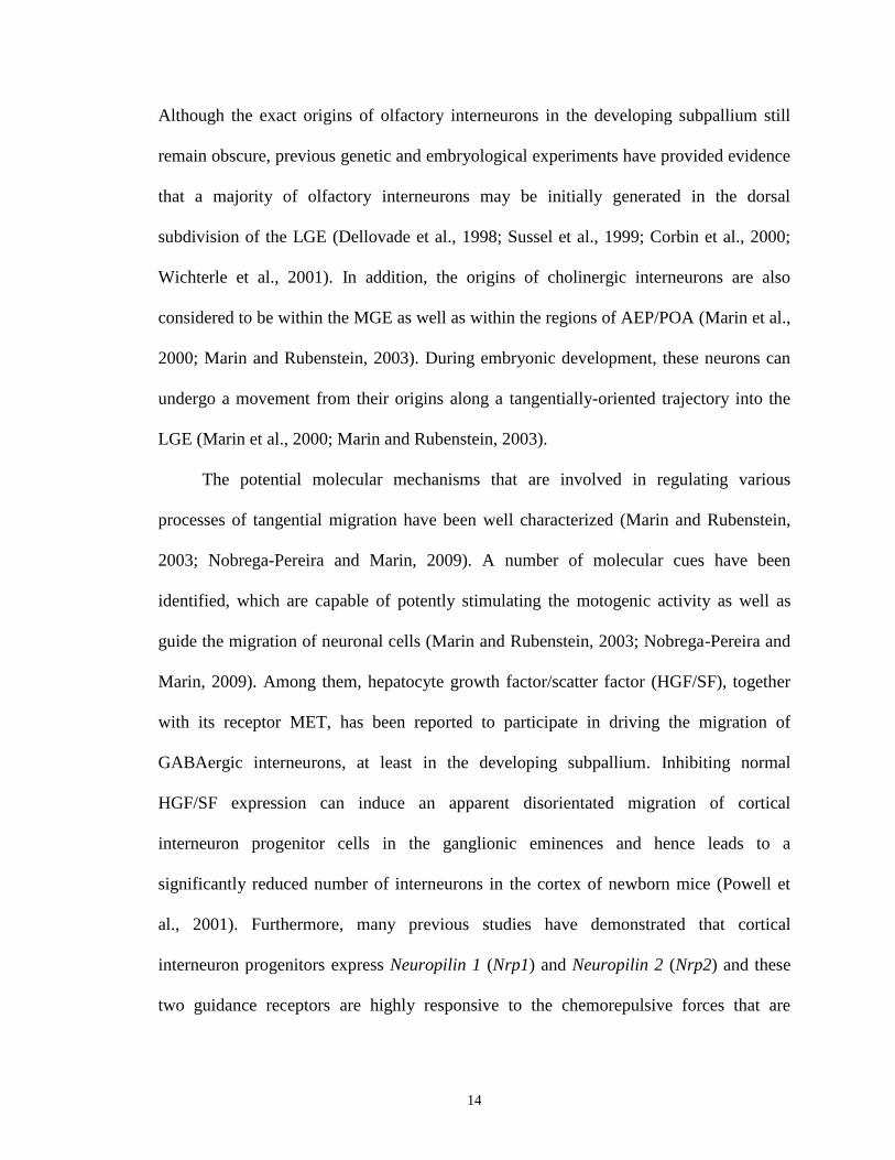
Figure 1.1. Anatomical organization of the mouse developing forebrain
(A) Schematic representation of a sagittal section of an E13.5 mouse brain showing
various major domains of the telencephalon and diencephalon. The mouse telencephalon
is composed of two primary structures: the pallium (dorsal portion), the subpallium
(ventral portion) and olifactory bulb, while the diencephalon contains two main
subregions, thalamus and hypothalamus. In the telencephalon, the pallium is
demonstrated in lighter gray color than the subpallium. (B-D) Serial transverse sections at
different levels illustrating the major subdivisions of the pallium, subpallium and
diencephalon. The planes of these sections are indicated in panel A. The subdivisions of
the pallium include medial pallium (MP), dorsal pallium (DP), lateral pallium (LP) and
ventral pallium (VP). The subpallium is composed of the following structures: lateral
ganglionic eminence (LGE), medial ganglionic eminence (MGE), septum (S), anterior
entopeduncular area (AEP), anterior preoptic area (POA) and eminentia thalami (ET).
The major diencephalic subdivisions are dorsal thalamus (DT), ventral thalamus (VT),
posterior preoptic area (POP), suprachiasmatic nucleus (SCH), pretectum (PT), PV
(paraventicular nucleus), AH (anterior hypothalamus), PEP (posterior entopeduncular
area) and TU (tuberal region). The pallium and subpallium of the telencephalon is
respectively demonstrated in yellow and green color, while the diencephalon is shown in
blue. Modified from Rossant and Tam (2002) and Marin and Rubenstein (2003).

5

6
Following the closure of the neural tube, distinct modes of growth and
morphogenesis will be separately initiated along both the A/P and D/V axes in the dorsal
and ventral portion of the developing telencephalon, ultimately leading to differential
structures between the pallium and subpallium. In the early developing telencephalon, the
establishment of different structures in the pallium and supallium as well as diverse
functional domains within them are mainly modulated by the combined activities of a
group of signaling molecules and transcription factors (Puelles et al., 2000; Rossant and
Tam, 2002; Hoch et al, 2009). Specifically, the signaling molecules (also defined as
“organizers”) generated from the signaling centers at various specific locations in the
developing telecephalon first establish and maintain correct spatio-temporal expression of
many regional transcription factors by localized expression of themselves or of their
activators (Puelles et al., 2000; Rossant and Tam, 2002; Hoch et al, 2009). As such, the
expression patterns of these transcription factors are built in a manner that is highly
correlated with morphological boundaries in the developing telencephalon (Rallu et al.,
2002; Schuurmans and Guillemot, 2002). These transcription factors will then exert their
functions in establishing the identities of distinct pallial and subpallial subdomains as
well as in producing different types of neurons within these subregions (Rallu et al.,
2002; Schuurmans and Guillemot, 2002; Wonders and Anderson, 2006). Indeed, a large
number of previous reports have described that different individual subdivisions within
the developing mouse subpallium (e.g. LGE, MGE, AEP/POA, and certain parts of the
septal and amygdaloid anlages) exhibit unique expression profiles of many transcription
factors that strictly resemble their regionalization and are believed to play central roles
either individually or in combination in controlling their formation (Bulfone et al., 1993a;

7
Bulfone et al., 1993b; Guillemont and Joyner, 1993; Porteus et al., 1994; Hsieh-Li et al.,
1995; Valerius et al., 1995; Liu et al., 1997; Hallonet et al., 1998; Eisenstat et al., 1999).
In addition, during embryonic development, these master regulatory genes are expressed
in a time-dependent manner. The expression of some genes, such as Otx2, Six3 and Vax1,
is detectable in progenitor cells as early as the time of early neural plate formation,
whereas others, including Dlx1, Dlx2, Gsh1, Gsh2, Mash1, Nkx2.1, and Islet1, start their
expression at different time points after the completion of neurulation, indicating that the
development of various subpallial subdomains may also occur in a stepwise fashion
(Bulfone et al., 1993a; Bulfone et al., 1993b; Guillemont and Joyner, 1993; Porteus et al.,
1994; Hsieh-Li et al., 1995; Valerius et al., 1995; Liu et al., 1997; Hallonet et al., 1998;
Eisenstat et al., 1999). Disrupting function of these genes has been revealed to induce
abnormal patterning and specification of the developing subpallial telencephalon. For
example, a ventral-to-dorsal transformation has been observed in the basal ganglia
(particulary in the LGE and MGE) of Nkx2.1 mutant mice (Kimura et al., 1996; Sussel et
al., 1999), whereas inactivation of Gsh2 impairs the formation of the LGE and striatum
and induces ectopic expression of many dorsal markers in the LGE (Szucsik et al., 1997;
Toresson et al., 2000; Yun et al., 2001).
As noted, the expression of these transcription factors is under the control of an
overall effect of various signaling proteins originally secreted from different “organizer”
centers. To date, a total of three types of signaling cues have been identified to be
critically implicated in the patterning and regionalization of the developing subpallium:
retinoic acid (RA), Fibroblast growth factors (Fgf) and Sonic hedgehog proteins (Shh),
(Puelles et al., 2000; Rossant and Tam, 2002; Hoch et al, 2009). Lateral RA signaling that

8
is originated from the olfactory placode area may play prominent roles in patterning of
progenitor areas at the intermediate level, such as the LGE (LaMantia et al. 1993; Pierani
et al. 1999). Indeed, Marklund et al. (2004) cultured the prospective intermediate
telencephalon from early chick embryos (~stage 14) and treated them with a RA receptor
antagonist (BMS-453) in vitro. They found that, following exposure to BMS-453, the
production and specification of a large percentage of LGE-derived cells can be
significantly blocked (Marklund et al., 2004). In addition, exposure of early-born cells
isolated from the dorsal telencephalon of stage 8 chick embryos to RA signaling is
sufficient to produce many intermediate features in these cells (Marklund et al., 2004).
Another signaling factor is Fgf8, which is originally produced in the regions of the
anterior neural ridge/cortical plate and is functionally involved in promoting the
patterning and specification of the most ventral subdomains in the subpallium (Rossant
and Tam, 2002; Lupo et al., 2006). Although targeted deletion of Fgf8 gene induces an
embryonic lethal phenotype (Sun et al., 1999), aberrant overexpression of Fgf8, together
with ectopic expression of several ventral markers, have been identified in the rostral
areas of the dorsal telencephalon of Gli3 mutant mice (Kuschel et al., 2003). Ectopic
expression of ventral markers has also been observed in the prospective dorsal
telencephalic explants dissected from early mouse embryos after exposure to Fgf8 signals
(Kuschel et al., 2003). Consistent with these results, the ventral telencephalon of
conditional Fgf8 knockouts is smaller and abnormally organized as compared to that of
their wild-type littermates (Storm et al., 2006). The patterning of subpallial structures can
be also influenced by Shh signaling that is derived from the telencephalic anlage as well
as the rostroventral parts of the telencephalon, such as MGE and POA (Rohr et al., 2001;

9
Lupo et al., 2006). Impaired Shh signals not only lead to the lack of several subpallial
structures but also cause the loss of many key ventral markers, such as Nkx2.1 and Dlx2
(Chiang et al., 1996; Muenke and Beachy, 2000). Ectopic expression of Shh signaling in
the pallium results in a ventral phenotype, such as ectopic expression of ventral markers
(e.g. Nkx2.1, Gsh2 and Dlx2) and impaired expression of some dorsal markers (e.g. Emx1
and Tbr1) (Kohtz et al., 1998; Gaiano et al., 1999; Corbin et al., 2000).
1.4. Cell migration in the developing telencephalon
1.4.1. Overview of cell migration in the developing telencephalon
After patterning of the telencephalon and specification of different types of neurons
within various regions of the developing telencephalon are accomplished, a process
called cell migration can be extensively observed in both the pallium and subpallium.
According to their distinct origins, different types of neurons basically utilize two general
modes of migration during embryonic development in order to reach and populate their
final destinations in the telencephalon as well as in other specific areas in the CNS
(Figure 1.2). The first is radial migration, occurring predominantly in the pallium as well
as in the mammalian striatum, where migrating glutamatergic and GABAergic projection
neurons move along the radial axis through their interaction with fiber tracts built by glial
cells. However, the underlying molecular mechanisms may be distinct depending on
specific local environment (Nadarajah and Parnavelas, 2002; Marin and Rubenstein,
2003). For instance, a major proportion of striatal GABAergic projection neurons is
found to be born in the proliferative VZ within the LGE and then radially migrate (at
~E12) towards the developing striatum in an outward direction that is perpendicular to
10
the ventricular surface (Halliday and Cepko, 1992; De Carlos et al., 1996; Hamasaki et
al., 2003). The second form of migration is tangential migration, which mainly happens
in the progenitor regions (e.g. LGE, MGE and caudal ganglionic eminence, CGE) within
the subpallium. Once the processes of proliferation and differentiation are completed,
different types of interneurons will tangentially migrate in multiple streams away from
the basal telencephalon in a direction that is orthogonal to that of radial migration along
both rostrocaudal and mediolateral axes, enter the cortex and eventually occupy their
final positions located in different layers of the cortex (radial migration is also involved at
this stage) (Nadarajah and Parnavelas, 2002; Marin and Rubenstein, 2003). Considering
its closer relevance to my thesis, I will be discussing only tangential migration with some
details in the following text.
1.4.2. Tangential migration in the developing telencephalon
As briefly noted above, in the developing mouse forebrain, distinct types of
interneurons originate from different progenitor domains within the subpallium and then
follow their own specific tangential migratory paths in order to ultimately locate and
establish their destined positions in various regions of the forebrain, primarily in the
pallium. For example, the progenitor cells that will later differentiate into diverse
subtypes of cortical GABAergic interneurons are not generated from the pallium; instead,
their origins can be traced back to the proliferative VZ within the LGE, MGE and CGE
of the basal telencephalon (Marin and Rubenstein, 2003). After leaving the progenitor
regions, the postmitotic immature cells will separate into several superficial or deep
migration streams, pass through the developing striatum and intrude into the neocortex in
11
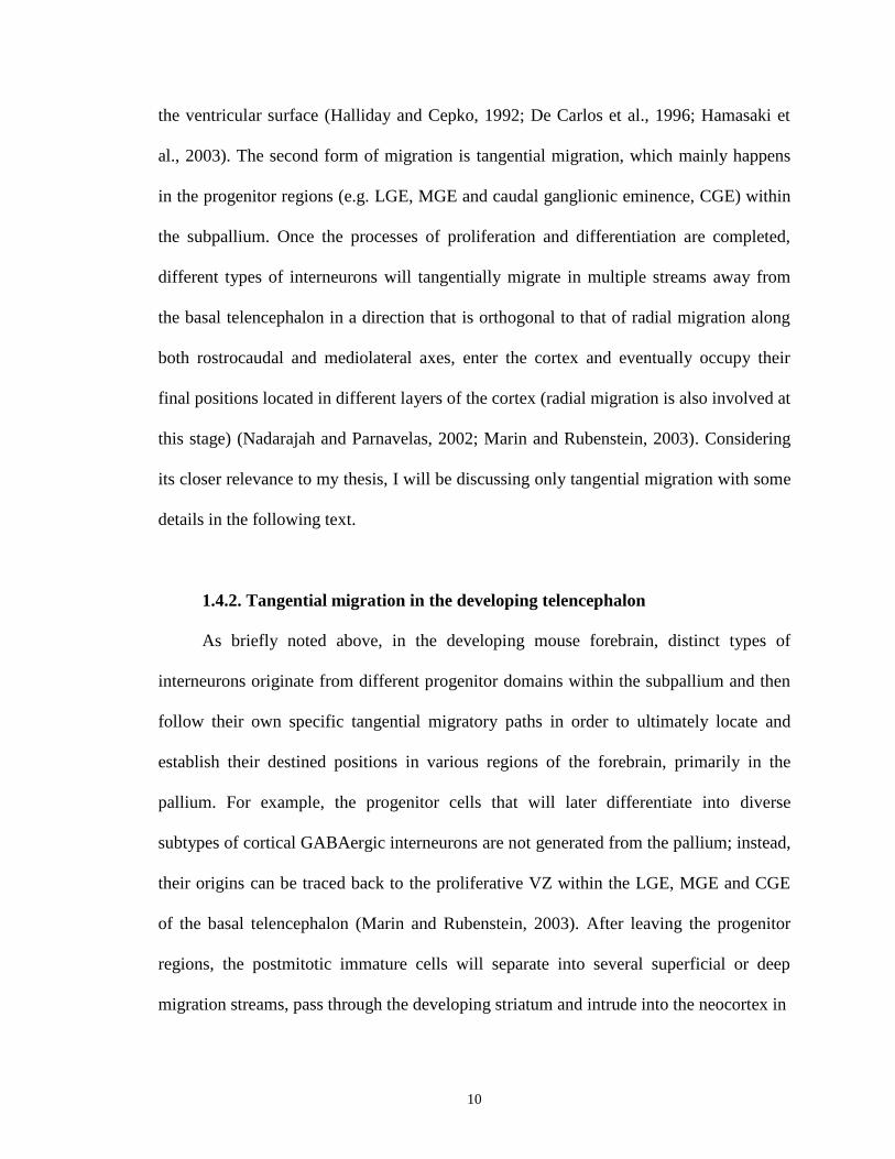
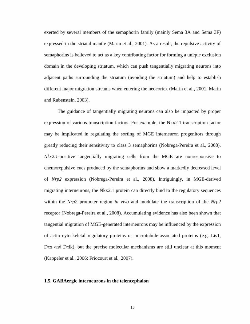
Figure 1.2. Neuronal migration in the mammalian telencephalon during early
embryogenesis
Different types of neurotransmitter are specified in distinct progenitor domains in
the developing telencephalon: glutamatergic excitatory neurons are mainly generated and
specified within progenitor domains in the pallium, while GABAergic inhibitory neurons
as well as cholinergic neurons are derived from progenitor cells within the LGE, MGE
and AEP/POA in the subpallium. During early embryogenesis, glutamatergic neurons
(blue arrows) migrate radially through different layers of the cortex; in contrast,
GABAergic (red arrows) and cholinergic neurons (black arrows on the right) migrate
tangentially from the MGE via different routes in order to reach their final destinations in
the cortex. Moreover, most GABAergic projection neurons mainly found in the LGE
(black arrow on the left) migrate radially from the LGE to the striatum in the developing
telencephalon. The black bar marks the boundary between the pallium and subpallium.
AEP, anterior entopeduncular area; BF, basal forebrain; DP, dorsal pallium; LGE, lateral
ganglionic eminence; LP, lateral pallium; MGE: medial ganglionic eminence; MP,
medial pallium; POA, pre-optic area; str, striatum; VP, ventral pallium. Modified from
Wullimann (2009).

12
13
a tangential manner that is vertical to the orientiation of radial glia fibers (Figure 1.2)
(Marin and Rubenstein, 2003). More specifically, three different major streams of
neurons near the corticostriatal notch have been revealed by previous cell tracing assays
before their invasion into the neocortex (Reviewed in Marin and Rubenstein, 2003;
Nadarajah and Parnavelas, 2002; Metin et al., 2006). The first one is found to be derived
from the MGE and mainly enter the preplate in the neocortex at ~E12. The cells
comprising this stream exhibit unique characteristics representing the typical Cajal–
Retzius cells (Lavdas et al 1999). The second tangential migration stream (also the most
dominant one) is made up of cells derived from the LGE, MGE and CGE, which mainly
travel through the intermediate zone (IZ) in the neocortex at ~E13-E15. At the later
stages (~E15 to P0), there exists another migration stream that is composed of cells
originating from the LGE and MGE and usually enter the cortex via the lower IZ and
SVZ (Anderson et al., 2001). Once arrived in the pallium, these migrating neurons will
turn and start moving radially (in waves) towards the pial surface in a fashion highly
similar to that of pallial projection neurons (Nadarajah and Parnavelas, 2002). It has been
gradually accepted that similar to pallial projection neurons, GABAergic cortical
interneurons are also settled in an `inside-out' pattern at various layers of the cortex.
Pallial projection neurons and cortical interneurons that are born at the same time points
are in general positioned at the same layer (Wichterle et al., 2001; Ang et al., 2003;
Valcanis and Tan, 2003; Hevner et al., 2004). Like GABAergic interneurons, progenitor
cells of olfactory interneurons are also born in the progenitor areas within the basal
ganglia, rather than in the olfactory bulb and, subsequently, migrate tangentially to take
up their positions in the olfactory bulb (Luskin, 1993; Lois and Alvarez-Buylla 1994).
14
Although the exact origins of olfactory interneurons in the developing subpallium still
remain obscure, previous genetic and embryological experiments have provided evidence
that a majority of olfactory interneurons may be initially generated in the dorsal
subdivision of the LGE (Dellovade et al., 1998; Sussel et al., 1999; Corbin et al., 2000;
Wichterle et al., 2001). In addition, the origins of cholinergic interneurons are also
considered to be within the MGE as well as within the regions of AEP/POA (Marin et al.,
2000; Marin and Rubenstein, 2003). During embryonic development, these neurons can
undergo a movement from their origins along a tangentially-oriented trajectory into the
LGE (Marin et al., 2000; Marin and Rubenstein, 2003).
The potential molecular mechanisms that are involved in regulating various
processes of tangential migration have been well characterized (Marin and Rubenstein,
2003; Nobrega-Pereira and Marin, 2009). A number of molecular cues have been
identified, which are capable of potently stimulating the motogenic activity as well as
guide the migration of neuronal cells (Marin and Rubenstein, 2003; Nobrega-Pereira and
Marin, 2009). Among them, hepatocyte growth factor/scatter factor (HGF/SF), together
with its receptor MET, has been reported to participate in driving the migration of
GABAergic interneurons, at least in the developing subpallium. Inhibiting normal
HGF/SF expression can induce an apparent disorientated migration of cortical
interneuron progenitor cells in the ganglionic eminences and hence leads to a
significantly reduced number of interneurons in the cortex of newborn mice (Powell et
al., 2001). Furthermore, many previous studies have demonstrated that cortical
interneuron progenitors express Neuropilin 1 (Nrp1) and Neuropilin 2 (Nrp2) and these
two guidance receptors are highly responsive to the chemorepulsive forces that are
15
exerted by several members of the semaphorin family (mainly Sema 3A and Sema 3F)
expressed in the striatal mantle (Marin et al., 2001). As a result, the repulsive activity of
semaphorins is believed to act as a key contributing factor for forming a unique exclusion
domain in the developing striatum, which can push tangentially migrating neurons into
adjacent paths surrounding the striatum (avoiding the striatum) and help to establish
different major migration streams when entering the neocortex (Marin et al., 2001; Marin
and Rubenstein, 2003).
The guidance of tangentially migrating neurons can also be impacted by proper
expression of various transcription factors. For example, the Nkx2.1 transcription factor
may be implicated in regulating the sorting of MGE interneuron progenitors through
greatly reducing their sensitivity to class 3 semaphorins (Nobrega-Pereira et al., 2008).
Nkx2.1-positive tangentially migrating cells from the MGE are nonresponsive to
chemorepulsive cues produced by the semaphorins and show a markedly decreased level
of Nrp2 expression (Nobrega-Pereira et al., 2008). Intriguingly, in MGE-derived
migrating interneurons, the Nkx2.1 protein can directly bind to the regulatory sequences
within the Nrp2 promoter region in vivo and modulate the transcription of the Nrp2
receptor (Nobrega-Pereira et al., 2008). Accumulating evidence has also been shown that
tangential migration of MGE-generated interneurons may be influenced by the expression
of actin cytoskeletal regulatory proteins or microtubule-associated proteins (e.g. Lis1,
Dcx and Dclk), but the precise molecular mechanisms are still unclear at this moment
(Kappeler et al., 2006; Friocourt et al., 2007).
1.5. GABAergic interneurons in the telencephalon
16
Although the neuronal network in the cerebral cortex is fairly complex, it is
generally established by two main classes of neurons: excitatory projection neurons and
inhibitory local-circuit neurons (also known as interneurons) (Di Cristo, 2007). While
projection neurons can send their axons to many different targets either within or farther
away from their local environment (e.g. the thalamus, brain stem and spinal cord),
inhibitory interneurons mostly set up synaptic connections with nearby projection
neurons, inhibit their excitatory activity and maintain the spatio-temporal balance of
excitation and inhibition essential for proper telencephalic function (Wonders and
Anderson, 2006; Di Cristo, 2007). Projection neurons utilize glutamate as their main
neurotransmitter and contribute to ~70-80% of all cortical neurons, whereas around 20-
30% of cortical neurons are made up of inhibitory interneurons that basically use GABA
as neurotransmitter (Parnavelas et al., 1977; Meinecke and Peters, 1987; De Felipe and
Farinas, 1992; Gupta et al., 2002). There also exists a comparatively smaller population
of dopaminergic and cholinergic neurons in the cortex than that of glutamatergic and
GABAergic cells (Wonders and Anderson, 2006; Di Cristo, 2007). It is worth noting that
~90% of neurons in the striatum of the mammalian telencephalon are projection neurons
but they use GABA, rather than glutamate, as their primary neurotransmitter and thus are
termed as GABAergic projection neurons (Kawaguchi et al., 1995; Kawaguchi, 1997a;
Gerfen, 1992). The remaining ~10% of neurons in the striatum are GABAergic
interneurons, which are mainly localized in the patch and matrix compartments of the
striatum and produce inhibitory signals to maintain the balance of striatal function
(Kawaguchi et al., 1995; Kawaguchi, 1997a; Gerfen, 1992). A very small proportion of

17
interneurons found in the striatum have also been shown to use dopamine or
acetylcholine as their main neurotransmitter (Gall et al., 1987; Kawaguchi et al., 1995).
Unlike projection neurons that have in general similar pyramidal morphology, such
as apical and basal dendrites and axons distantly projecting into various sites within the
white matter, GABAergic interneurons display a remarkable heterogeneity and different
subtypes of GABAergic interneurons are usually classified by their distinct morphology,
connectivity patterns, electrophysiological features and biochemical contents (reviewed
in Markram et al., 2004; Flames and Marin, 2005; Wonders and Anderson, 2006; Gelman
and Marin, 2010). More specifically, GABAergic interneurons can be categorized into
the following subgroups according to their morphological shapes, connectivity patterns,
electrophysiological properties and their expression of a group of molecular markers
reflecting their endogenous biochemical content. Based on the characteristics of axons
and dendrites, there are basket cells, bipolar cells, double bouquet cells, bitufted cells,
Chandelier cells and Martinotti cells. Based on the location of the branching terminus of
their axons, interneurons can be classified as axon-targeting, dendrite-targeting and soma-
targeting cells (or any combination of them). Based on their distinct intrinsic firing
patterns, GABAergic interneurons can be may further subdivided as fast spiking, burst
spiking non-pyramidal, regular spiking non-pyramidal, irregular spiking and late spiking
cells. At different stages of either embryonic or adult neurogenesis, GABAergic
interneurons are usually classified by detecting their distinct expression patterns of three
calcium-binding proteins, calbindin (CB), calretinin (CR) and parvalbumin (PV), and
four neuropetides, somatostatin (SOM), neuropeptide Y (NPY), vaso-active intestinal
peptide (VIP) and cholecystokinin (CCK) (Markram et al., 2004; Wonders and Anderson,

18
2006; Gelman and Marin, 2010). It should be emphasized here that under most situations,
it is very difficult to precisely distinguish between distinct subtypes of GABAergic
interneurons by only checking the expression of one or two of these markers due to the
overlaps of their expression profiles. For example, the expression pattern of CB, PV,
SOM and CV overlap each other to varying degrees (Kubota et al., 1994; Kawaguchi and
Kubota, 1997b). Thereby, using one or more combinations of these molecular markers is
required to better define each interneuron subtype, although some previous studies have
provided evidence that different interneuron subtypes, such as PV-, CR- and SOM-
positive cells, are comprised of largely non-overlapping populations of cells at least in the
sensory cortex and suggest that this phenomenon may possibly be applicable to most
mammalian species (Kubota et al., 1994; Cauli et al., 1997; Tamamaki et al., 2003).
1.6. Development of the zebrafish telencephalon
1.6.1. Advantages of using zebrafish as a model organism
Teleosts comprise a large and extremely diverse group of fish with bony skeletons,
covering approximately 50% of the existing vertebrates (Mueller and Wullimann, 2009).
Zebrafish (Danio rerio) is a small, freshwater teleost fish that mainly lives in tropical
regions, particularly in India and Southeast Asia (Eisen, 1996). In addition to its
applicable value in fisheries research, in the past two decades, zebrafish has rapidly
emerged as a powerful model organism not only appropriate for developmental and
genetic studies, but also for many other research areas, such as carcinogenesis,
cardiovascular diseases and biotechnique development (Dodd et al., 2000; Pyati et al.,
2007; Ingham, 2009). Compared to mammals, such as the mouse, zebrafish have a
19
number of unique characteristics that are especially suitable for developmental biology
studies. First is their small body size and short generation time. The size of zebrafish
embryos is only ~0.7 mm in diameter during fertilization and quickly reaches 3.5 mm
within 3 days. Patterning and segmentation of the body plan can be completed within the
first day post fertilization (dpf) and the process of primary organogenesis starts at 2 dpf.
The vast majority of organs become functional at 3-5 dpf and sexual maturity is
accomplished within the first three months of their life. Second, zebrafish have high
fecundity. Their embryos are externally fertilized and breeding of one single pair can
yield up to hundreds of spawned eggs per day. Third, zebrafish embryos and larvae are
optically transparent. By using live-imaging techniques, cellular morphology and
movement can be easily and non-invasively observed/traced. Fourth, the genetic
tractability of zebrafish, such as high chromosomal integration frequency and good
germline transmission when generating transgenic models, allows and facilitates the
investigation of a wide variety of developmental events (Westerfield, 1995; Detrich et al.,
1999; Kawakami, 2004).
1.6.2. Eversion versus evagination: a different mode of telencephalic
development in zebrafish (and other teleost) as compared to most other vertebrate
lineages
Similar to that of mammals, the developing zebrafish telencephalon is also made
up of four major subdivisions in the pallium (lateral, ventral, medial and dorsal pallium,
LP, VP, MP and DP) and a relatively larger ventral subpallium, where increasing
numbers of glutamatergic and GABAergic neurons are respectively generated and
20
differentiatied starting at ~ 2 dpf during early neurogenesis (Mueller and Wullimann,
2009). Despite the fact that the first steps of neural tube formation are in general similar
among all vertebrates, the modes of telencephalic morphogenesis differ between teleosts
and most other vertebrate groups. Distinct from the evaginated pattern of telencephalic
development found in other vertebrates, where the telencephalon is suggested to develop
as two hemispheres with a ventricle in the middle, the telencephalon of teleosts (e.g.
zebrafish) undergoes an uncommon process of morphogenesis termed as eversion
(occuring predominantly in the regions of MP and DP) and leads to a remarkable change
of anatomical topography in the telencephalon (Figure 1.3) (Wullimann and Mueller,
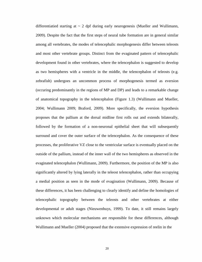
2004; Wullimann 2009; Braford, 2009). More specifically, the eversion hypothesis
proposes that the pallium at the dorsal midline first rolls out and extends bilaterally,
followed by the formation of a non-neuronal epithelial sheet that will subsequently
surround and cover the outer surface of the telencephalon. As the consequence of these
processes, the proliferative VZ close to the ventricular surface is eventually placed on the
outside of the pallium, instead of the inner wall of the two hemispheres as observed in the
evaginated telencephalon (Wullimann, 2009). Furthermore, the position of the MP is also
significantly altered by lying laterally in the teleost telencephalon, rather than occupying
a medial position as seen in the mode of evagination (Wullimann, 2009). Because of
these differences, it has been challenging to clearly identify and define the homologies of
telencephalic topography between the teleosts and other vertebrates at either
developmental or adult stages (Nieuwenhuys, 1999). To date, it still remains largely
unknown which molecular mechanisms are responsible for these differences, although
Wullimann and Mueller (2004) proposed that the extensive expression of reelin in the

21
Figure 1.3. Two different modes of telencephalic development from the neural tube in
vertebrates
The mode of evagination (left) occurs in a vast majority of vertebrates except for
the ray-finned fish. In this process, the telencephalon gradually evolves as two identical
hemispheres enclosing a central ventricle. The process of eversion (right) has been
proposed to happen duing the telecephalic development of most ray-finned fish, including
teleosts. Different from evagination, the pallium in the mode of eversion rolls out
bilaterally and eventually makes the proliferative domains that is close to the ventricular
surface (labeled with “x”) outside of the pallium. DP, dorsal pallium; GP, globus
pallidus; LP, lateral pallium; MP, medial pallium; Sep, Septum; Str, striatum; VP, ventral
pallium. Note that with respect to the telencephalic development in teleosts, several other
hypothetical models have been proposed, such as the partial pallial eversion model
(Wullimann and Mueller, 2004) and a new three-dimensional eversion model (Yamamoto
et al., 2007). Adapted from Niewenhuys and Meek (1990).
22

23
pallial cells in the telencephalon of teleosts might be one of the potential contributing
factors. It is also worthy of mentioning that apart from the simple eversion model, several
other hypothetical models, such as a partial eversion model and a more complex three-
dimensional eversion model, have been recently proposed (Wullimann and Mueller,
2004; Yamamoto et al., 2007), which definitely will form a good basis for a better
understanding of the different telencephalic topography between teleosts and other
vertebrates.
1.6.3. Conserved genetic cascades in the developing zebrafish subpallium as
compared to the mouse
While the fundamental mode of telencephalic development is divergent between
mouse and zebrafish, researchers have made important attempts to compare the
embryonic forebrain development between these two distantly related vertebrate species
with an aim to identify some homologies between their distinct morphological structures
(Mueller et al., 2006; Mueller et al., 2008). By examining the expression patterns of
common developmental genes as well as several classic molecular markers for neuronal
proliferation and neurogenesis, they discovered a number of anatomical correspondences
in both the pallium and subpallium between the mouse and zebrafish forebrain
(Wullimann and Mueller, 2004; Wullimann, 2009). Consistent with observations of their
mouse orthologs in the developing mouse subpallium, the proneural basic helix-loop-
helix (bHLH) gene, ascl1a (also known as zash1a, an orthologous gene of the mouse
Mash1) and the regulatory dlx1a/dlx2a genes are found to be specifically expressed
within restricted domains (close to the ventricular surface) in the zebrafish subpallium

24
(Figure 1.4A) (Quint et al., 2000; Wullimann and Mueller, 2002; Mueller et al., 2008).
Likewise, similar to the expression patterns of their orthologs in the mouse subpallium,
the expression domains of LIM-homeobox genes lhx6 and lhx7 are predominantly located
within in the ventral subpallium (septum, Sv) as well as within the ventral subdivision of
the dorsal subpallium (Sdv) in the developing zebrafish telenephalon, but not in the
dorsal subdivision of the dorsal subpallium (Sdd) (Figure 1.4B; Mueller et al., 2008). It is
worth noting that Sdv and Sdd have been proposed as corresponding homologous
structures to the MGE and LGE within the embryonic mouse subpallium (Figure 1.4B;
Wullimann, 2009). In addition, nkx2.1, whose mouse orthologous gene serves as an
essential modulator gene indispensable for proper regional specification within the mouse
subpallium, is also found to be expressed in the early developing zebrafish subpallium
(Rohr et al., 2001), even though its detailed expression pattern still needs to be further
determined. This regional homology between the mouse and zebrafish telencephalon was
also seen in the expression pattern of the neurotransmitter GABA (Mueller et al., 2006).
In short, these data strongly suggest that despite the difference in the telencephalic
development between mouse and zebrafish, the genetic pathways underlying this process
may be highly conserved between these two species.
1.7. The Dlx gene family
1.7.1. Origin, genomic organization and evolution of the Dlx genes
1.7.1.1. The Drosophila distal-less (Dll) gene
The Dlx genes of vertebrates are related in sequence to the distal-less (Dll) gene
that was initially discovered and characterized in Drosophila (Panganiban and

25
Figure 1.4. Schematic diagrams summarizing expression patterns of various key
regulatory genes in the developing zebrafish telencephalon
(A) Transverse section of a 3dpf zebrafish telencephalon showing distinct
expression domains of a group of important regulatory genes that are commonly used to
define different major subdivisions in the developing zebrafish forebrain, such as zash1a
(ascl1a) and dlx1a/dlx2a expression in the subpallium. Adapted from Wullimann and
Mueller, 2004. (B) Structural comparison between the early mouse and zebrafish
telencephalon according to the expression patterns of several key regulatory genes (lhx6,
dlx2a, lhx7, tbr2 and gad67). The proposed homologous domains between the mouse and
zebrafish telencephalon are indicated by solid lines between the corresponding structures.
Dl, lateral zones of the dorsal telencephalon; Dm, medial zones of the dorsal
telencephalon; Dp, posterior zones of the dorsal telencephalon; DP, dorsal pallium; LP,
lateral pallium; LGE, lateral ganglionic eminence; MGE, medial ganglionic eminence;
MP, medial pallium; Sdd, dorsal subdivision of dorsal subpallium; Sdv, ventral
subdivision of dorsal subpallium; Sv, ventral subpallium; VP, ventral pallium. Adapted
from Wullimann (2009).

26

27
Rubenstein, 2002). The Dll gene was found to encode a homeodomain-containing
transcription factor responsible for controlling proper proximo-distal (P/D) patterning as
well as normal specification and organization of several distal appendages, including the
limbs, antennae and mouth, during Drosophila development. Dll null flies die at the early
stage of embryogenesis due to the lack of rudimentary larval limbs and antennae, which
subsequently serves as the ears and nose of the fly (Cohen et al., 1989; Cohen and
Jurgens, 1989; Wu and Cohen, 1999). Consistently, ectopic Dll expression can result in
abnormal patterning and formation of the limbs and antennae, although the specific
phenotypes vary with the specific location where it is expressed (Panganiban and
Rubenstein, 2002). In addition to its key role in regulating proper distal appendage
development, Dll may be also involved in the formation of certain parts of the nervous
system in fly. While its specific functional implication in these regions still remains
unknown, Dll expression has been clearly detected in the two optic lobes of the brain and
subsets of glial cells in the ventral nerve cord (Kaphingst and Kunes, 1994; Panganiban
and Rubenstein, 2002; Skeath and Panganiban, unpublished observation).
1.7.1.2. Genomic organization of the Dlx genes
Vertebrates have multiple Dlx genes that are typically organized as convergently
transcribed bigene clusters with overlapping expression patterns (reviewed in Zerucha
and Ekker, 2000; Panganiban and Rubenstein, 2002). For example, a total of six Dlx
genes have been identified in mouse and human (Price et al., 1991; Robinson et al., 1991;
Simeone et al., 1994; Weiss et al., 1994; Nakamura et al., 1996) and they are arranged as
three bigene pairs (Dlx1/Dlx2, Dlx3/Dlx4 and Dlx5/Dlx6) separated by relatively short
28
intergenic sequences (<12 kb) (Ghanem et al., 2003). Each of these bigene Dlx clusters is
also physically linked to a Hox gene cluster on distinct chromosomes (Stock et al., 1996).
Unlike mammals, some teleost fish including zebrafish contain at least eight dlx genes.
Six of them are found in the bigene arrangement as dlx1a/dlx2a, dlx3b/dlx4b and
dlx5a/dlx6a similar to the six mammalian orthologs (Ekker et al., 1992; Akimenko et al.,
1994; Stock et al., 1996; Ellies et al., 1997a). However, the other two additional dlx genes
(dlx2b and dlx4a) are not linked to other known dlx genes (Stock et al., 1996; Ellies et al.,
1997a). All vertebrate Dlx genes consist of three exons and two introns and encode 243 to
333-amino acid protein products containing a highly conserved 61 amino acid
homeodomain (Liu et al., 1997). According to sequence similarity found in the
homeodomain and carboxyl terminus, the six common Dlx genes can be generally
classified into two subfamilies or clades: Dlx1/4/6 and Dlx2/3/5 (Stock et al., 1996; Ellies
et al., 1997a). However, the question of whether there is any functional difference
between these two groups of Dlx genes remains to be answered.
1.7.1.3. Evolution of the vertebrate Dlx gene family
Although only one Dll gene has been identified in the genome of Drosophila and
amphioxus, vertebrates have been shown to possess several Dlx genes. The tunicate
Ciona intestinalis has three Dlx genes which are organized as one convergently
transcribed bigene pair, Ci-Dll-A/Ci-Dll-B, and a third unlinked gene, Ci-Dll-C
(Caracciolo et al., 2000; Irvine et al., 2007). The genome of lampreys contains at least six
Dlx genes (LjDlxA-F), two of which exist as one bigene pair (LjDlxA/LjDlxD) (Myojin et
al., 2001; Neidert et al., 2001; Sumiyama et al., 2003; Kuraku et al., 2009). Similarly,

29
mammals, such as mouse and human, have six Dlx genes arranged as three bigene
clusters as noted above (Price et al., 1991; Robinson et al., 1991; Simeone et al., 1994;
Weiss et al., 1994; Nakamura et al., 1996; Stock et al., 1996). Based on the highly
conserved genomic organization of the Dlx gene family and the increased number of Dlx
genes over evolutionary time, a hypothesized model regarding Dlx gene evolution has
been proposed: an inverted tandem duplication of the single ancestral Dlx gene plus two
successive rounds of genome duplication events and a subsequent loss of one Dlx bigene
pair may together contribute to the present organization of six Dlx genes as commonly
seen in mammals (Figure 1.5) (Zerucha and Ekker, 2000; Sumiyama et al., 2003). With
respect to the eight dlx genes found in zebrafish or other teleost fish, one explanation
would be that an additional genome duplication event specific to certain teleost lineages
may likely have occurred that ultimately generates two extra dlx genes in the genome.
(Figure 1.5) (Stock et al., 1996; Amores et al., 1998; Robinson-Rechavi et al., 2001).
Since Dlx1 shares the highest sequence similarity with the Drosophila Dll gene when
compared to other vertebrate Dlx genes, Dlx1 has been thought to act as a founding gene
during the evolutionary process of the Dlx gene family (Holland et al., 1996; Stock et al.,
1996).
1.7.2. Expression and function of Dlx genes in the forebrain
The Dlx genes are expressed in a number of organs and tissues mostly with highly
well-organized morphological structures, including the forebrain, branchial arches,
sensory organs, limbs/fin buds, bone/cartilage, hematopoeitic and immune systems, and
are pivotal for their proper development during embryogenesis and/or later
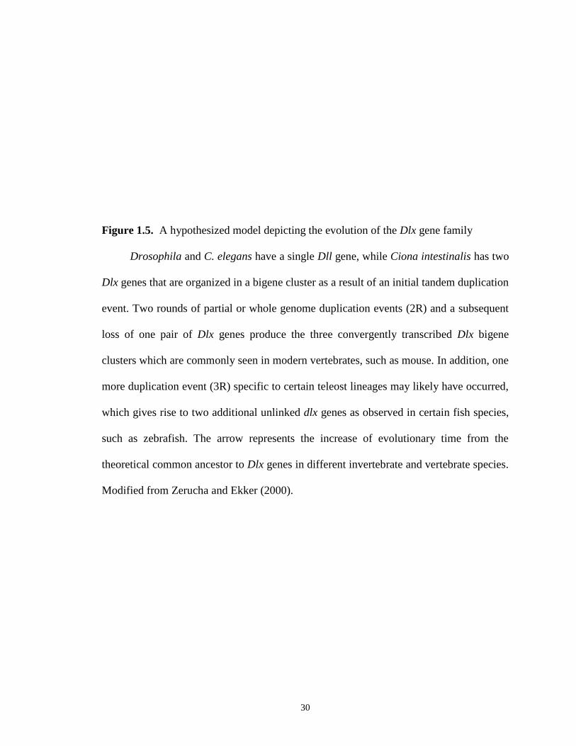
30
Figure 1.5. A hypothesized model depicting the evolution of the Dlx gene family
Drosophila and C. elegans have a single Dll gene, while Ciona intestinalis has two
Dlx genes that are organized in a bigene cluster as a result of an initial tandem duplication
event. Two rounds of partial or whole genome duplication events (2R) and a subsequent
loss of one pair of Dlx genes produce the three convergently transcribed Dlx bigene
clusters which are commonly seen in modern vertebrates, such as mouse. In addition, one
more duplication event (3R) specific to certain teleost lineages may likely have occurred,
which gives rise to two additional unlinked dlx genes as observed in certain fish species,
such as zebrafish. The arrow represents the increase of evolutionary time from the
theoretical common ancestor to Dlx genes in different invertebrate and vertebrate species.
Modified from Zerucha and Ekker (2000).
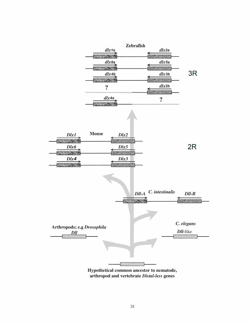
31
32
developmental stages (reviewed in Bendall and Abate-Shen, 2000; Zerucha and Ekker,
2000; Panganiban and Rubenstein, 2002). In general, there are three major molecular
characteristics of Dlx expression in these tissues: 1) their expression patterns appear to be
evolutionarily conserved among many divergent vertebrate species including human,
mouse and zebrafish (Akimenko et al. 1994; Ellies et al. 1997b; Zerucha and Ekker,
2000; Panganiban and Rubenstein, 2002); 2) endogenous expression of distinct Dlx
bigene clusters follows a nested spatial-temportal pattern along the P/D axis, which is
particularly apparent in the forebrain and branchial arches (Liu et al., 1997; Qiu et al.,
1997; Eisenstat et al., 1999); 3) although subtle differences in the expression of various
Dlx genes have been observed (and will be further discussed below) that may potentially
contribute to their distinct functions, the expression patterns of either paralogous Dlx
genes from the same bigene pair or those from different clusters are highly overlapping,
which make it a very difficult task to distinguish them from one another by simply
judging the expression data (Akimenko et al., 1994; Ellies et al., 1997a; Quint et al.,
2000). Indeed, a series of loss-of-function studies in mice have revealed that single Dlx
gene knockouts have relatively milder abnormal phenotypes as compared to that of
double Dlx null mutants (Dlx1/2-/-
and Dlx5/6-/-
), which exhibit more severe defects along
with some novel malformations in the forebrain and branchial arches (Qiu et al., 1995;
Anderson et al., 1997a; Anderson et al., 1997b; Qiu et al., 1997; Acampora et al., 1999;
Depew et al., 1999; Cobos et al., 2005a; Cobos et al., 2007; Robledo et al., 2002; Wang
et al., 2010). Here, I am only focusing on the expression pattern and functional roles of
Dlx genes in the forebrain.

33
Among the six mammalian Dlx genes, four Dlx genes (Dlx2, Dlx1, Dlx5 and Dlx6)
are sequentially expressed in the embryonic mouse forebrain (Bulfone et al., 1993b; Liu
et al., 1997; Eisenstat et al., 1999; Zerucha et al., 2000). Five out of the eight orthologous
dlx genes from zebrafish are expressed in the developing zebrafish forebrain: dlx1a,
dlx2a, dlx5a, dlx6a, and dlx2b (Akimenko et al. 1994; Ellies et al. 1997b). Dlx gene
expression is specifically restricted to two major domains in the developing forebrain: the
ventral telencephalon (basal ganglia in the subpallium) and the diencephalon, including
both the prethalamus (ventral thalamus) and hypothalamus (Robinson et al., 1991;
Bulfone et al., 1993a). These two specific areas have been proposed to be homologous
between the mouse and zebrafish forebrain based on the expression data as discussed in
previous sections (Mueller and Wullimann, 2009; Wullimann, 2009) and have also been
identified in the forebrain of other vertebrates, such as chicks, frogs and lamprey
(Fernandez et al., 1998; Puelles et al., 2000; Myojin et al., 2001). In the developing
ventral telencephalon of the mouse, various Dlx genes display distinct, but highly
overlapping, spatial expression patterns, indicative of a possible functional redundancy
between each individual Dlx gene, at least in certain populations of cells (Figure 1.6):
Dlx1 and Dlx2 are mainly expressed in the least mature early-born progenitor cells in the
VZ and SVZ as well as in a certain number of more differentiated cells scattered in the
mantle zone (MZ); Dlx5 is predominantly expressed in the SVZ and expressed at
intermediate levels in the MZ; Dlx6 is mostly expressed in the most differentiated
postmitotic neurons in the MZ and moderate Dlx6 expression is also found in cells in the
SVZ (Bulfone et al., 1993b; Liu et al., 1997; Eisenstat et al., 1999; Zerucha et al., 2000).
In the developing mouse forebrain, Dlx genes encode a group of transcription

34
Figure 1.6. Expression domains of the Dlx genes in the mouse ventral telencephalon
Representative schematic of a transverse section of an E12.5 mouse
telencephalon showing the combined expression areas of Dlx1/Dlx2 and Dlx5/Dlx6
transcripts (Yellow regions) in the subpallium. The arrows indicate two major tangential
migration streams from the subpallium to the neocortex. Dlx2, Dlx1, Dlx5 and Dlx6 are
sequentially expressed during forebrain development and exhibit distinct spatial but
highly overlapping expression patterns in the ventral telencephalon. A majority of
progenitor cells within the proliferative regions (VZ and SVZ) of the basal ganglia
express one or more Dlx genes during the proliferation and/or differentiation process.
Dlx1 and Dlx2 expression are mainly found in the SVZ as well as some scattered cells in
the VZ and MZ. Dlx5 is mainly expressed in the SVZ with intermediate expression in the
MZ. A few scattered Dlx5-positive cells can also be identified in the VZ. Dlx6 is mainly
expressed in the MZ and moderately expressed in the SVZ. The number of cells
expressing various Dlx genes in the VZ, SVZ and MZ is represented by the color
(uniform expression: dark color; intermediate expression: light color; scattered
expression: dots). Abbreviations: LGE: lateral ganglionic eminence; LV: lateral ventricle;
MGE: medial ganglionic eminence; MZ: mantle zone; NCX: neocortex; PCX:
palliocortex; SVZ: subventricular zone; VZ: ventricular zone; III: third ventricle.
Adapted from Panganiban and Rubenstein (2002).
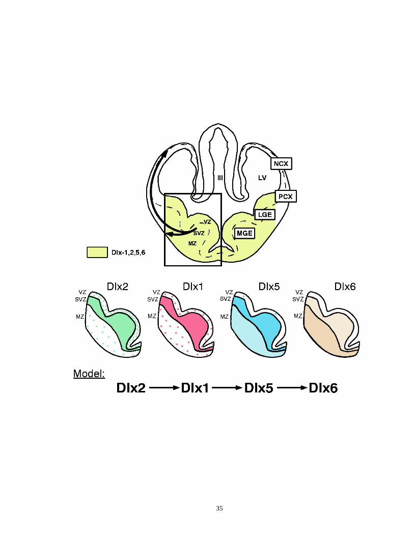
35
36
factors that play a vital role (amongst others) in manipulating the proliferation,
differentiation as well as migration of subpallim-derived GABAergic projection neurons
and interneurons that will later populate many different areas in the forebrain, such as the
cortex, hippocampus, olfactory bulb and striatum (Anderson 1997a; Bulfone et al., 1998;
Marin et al., 2000; Pleasure et al., 2000; Anderson et al., 2001; Stuhmer et al., 2002a).
Although several dlx genes are also expressed in the zebrafish forebrain, there is
presently no systematic study reporting their specific function in this tissue, mainly due to
the lack of simple technologies to implement targeted deletions or mutations of dlx genes
in zebrafish. The function of Dlx genes during mouse forebrain development has been
primarily assayed in the models of single and double Dlx knockout mice (Table 1.1).
While Dlx single knockouts die very quickly after birth (Dlx2 and Dlx5 mutants) or
within the first three postnatal weeks (Dlx1 mutants maintained in a C57BL/6J
background) owing to either craniofacial defects or deficiency in the enteric nervous
system (Qiu et al., 1995; Qiu et al., 1997; Acampora et al., 1999), forebrain development
in these mutant animals seemingly remains largely intact when compared to that of wild-
type littermates. However, Cobos et al. (2005) reported that postnatal Dlx1 mutants (> 1
month old) maintained in a C57BL/6J×CD1 background have a specific loss of several
major GABAergic interneurons subtypes (CR-, SOM- and NPY-expressing cells) in the
cortex and hippocampus and this phenotype occurs in a time-dependent manner. These
mice also exhibit a hyperexcitable and epileptic phenotype possibly as a result of reduced
number of GABAergic interneurons and a decreased synaptic inhibition mediated by
GABA (Cobos et al., 2005a). Moreover, recent observations in adult Dlx1-/-
mutant mice
revealed that they have significantly increased activities in several behavioral analyses,
37
such as open field and tail suspension tests, in comparison to wild-type controls (Mao et
al., 2009). They also have a reduced response to fear conditioning (Mao et al., 2009).
Dlx2 single mutants have normal morphology and histology of the forebrain during
embryogenesis but a reduced number of dopaminergic neurons are found in the olfactory
bulb (Qiu et al., 1995). Unfortunately, the potential forebrain phenotypes of Dlx2-/-
mutants at subsequent postnatal time points cannot be further evaluated because of their
death soon after birth (Qiu et al., 1995). Likewise, no apparent abnormalities have been
seen in the forebrain of Dlx5 single mutants except for a decrease of GABAergic neurons
in the olfactory bulb (Acampora et al., 1999; Long et al., 2005). To date, the phenotypes
of mice lacking the Dlx6 gene have not yet been documented.
In contrast to Dlx single mutants, inactivation of both Dlx1 and Dlx2 (although
these mice die very shortly after birth) leads to a major block of the production and
differentiation of late-born striatal projection neurons (Anderson et al. 1997a; Marin et al.
2000). Most of these neurons remain in the progenitor domains in the ganglionic
eminences, rather than radially migrate to their destinations in the striatum (Anderson et
al. 1997a; Marin et al. 2000). Similar deficiencies in neurogenesis and neuronal
differentiation were also observed in GABAergic, dopaminergic and cholinergic
interneuron progenitors born in the LGE and MGE (Anderson et al., 1997a; Bulfone et
al., 1998; Marin et al., 2000; Pleasure et al., 2000). These cells accumulate in the basal
ganglia and fail to migrate tangentially to the neocortex and hippocampus as well as to
the olfactory bulb via the rostral migration stream (Anderson et al., 1997a; Bulfone et al.,
1998; Marin et al., 2000; Pleasure et al., 2000). Thus, compared to wild-type mice,
Dlx1/Dlx2 double knockouts display markedly decreased numbers of GABAergic and
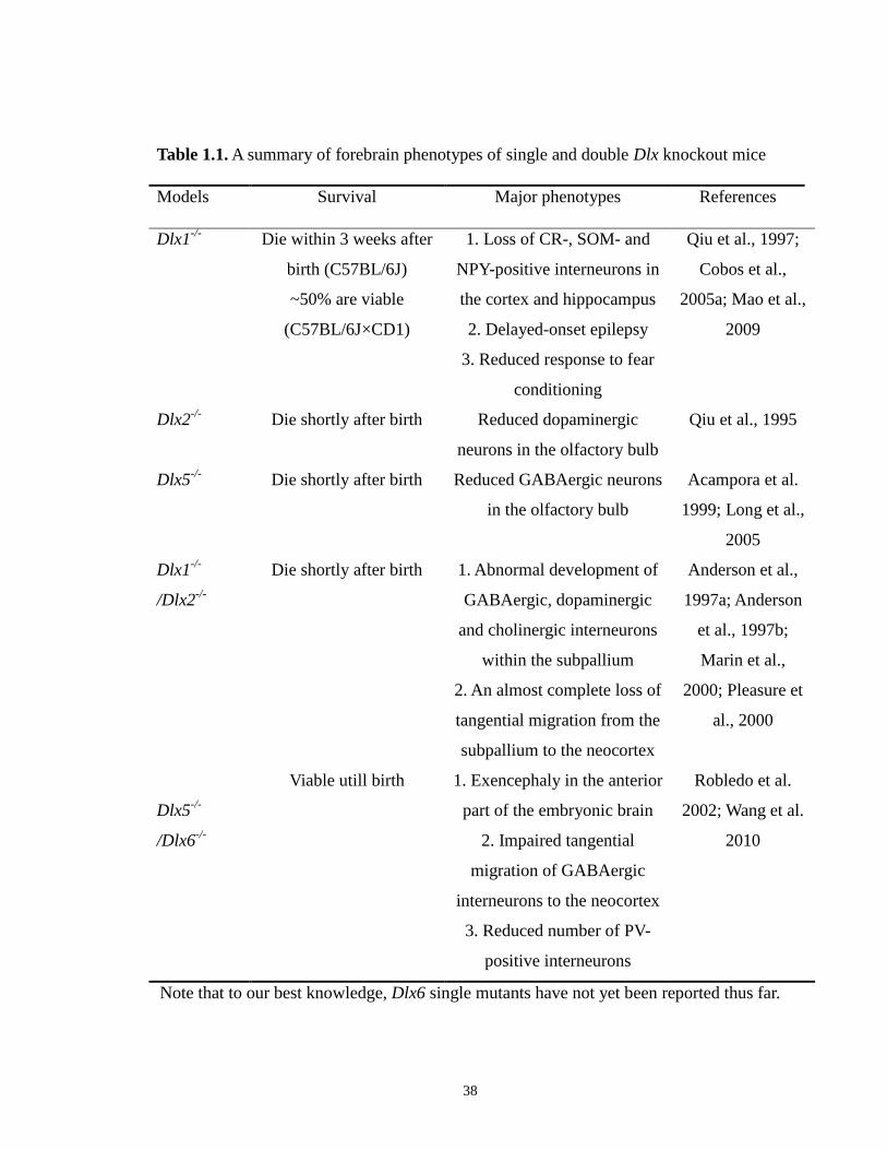
38
Table 1.1. A summary of forebrain phenotypes of single and double Dlx knockout mice
Models Survival Major phenotypes References
Dlx1-/-
Dlx2-/-
Dlx5-/-
Dlx1-/-
/Dlx2-/-
Dlx5-/-
/Dlx6-/-
Die within 3 weeks after
birth (C57BL/6J)
~50% are viable
(C57BL/6J×CD1)
Die shortly after birth
Die shortly after birth
Die shortly after birth
Viable utill birth
1. Loss of CR-, SOM- and
NPY-positive interneurons in
the cortex and hippocampus
2. Delayed-onset epilepsy
3. Reduced response to fear
conditioning
Reduced dopaminergic
neurons in the olfactory bulb
Reduced GABAergic neurons
in the olfactory bulb
1. Abnormal development of
GABAergic, dopaminergic
and cholinergic interneurons
within the subpallium
2. An almost complete loss of
tangential migration from the
subpallium to the neocortex
1. Exencephaly in the anterior
part of the embryonic brain
2. Impaired tangential
migration of GABAergic
interneurons to the neocortex
3. Reduced number of PV-
positive interneurons
Qiu et al., 1997;
Cobos et al.,
2005a; Mao et al.,
2009
Qiu et al., 1995
Acampora et al.
1999; Long et al.,
2005
Anderson et al.,
1997a; Anderson
et al., 1997b;
Marin et al.,
2000; Pleasure et
al., 2000
Robledo et al.
2002; Wang et al.
2010
Note that to our best knowledge, Dlx6 single mutants have not yet been reported thus far.
39
cholinergic striatal interneurons, olfactory bulb interneurons (>95% are GABAergic and
dopaminergic), hippocampal interneurons (>95% are GABAergic) and cortical
interneurons (>75% are GABAergic) (Anderson et al., 1997a; Anderson et al., 1997b;
Bulfone et al., 1998; Marin et al., 2000; Pleasure et al., 2000; Long et al., 2007). This
defective neuronal migration seen in Dlx1/Dlx2-/-
mutants has been recently shown to be
likely due to increased neurite length and reduced branching of migrating cells (Cobos et
al., 2007). Furthermore, both Dlx5 and Dlx6 expression are almost completely absent in
the forebrain of Dlx1/Dlx2 double knockouts, indicating that Dlx1 and Dlx2 are upstream
genetic regulators of Dlx5 and Dlx6 genes (Anderson et al. 1997a; Zerucha et al. 2000).
This interaction between Dlx1/Dlx2 and Dlx5/Dlx6 may be mediated by a cis-regulatory
element (CRE) named I56i that is located in the intergenic sequence between the
Dlx5/Dlx6 bigene cluster (Zerucha et al., 2000, more details on I56i below). While the
phenotype in the forebrain of Dlx5/Dlx6 null mice cannot be assessed in detail because of
the exencephaly that also results in the anterior parts of the forebrain, recent laboratory
work from Dr. Rubenstein’s group showed that simultaneous deletion of Dlx5 and Dlx6
genes can significantly slow down the movement of tangentially migrating GABAergic
interneurons from the subpallium to the neocortex (Wang et al., 2010; Wang and
Rubenstein, personal communication). However, the impact on their migration is not as
severe as that seen in Dlx1/Dlx2-/-
mutants (Anderson et al., 1997a; Bulfone et al., 1998;
Marin et al., 2000; Pleasure et al., 2000). Dlx5/Dlx6-/-
mutants also have a selectively
reduced number of PV-expressing cortical interneurons with increased dendrite
branching (Wang et al., 2010). Given that Dlx1 is preferentially involved in the
production and maturation of CR-, SOM- and NPY-positive subclasses of cortical
40
interneurons (but not PV-expressing cells) (Cobos et al., 2005a), it is reasonable to infer
that different individual Dlx genes may be specifically required for proper development
and/or function of distinct subtypes of GABAergic interneurons.
1.7.3. Regulation of Dlx gene expression
1.7.3.1. Upstream regulators of Dlx genes in the forebrain
To better understand the contributions of Dlx genes to the complicated genetic
regulatory network during the forebrain development, it is of significant importance to
identify and characterize their upstream transcriptional or post-transcriptional
modulators. Unfortunately, so far, only a few signaling molecules and transcription
factors have been revealed to participate in regulating Dlx expression in the developing
forebrain. Although the mechanism by which they interact with each other is still unclear,
administration of RA to early mouse and zebrafish embryos, before the occurrence of
neural crest cell migration, leads to a rapid and marked inhibition of expression of several
Dlx genes in ectomesenchymal cells (Ellies et al., 1997b; Vieux-Rochas et al., 2007). In
addition, Gaiano et al. (1999) reported that ectopic expression of Shh in the pallium of
the early developing telencephalon starting at ~E8.5 stimulates and induces ectopic dorsal
expression of several ventral markers including Dlx2 at the sites of ectopic Shh. The level
of Dlx2 expression was found to be significantly declined in the embryonic forebrain of
Shh-/- mutant mice (Ohkubo and Rubenstein, unpublished data). Other known upstream
regulators of Dlx genes include the bHLH transcription factor Mash1 and the Fez1
forebrain-specific zinc finger protein (Long et al., 2009a; Yang et al., 2001). Previous in
vitro assays have shown that when Mash1 is ectopically expressed in the developing
41
mouse pallium, ectopic Dlx1 expression can be induced in the same domains (Fode et al.,
2000; Stuhmer et al., 2002b). Further to these data, our laboratory recently found that the
Mash1 protein could physically bind to an E-box sequence present in an I12b enhancer
element located in the Dlx1/Dlx2 intergenic region and regulate Dlx1/Dlx2 expression
(Poitras et al., 2007, more information on I12b will be provided below). Forced
overexpression of Fez1 results in ectopic expression of both dlx2a and dlx6a in the early
embryonic zebrafish forebrain, whereas morpholino-based inhibition of Fez1 expression
significantly reduces dlx2a expression in the ventral forebrain as compared to that in
control embryos (Yang et al., 2001).
1.7.3.2. Downstream targets of Dlx genes in the forebrain
Although increasing evidence has shown that Dlx proteins can directly target Dlx
genes by themselves and regulate their own expression through either cross-regulatory or
auto-regulatory mechanisms, up to the present, our general knowledge regarding other
direct transcriptional downstream targets of various Dlx genes in the forebrain is still very
limited. In recent years, a lot of effort has been made to identify additional downstream
genes that can be regulated by Dlx genes mostly through conducting microarray analyses
in Dlx knockout mice. Based on gene expression array analysis in Dlx1/Dlx2 double
mutants, Long and coworkers identified a large body of potential downstream candidate
genes of Dlx1 and Dlx2 in the the subpallial ganglionic eminences, olfactory bulb and
striatum (Long et al., 2007; Long et al., 2009a; Long et al., 2009b). Likewise, a
microarray assay comparing gene expression profiles between Dlx1/2-/-
and Dlx1/2+/-
MGE cells also screened many potential Dlx target genes, such as Pax3, that are involved

42
in the regulation of neuronal differentiation and migration (Cobos et al., 2007). More
specifically, Dlx genes have been demonstrated to act as upstream modulators of Gad1
and Gad2 genes, which encode a key rate-limiting enzyme controlling GABA synthesis.
Stuhmer et al. (2002b) showed that ectopic Dlx2 and Dlx5 expression in vitro are
sufficient to induce ectopic expression of both Gad1 and Gad2 in many domains of the
mouse forebrain. In addition, there have been studies reporting that Dlx1, Dlx2 and Dlx5
proteins can activate the reporter gene expression via acting through an enhancer element
identified within the Gad1 locus in vitro (reviewed in Panganiban and Rubenstein, 2002).
Consistently, the level of Gad1 transcript is significantly down-regulated in Dlx1/Dlx2
null mice (Long et al., 2009a; Long et al., 2009b).
Nrp2 is another well-established direct downstream target of Dlx genes. Chromatin
immunoprecipitation (ChIP) analysis using E13.5 ganglionic eminences revealed that
Dlx1 and Dlx2 proteins can directly bind to a Nrp2 promoter region 2 (Nrp2-ii) in vivo
(Le et al., 2007). The authors showed that both recombinant Dlx1 and Dlx2 proteins can
also bind to this specific region of Nrp2 promoter in vitro and inhibit the transcription of
Nrp2 (Le et al., 2007). These results are further supported by the evidence that disruption
of Dlx1 and Dlx2 function causes elevated and ectopic Nrp2 expression in the ventral
telencephalon and GABAergic interneurons abnormally overexpressing Nrp2 accumulate
in the LGE and MGE instead of migrating to the neocortex (Marin et al., 2001; Marin and
Rubenstein 2003). Moreover, the Aristaless (Arx) gene, another key player involved in
promoting tangential migration of GABAergic interneurons, has been shown to be a
direct target of the Dlx2 protein (Colasante et al., 2008). Very interestingly, the
43
Aristaless-like gene in Drosophila is also found to be a downstream target of the Dll gene
(Depew et al., 2002).
1.7.3.3. Cis-regulatory elements (CREs) of Dlx genes
The highly conserved genomic organization of multiple Dlx bigene clusters among
different distant vertebrate species provides us with hints that this genomic arrangement
may have important implications and thus have gained selective advantage through the
evolutionary process. This viewpoint is further supported by the fact that the expression
patterns of orthologous Dlx genes in distant vertebrates also appear to be highly
conserved in many tissues, including the forebrain, even after a number of genomic
duplication events during evolution. One possible explanation for these phenomena
would be that the corresponding regulatory mechanisms essential for maintaining Dlx
expression and function, such as their cis- and trans-acting elements or certain key
signaling pathways, may have been retained by selective pressure during evolution of the
Dlx gene family. For example, it is likely that sharing of different CREs within the locus
of a given Dlx bigene cluster (either individually or in combination) may not only help
maintain the organization of the two physically-linked Dlx genes as a pair through
evolution, but also contribute, at least in part, to the overlapping expression pattern and
possibly redundant activitiies of paralogous Dlx genes.
In the past ten years, our laboratory has employed phylogenetic footprinting to
investigate the noncoding sequences surrounding the Dlx1/Dlx2 and Dlx5/Dlx6 loci and
screened several well-conserved CREs across the genomes of five highly divergent
vertebrates (mouse, human, zebrafish and two species of pufferfish). Two conserved
44
CREs, I56i (~440 bp) and I56ii (~310 bp), were identified in the intergenic region of the
Dlx5/Dlx6 bigene cluster of the five different species (I56i: sequence identity > 81%,
I56ii: sequence identity > 84%), both of which are able to target expression of reporter
transgenes to the basal telencephalon and diencephalon of transgenic mouse and
zebrafish in a pattern that highly resembles that of endogenous Dlx expression (Figure
1.7A-B) (Zerucha et al., 2000; Ghanem et al., 2003). Transgenic experiments showed that
the I56i CRE can also drive reporter gene expression to branchial arches, specifically to
the mandibular component of the first branchial arch and the second branchial arch
(Figure 1.7B) (Zerucha et al., 2000; Ghanem et al., 2003; Park et al., 2004). In addition,
co-transfection assays in cultured cells and DNA-protein binding analyses revealed that
Dlx1/Dlx2/Dlx5 protein can activate transcription through binding to putative Dlx
binding sites within the I56i sequence, suggesting that I56i CRE may be important for
establishing cross-regulatory interactions between various Dlx genes (Zerucha et al,
2000; Stuhmer et al., 2002b). Interestingly, similar to a significant reduction of
endogenous Dlx5/Dlx6 expression, the expression of a lacZ reporter gene under control
of I56i CRE is barely detectable in Dlx1/Dlx2-/-
mice (Anderson et al., 1997b; Zerucha et
al., 2000). These compelling findings suggest that Dlx1 and Dlx2 may act as upstream
modulators of Dlx5 and Dlx6 by functioning via the I56i enhancer (Anderson et al.,
1997b; Zerucha et al., 2000).
Data from Kohtz’ group also pointed out that Evf2, an ultraconserved noncoding
RNA that is partially encoded within I56i, is able to recruit Dlx and Mecp2 proteins to
I56i by forming a stable complex, impact its transcriptional activity and then regulate
Dlx5, Dlx6 and Gad67 expression (Feng et al., 2006; Bond et al., 2009). Evf2TS/TS
mutant

45
mice (P0) completely lacking the ultraconserved region of Evf2 have reduced levels of
Gad67 expression and a marked decrease (40-65%) of GABAergic interneurons in the
dentate gyrus and CA1 and CA3 regions of the hippocampus (Bond et al., 2009).
Hamilton et al. (2005) searched sequence variances within DLX1/DLX2 and
DLX5/DLX6 loci in patients with autism and found a total of 31 single nucleotide
polymorphisms (SNPs) and two insertion/deletion polymorphisms. Interestingly, one of
them is an adenine to guanine alteration at position 182 within one of the two putative
DLX binding sites identified in I56i CRE (Zerucha et al., 2000). By using a transgenic
approach in mice, we recently demonstrated that this SNP significantly affects the
endogeneous activity of I56i CRE and results in a remarkable reduction in reporter gene
expression in the progenitor regions of the early developing telencephalon, in the
tangentially migrating streams of GABAergic neurons as well as in various GABAergic
interneuron subtypes in the somatosensory cortex of adult animals (Poitras et al., 2010).
Our in vitro analyses, such as EMSA and co-transfection assays, also showed that this
SNP is sufficient enough to impair the physical binding of several Dlx proteins to I56i
and significantly decreases the transcriptional activity of the I56i enhancer in response to
Dlx2 and Dlx5 proteins (Poitras et al., 2010). These novel findings suggest that disrupted
activity of individual Dlx CREs due to the presence of a single nucleotide alteration can
destroy proper Dlx function and trigger abnormal development of GABAergic neurons,
which has been found to be related to many human neurological disorders.
Likewise, two conserved CREs, I12a (~550 bp) and I12b (~400 bp), were also
discovered in the intergenic region of Dlx1/Dlx2 bigene cluster with a high sequence
identity among human, mouse, zebrafish and two species of pufferfish (I12a: sequence
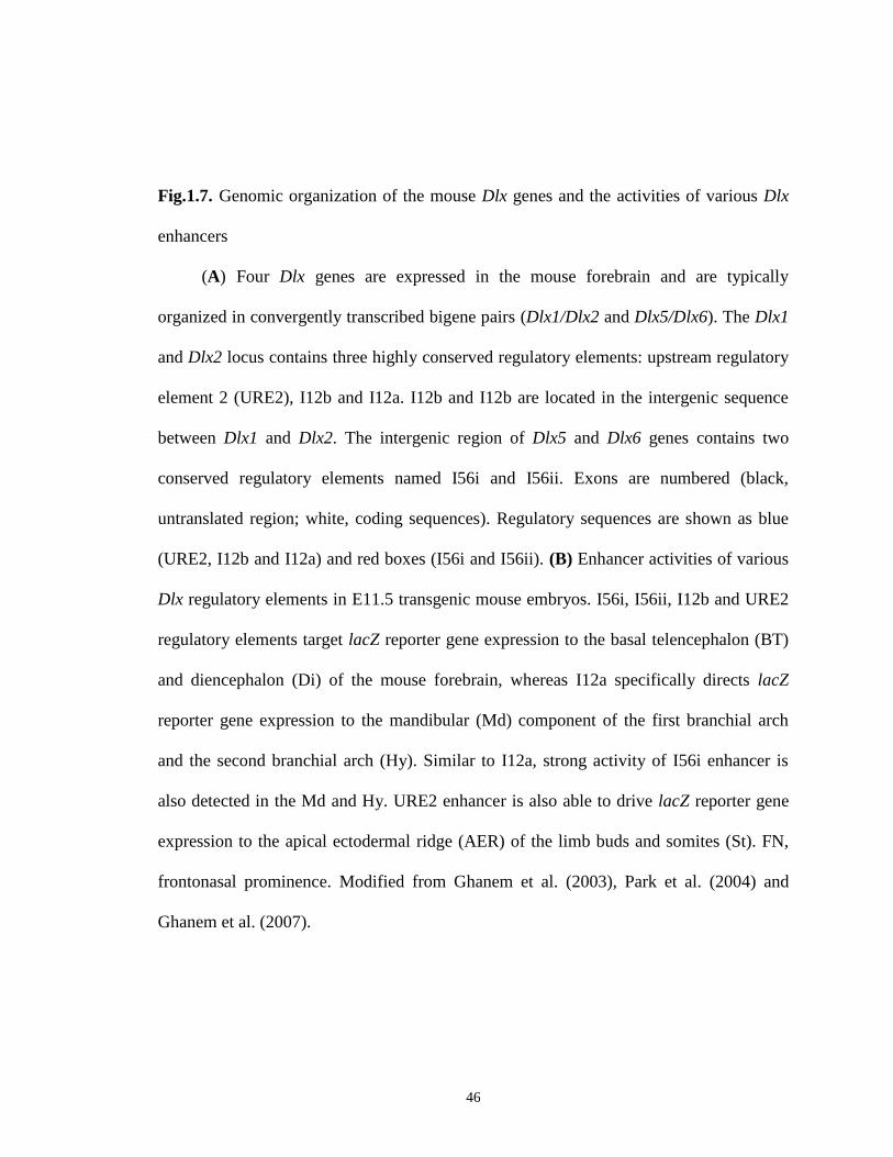
46
Fig.1.7. Genomic organization of the mouse Dlx genes and the activities of various Dlx
enhancers
(A) Four Dlx genes are expressed in the mouse forebrain and are typically
organized in convergently transcribed bigene pairs (Dlx1/Dlx2 and Dlx5/Dlx6). The Dlx1
and Dlx2 locus contains three highly conserved regulatory elements: upstream regulatory
element 2 (URE2), I12b and I12a. I12b and I12b are located in the intergenic sequence
between Dlx1 and Dlx2. The intergenic region of Dlx5 and Dlx6 genes contains two
conserved regulatory elements named I56i and I56ii. Exons are numbered (black,
untranslated region; white, coding sequences). Regulatory sequences are shown as blue
(URE2, I12b and I12a) and red boxes (I56i and I56ii). (B) Enhancer activities of various
Dlx regulatory elements in E11.5 transgenic mouse embryos. I56i, I56ii, I12b and URE2
regulatory elements target lacZ reporter gene expression to the basal telencephalon (BT)
and diencephalon (Di) of the mouse forebrain, whereas I12a specifically directs lacZ
reporter gene expression to the mandibular (Md) component of the first branchial arch
and the second branchial arch (Hy). Similar to I12a, strong activity of I56i enhancer is
also detected in the Md and Hy. URE2 enhancer is also able to drive lacZ reporter gene
expression to the apical ectodermal ridge (AER) of the limb buds and somites (St). FN,
frontonasal prominence. Modified from Ghanem et al. (2003), Park et al. (2004) and
Ghanem et al. (2007).

47
48
identity > 92%, I12b: sequence identity > 77%, Ghanem et al., 2003) (Figure 1.7A). The
I12b CRE was found to specifically target reporter gene expression to the basal
telencephalon and diencephalon in the mouse forebrain (Figure 1.7B) (Ghanem et al.,
2003), whereas I12a targets lacZ expresnsion to the mandibular part of the first branchial
arch and the second branchial arch, similar to the areas that are targeted by I56i in the
branchial arches (Figure 1.7B) (Park et al., 2004). The expression patterns of I12b-lacZ
and I12a-lacZ transgenes faithfully recapitulate the patterns of endogenous Dlx1/Dlx2
expression in the forebrain and branchial arches. An additional CRE, named URE2
(Upstream Regulatory Element 2, ~900 bp), located at ~12 kb upstream of the Dlx1 start
codon, was identified with > 75% sequence identity between mouse and zebrafish
(Ghanem et al., 2007; MacDonald et al., 2010b) (Figure 1.7A). Analyses in transgenic
mice showed that URE2 CRE not merely drives reporter gene expression to the basal
telencephalon and diencephalon, but also to the apical ectodermal ridge (AER) of the
limb buds and to the somites (Figure 1.7B) (Ghanem et al., 2007). Despite the fact that
the forebrain expression patterns of the lacZ reporter gene targeted by I56i, I56ii, I12b
and URE2 appear to be highly similar in whole-mount mouse embryos, further studies on
transverse telencephalic sections at various stages revealed some differences between
I56i-lacZ, I12b-lacZ and URE2-lacZ transgene expression (Ghanem et al., 2003; 2007).
Indeed, a detailed comparison between I56i, I12b and URE2 enhancer activity showed
that these three individual CREs target overlapping but distinct populations of
GABAergic interneurons in the ganglionic eminences as well as in the cortex of adult
animals (Ghanem et al., 2007; Potter et al., 2009). Moreover, a recent study regarding the
functional importance of I12b CRE showed that the proneural transcripton factor Mash1
49
can modulate Dlx1/Dlx2 expression as a key upstream regulator through directly binding
to I12b CRE (Poitras et al., 2007). I12b was also reported to be a direct target of Dlx2
protein and correct Dlx1 and Dlx2 expression might be importantly involved in
maintaining the proper activity of I12B CRE (Potter et al., 2009).
A total of five conserved CREs with size ranging from ~180 to ~358 bp have been
found in the intergenic locus between Dlx3 and Dlx4 genes and they all display high
sequence identity (over 82%) between mouse and human genome (Sumiyama et al.,
2002). I37-2, one of these five CREs, is physically close to Dlx4 gene and contains two
potential homeodomain binding sites as well as two putative Dlx binding sites
(Sumiyama and Ruddle, 2003). In transgenic mouse assays, this enhancer can target
reporter gene expression to mesenchymal cells in the first and second branchial arches
and the expression domains closely mimic that of endogenous Dlx3/Dlx4 expression in
the branchial arches (Sumiyama and Ruddle, 2003). Moreover, at least one of these five
CREs was also found in the zebrafish dlx3b/dlx4b intergenic sequence (M. Ekker,
unpublished observation).
1.7.3.4. Consensus Dlx-binding motifs present in various Dlx CREs
So far, several potential binding motifs that can be recognized and bound by Dlx
proteins have been identified and characterized. One representative example is an optimal
consensus DNA sequence, namely (A/C/G)TAATT(G/A)(C/G), which was originally
found as a potential binding module for the Xenopus Dlx3 protein and then was proved to
be a binding domain for many other Dlx proteins in vitro (Feledy et al., 1999). Thus, the
authors claim that most of Dlx proteins, if not all, may share similar DNA-binding
50
activity at least under certain circumstances (Feledy et al., 1999). Although the various
Dlx CREs identified by our laboratory exhibit some overlapping activities when tested in
transgenic mouse and zebrafish, these regulatory elements show only limited sequence
similarity (Zerucha et al., 2000; Ghanem et al., 2003; Ghanem et al., 2007), supporting a
notion that these individual regulatory elements may be responsive to and modulated by
different transcription factors, in addition to Dlx proteins. Previous in vitro analyses in
culture cells and embryonic brain slices revealed that zebrafish dlx1a/2a/3b/5a/6a
proteins as well as mouse Dlx1/2/5 proteins can activate the transcription of reporter
genes by acting on the two TAATT Dlx-binding sites found in the I56i CRE (Zerucha et
al. 2000; Stuhmer et al. 2002b). Mutagenesis of one or both of these two motifs could
significantly disrupt the binding of Dlx proteins to I56i CRE and hence cause a
remarkably decreased reporter gene activity in the forebrain of transgenic animals
(Zerucha et al., 2000). Additionally, DNA I footprinting assays using nuclear extracts
from E13.5 mouse telencephalon and two DNA fragments comprising I12b CRE
suggested that I12b contains six protein-protected areas (footprints), each of which
indicates a putative protein-DNA binding domains (Poitras et al., 2007). Among them,
two putative TAATT Dlx binding sites and a CANNTG E-box binding site (that can be
directly bound by the transcriptioin factor Mash1, as mentioned earlier) were shown to be
important for correct lacZ reporter gene expression in the basal telecephalon and
diencephalon of transgenic mice (Poitras et al., 2007; Potter et al., 2008). Mutations of
the three binding sites present in I12b result in loss of reporter gene expression, to
varying degrees, in many domains of the telencephalon and diencephalon (Poitras et al.,
2007). In particular, simultaneous disruption of the two Dlx binding motifs can lead to an
51
almost entire loss of basal telencephalic and diencephalic expression (Poitras et al.,
2007). Aside from the consensus sequence motifs discussed above, researchers also
identified a number of TAAT/ATTA core binding motifs at several locations in the
sequences of both I56i and I56ii CREs (Zerucha et al., 2000; Ghanem et al., 2003; Zhou
et al., 2004), which may play a potential role in recruiting Dlx proteins or other
homeodomain-containing transcription factors (e.g. Hox and Islet1 proteins) probably
involved in regulating Dlx function (Gehring et al., 1994; Pankratova and Polanovsky,
1998).
52
Statement of Inquiry
During the process of embryogenesis, the CNS including the brain and spinal cord
is gradually established and organized by numerous different histological structures,
which are originally formed by diverse types of neuronal cells. The correct differentiation
of multipotential progenitor cells into specific types of neuronal populations at different
developmental stages is under the control of a complicated and precisely-regulated
genetic network. The vertebrate Dlx genes are typically organized as multiple bigene
pairs and are sequentially expressed in a spatial and temporal pattern in the developing
forebrain. Specifically, various Dlx genes encode a group of homeodomain-containing
transcription factors that play essential roles in regulating proper differentiation,
migration and survival of GABAergic neurons that will later populate different regions in
the telencephalon and diencephalon. Furthermore, the forebrain expression pattern of Dlx
genes is highly overlapping with that of the two Gad genes coding enzymes responsible
for synthesizing the inhibitory neurotransmitter GABA and expression of several Dlx
genes has been shown to be sufficient to induce Gad expression both in vitro and in vivo.
Proper Dlx forebrain expression is modulated at least in part by several regulatory
elements that are originally identified in their surrounding sequences. These regulatory
elements appear to be highly conserved among various distantly related vertebrate
species, some of which are even ultraconserved, suggesting that they might have
undergone a strong evolutionary selection together with the Dlx genes over evolutionary
time. Using transgenic analysis in mice, our laboratory has found that at least four of
those elements, URE2 and I12b in the Dlx1/Dlx2 loci and I56i and I56ii in the Dlx5/Dlx6
loci, act as forebrain-specific enhancers by targeting reporter gene expression in a pattern

53
that strictly resembles endogenous Dlx forebrain expression. Moreover, a detailed
comparison between URE2, I12b and I56i enhancer activity revealed that these enhancers
may regulate Dlx expression in distinct but highly overlapping subpopulations of
GABAergic interneurons in the developing telencephalon. However, our overall
understanding of Dlx gene regulation via these various individual enhancers during
forebrain development is still very limited in many aspects.
In my PhD project, I have attempted to address several unresolved questions
regarding Dlx regulation in the developing mouse and zebrafish forebrain: 1) Does
another enhancer I56ii possess a similar activity when compared to that of the other three
forebrain-specific enhancers (I56i, I12b and URE2; note that I12a was not included for
comparison since it is only active in the branchial arches, Park et al., 2004)? 2) Does
sequence conservation of these regulatory elements indicate a degree of similar function
across the developing forebrain of distantly related vertebrates (e.g. zebrafish), at least
during the development of GABAergic neurons? 3) What is the relative contribution of
the various forebrain individual enhancers to overall Dlx expression? 4) What are the
potential impacts of loss of an individual Dlx enhancer on the process of forebrain
development?
In chapter II of my doctoral thesis, I have specifically answered the first question.
In collaboration with Dr. Noël Ghanem, I have conducted a detailed spatio-temporal
analysis of I56ii activity in the developing telencephalon between E10.5 and E15.5, and
compared its regulatory activity with that of the other three Dlx enhancers using lacZ
reporter genes in transgenic mice. We found that unlike the other three enhancers, I56ii
does not label interneuron progenitors in the subpallial telencephalon; instead, I56ii-
54
positive cells are a subpopulation of GABAergic projection neurons expressing the
striatal markers Meis2 and Islet1.
In chapter III, I aimed to address the above-noted second question with a focus on
the potential implications of I56i and I56ii enhancers in the dlx-mediated regulatory
pathways during zebrafish GABAergic neuron development. I have established two
independent lines of transgenic zebrafish with reporter constructs under the control of
different combinations of I56i and I56ii enhancers and found that these regulatory
elements exhibit a fairly conserved activity in the zebrafish forebrain when compared to
their corresponding mouse orthologs. We attempted to knock down dlx1a/dlx2a and
dlx5a/dlx6a expression by using morpholino oligonucleotides and found that proper dlx
expression in the forebrain by acting through the I56i and I56ii enhancers is essential for
proper GABAergic neuron production in the diencephalon of zebrafish forebrain but
seems less important for those in the telencephalon, a result that is not consistent with
that observed in mice. Therefore, we propose that the dlx-mediated genetic cascades
controlling GABAergic neuron development are evolutionarily conserved in the
prethalamus but maybe have undergone divergence in the telencephalon between
zebrafish and mammals over evolutionary time. Also, other regulatory programs in
addition to the dlx-mediated ones may be functionally involved in controlling the
establishment of diverse GABAergic interneuron subtypes in the developing zebrafish
forebrain.
Finally, in the study reported in chapter IV, I answered partially the third and
fourth questions with the purpose of understanding the specific contribution of the I12b
enhancer to overall Dlx regulation as well as to the process of GABAergic neuron
55
generation. Working with Dr. Luc Poitras, I have successfully generated a mutant mouse
strain with a targeted deletion of the I12b enhancer. My data demonstrated that although
mice homozygous for I12b deletion are viable, develop normally, and display no overt
morphological abnormalities in the forebrain, targeted disruption of this element leads to
a significant decline of Dlx1 and Dlx2 levels and seemingly could interfere with normal
cell proliferation in the ventral telencephalon, at least in the progenitor domains in the
LGE and MGE. These initial phenotype analyses will be of important resource for our
future studies and will eventually help us better understand the relative contribution of
I12b to Dlx forebrain expression as well as to the regulatory cascades associated with this
enhancer during vertebrate forebrain development.
56
2. Characterization of a distinct subpopulation of striatal projection
neurons expressing the Dlx genes in the basal ganglia through the
activity of the I56ii enhancer
Noël Ghanem a,1
, Man Yu a,1
, Luc Poitras a, John L.R. Rubenstein
b and Marc Ekker
a,*
a Center for Advanced Research in Environmental Genomics (CAREG), Department of
Biology, University of Ottawa, Ottawa, ON, Canada K1N 6N5
b Nina Ireland Laboratory of Developmental Neurobiology, Center for Neurobiology and
Psychiatry, Department of Psychiatry, University of California, San Francisco, CA
94143, USA
1 These authors contributed equally to this work.
* Corresponding author: Marc Ekker, Department of Biology, University of Ottawa, 30
Marie Curie, Ottawa, ON, Canada K1N 6N5. Tel.: 613-562-5800, ext. 2605; Fax: 613-
562-5486; E-mail address: [email protected]
Keywords: Dlx genes, enhancers, GABAergic neurons, ganglionic eminence, homeobox,
mice, migration, progenitors, striatal projection neurons

57
Abstract
Regulation of region-specific neuronal differentiation and migration in the embryonic
forebrain is a complex mechanism that involves a variety of transcription factors such as
the Dlx genes. At least four cis-acting regulatory elements (CREs) are responsible for the
Dlx transcriptional regulation in the subcortical telencephalon and the rostral
diencephalon. These include I12b and URE2 in the Dlx1/2 bigene cluster, and, I56i and
I56ii in the Dlx5/6 cluster. We previously reported that URE2, I12b, and I56i, mark
different progenitor cell populations in the ganglionic eminences as well as different
subtypes of adult cortical interneurons. Here, we carried out a detailed spatial and
temporal analysis of the I56ii CRE activity in the developing telencephalon between
E10.5 and E15.5, and compared its activity with the other three Dlx CREs using lacZ
reporter genes in transgenic mice. We show that I56ii marks distinct group(s) of neurons
located in the superficial mantle of the LGE and MGE between E11.5 and E13.5. The
I56ii-positive cells are Dlx- and GABA-immunoreactive. However, unlike the other
CREs, I56ii does not label interneuron progenitors in the basal ganglia, nor tangentially
migrating cells to the cortex at E13.5. Instead, I56ii-positive cells mark a
subpopulation(s) of post-mitotic projection neurons that tangentially migrate from the
LGE to the deep mantle of the MGE and reside between the subventricular zone and the
globus pallidus during midgestation. The majority of these neurons express the striatal
markers Meis2 and Islet1. Moreover, both Meis2 and Islet1 activate transcription of a
reporter gene containing the I56ii sequence in co-transfection assays, indicating that these
transcriptional factors may be potential upstream modulators of the Dlx genes in vivo.
58
2.1. Introduction
Over the past fifteen years, genetic studies have focused on elucidating the
mechanisms underlying the development of the telencephalon, one of the most complex
and diverse regions in the central nervous system (CNS). Several models of telencephalic
organization and development have emerged implicating a diverse array of signaling
molecules and transcription factors that control cell fate specification and proliferation as
well as differentiation and migration [for reviews; (Schuurmans and Guillemot, 2002;
Zaki et al., 2003; Puelles and Rubenstein, 2003; Guillemot, 2007)]. Hence, programs of
regional identity as defined by the expression of distinct transcription factors in different
progenitor regions control most aspects of histogenesis within the developing
telencephalon. As a result, the embryonic telencephalon is divided into the pallium
(primordium of neocortex, hippocampus, piriform cortex and amygdala) and the
subpallium (primordium of basal ganglia), which are, in turn, subdivided into distinct
progenitor domains (Puelles et al., 1999; Puelles et al., 2000; Marin et al., 2003). Post-
mitotic (immature) neurons are generally derived from progenitor cells located nearby in
the ventricular zone (VZ) of the neuroepithelium before they translocate to adjacent
locations in the mantle through radial migration like during the generation of cortical
glutamatergic neurons (Rakic and Lombroso, 1998). However, in some cases, cell types
generated in specific progenitor zone(s) migrate orthogonally to the radial axis of the
neural tube and intermingle with other neurons in a common final destination, where that
cell type apparently is not made, at least in large numbers. This process, known as
tangential migration, is widely spread in the developing CNS and contributes to the
complexity of the neuronal network found in the vertebrate brain (Corbin et al., 2001;
59
Marin and Rubenstein, 2001). For instance, the majority of cortical GABAergic
interneurons (De Carlos et al., 1996; Anderson et al., 1997a; Tamamaki et al., 1997;
Lavdas et al., 1999; Anderson et al., 2001; Cobos et al., 2001; Wichterle et al., 2001;
Gorski et al., 2002; He et al., 2001; Nery et al., 2002) and cortical oligodendrocytes
(Tekki-Kessaris et al., 2001; Thomas et al., 2000) in mouse and chick are born to ventral
progenitors located in the subpallium and reach their final destination by tangential
migration.
Several genes including Lhx6, Arx and the Dlx genes are involved in the regulation
of neuronal migration in the ventral telencephalon (Cobos et al., 2007; Colombo et al.,
2007; Liodis et al., 2007). The Dlx genes are transcription factors that are required for the
proper differentiation and migration of most ventrally-derived neurons including striatal
projection neurons and cortical interneurons. Thus, Dlx1/Dlx2 null mice display a major
block of cell differentiation in the lateral and medial ganglionic eminences (LGE and
MGE), and lack both radial and tangential migration of several types of GABAergic,
dopaminergic and cholinergic interneurons derived from the subpallium (Anderson et al.,
1997a; Anderson et al., 1997b; Bulfone et al., 1998; Marin et al., 2000; Pleasure et al.,
2000; Anderson et al., 2001; Marin and Rubenstein, 2001; Yun et al., 2002; Long et al.,
2007). Four Dlx genes, Dlx1, Dlx2, Dlx5 and Dlx6, are expressed in the forebrain and
show highly overlapping expression patterns but with subtle spatio-temporal differences
in the telencephalon and diencephalon, suggesting the presence of both redundant and
unique functions among these genes (Panganiban and Rubenstein, 2002). Towards
elucidating the biochemical mechanisms that control development of ventral progenitors
in the telencephalon, our laboratory investigated the regulatory elements and mechanisms
60
controlling Dlx gene expression and showed that shared regulatory mechanisms may
underlie the functional redundancy among Dlx paralogs (Zerucha et al., 2000; Ghanem et
al., 2003). Hence, we identified and characterized four conserved regulatory elements
(CREs) acting as forebrain-specific enhancers for the Dlx genes in vertebrates: I56i and
I56ii in the Dlx5/Dlx6 bigene cluster (Zerucha et al., 2000), and, URE2 and I12b in the
Dlx1/Dlx2 cluster (Ghanem et al., 2003; Ghanem et al., 2007). Furthermore, we reported
that URE2, I12b and I56i, mark different progenitor cell populations in the ganglionic
eminences and different subtypes of adult cortical interneurons (Ghanem et al., 2007).
These findings suggest that distinct Dlx functions could be mediated by different
regulatory elements and/or mechanisms.
Here we expand our previous findings by demonstrating the existence of a distinct
regulatory role played by I56ii when compared with the three Dlx CREs described earlier.
We carried out a detailed spatial and temporal analysis of the lacZ reporter transgene
expression driven by the I56ii CRE between E10.5 and E15.5 and compared its activities
with that of three previously described Dlx CREs. We report that, unlike the other CREs,
I56ii is not active in GABAergic interneuron progenitors in the basal ganglia, nor in
tangentially migrating cells to the cortex. Instead, it targets lacZ expression specifically to
subpopulation(s) of post-mitotic projection neurons that are probably derived from LGE
progenitors and have tangentially migrated to the deep mantle of the LGE and MGE
between E11.5 and E13.5. In addition, we also identify that the I56ii-positive neurons
express two striatal markers, Meis2 and Islet1, during midgestation, both of which can
activate transcription via I56ii in co-transfection assays in vitro, suggesting these
transcriptional factors may be potential upstream regulators of Dlx genes in vivo.
61
Together, our data reflect a complex and dynamic regulation of Dlx gene expression
during the early stages of embryonic development through several regulatory elements
with overlapping and distinct function(s).
2.2. Material and methods
2.2.1. Transgenic animals
For transgenic mice, sequences containing the four mouse enhancers (URE2, I12b,
I56i and I56ii) were subcloned separately into the p1229/p1230 vectors (Yee and Rigby
et al., 1993) that contain a human β-globin minimal promoter and the lacZ reporter gene.
Subclonings were done using a PCR-based approach or using convenient restriction sites
and followed by sequencing to verify the integrity of each insert. The p1230-based I12b-
alkaline phosphatase (I12b-AP) construct was generated as described earlier (Ghanem et
al., 2007). At least two independent transgenic lines that show lacZ or AP staining were
generated with each construct. Staining results on whole mount embryos and on coronal
sections of the forebrain were replicated from independent lines. Transgenic animals
were produced and analyzed as previously described (Zerucha et al., 2000).
2.2.2. Histology
E10 to E12.5 mouse embryos were fixed for 45min to 2h in 4% cold
paraformaldehyde (PFA) in 1×PBS at 4°C, then, washed and stained for β-galactosidase
activity overnight (O/N) at 28°C in a solution of 1 mg/ml X-gal, 5 mM K3Fe(CN)6, 5 mM
K4Fe(CN)6, 2 mM MgCl2, and 0.02% NP-40 in PBS. The stained brains were dissected
and equilibrated in 20% sucrose solution O/N at 4°C prior to sectioning. Brains were
62
embedded in Tissue-Tek media the following day, cryoprotected and processed for frozen
sectioning at 20-50μm using a cryostat (Leica CM3050 S). Sections were mounted with
Aquatex (EM Science, VWR). E13.5-P0 mouse brains were fixed for 2h to O/N in 4%
cold PFA in PBS at 4°C, dissected and sectioned prior to staining. LacZ staining and
mounting was performed as described above.
2.2.3. Double immunohistochemistry
Frozen sections of E10.5-E15.5 mouse brain were dried for 1h at room
temperature, then, washed for 3×5 min each in 0.1M phosphate buffer (PB) to eliminate
residues from tissue protection medium. Immunostaining was performed as described
earlier (Ghanem et al., 2007). The following antibodies have been applied in this study:
guinea pig anti-β-gal (1:1000, a generous gift from Dr. Thomas Sargent), rabbit pan-Dll
antibody (anti-Dlx; 1:100, a kind gift from Drs. Grace Panganiban and J.L.R.R.), rabbit
anti-human placental alkaline phosphatase (PLAP, 1:100, Serotec), mouse anti-PCNA
(1:300; Vector Laboratories), mouse anti-MAP2 (1:300, Sigma), rabbit anti-active
caspase 3 (1:500, BD Biosciences), rabbit anti-GABA (1:4000, Sigma), rabbit anti-
calbindin (1:1000, Chemicon), rat anti-somatostatin (1:100, Chemicon), rabbit anti-NPY
(1:4000, Immunostar), rabbit anti-TH (1:350, Chemicon), mouse anti-Islet1 (1:100,
Developmental Studies Hybridoma Bank), and goat anti-Meis2 (1:100, Santa Cruz
Biotechnology). Secondary antibodies were all purchased from Invitrogen: goat anti-
rabbit Alexa Fluor 488, goat anti-mouse Alexa Fluor 488, goat anti-rat Alexa Fluor 488,
donkey anti-goat Alexa Fluor 488, and goat anti-guinea pig Alexa Fluor 594. For PCNA
63
detection, sodium citrate antigen retrieval, pH 6.0, was performed on sections before
incubation with PCNA antibodies.
2.2.4. In situ RNA hybridization
In situ hybridization on frozen tissue sections and digoxigenin RNA probe labeling
were performed according to the procedures described in (Wallace and Raff, 1999).
Hybridized probes were detected with an AP-conjugated anti-digoxigenin Fab fragment
antibody (1:2000, Roche) and visualized with the NBT/BCIP substrate system. Antisense
riboprobes for Dlx5 and Dlx6 were prepared as previously described (Quint et al., 2000;
Cobos et al., 2006; Long et al., 2007).
2.2.5. Co-transfection and chloramphenicol acetyltransferase (CAT) assays
The mouse I56ii CRE was amplified by PCR and inserted upstream of the
thymidine kinase (tk) minimal promoter in a pBLCAT2 vector, we called it mI56ii-
pBLCAT2. We also created, using the overlapping-PCR method, a modified mI56ii-
pBLCAT2 plasmid (mI56ii-mutant-pBLCAT2) containing the mutagenized Meis2
consensus binding site (TGTC to TCTA) in mI56ii sequence (position nt. 262 to nt. 265,
Supplementary Figure 2.1). The mouse Islet1 cDNA encompassing the full-length coding
sequence (kindly provided by Dr. Steve M Sperber, National Institute of Child Health
and Human Development) was subcloned into the EcoRI site of a pCS2+ expression
vector, designated as Islet1-pCS2+. The Meis2-pCS2+ expression vector was previously
generated in our laboratory (Zerucha et al., 2000). The pSV-β-Gal reporter plasmid
encoding β-galactosidase was purchased from Promega and used as an internal control.
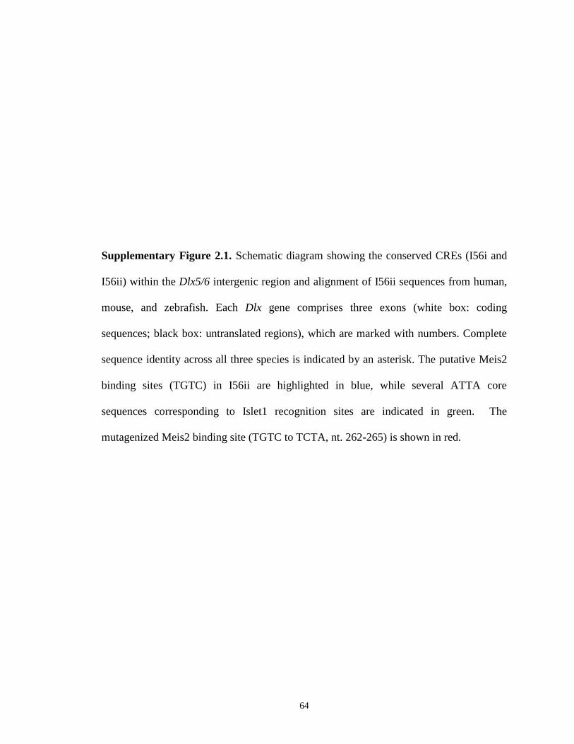
64
Supplementary Figure 2.1. Schematic diagram showing the conserved CREs (I56i and
I56ii) within the Dlx5/6 intergenic region and alignment of I56ii sequences from human,
mouse, and zebrafish. Each Dlx gene comprises three exons (white box: coding
sequences; black box: untranslated regions), which are marked with numbers. Complete
sequence identity across all three species is indicated by an asterisk. The putative Meis2
binding sites (TGTC) in I56ii are highlighted in blue, while several ATTA core
sequences corresponding to Islet1 recognition sites are indicated in green. The
mutagenized Meis2 binding site (TGTC to TCTA, nt. 262-265) is shown in red.
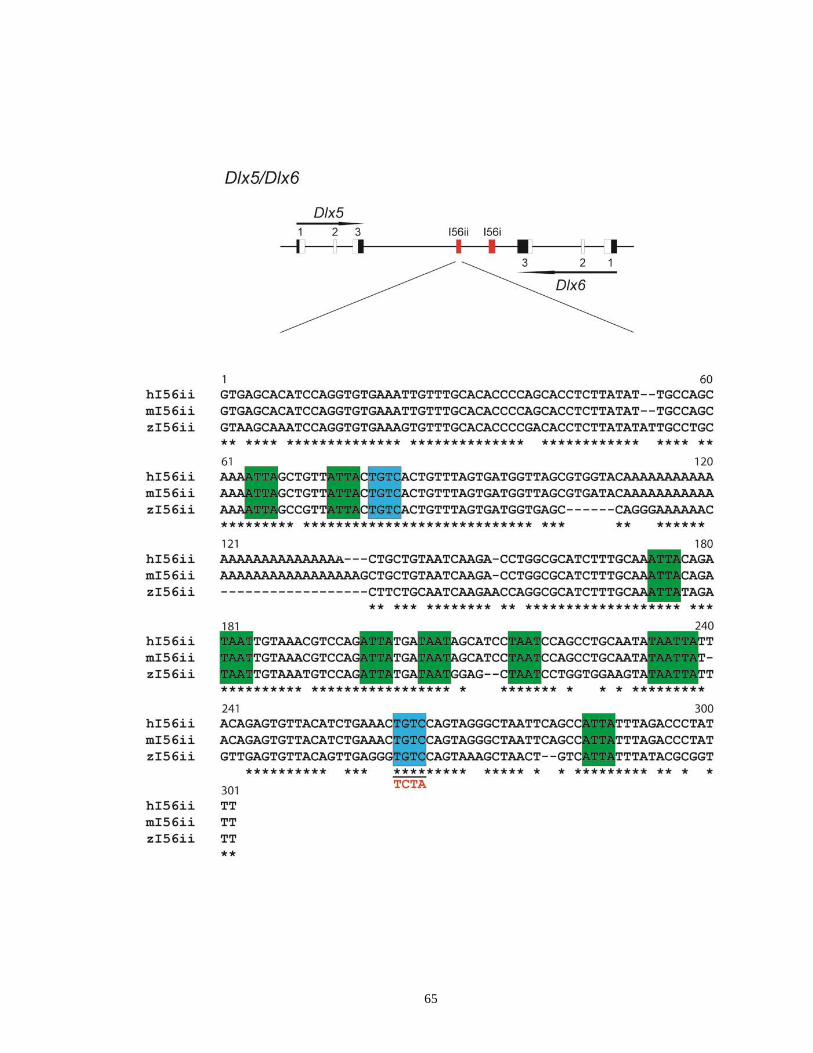
65
66
Co-transfection assays were performed as follows: briefly, P19 murine embryonic
carcinoma cells were seeded at a density of 3 × 10⁵ in 60-mm-diameter tissue culture
plates for 24-36h prior to transfection. Cells were subsequently transfected with 2µg of
mI56ii-pBLCAT2, mI56ii-mutant-pBLCAT2, or enhancer-less pBLCAT2, 2µg of each
expression vector (Meis2-pCS2+, Islet1-pCS2+, or empty pCS2+), and 2µg of pSV-β-
Gal using the lipofectamine 2000 reagent (Invitrogen). The CAT activity in cell extracts
was analyzed 48h post-transfection on TCL plates by applying the chloroform:methanol
solvent system and was quantified by a PhosphorImager as described earlier (Zerucha et
al., 2000). After normalizing to the β-galactosidase activity, the relative CAT activity for
each expression vector was measured and compared to the basal level of activity seen in
the control. The statistical analysis of the relative CAT activities among different groups
was performed by using the Student t-test. All experiments were repeated at least two
times in triplicate. P values < 0.05 were considered statistically significant.
2.3. Results
2.3.1. Spatio-temporal comparisons of lacZ expression in four Dlx CRE lines
during the development of the telencephalon
Divergence in the sequences of the four Dlx forebrain CREs namely, URE2, I12b,
I56i and I56ii, suggests that each of them is involved in distinct aspects of Dlx regulation
either spatially and/or temporally. Previously, we showed that URE2, I12b and I56i, have
some distinct and overlapping regulatory properties in the developing telencephalon
despite highly overlapping activities (Ghanem et al., 2007). To test whether I56ii
promotes expression with a distinct pattern, we conducted a detailed spatial and temporal

67
analysis of lacZ reporter gene expression driven by this CRE in the telencephalon. We
then compared the lacZ reporter expression of I56ii-lacZ, and, I56i-lacZ, URE2-lacZ and
I12b-lacZ on coronal sections between E10.5 and E15.5. All results were confirmed in at
least two independent transgenic lines for each CRE.
E10-10.5
The onset of lacZ expression conferred by all four CREs is detected around E10 in
a small cluster of cells located in the region of the primordia of the prethalamus
(previously known as the ventral thalamus) (data not shown). This time point is
comparable to the onset of endogenous Dlx1 and Dlx2 expression in the prethalamus at
E9.0 and E9.5, respectively, as determined by in situ RNA hybridization (Bulfone et al.,
1993b; Price et al., 1991) and by RNA blot analysis (McGuinness et al., 1996). This is
also consistent with the onset of Dlx5 and Dlx6 expression which occurs around E9.5
(Simeone et al., 1994).
Shortly after their onset, lacZ expression in all four CRE lines becomes visible in
the two domains where endogenous Dlx genes are expressed in whole mount embryos.
Domain I (diencephalon) includes expression in the prethalamus and a Dlx+ longitudinal
domain in the hypothalamus. Domain II encompasses most of the subpallial
telencephalon (Figure 2.1A-D). In URE2-lacZ, I12b-lacZ, and I56i-lacZ transgenics, β-
galactosidase-positive cells are largely absent from the ventricular zone (VZ) at E10.5 on
coronal sections except for scattered cells in the URE2 and I56i lines (Figure 2.1E,G;
arrowheads), and are concentrated as a thick subpial layer (Figure 2.1E-G). At this age,
the subpial tissue consists of the emerging subventricular zone (SVZ) and mantle zone

68
(MZ) (Yun et al., 2002). Unlike expression from the other three CREs, I56ii-lacZ
transgenics produce only a few lacZ positive cells that are present in the subpial region of
the LGE but not the MGE at this age (Figure 3.1H, arrows).
E11.5
Transgenic animals produced with all four reporter constructs show stronger lacZ
expression in the subpallial telencephalon at E11.5 compared to E10.5. Transgene
expression in the forebrain is restricted to regions where endogenous Dlx genes are
expressed. Expression patterns of the four transgenes overlap extensively, even though
discrete and reproducible differences in enhancer activity are observed, especially
between the I56ii-lacZ transgene and the other CREs (Figure 2.2A-D and Supplementary
Figure 2.2). Therefore, URE2-lacZ, I12b-lacZ, and I56i-lacZ are characterized by a
nearly homogeneous expression in the SVZ and MZ in most areas of the subpallium
(Figure 2.2A-C and Supplementary Figure 2.2A-C, E-G) with the exception of the VZ in
I12b-lacZ and I56i-lacZ lines (Figure 2.2B, C, and supplementary Figure 2.2B, C, F, G),
and, the ventral MGE (vMGE) in URE2-lacZ lines (Figure 2.2A and Supplementary
Figure 2.2A,E). In contrast, expression of the I56ii-lacZ transgene is only detected in a
subset of cells in the MZ that are largely limited to parts of the ventral LGE (vLGE),
dorsal and medial MGE (dMGE and mMGE), pre-optic area (POA) and caudal
ganglionic eminence (CGE) (Figure 2.2D and Supplementary Figure 2.2D,H). Expression
of lacZ from all four transgenes is sparse in the septum at this age (Supplementary Figure
2.2A-D), as is the case for endogenous Dlx expression.
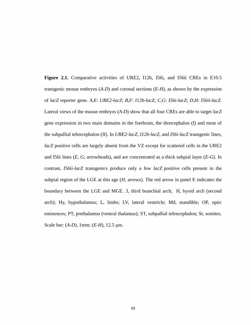
69
Figure 2.1. Comparative activities of URE2, I12b, I56i, and I56ii CREs in E10.5
transgenic mouse embryos (A-D) and coronal sections (E-H), as shown by the expression
of lacZ reporter gene. A,E: URE2-lacZ; B,F: I12b-lacZ; C,G: I56i-lacZ; D,H: I56ii-lacZ.
Lateral views of the mouse embryos (A-D) show that all four CREs are able to target lacZ
gene expression in two main domains in the forebrain, the diencephalon (I) and most of
the subpallial telencephalon (II). In URE2-lacZ, I12b-lacZ, and I56i-lacZ transgenic lines,
lacZ positive cells are largely absent from the VZ except for scattered cells in the URE2
and I56i lines (E, G; arrowheads), and are concentrated as a thick subpial layer (E-G). In
contrast, I56ii-lacZ transgenics produce only a few lacZ positive cells present in the
subpial region of the LGE at this age (H, arrows). The red arrow in panel E indicates the
boundary between the LGE and MGE. 3, third branchial arch; H, hyoid arch (second
arch); Hy, hypothalamus; L, limbs; LV, lateral ventricle; Md, mandible; OP, optic
eminences; PT, prethalamus (ventral thalamus); ST, subpallial telencephalon; St, somites.
Scale bar: (A-D), 1mm; (E-H), 12.5 μm.
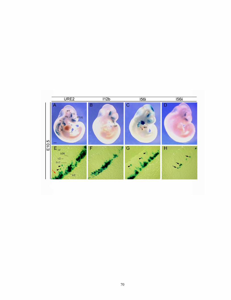
70

71
Figure 2.2. Different enhancer activities of URE2, I12b, I56i, and I56ii in the subpallial
telencephalon of transgenic mice. Coronal hemisections showing lacZ expression under
the control of each enhancer at E11.5 (A-D) and E13.5 (E-H) in medial levels. A,E:
URE2-lacZ; B,F: I12b-lacZ; C,G: I56i-lacZ; D,H: I56ii-lacZ. At both E11.5 and E13.5,
URE2-lacZ, I12b-lacZ-, and I56i-lacZ- positive cells display a nearly homogeneous
pattern of expression in most areas of the subpallium (A-C, E-G). In contrast, expression
of the I56ii-lacZ transgene is mainly restricted to a subset of cells in the superficial
mantle of the vLGE, MGE, AEP and POA (D, H, arrows), the regions where the other
three CRE lines produce weak or no lacZ expression (E-G, arrows).Unlike in the URE-
lacZ, I12b-lacZ, and I56i-lacZ lines, I56ii-lacZ does not label cells migrating tangentially
to the cortex at E13.5 (H versus E-G, arrowheads). DP, dorsal telencephalon; LV, lateral
ventricle. Scale bar: (A-H), 250 μm.

72
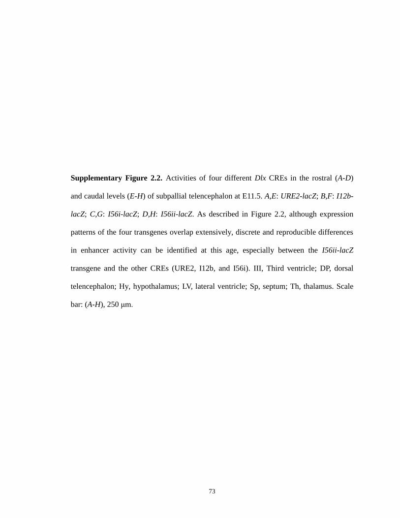
73
Supplementary Figure 2.2. Activities of four different Dlx CREs in the rostral (A-D)
and caudal levels (E-H) of subpallial telencephalon at E11.5. A,E: URE2-lacZ; B,F: I12b-
lacZ; C,G: I56i-lacZ; D,H: I56ii-lacZ. As described in Figure 2.2, although expression
patterns of the four transgenes overlap extensively, discrete and reproducible differences
in enhancer activity can be identified at this age, especially between the I56ii-lacZ
transgene and the other CREs (URE2, I12b, and I56i). III, Third ventricle; DP, dorsal
telencephalon; Hy, hypothalamus; LV, lateral ventricle; Sp, septum; Th, thalamus. Scale
bar: (A-H), 250 μm.

74
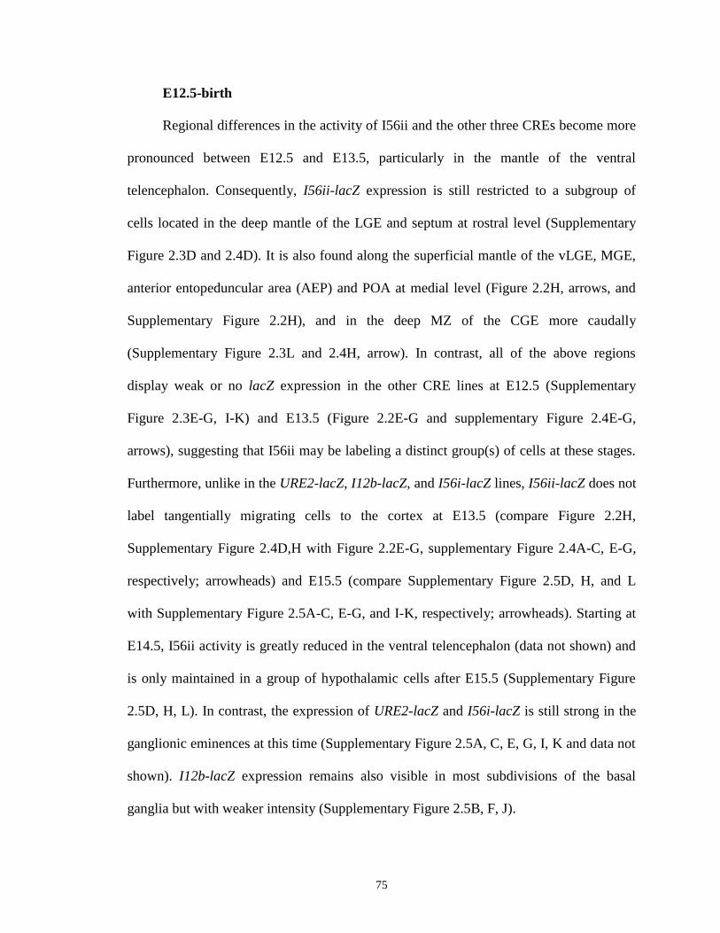
75
E12.5-birth
Regional differences in the activity of I56ii and the other three CREs become more
pronounced between E12.5 and E13.5, particularly in the mantle of the ventral
telencephalon. Consequently, I56ii-lacZ expression is still restricted to a subgroup of
cells located in the deep mantle of the LGE and septum at rostral level (Supplementary
Figure 2.3D and 2.4D). It is also found along the superficial mantle of the vLGE, MGE,
anterior entopeduncular area (AEP) and POA at medial level (Figure 2.2H, arrows, and
Supplementary Figure 2.2H), and in the deep MZ of the CGE more caudally
(Supplementary Figure 2.3L and 2.4H, arrow). In contrast, all of the above regions
display weak or no lacZ expression in the other CRE lines at E12.5 (Supplementary
Figure 2.3E-G, I-K) and E13.5 (Figure 2.2E-G and supplementary Figure 2.4E-G,
arrows), suggesting that I56ii may be labeling a distinct group(s) of cells at these stages.
Furthermore, unlike in the URE2-lacZ, I12b-lacZ, and I56i-lacZ lines, I56ii-lacZ does not
label tangentially migrating cells to the cortex at E13.5 (compare Figure 2.2H,
Supplementary Figure 2.4D,H with Figure 2.2E-G, supplementary Figure 2.4A-C, E-G,
respectively; arrowheads) and E15.5 (compare Supplementary Figure 2.5D, H, and L
with Supplementary Figure 2.5A-C, E-G, and I-K, respectively; arrowheads). Starting at
E14.5, I56ii activity is greatly reduced in the ventral telencephalon (data not shown) and
is only maintained in a group of hypothalamic cells after E15.5 (Supplementary Figure
2.5D, H, L). In contrast, the expression of URE2-lacZ and I56i-lacZ is still strong in the
ganglionic eminences at this time (Supplementary Figure 2.5A, C, E, G, I, K and data not
shown). I12b-lacZ expression remains also visible in most subdivisions of the basal
ganglia but with weaker intensity (Supplementary Figure 2.5B, F, J).

76
In summary, our data suggest that I56ii CRE displays a different activity compared
with URE2, I12b and I56i, that is confined to group(s) of cells lining the mantle of the
ganglionic eminences between E11.5 and E13.5.
2.3.2. I56ii marks a subgroup of striatal projection neurons expressing Meis2
and Islet1
We sought to characterize the identity of the I56ii-lacZ positive cells. First, we
checked that I56ii-lacZ-expressing cells are Dlx-positive between E11.5 and E13.5
comparing sections stained with X-gal with consecutive sections hybridized to Dlx5 or
Dlx6 cRNA probes (Figure 2.3 and data not shown). We also applied double-labeling
with a pan-Dll antibody and the β-galactosidase antibody (Figure 2.4A-D and data not
shown). Interestingly, we found that a stripe of cells that extends from the ventral MGE
towards the LGE and expresses the reporter transgene also express relatively high levels
of Dlx6 transcripts (Figure 2.3C,E; arrowheads).
Then, in order to test whether I56ii and I12b/I56i are active in the same or distinct
group(s) of cells, we crossed the I56ii-lacZ line with an alkaline phosphatase (AP)
reporter line under the control of I12b (I12b-AP). We then performed double
immunohistochemistry on brain sections from double hemizygote embryos (AP+/lacZ+)
at E12.5 and E13.5 using β-gal and AP antibodies. As a result, we found that I56ii-lacZ
positive cells almost never overlap with cells expressing the I12b-AP transgene at all
levels and ages examined (Figure 2.4E-H and data not shown).
Next, we investigated whether I56ii-lacZ positive cells proliferate and whether they
undergo apoptosis after E13.5. We found that between E11.5 and E13.5, most I56ii-lacZ
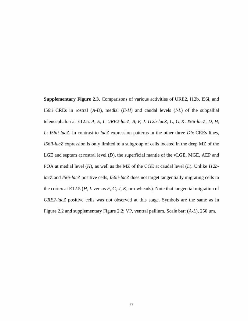
77
Supplementary Figure 2.3. Comparisons of various activities of URE2, I12b, I56i, and
I56ii CREs in rostral (A-D), medial (E-H) and caudal levels (I-L) of the subpallial
telencephalon at E12.5. A, E, I: URE2-lacZ; B, F, J: I12b-lacZ; C, G, K: I56i-lacZ; D, H,
L: I56ii-lacZ. In contrast to lacZ expression patterns in the other three Dlx CREs lines,
I56ii-lacZ expression is only limited to a subgroup of cells located in the deep MZ of the
LGE and septum at rostral level (D), the superficial mantle of the vLGE, MGE, AEP and
POA at medial level (H), as well as the MZ of the CGE at caudal level (L). Unlike I12b-
lacZ and I56i-lacZ positive cells, I56ii-lacZ does not target tangentially migrating cells to
the cortex at E12.5 (H, L versus F, G, J, K, arrowheads). Note that tangential migration of
URE2-lacZ positive cells was not observed at this stage. Symbols are the same as in
Figure 2.2 and supplementary Figure 2.2; VP, ventral pallium. Scale bar: (A-L), 250 μm.
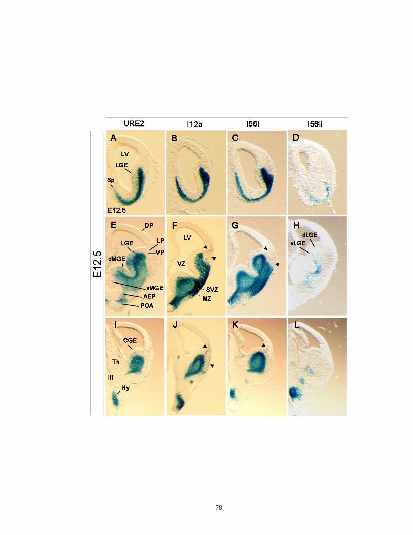
78

79
Supplementary Figure 2.4. Enhancer activities of URE2, I12b, I56i, and I56ii in rostral
(A-D) and caudal levels (E-H) of the subpallial telencephalon at E13.5. A,E: URE2-lacZ;
B,F: I12b-lacZ; C,G: I56i-lacZ; D,H: I56ii-lacZ. Similar to results obtained at E12.5 (see
Supplementary Figure 2.3), I56ii-lacZ activity is continuously maintained in the
subpopulation of cells located in the deep MZ of the LGE and septum at rostral level (D)
and in the deep MZ of the CGE more caudally (H, arrow) at E13.5. I56ii-lacZ does not
label tangentially migrating cells to the cortex at E13.5 (see also Figure 2.2). The cells
migrating to cortex are indicated with arrowheads. Symbols are the same as in Figure 2.2
and Supplementary Figure 2.2. Scale bar: (A-H), 250 μm.

80
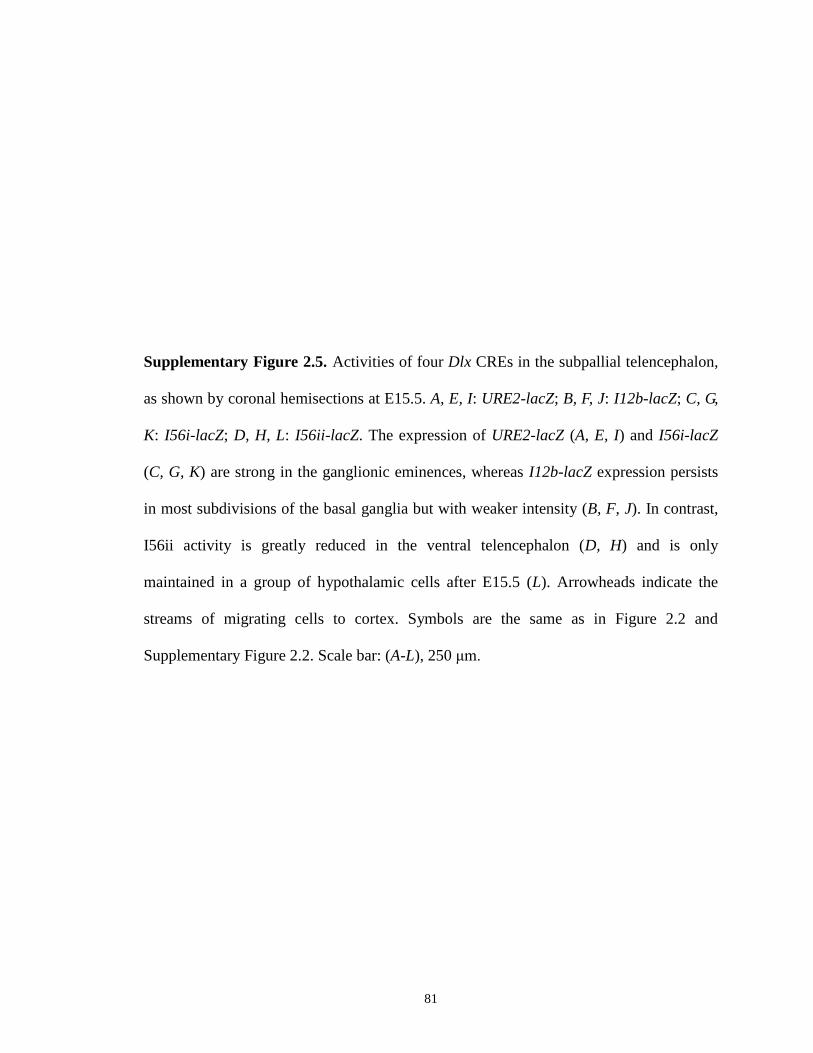
81
Supplementary Figure 2.5. Activities of four Dlx CREs in the subpallial telencephalon,
as shown by coronal hemisections at E15.5. A, E, I: URE2-lacZ; B, F, J: I12b-lacZ; C, G,
K: I56i-lacZ; D, H, L: I56ii-lacZ. The expression of URE2-lacZ (A, E, I) and I56i-lacZ
(C, G, K) are strong in the ganglionic eminences, whereas I12b-lacZ expression persists
in most subdivisions of the basal ganglia but with weaker intensity (B, F, J). In contrast,
I56ii activity is greatly reduced in the ventral telencephalon (D, H) and is only
maintained in a group of hypothalamic cells after E15.5 (L). Arrowheads indicate the
streams of migrating cells to cortex. Symbols are the same as in Figure 2.2 and
Supplementary Figure 2.2. Scale bar: (A-L), 250 μm.
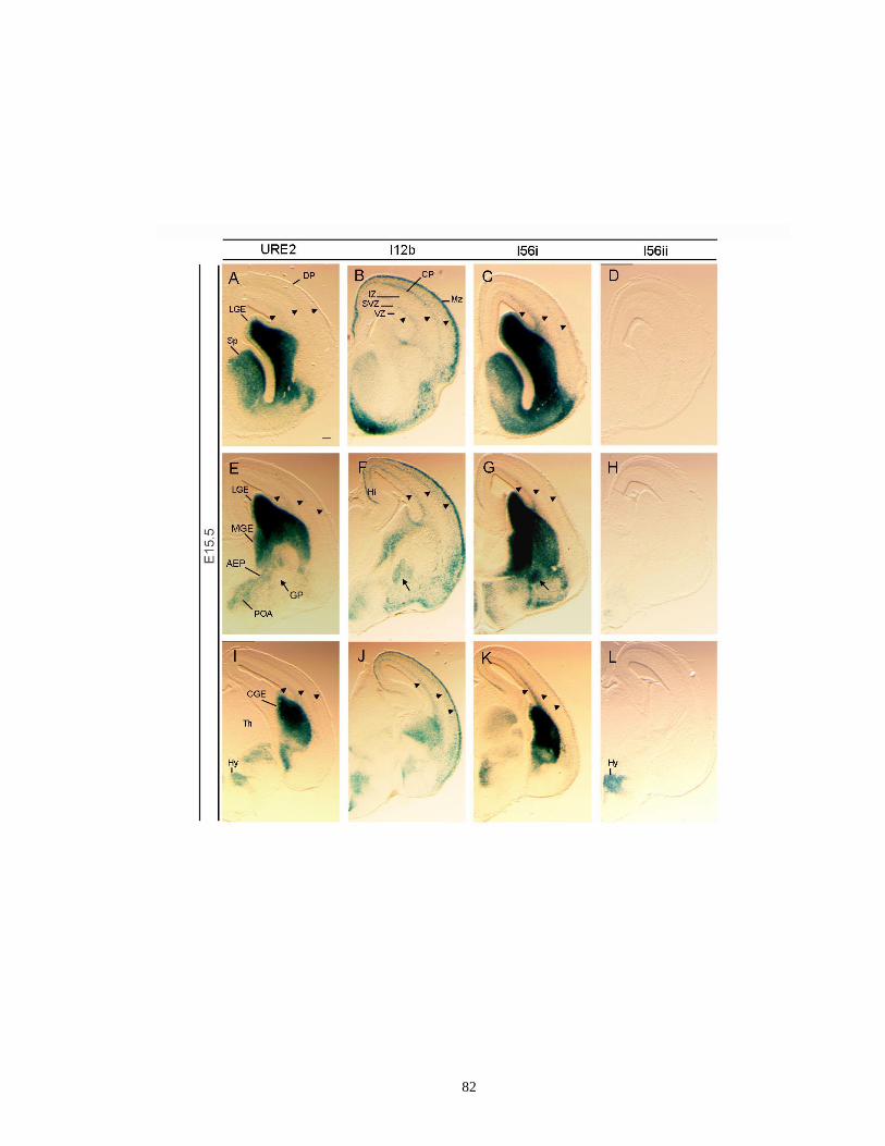
82
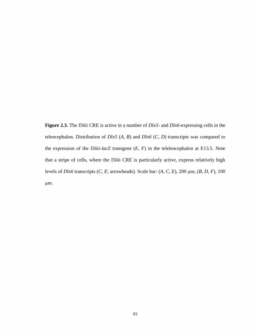
83
Figure 2.3. The I56ii CRE is active in a number of Dlx5- and Dlx6-expressing cells in the
telencephalon. Distribution of Dlx5 (A, B) and Dlx6 (C, D) transcripts was compared to
the expression of the I56ii-lacZ transgene (E, F) in the telelencephalon at E13.5. Note
that a stripe of cells, where the I56ii CRE is particularly active, express relatively high
levels of Dlx6 transcripts (C, E; arrowheads). Scale bar: (A, C, E), 200 μm; (B, D, F), 100
μm.

84
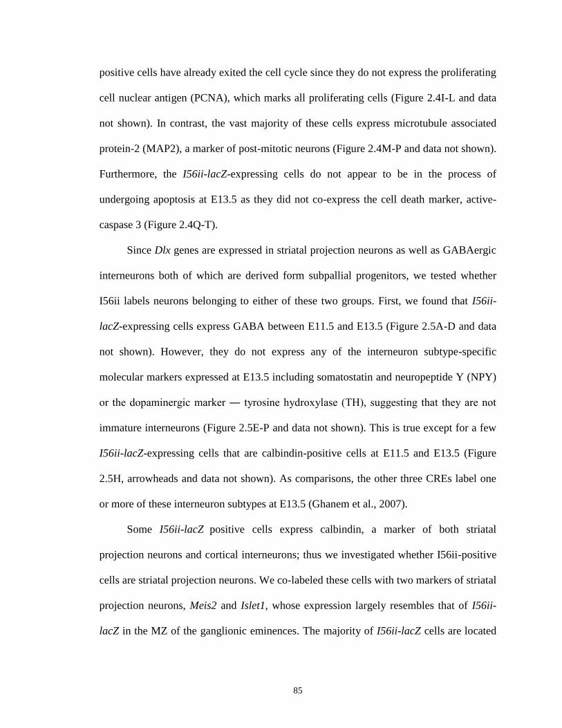
85
positive cells have already exited the cell cycle since they do not express the proliferating
cell nuclear antigen (PCNA), which marks all proliferating cells (Figure 2.4I-L and data
not shown). In contrast, the vast majority of these cells express microtubule associated
protein-2 (MAP2), a marker of post-mitotic neurons (Figure 2.4M-P and data not shown).
Furthermore, the I56ii-lacZ-expressing cells do not appear to be in the process of
undergoing apoptosis at E13.5 as they did not co-express the cell death marker, active-
caspase 3 (Figure 2.4Q-T).
Since Dlx genes are expressed in striatal projection neurons as well as GABAergic
interneurons both of which are derived form subpallial progenitors, we tested whether
I56ii labels neurons belonging to either of these two groups. First, we found that I56ii-
lacZ-expressing cells express GABA between E11.5 and E13.5 (Figure 2.5A-D and data
not shown). However, they do not express any of the interneuron subtype-specific
molecular markers expressed at E13.5 including somatostatin and neuropeptide Y (NPY)
or the dopaminergic marker ― tyrosine hydroxylase (TH), suggesting that they are not
immature interneurons (Figure 2.5E-P and data not shown). This is true except for a few
I56ii-lacZ-expressing cells that are calbindin-positive cells at E11.5 and E13.5 (Figure
2.5H, arrowheads and data not shown). As comparisons, the other three CREs label one
or more of these interneuron subtypes at E13.5 (Ghanem et al., 2007).
Some I56ii-lacZ
positive cells express calbindin, a marker of both striatal
projection neurons and cortical interneurons; thus we investigated whether I56ii-positive
cells are striatal projection neurons. We co-labeled these cells with two markers of striatal
projection neurons, Meis2 and Islet1, whose expression largely resembles that of I56ii-
lacZ in the MZ of the ganglionic eminences. The majority of I56ii-lacZ cells are located

86
in a deep mantle layer that is intercalated between the SVZ and more superficial parts of
the mantle of the ventral LGE (striatum) and the MGE (globus pallidus) (note:
superficial, as in the cortex, refers to being close to pia). This region also expresses the
Islet1 (Figure 2.6A-D) and Meis2 (Figure 2.6I-L) transcription factors at E13.5. In
contrast, we did not detect I56i-lacZ- nor I12b-lacZ-expressing cells that were co-labeled
with anti-Islet1 or anti-Meis2 antibodies at this age (Figure 2.6E-H, M-P, and
Supplementary Figure 2.6E-H, M-P). Furthermore, we found that only a few URE2-lacZ
positive cells express either of these two transcription factors in the mantle of the MGE
(Supplementary Figure 2.6C, D, K, L). Note that, some URE2-lacZ positive cells located
in the mantle of the dorsal LGE also express either Islet1 or Meis2; however, I56ii is not
active in this region (Supplementary Figure 2.6C, D, K, L).
Taken together, our results lead to the conclusion that I56ii-lacZ-expressing cells in
the subpallial telencephalon are a subpopulation(s) of post-mitotic projection neurons that
may be derived from ventral LGE progenitor cells, as suggested by their expression of
two striatal markers, Meis2 and Islet1. They constitute a distinct population of Dlx-
expressing cells compared to those where the other CREs, URE2, I12b, and I56i, are
active.
2.3.3. Meis2 and Islet1 proteins can bind and activate reporter gene
transcription via the I56ii enhancer sequence
To further clarify the existence of a potential regulation of the Dlx genes by Meis2
and Islet1 proteins, we searched the I56ii sequence using the genomatix software and
identified two putative binding sites (TGTC) for Meis homeodomain proteins as well as

87
Figure 2.4. Characterization of the I56ii CRE activity in the LGE and MGE at E13.5. A-
D: Double-immunohistochemistry showing that I56ii-lacZ-expressing cells are Dlx-
positive by co-labeling using a pan-Dll antibody and a β-galactosidase specific antibody;
E-H: I56ii-lacZ positive cells do not overlap with I12b-AP-expressing cells, indicating
that I56ii and I12b/I56i are active in distinct subpopulation(s) of cells at this time point; I-
L: At E13.5, most I56ii-lacZ positive cells have already exited the cell cycle because they
do not express the PCNA, which marks all the proliferating cells; M-P: I56ii-lacZ
positive cells are post-mitotic cells since they are co-localized with the molecular marker
MAP2; Q-T: These cells are not apoptotic as they do not co-express the cell death
marker, active-caspase 3. Cells expressing I56ii-lacZ are shown in red; while cells
expressing Dlx, AP, PCNA, MAP2 or active-caspase 3 are shown in green, respectively.
D, H, L, P, T are high-magnification pictures of boxes shown in C, G, K, O, S. Scale bars:
(A-C, E-G, I-K, M-O, Q-S), 12.5 μm; (D, H, L, P, T), 8.7 μm.
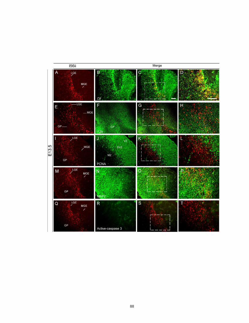
88

89
Figure 2.5. I56ii-lacZ positive cells are immunoreactive for GABA (A-D) at E13.5. In
contrast to the other Dlx CREs (URE2, I12b, and I56i, Ghanem et al., 2007), I56ii is not
active in interneuron progenitor cells (calbindin: E-H; somatostatin: I-L; NPY: M-P) in
the basal ganglia or tangentially migrating cells to the cortex at E13.5, except for a few
I56ii-lacZ-expressing cells that are calbindin-positive (H, arrowheads). Cells expressing
I56ii-lacZ are shown in red; whereas cells expressing GABA, calbindin, somatostatin or
NPY are shown in green, respectively. D, H, L, P are high-magnification pictures of
boxes shown in C, G, K, O. Scale bars: (A-C, E-G, I-K, M-O), 12.5 μm; (D, H, L, P), 8.7
μm.

90

91
Figure 2.6. The I56ii CRE is active in a subset of cells expressing Islet1 and Meis2. The
vast majority of I56ii-lacZ-expressing cells located in a deep mantle layer that is
intercalated between the SVZ and more superficial parts of the mantle of the ventral LGE
(striatum) and the MGE (globus pallidus) express two markers of striatal projection
neurons, Islet1 (A-D) and Meis2 (I-L) at E13.5. No such co-localization is observed for
the I56i-lacZ positive cells at this stage (Islet1: E-H; Meis2: M-P). Cell expressing I56ii-
lacZ or I56i-lacZ are shown in red; Islet1- or Meis2- positive cells are shown in green. D,
H, L, P: high magnifications of boxes shown in C, G, K, O, respectively. Scale bars: (A-C,
E-G, I-K, M-O), 12.5 μm; (D, H, L, P), 8.7 μm.

92
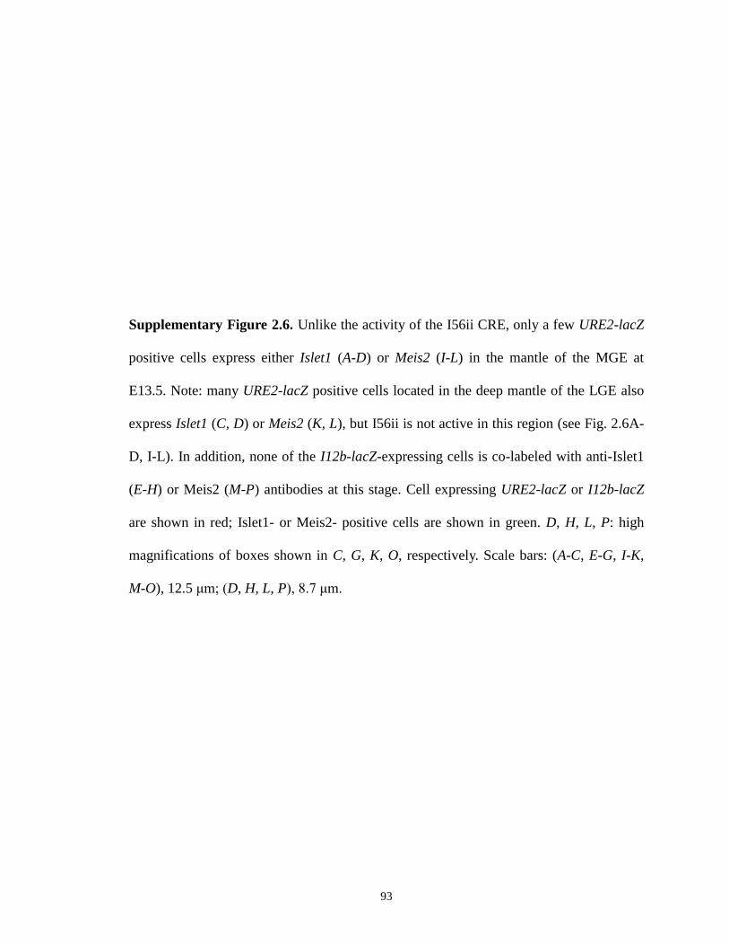
93
Supplementary Figure 2.6. Unlike the activity of the I56ii CRE, only a few URE2-lacZ
positive cells express either Islet1 (A-D) or Meis2 (I-L) in the mantle of the MGE at
E13.5. Note: many URE2-lacZ positive cells located in the deep mantle of the LGE also
express Islet1 (C, D) or Meis2 (K, L), but I56ii is not active in this region (see Fig. 2.6A-
D, I-L). In addition, none of the I12b-lacZ-expressing cells is co-labeled with anti-Islet1
(E-H) or Meis2 (M-P) antibodies at this stage. Cell expressing URE2-lacZ or I12b-lacZ
are shown in red; Islet1- or Meis2- positive cells are shown in green. D, H, L, P: high
magnifications of boxes shown in C, G, K, O, respectively. Scale bars: (A-C, E-G, I-K,
M-O), 12.5 μm; (D, H, L, P), 8.7 μm.

94
95
several potential core binding sites (ATTA) for Islet proteins (Supplementary Figure 2.1).
It is noteworthy that these ATTA-core motifs are also shared by a large number of other
homeodomain proteins, such as Hox and Oct proteins (Gehring et al., 1994; Pankratova
and Polanovsky, 1998). To explore whether Meis2 or Islet1 binds and activates
transcription via these binding sites, we conducted transient co-transfection reporter
assays. As noted above, P19 embryonic carcinoma cells were transfected with the mI56ii-
pBLCAT reporter construct, together with Meis2-pCS2+ or Islet1-pCS2+ expression
vectors (see material and methods for experimental details). We then quantified the
relative CAT activity in the presence of Meis2 or Islet1, and found that it was
significantly increased by about 4.50- and 3.53-fold, respectively, when compared to
controls and after normalization with the β-galactosidase expression, even though the
increase in both cases was not statistically significant (P = 0.052 and P = 0.081; Figure
2.7). Co-transfection of a modified mI56ii-pBLCAT vector containing the mutant Meis2
binding site (mI56ii-mutant-pBLCAT2) significantly decreased the transcriptional
activation of the reporter gene in the same assay and virtually no activation by Meis2 was
obtained with an enhancer-less vector (empty pBLCAT2) (Figure 2.7). These data
suggest that both Meis2 and Islet1 proteins are potential upstream regulators of Dlx gene
expression in vivo and this regulation could be mediated by the I56ii CRE.
2.4. Discussion
In the present report, we analyzed the regulatory role of the I56ii CRE in the mouse
telencephalon using lacZ reporter transgenes. We report that the I56ii CRE shows a
strikingly different activity during midgestation when compared with the other three Dlx

96
CREs that are also active in the forebrain: I56i from the Dlx5/6 bigene cluster and, URE2
and I12b, from the Dlx1/2 cluster. Thus, the I56ii-lacZ-expressing cells display the
following properties: 1) They express the transgene more transiently, between E11.5 and
E13.5. Their expression is restricted to a deep mantle layer that is intercalated between
the subpallial SVZ and more superficial parts of the mantle of the ventral LGE (striatum)
and the MGE (globus pallidus). The other three Dlx CREs are weakly or not active in
these cells (Figure 2.2, arrows); 2) Unlike the other three Dlx CREs, the I56ii-lacZ-
expressing cells do not express markers of interneurons including somatostatin and NPY
(Figure 2.5 and data not shown) and do not tangentially migrate to the cortex. Rather, the
I56ii-lacZ-expressing cells are a subpopulation of post-mitotic GABAergic projection
neurons expressing two striatal markers, Meis2 and Islet1 (Figures 2.4-2.6); and, 3) Both
Meis2 and Islet1 proteins can induce transcription in vitro via one or more putative
binding sites found in the I56ii sequence (Figure 2.7).
2.4.1. Comparison of the lacZ reporter gene expression driven by the four Dlx
CREs with endogenous Dlx gene expression
The onset of expression of the lacZ transgene in all four CRE lines is detected in a
group of prethalamic cells around ~E10 and, therefore, slightly contrasts with the onset of
the endogenous Dlx gene expression which occurs between E9-9.5. This difference could
be due to low levels of the β-galactosidase protein that are undetectable before E10
and/or to the existence of other regulatory sequence(s) that are necessary to refine the
temporal expression of the Dlx genes. In all four transgenic lines, the β-galactosidase-
positive cells are Dlx-positive as suggested by their co-labeling with a pan-Dll antibody
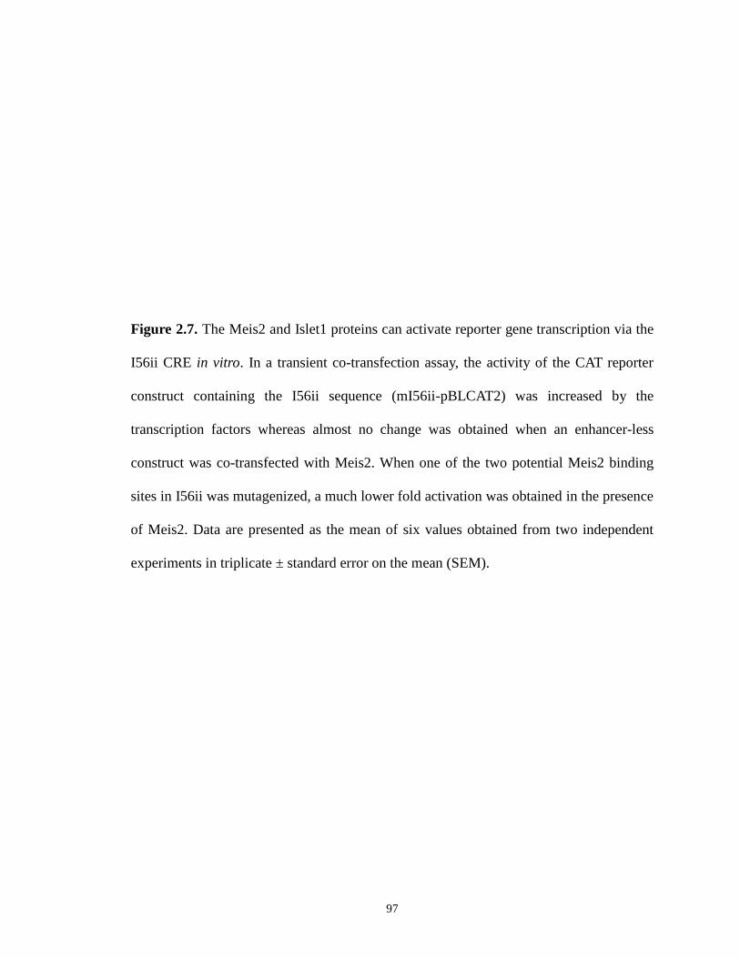
97
Figure 2.7. The Meis2 and Islet1 proteins can activate reporter gene transcription via the
I56ii CRE in vitro. In a transient co-transfection assay, the activity of the CAT reporter
construct containing the I56ii sequence (mI56ii-pBLCAT2) was increased by the
transcription factors whereas almost no change was obtained when an enhancer-less
construct was co-transfected with Meis2. When one of the two potential Meis2 binding
sites in I56ii was mutagenized, a much lower fold activation was obtained in the presence
of Meis2. Data are presented as the mean of six values obtained from two independent
experiments in triplicate ± standard error on the mean (SEM).
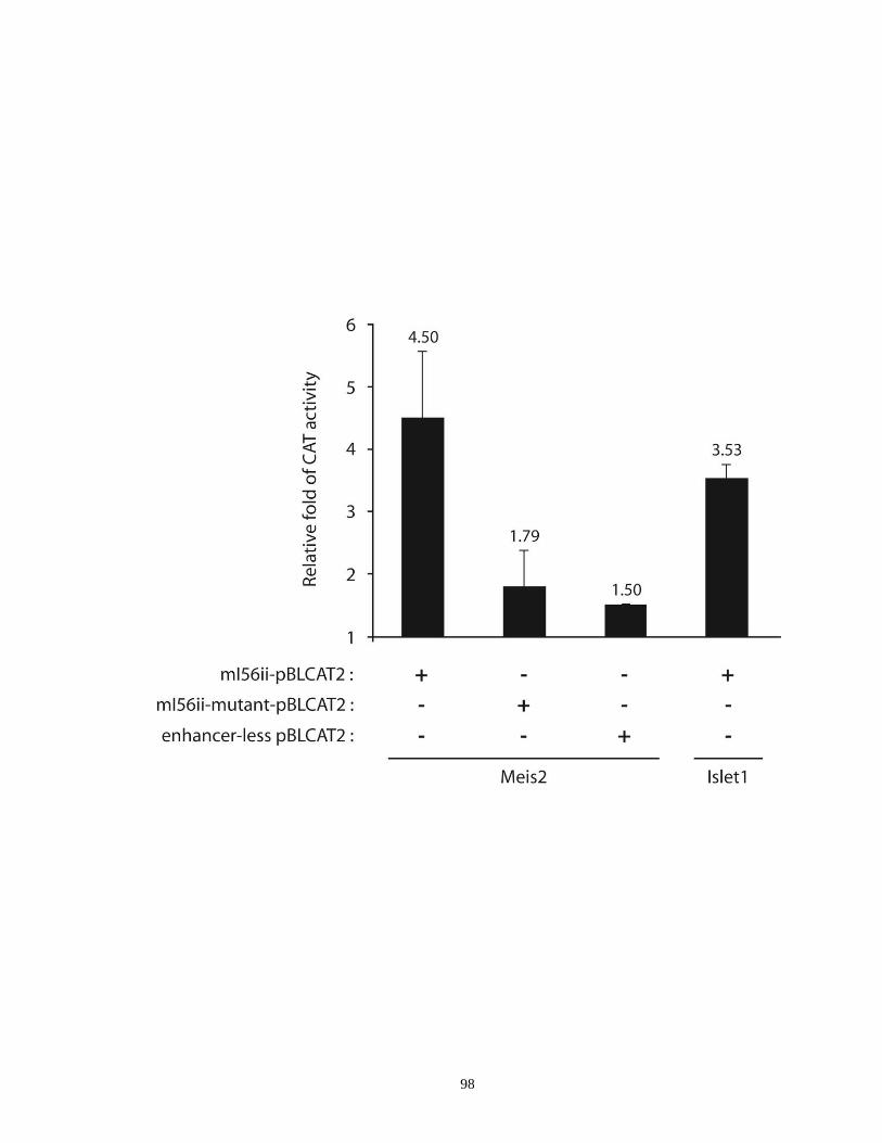
98

99
(Figure 2.4A- D and data not shown; Ghanem et al., 2007). In addition, the patterns of
enhancer activity between E10.5 and E15.5 overlap with the endogenous patterns of Dlx
expression (Figure 2.3 and data not shown). However, the transgene expression in all
lines examined lack one feature of telencephalic Dlx expression at E10.5: previous
studies showed that the dorsal LGE (dLGE) has a high concentration of Dlx2+ cells in the
VZ (Eisenstat et al., 1999; Yun et al., 2002). As none of the transgenic lines studied here
show this property, it suggests that this expression is conferred by an unknown enhancer
element. Furthermore, the β-galactosidase-positive neurons are still found in the mouse
neocortex at birth as well as one month after birth in all CRE lines (Ghanem et al., 2007)
except in the I56ii lines where lacZ expression is maintained only in a group of
hypothalamic cells after E14.5 (Supplementary Figure 2.5D, H, L and data not shown).
2.4.2. I56ii CRE displays a differential activity in comparison with the I56i,
I12b and URE2 CREs
Using double labeling, we showed that I56ii-lacZ- and I12b-AP-expressing cells do
not overlap between at E12.5 and E13.5 in the ganglionic eminences (Figure 2.4E-H and
data not shown). We previously showed that I12b and I56i are co-active in more than
80% of the progenitor population(s) found in the proliferative zones between E11.5 and
E13.5 (Ghanem et al., 2007). Therefore, we can infer that I56ii and I56i are likely to be
active in distinct group(s) of cells at these time points. Several lines of additional
evidence support our conclusion: 1) We found that I56ii-positive cells are Dlx+/GABA+.
They also specifically express Meis2 and Islet1, two markers of striatal projection
neurons that are primarily derived from the LGE. In contrast, none of the other three
100
lacZ-CRE transgenes co-expresses Meis2 or Islet1 in the superficial mantle of the
ganglionic eminences, except for a few cells weakly expressing URE2-lacZ (Figure 2.6
and Supplementary Figure 2.6); 2) We showed that URE2, I12b, and I56i are active in
distinct subpopulations of immature cortical and hippocampal interneurons that
tangentially migrate from subpallium, including populations expressing calbindin,
somatostatin and NPY at E13.5 (Ghanem et al., 2007 and this study, data not shown). In
addition, we showed that expression from the elements continue to mark different
subtypes of adult interneurons (Ghanem et al., 2007). In contrast, the I56ii CRE is
inactive in cells migrating tangentially to the cortex and its activity is not detectable past
E14.5 in the basal ganglia as discussed earlier.
2.4.3. I56ii marks a subgroup of striatal projection neurons that are probably
derived from the ventral LGE that tangentially migrate into the pallium
Based on their profile and pattern of expression as well as the orientation of their
migratory processes, we suggest that the I56ii-lacZ-expressing cells are GABAergic
projection neurons that may derive from progenitors found in the ventral LGE and then
migrate tangentially following a dorsal-to-ventral route before they finally settle down
between the SVZ and the globus pallidus in the deep mantle of the MGE (Figures 2.2 and
2.6).
There is recent evidence of a novel dorsal-to-ventral tangential migration of neural
stem cells in the perinatal forebrain (Willaime-Morawek et al., 2006). Furthermore,
tangentially migrating cells can translocate between subdivisions within the subpallium
itself including the LGE and MGE as well as the AEP and POA. For instance, progenitor

101
cells found in the AEP/POA region migrate dorsally into the striatum to become
cholinergic interneurons (Marin et al., 2000; Zhao et al., 2003). Also, it was shown
recently that a subgroup of projection neurons, namely “corridor cells”, tangentially
migrate from the LGE to the MGE around E13.5 to establish a permissive route that is
essential for thalamocortical axon guidance in mouse (Lopez-Bendito et al., 2006).
Furthermore, the so-called “corridor cells” are GABAergic neurons that, like the I56ii-
lacZ-expressing cells described here, specifically express LGE markers such as Islet1,
Ebf1 and Meis2 (Lopez-Bendito et al., 2006). It will be interesting to determine in the
future whether I56ii is a marker of these “corridor cells” during midgestation.
2.4.4. Meis2 and Islet1 are potential upstream regulators of Dlx genes
Our data suggest that the Dlx genes may be potentially regulated by Meis2 and
Islet1 to possibly maintain their activities via the I56ii sequence, at least between E11.5
to E13.5. This observation is supported by the ability of both proteins to activate the
transcription (by about 3.5-4.5 fold) of a reporter gene via I56ii in transient co-
transfection assays in vitro (Figure 2.7). There are two potential binding sites for Meis2
in I56ii (Supplementary Figure 3.1) and when one of the two sites is mutated in the above
sequence, activation by Meis2 is almost totally abolished. Future experiments such as
chromatin immunoprecipitation (ChIP) could determine whether this interaction also
takes place in vivo. As for Islet1, it was not possible to identify a precise binding site in
I56ii as the binding preferences of this transcriptional activator are poorly defined as the
ATTA sequence which is found in several locations in I56ii (Supplementary Figure 2.1)
and which is shared by numerous homeodomain transcription factors.

102
In conclusion, we showed in the present study that I56ii-lacZ-expressing cells in
the ventral forebrain are a subpopulation(s) of post-mitotic projection neurons that may
be derived from progenitor cells located in the LGE as suggested by their expression of
two striatal markers, Meis2 and Islet1. They constitute a distinct population of Dlx-
expressing cells compared to those where the other CREs, URE2, I12b and I56i, are
active. Therefore, the distinct activities of the four Dlx CREs, we have identified thus far,
reflect a complex and dynamic spatio-temporal regulation of Dlx gene expression during
the early stages of embryonic development. Moreover, it highlights that the Dlx genes
may have distinct functions in distinct progenitor populations born and derived from the
basal ganglia.
Acknowledgements
We thank Dr. Thomas Sargent laboratory for providing the guinea pig lacZ antibody and
Dr. Steve M. Sperber for providing the Islet1 cDNA clones. We are grateful to Adrianna
Gambarotta and Gary Hatch for creating and maintaining the transgenic mice. This work
was supported by the Canadian Institutes of Health Research (CIHR) Grant, MOP14460.
N.G. was supported in part by a scholarship from the Lebanese University, Beyrouth.
M.Y. was supported by a University of Ottawa International Scholarship and an Ontario
Graduate Scholarship (OGS). J.L.R.R. was supported by: Nina Ireland, NIMH RO1
MH49428-01 and K05MH065670. M.E. was supported by an Investigator Award of the
CIHR.

103
3. Roles of the cis-regulatory elements, I56i and I56ii, from the
dlx5a/dlx6a intergenic region, during zebrafish GABAergic neuron
development and their cross-regulatory interaction
Man Yu a,b
, Yanwei Xi a, Ryan B. MacDonald
a, Marc Ekker
a,b,*
a Centre for Advanced Research in Environmental Genomics (CAREG), Department of
Biology, University of Ottawa, 20 Marie Curie, Ottawa, ON, Canada K1N 6N5
b Department of Cellular and Molecular Medicine, University of Ottawa, 451 Smyth
Road, Ottawa, ON, Canada K1H 8M5
* Corresponding author. Department of Biology, University of Ottawa, 30 Marie Curie,
Ottawa, ON, Canada K1N 6N5. Tel.: +1-613-5625800, ext. 2605; Fax: +1-613-5625486;
E-mail address: [email protected]
Keywords: cis-regulatory elements; dlx genes; forebrain; GABAergic neuron; gene
regulation; transgenesis; zebrafish (Danio rerio)

104
Abstract
During vertebrate forebrain formation, Dlx homeobox genes play essential roles in the
differentiation, migration and survival of subpallium-derived progenitors that will later
give rise to diverse subtypes of γ-aminobutyric acid (GABA)-expressing neurons,
including inhibitory cortical interneurons. They also participate in the regulation of the
two Gad genes that encode the enzymes necessary for GABA synthesis. In mice, at least
four cis-regulatory elements (CREs) control Dlx expression in the telencephalon and
diencephalon: URE2 and I12b in the Dlx1/Dlx2 bigene cluster, and, I56i and I56ii in the
Dlx5/Dlx6 bigene cluster. However, little is known so far with respect to the function of
orthologous dlx genes and their regulatory elements during zebrafish GABAergic neuron
development. To investigate whether similar dlx-mediated pathways exist in the early
developing zebrafish forebrain, we characterized two independent lines of transgenic
zebrafish carrying GFP reporter genes: one under the control of a 1.4kb dlx5a/dlx6a
intergenic sequence (encompassing I56i and I56ii) and one with a 1.1kb fragment
containing only I56i CRE, respectively. Our comparative analysis showed that GFP
expression patterns of these two transgenic lines are largely overlapping throughout the
basal forebrain. Intriguingly, compared to the 1.4kbdlx5a/dlx6a:GFP fish, populations of
GFP-positive cells detected in the mesencephalic tectum are almost completely unlabeled
in the 1.1kbI56i:GFP embryos, probably due to the lack of I56ii CRE activity. Despite
that difference, almost all GFP-expressing cells are GABA-positive. Co-expression of the
GFP transgene with various GABAergic interneuron markers was only observed in
certain domains in the ventral portion of the telencephalon and diencephalon as well as in
the midbrain tectum. Simultaneous inactivation of dlx1a/dlx2a or dlx5a/dlx6a function

105
led to a marked loss of dlx CRE activity specifically restricted to the diencephalon and
midbrain tectum, but a comparatively milder extent of reduction in the telencephalon,
suggesting the cross-regulation of dlx5a/dlx6a by dlx1a and dlx2a is similar, at least in
part, to that previously reported for their mouse orthologs. Together, our data suggest that
dlx genes and their CREs are involved in a conserved genetic pathway necessary for
proper GABAergic neuron production and dlx expression in the developing zebrafish
forebrain, particularly in the diencephalon.
3.1. Introduction
The Dlx gene family encodes homeodomain-containing transcription factors that
are implicated in the development of the mammalian forebrain (Zerucha and Ekker,
2000; Panganiban and Rubenstein, 2002). In the developing mouse forebrain, four Dlx
genes (Dlx1, Dlx2, Dlx5 and then Dlx6) are sequentially expressed starting at around
embryonic day 9.5 (E9.5) and exhibit highly overlapping but distinct expression patterns
in the ganglionic eminences of the ventral telencephalon as well as in the ventral
diencephalon (prethalamus and hypothalamus) (Liu et al., 1997; Yang et al., 1998;
Eisenstat et al., 1999). Specifically, Dlx1 and Dlx2 are predominantly expressed in the
least mature early-born progenitor cells within the ventricular and subventricular zones
(VZ and SVZ), whereas Dlx5 and Dlx6 genes are mainly expressed in the more
differentiated postmitotic cells within the SVZ and mantle zone (MZ) (Eisenstat et al.,
1999; Panganiban and Rubenstein, 2002). The forebrain expression patterns of Dlx genes
largely overlap with that of the two glutamic acid decarboxylase (Gad) genes, Gad1 and
Gad2, which encode the key enzymes required for the production of the neurotransmitter

106
γ-amino butyric acid (GABA) (Yun et al. 2002; Long et al., 2009a). Ectopic expression
of Dlx proteins in vitro is able to induce ectopic Gad expression in many domains of the
developing mouse forebrain (Anderson et al., 1999; Stuhmer et al., 2002b; Yun et al.,
2002). Previous loss-of-function studies have shown that Dlx genes are particularly
responsible for the proper proliferation, differentiation, and survival of GABAergic
interneuron progenitor cells and those cells will later migrate and populate various areas
of the forebrain (Panganiban and Rubenstein, 2002; Wonders and Anderson, 2006). For
example, inactivation of both Dlx1 and Dlx2 genes leads to a major deficiency in the
generation and differentiation of GABA-expressing striatal projection neurons and
GABAergic interneuron progenitors born in the ganglionic eminences (Anderson et al.,
1997a; Anderson et al., 1997b; Marin et al., 2000; Pleasure et al., 2000; Long et al.,
2007). These cells accumulate in the basal ganglia and fail to migrate to the neocortex,
olfactory bulb and hippocampus through different migratory routes (Anderson et al.,
1997a; Anderson et al., 1997b; Marin et al., 2000; Pleasure et al., 2000; Long et al.,
2007). Likewise, simultaneous inactivation of Dlx5 and Dlx6 function results in
exencephaly of the anterior portion of the embryonic forebrain and the mutants have a
decreased number of PV-expressing cortical interneurons with increased dendrite
branching (Wang et al., 2010).
Vertebrates have multiple Dlx genes that are generally arranged as convergently
transcribed bigene clusters on distinct chromosomes (Zerucha and Ekker, 2000). The two
genes of a bigene pair are separated by a short intergenic sequence (~3.5 to 16 kb)
(Zerucha and Ekker, 2000). Mammals have six Dlx genes organized as three bigene pairs
(Dlx1/Dlx2, Dlx3/Dlx4 and Dlx5/Dlx6), whereas some teleost fish including zebrafish

107
have eight dlx genes, among which six (dlx1a/dlx2a, dlx3b/dlx4b and dlx5a/dlx6a) are
organized in a manner similar to their mammalian counterparts (Stock et al., 1996;
Amores et al., 1998; Quint et al., 2000). The other two additional dlx genes (dlx2b and
dlx4a) are not linked with other dlx genes and are considered as the respective duplicates
of ancestral Dlx2 and Dlx4 genes, following the occurrence of a teleost-specific whole or
partial genome duplication event (Amores et al., 1998). Consistent with mouse Dlx
forebrain expression, five zebrafish dlx genes (dlx1a/dlx2a, dlx2b and dlx5a/dlx6a) are
expressed in a highly similar pattern in the developing zebrafish telencephalon and
diencephalon (Akimenko et al. 1994; Ellies et al. 1997; MacDonald et al., 2010a). In
addition, dlx1a and dlx2a expression has been mainly found in the cell populations that
are located more close to the ventricle as compared to that of dlx5a and dlx6a, a situation
that resembles the spatio-temporal forebrain expression of mouse Dlx genes (Zerucha et
al., 2000; MacDonald et al., 2010a). Proper Dlx expression in the developing mouse and
zebrafish forebrain is transcriptionally regulated, at least in part, by a number of highly
conserved cis-regulatory elements (CREs, enhancers) present in their surrounding
genome regions (Zerucha et al., 2000; Ghanem et al., 2003; Ghanem et al., 2007): URE2
located upstream of Dlx1 (dlx1a), I12a and I12b within the Dlx1/Dlx2 (dlx1a/dlx2a)
intergenic region, I56i and I56ii within the Dlx5/Dlx6 (dlx5a/dlx6a) intergenic region.
Our previous studies using transgenic mice have shown that URE2, I12b and I56i CREs
target GABA-expressing progenitors in the ganglionic eminences in the subpallial
telecephalon as well as diverse subtypes of GABAergic interneurons in the adult cortex
(Ghanem et al., 2007). In contrast, I56ii activity is mainly restricted to a subpopulation of
108
GABAergic striatal projection neurons at E11.5-E13.5 during early embryogenesis
(Ghanem et al., 2008).
To date, the roles played by dlx genes and their various individual CREs in the
genetic cascades involved in GABAergic neuron formation are not well understood,
especially in non-mammalian vertebrates. Using triple fluorescence in situ hybridization
techniques, our laboratory has shown that dlx genes are expressed in highly overlapping
domains with that of the gad genes in the developing zebrafish subpallium (MacDonald
et al., 2010a). The spatial-temporal forebrain expression of dlx and gad1 genes and the
subpallial-to-pallial migration of dlx/gad1-expressing neurons are consistent with
previous studies done in the mouse, indicating that these genes may be involved in a
conserved genetic pathway mediating GABAergic neuron development (Panganiban and
Rubenstein, 2002; Marin and Rubenstein, 2003; Mueller et al., 2008; MacDonald et al.,
2010a). To further understand the regulatory roles of the dlx CREs during zebrafish
GABAergic neuron generation, we characterized two stable lines of transgenic zebrafish:
one in which GFP is under the control of a 1.4kb dlx5a/dlx6a intergenic sequence
(encompassing I56i and I56ii; Ghanem et al., 2003) and a newly-produced line containing
a 1.1kb fragment encompassing only I56i CRE. In addition to a comparative analysis
conducted between the two transgenic lines, we also sought to determine whether any
potential association exists between GFP expression and several GABAergic interneuron
markers using double immunohistochemical staining in an effort to evaluate the
functional importance of these two enhancers in the differentiation of zebrafish
GABAergic neurons. The possible auto- and/or cross-regulation of dlx genes during
109
zebrafish GABAergic neuron development was also investigated in our transgenic
models by employing morpholino-mediated knockdown assays.
3.2. Materials and methods
3.2.1. Animal maintenance
Adult wild-type and transgenic zebrafish were maintained in circulating system
water at 28.5°C under a constant photoperiod (a light/dark cycle of 14/10h) according to
standard methods (Westerfield, 2000). Fish were fed once or twice daily with No.1
crumble (ZeiglerTM
, Aquatic Habitats). Embryo-collection traps were placed inside each
9L acrylic tank containing 20-30 fish in the evening and embryos were harvested the next
morning. Embryos were raised in an incubator at 28.5°C, staged in days post fertilization
(dpf) according to specific criteria outlined by Kimmel et al. (1995) and euthanized with
an overdose of tricainemesylate (ethyl 3-amino-benzoate methanesulfonate, Sigma) when
needed. All experimental procedures in this study were carried out in accordance with the
guidelines of the Canadian Council on Animal Care (CCAC) and were approved by the
University of Ottawa Animal Care Committee.
3.2.2. Construction of dlx CRE transgene vectors
The 1.4kbdlx5a/dlx6a:GFP transgene construct has been previously generated in
the laboratory as described in (Zerucha et al., 2000; Ghanem et al., 2003). In brief, a
~1.4kb dlx5a/dlx6a intergenic region containing both I56i and I56ii CREs was subcloned
into a modified SP72-pEGFP-N1 vector using KpnI/ClaI restriction sites and placed
downstream of a ~3.5kb fragment from the immediate 5’ flanking region of zebrafish
110
dlx6a, including part of the 5’UTR. This dlx6a 5’ flanking fragment per se does not
generate tissue-specific expression pattern in transgenic zebrafish (Ghanem et al., 2003).
The 1.1kbI56i:GFP transgene construct was made up of a ~1.1kb region from the
dlx5a/dlx6a intergenic locus containing only I56i CRE and its flanking sequences. This
fragment shares the same sequence with the 1.4kb dlx5a/dlx6a intergenic segment with
the only difference being the lack of the ~300 bp I56ii CRE (Figure 3.1). Specifically, a
~1.1kb SalI-SalI I56i fragment was first PCR-amplified from the 1.4kbdlx5a/dlx6a:GFP
transgene plasmid using the following primer pair (forward: 5’-GTC GAC GCT CAA
TTA TTA AGG TAT TGA CAA-3’; reverse: 5’-GTC GAC ACA AGC GGT TAC AAC
GCT ATC-3’) and was cloned into the pDrive vector (Qiagen). After a SalI digestion, the
linearized I56i fragment was separated in 1% agarose gel, recovered and purified using a
gel extraction kit (Qiagen). This fragment was then ligated into the unique SalI site
immediately upstream of a β-globin minimal promoter linked to the coding sequence of a
GFP reporter cassette in a Tol2 transposon plasmid that was modified from a SP72 vector
(Promega) (Kawakami, 2004; MacDonald et al., 2010a). The resulting I56i-β-globin-GFP
sequence was eventually flanked at both ends by Tol2 transposable element recognition
sites. The 300bpI56ii:GFP transgene vector that is under the control of a ~300 bp
zebrafish I56ii sequence was generated in the same way by PCR amplification and
subcloning into the Tol2 transposon plasmid.
3.2.3. Generation and visualization of transgenic zebrafish
The tg(1.4kbdlx5a/dlx6a:GFP) transgenic line has been previously established and
expresses GFP in the domains of dlx expression in the telencephalon and diencephalon

111
Figure 3.1. A schematic drawing of GFP transgene constructs used to establish
stable transgenic zebrafish lines
Similar to mammalian Dlx genes, zebrafish dlx5a/dlx6a genes are physically linked in a
bigene arrangement and contain two highly conserved intergenic CREs named I56i and
I56ii. The tg(1.4kbdlx5a/dlx6a:GFP) vector is controlled by a 1.4kb dlx5a/dlx6a
intergenic sequence encompassing both I56i and I56ii CREs, while the
tg(1.1kbI56i:GFP) construct is under the regulation of a ~1.1kb fragment containing all
elements of the 1.4kb sequence, except for the I56ii CRE. Exons are numbered (black,
untranslated region; white, coding sequences). I56i and I56ii regulatory elements are
shown as red boxes.
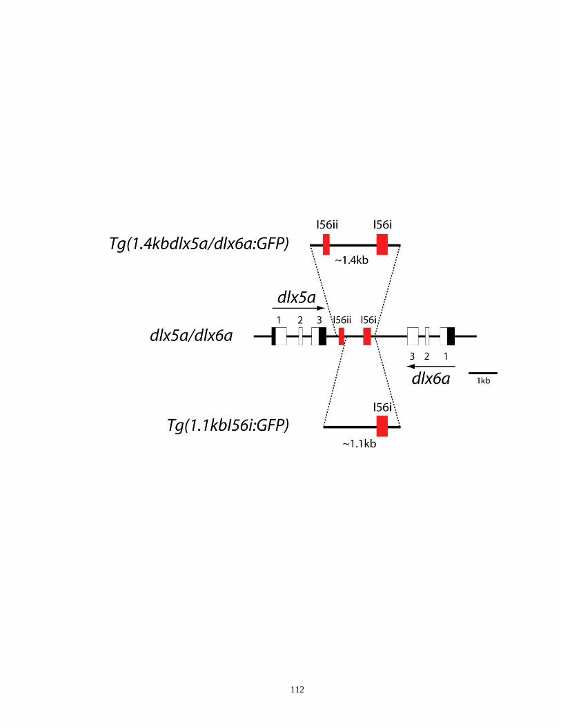
112
113
(Zerucha et al., 2000; Ghanem et al., 2003). The tg(1.1kbI56i:GFP) line was generated
by means of Tol2-mediated transgenesis. For the production of transgenic zebrafish, Tol2
transposase mRNA was first synthesized and purified with the mMessage SP6 Kit
(Ambion) from the pCS2-TP vector after digestion with Not1 according to the
manufacturer’s instructions. The 1.1kbI56i:GFP construct was then co-injected at
approximately 50 ng/µl in standard DNA microinjection buffer (0.2 mM KCl, 0.1%
phenol red), along with 50 ng/µl of in vitro transcribed Tol2 transposase mRNA, into
fertilized one-cell stage embryos following procedures described previously (Fisher et al.,
2006). Injected embryos were screened and those with strong GFP expression were raised
to sexual maturity. Founder transgenic zebrafish (F0) were interbred with each other or
outcrossed in pairs with wild-type fish. At least 100 embryos for each pair were screened
by GFP fluorescence starting at 1-3 dpf. F1 progeny expressing strong GFP were selected
and allowed to grow up to adulthood for establishing stable transgenic lines. Four
independent transgenic lines (#11, #15, #17 and #21) were obtained and all gave identical
and consistent GFP expression patterns in the developing forebrain, albeit with varying
GFP signal intensity. The 300bpI56ii:GFP transgene construct was injected into one-cell
stage embryos in the same manner as the 1.4kbdlx5a/dlx6a:GFP vector and expression of
the 1.4kbdlx5a/dlx6a:GFP construct was examined in primary transgenic embryos.
Screening for GFP-positive offspring was performed on a Nikon NBZ 1500 dissecting
fluorescence microscope and image acquisition was done with a Nikon DXM 1200C
digital camera.
3.2.4. Immunohistochemistry
114
For immunostaining assays, zebrafish embryos were collected at different time
points (3 and 5 dpf) and fixed in 4% paraformaldehyde (PFA)/1×PBS O/N at 4°C.
Samples were then equilibrated in 30% sucrose/1×PBS O/N at 4°C prior to
cryosectioning. The next day, fixed embryos were oriented and embedded in Tissue
Freezing Medium (Triangle Biomedical Sciences) and transverse sections of 10-12 µm
thickness were put onto Superfrost Plus slides (Fisher) using a Leica CM1850 standard
cryostat (Leica Microsystems) at -20°C. After washing 3×10 min with 1×PBS to remove
cryoprotectant, tissue sections were preincubated with a blocking solution containing 1%
bovine serum albumin (BSA), 10% newborn calf serum and 0.03% Tween 20 for 2h at
RT. The solution was subsequently replaced with a fresh aliquot containing the primary
antibody (for dual-labeling, two primary antibodies were added simultaneously) and
incubation was carried out in a humid chamber O/N at 4°C. Following 3×15 min washes
in 1×PBS, sections were incubated in the dark with the appropriate secondary
fluorescence-coupled antibody for 2h at RT and the slides were cover-slipped with a
Vectashield mounting medium (Vector Labs). Histological sections were analyzed with a
Zeiss Axiophot fluorescence microscope by using the appropriate filter sets and
photographed with a Zeiss Axiocam digital camera.
The following primary antibodies were used in this study: rabbit polyclonal anti-
GFP (1:500, Invitrogen), mouse monoclonal anti-GFP (1:500, Invitrogen), rabbit
polyclonal anti-calretinin (CR, 1:1000, Swant), rabbit polyclonal anti-calbindin (CB,
1:1000, Swant), mouse monoclonal anti-parvalbumin (PV, 1:500, Sigma). The following
secondary antibodies were applied: Alexa Fluor 488® goat anti-rabbit IgG (H+L) (1:300
Invitrogen), Alexa Fluor 488® goat anti-mouse IgG (H+L) (1:300, Invitrogen),
115
AlexaFluor 594®
goat anti-rabbit IgG (H+L) (1:300, Invitrogen), Alexa Fluor 594® goat
anti-mouse IgG (H+L) (1:300, Invitrogen).
3.2.5. Morpholino knockdown of dlx mRNA expression
Antisense morpholino oligonucleotides (MOs) complementary to the translation
initiation sites of zebrafish dlx1a, dlx2a, dlx5a and dlx6a mRNA as well as a standard
control morpholino were purchased from Gene Tools (Philomath, OR) and were diluted
with nuclease-free H2O. Serial concentrations of MOs (1, 2 and 4 ng/nl) were then
prepared in 1×Danieau buffer [58 mM NaCl, 0.7 mM KCl, 0.4 mM MgSO4, 0.6 mM
Ca(NO3)2, 5.0 mM HEPES pH 7.6] and phenol red was added to the MO injection
solution at a final concentration of 0.05%. Approximately 1 nl of MO was microinjected
into one cell-stage transgenic zebrafish embryos using a Narishige IM300 microinjector.
Injected embryos were incubated in 30 ml Petri dishes with E3 embryo medium
containing 0.03% methylene blue at 28.5°C. The morphology of morphants and their
GFP expression patterns were examined under a Nikon NBZ1500 dissecting microscope
and pictures were taken using a Nikon DXM 1200C digital camera. For double MO
knockdown experiments, two MOs (dlx1a+dlx2a or dlx5a+dlx6a) were always co-
injected in equal amounts. Standard control MOs were injected at the same
concentrations simultaneously. The following translation blocking MOs were used: dlx1a
(Sperber et al., 2008), dlx2a (Sperber et al., 2008), dlx5a (Walker et al., 2007), dlx6a (5’-
TGG TCA TCATCA AAT TTT CTG CTT T-3’). The sequence for the standard control
MO is 5’-CCT CTT ACC TCA GTT ACA ATT TAT A-3’.
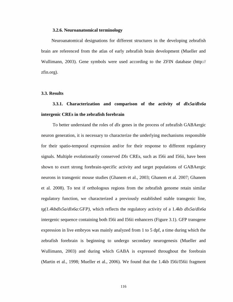
116
3.2.6. Neuroanatomical terminology
Neuroanatomical designations for different structures in the developing zebrafish
brain are referenced from the atlas of early zebrafish brain development (Mueller and
Wullimann, 2003). Gene symbols were used according to the ZFIN database (http://
zfin.org).
3.3. Results
3.3.1. Characterization and comparison of the activity of dlx5a/dlx6a
intergenic CREs in the zebrafish forebrain
To better understand the roles of dlx genes in the process of zebrafish GABAergic
neuron generation, it is necessary to characterize the underlying mechanisms responsible
for their spatio-temporal expression and/or for their response to different regulatory
signals. Multiple evolutionarily conserved Dlx CREs, such as I56i and I56ii, have been
shown to exert strong forebrain-specific activity and target populations of GABAergic
neurons in transgenic mouse studies (Ghanem et al., 2003; Ghanem et al. 2007; Ghanem
et al. 2008). To test if orthologous regions from the zebrafish genome retain similar
regulatory function, we characterized a previously established stable transgenic line,
tg(1.4kbdlx5a/dlx6a:GFP), which reflects the regulatory activity of a 1.4kb dlx5a/dlx6a
intergenic sequence containing both I56i and I56ii enhancers (Figure 3.1). GFP transgene
expression in live embryos was mainly analyzed from 1 to 5 dpf, a time during which the
zebrafish forebrain is beginning to undergo secondary neurogenesis (Mueller and
Wullimann, 2003) and during which GABA is expressed throughout the forebrain
(Martin et al., 1998; Mueller et al., 2006). We found that the 1.4kb I56i/I56ii fragment

117
specifically drives GFP expression in the telencephalon starting at approximately 1 dpf
(Figure 3.2A,B), and in certain domains of the ventral diencephalon (prethalamus and
hypothalamus) starting at 2 dpf (Figure 3.2C,D). GFP expression in the prethalamus and
hypothalamus becomes more apparent from 3 dpf onwards (Figure 3.2E-H). Scattered
populations of GFP-positive cells were also clearly seen in the mesencephalic tectum
(also known as optic tectum, Mueller and Wullimann, 2003) starting at 2 dpf and
persisted until at least 5 dpf (Figure 3.2C,E,G, arrows). Our immunohistochemistry
analysis on 1.4kbdlx5a/dlx6a:GFP brain sections revealed that overall GFP expression
patterns in the telencephalon and ventral diencephalon (Figure 3.3A,E,I,M) closely mimic
the endogenous dlx5a/dlx6a expression (Mione et al., 2008; MacDonald et al., 2010a).
To confirm whether the zebrafish I56i and I56ii CREs target dlx expression to cells
that are GABAergic neurons, we co-labeled 3 dpf transverse sections with antibodies
against GFP and GABA and observed an extensive co-expression between GFP and
GABA at different planes throughout the forebrain (Figure 3.3). It should be noted that a
small number of GFP-positive cells located close to the ventral midline of the
telencephalon and diencephalon, an area known to be a proliferative region (Mueller and
Wullimann, 2003), remain free of GABA staining (Figure 3.3D,H,L,P, arrows),
indicating those cells are probably still in the cell cycle and have not yet acquired the
GABAergic phenotype. These results demonstrate that similar to their corresponding
mouse orthologs, zebrafish dlx5a/dlx6a intergenic CREs are also active in GABAergic
neurons during zebrafish forebrain development. In addition to their highly conserved
sequences, zebrafish and mammalian Dlx CREs may possibly share at least a certain

118
Figure 3.2. Comparison of reporter gene expression patterns in the
1.4kbdlx5a/dlx6a:GFP (A-H) and 1.1kbI56i:GFP live embryos (I-P) at various
developmental stages (1, 2, 3 and 5 dpf)
In the 1.4kbdlx5a/dlx6a:GFP fish, robust GFP expression is first found in the
telencephalon starting at 1 dpf (A,B), while GFP expression in the ventral diencephalon
(prethalamus and hypothalamus) appears at ~2 dpf (C,D) and becomes more apparent
after 3 dpf (E-H). Scattered populations of GFP-positive cells are also clearly identified
in the midbrain tectum starting at 2 dpf and are present until 5 dpf (C,E,G, arrows).
Although GFP expression patterns found throughout the ventral telencephalon and
diencephalon in the 1.1kbI56i:GFP fish (I-P) largely resembles those detected in the
tg(1.4kbdlx5a/dlx6a:GFP) line at the same time points, 1.1kbI56i:GFP larvae completely
lack GFP expression in the tectum (K,M,O) and seem to have fewer GFP-positive cells
in the telencephalon and diencephalon (L,N,P). Note that all injected embryos were
treated with 0.003% 1-phenyl-2-thiourea (PTU) to inhibit pigmentation.
(A,C,E,G,I,K,M,O): lateral views, anterior to the left, dorsal up; (B,J): dorsal views,
anterior down; (D,L): ventral views, anterior to the left; (F,H,N,P): dorsal views, anterior
to the left. Scale bars: 50 μm (A,B,I,J) and 100 μm (C,D,K,L; E-H, M-P).
Abbreviations: hb, hindbrain; pTh, prethalamus; hy: hypothalamus; tec, tectum; tel,
telencephalon.

119

120
Figure 3.3. Double immunostaining on transverse sections showing colocalization of
GFP and GABA in the 1.4kbdlx5a/dlx6a:GFP transgenic embryos at 3 dpf
A vast majority of GFP-positive cells (A,E,I,M) in the ventral telencephalon and
diencephalon as well as in the midbrain tectum co-express GABA (B,F,J,N), indicating
dlx5a/dlx6a regulatory elements are specifically active in GABAergic neurons. Note that
a small number of GFP-positive cells mainly located close to the ventricle in the ventral
telencephalon and diencephalon are free of GABA labeling (D,H,L,P, arrows), consistent
with the presence of large proliferative zones. Dorsal is upward in all the panels.
C,G,K,O are merged images of (A,B), (E,F), (I,J) and (M,N), respectively. D, H, L and
P are higher magnification pictures of C, G, K and O showing double-labelling of GFP
and GABA. Abbreviations: di, diencephalon; tec, midbrain tectum; tel, telencephalon.
121
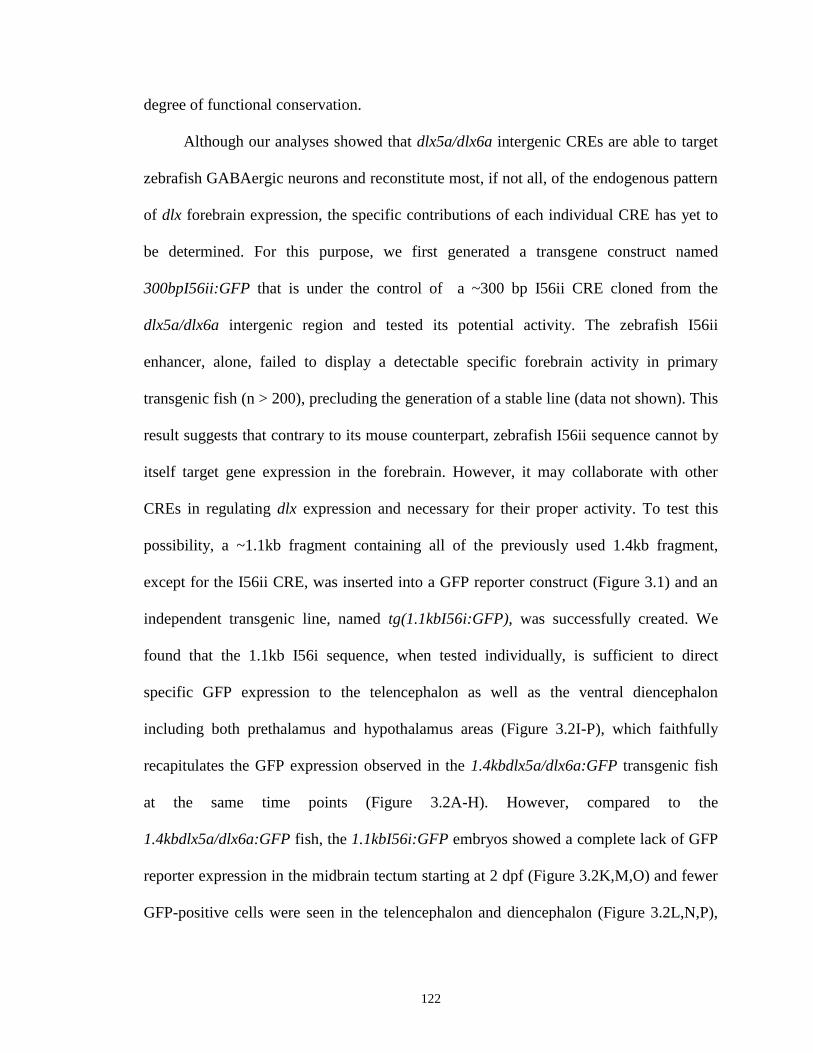
122
degree of functional conservation.
Although our analyses showed that dlx5a/dlx6a intergenic CREs are able to target
zebrafish GABAergic neurons and reconstitute most, if not all, of the endogenous pattern
of dlx forebrain expression, the specific contributions of each individual CRE has yet to
be determined. For this purpose, we first generated a transgene construct named
300bpI56ii:GFP that is under the control of a ~300 bp I56ii CRE cloned from the
dlx5a/dlx6a intergenic region and tested its potential activity. The zebrafish I56ii
enhancer, alone, failed to display a detectable specific forebrain activity in primary
transgenic fish (n > 200), precluding the generation of a stable line (data not shown). This
result suggests that contrary to its mouse counterpart, zebrafish I56ii sequence cannot by
itself target gene expression in the forebrain. However, it may collaborate with other
CREs in regulating dlx expression and necessary for their proper activity. To test this
possibility, a ~1.1kb fragment containing all of the previously used 1.4kb fragment,
except for the I56ii CRE, was inserted into a GFP reporter construct (Figure 3.1) and an
independent transgenic line, named tg(1.1kbI56i:GFP), was successfully created. We
found that the 1.1kb I56i sequence, when tested individually, is sufficient to direct
specific GFP expression to the telencephalon as well as the ventral diencephalon
including both prethalamus and hypothalamus areas (Figure 3.2I-P), which faithfully
recapitulates the GFP expression observed in the 1.4kbdlx5a/dlx6a:GFP transgenic fish
at the same time points (Figure 3.2A-H). However, compared to the
1.4kbdlx5a/dlx6a:GFP fish, the 1.1kbI56i:GFP embryos showed a complete lack of GFP
reporter expression in the midbrain tectum starting at 2 dpf (Figure 3.2K,M,O) and fewer
GFP-positive cells were seen in the telencephalon and diencephalon (Figure 3.2L,N,P),
123
most likely due to the loss of the regulatory function of the I56ii CRE. These
observations are supported by immunostaining assays on tissue sections showing that
GFP expression in the midbrain tectum is almost entirely absent in the 1.1kbI56i:GFP
embryos (Figure 3.4E,I), whereas expression patterns in the ventral telencephalon and
diencephalon are essentially identical between the two constructs (Figure 3.3A,E,I,M and
Figure 3.4A,E,I). Furthermore, the 1.1kbI56i:GFP fish appear to have a markedly
reduced number of GFP-positive cells in the ventral telencephalon at the rostral level
(Figure 3.4A) as well as a loss of a small subpopulation of GFP-expressing cells in the
ventral diencephalon (prethalamus, Figure 3.4I) as compared to the corresponding
regions in 1.4kbdlx5a/dlx6a:GFP fish (Figure 3.3A and Figure 3.3I). In the
1.1kbI56i:GFP fish, robust GFP expression is also found in the forebrain bundles
assembled bilaterally in the basal forebrain (Figure 3.4A, arrows). As expected, the
1.1kbI56i-directed GFP expression within domains of the ventral telencephalon and
diencephalon is highly co-localized with that of GABA-positive neurons (Figure 3.4),
consistent with the observations made in the 1.4kbdlx5a/dlx6a:GFP fish (Figure 3.3).
These findings support a notion that whereas the overall activity of intergenic regulatory
elements associated with the dlx5a/dlx6a locus is primarily restricted to GABAergic
neurons, the different individual enhancers, I56i and I56ii, seem to perform distinct, non-
redundant roles in modulating the development of different subpopulation of GABA-
expressing cells, with I56ii necessary for expression in the tectum.
3.3.2. dlx5a/dlx6a intergenic CREs do not extensively mark diverse
GABAergic interneuron subtypes
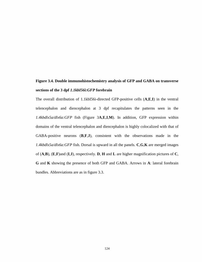
124
Figure 3.4. Double immunohistochemistry analysis of GFP and GABA on transverse
sections of the 3 dpf 1.1kbI56i:GFP forebrain
The overall distribution of 1.1kbI56i-directed GFP-positive cells (A,E,I) in the ventral
telencephalon and diencephalon at 3 dpf recapitulates the patterns seen in the
1.4kbdlx5a/dlx6a:GFP fish (Figure 3A,E,I,M). In addition, GFP expression within
domains of the ventral telencephalon and diencephalon is highly colocalized with that of
GABA-positive neurons (B,F,J), consistent with the observations made in the
1.4kbdlx5a/dlx6a:GFP fish. Dorsal is upward in all the panels. C,G,K are merged images
of (A,B), (E,F)and (I,J), respectively. D, H and L are higher magnification pictures of C,
G and K showing the presence of both GFP and GABA. Arrows in A: lateral forebrain
bundles. Abbreviations are as in figure 3.3.

125
126
In mice, Dlx enhancers have been shown to be highly active in various major
subtypes of GABAergic interneurons (Ghanem et al., 2007), suggesting that proper
function of these CREs might be important to target Dlx gene expression to specific
populations of differentiating GABAergic neurons. To further examine this possibility in
the zebrafish forebrain, we thereby utilized a double-immunohistochemistry analysis in
the 1.4kbdlx5a/dlx6a:GFP transgenic fish in order to evaluate whether dlx5a/dlx6a CREs
can target transgene expression to groups of cells that express particular GABAergic
neuron subtypes. Surprisingly, comparison of GFP expression with commonly used
interneuron markers such as calbindin (CB), calretinin (CR) and parvalbumin (PV),
revealed that a vast majority of GFP-positive cells within the telencephalon and
diencephalon of 3 dpf embryos are not co-labeled by the three interneuron markers that
we tested (Figure 3.5). Nonetheless, we did identify some co-localization between GFP
and these molecular markers in certain populations of cells. For instance, many GFP-
expressing cells appear to coexpress CR in the ventral diencephalon (prethalamus, Figure
3.5L) as well as in the optic tectum (Figure 3.5P). A number of GFP-expressing cells in
the ventral telencephalon (Figure 3.5T) and in the rostral hypothalamic region (Figure
3.5X) were found to PV-positive. In the case of GFP and CB coexpression, a few
scattered cells in the basal forebrain and in the midbrain tectum were found to be double-
positive (Figure 3.5D,H, arrowheads). Similar results were also observed at 5 dpf and
later developmental stages (data not shown). These data are, therefore, at least in part
contrasting with our earlier studies in mice (Ghanem et al., 2007).
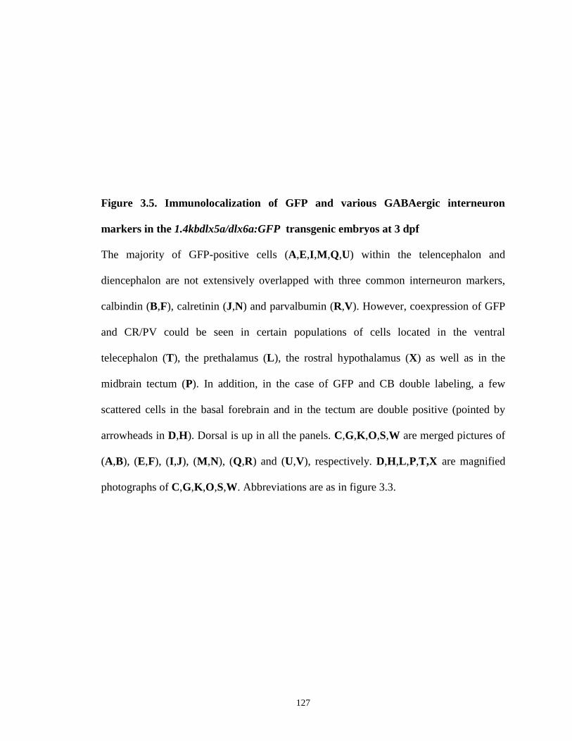
127
Figure 3.5. Immunolocalization of GFP and various GABAergic interneuron
markers in the 1.4kbdlx5a/dlx6a:GFP transgenic embryos at 3 dpf
The majority of GFP-positive cells (A,E,I,M,Q,U) within the telencephalon and
diencephalon are not extensively overlapped with three common interneuron markers,
calbindin (B,F), calretinin (J,N) and parvalbumin (R,V). However, coexpression of GFP
and CR/PV could be seen in certain populations of cells located in the ventral
telecephalon (T), the prethalamus (L), the rostral hypothalamus (X) as well as in the
midbrain tectum (P). In addition, in the case of GFP and CB double labeling, a few
scattered cells in the basal forebrain and in the tectum are double positive (pointed by
arrowheads in D,H). Dorsal is up in all the panels. C,G,K,O,S,W are merged pictures of
(A,B), (E,F), (I,J), (M,N), (Q,R) and (U,V), respectively. D,H,L,P,T,X are magnified
photographs of C,G,K,O,S,W. Abbreviations are as in figure 3.3.
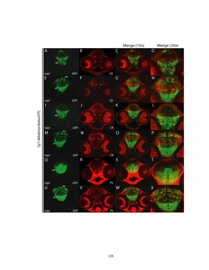
128
129
3.3.3. Loss of dlx1a/dlx2a or of dlx5a/dlx6a function dramatically affects
enhancer activity in the zebrafish diencephalon
In mice, Dlx genes have been revealed to regulate their own expression or
expression of their paralogous genes through acting on the CREs within their loci or
those from other Dlx bigene clusters (Zerucha et al., 2000; Zhou et al., 2004; Poitras et
al., 2007; Potter et al., 2009). To explore whether auto- or cross-regulatory loops between
distinct dlx bigene pairs exist in zebrafish as is the case in mice, different combinations of
translation blocking MOs against dlx mRNA transcripts were injected into fertilized one-
cell stage embryos from the 1.4kbdlx5a/dlx6a:GFP and 1.1kbI56i:GFP transgenic fish
and the effects on the reporter activity were assessed (Table 3.1). MO-treated embryos
were examined at 3 dpf, a stage when all dlx genes are highly expressed and various
distinct regions of the developing forebrain become identifiable. We found that inhibition
of single dlx gene (dlx1a, dlx2a, dlx5a or dlx6a) did not result in a marked loss or visible
alteration of GFP expression (data not shown), indicating a potential functional
redundancy among dlx genes. In contrast, the 1.4kbdlx5a/dlx6a:GFP embryos that
received the dlx1a+dlx2a MOs displayed an extensive depletion of GFP expression in the
entire diencephalon and in the midbrain tectum (Figure 3.6A,B, arrows). However,
telencephalic GFP expression in a majority of morphants remained largely intact (Figure
3.6A,B). Likewise, compared with the embryos injected with control MO, the
simultaneous loss of dlx1a and dlx2a function in the 1.1kbI56i:GFP embryos caused a
marked decrease in reporter transgene expression in the diencephalon (Figure 3.6D,
arrows) and a reduction of GFP expression was also noticed in the telencephalon of some
fish (Figure 3.6D).
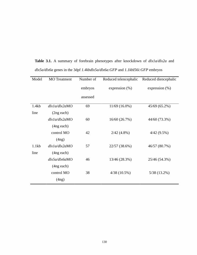
130
Table 3.1. A summary of forebrain phenotypes after knockdown of dlx1a/dlx2a and
dlx5a/dlx6a genes in the 3dpf 1.4kbdlx5a/dlx6a:GFP and 1.1kbI56i:GFP embryos
Model MO Treatment Number of
embryos
assessed
Reduced telencephalic
expression (%)
Reduced diencephalic
expression (%)
1.4kb
line
1.1kb
line
dlx1a/dlx2aMO
(2ng each)
dlx1a/dlx2aMO
(4ng each)
control MO
(4ng)
dlx1a/dlx2aMO
(4ng each)
dlx5a/dlx6aMO
(4ng each)
control MO
(4ng)
69
60
42
57
46
38
11/69 (16.0%)
16/60 (26.7%)
2/42 (4.8%)
22/57 (38.6%)
13/46 (28.3%)
4/38 (10.5%)
45/69 (65.2%)
44/60 (73.3%)
4/42 (9.5%)
46/57 (80.7%)
25/46 (54.3%)
5/38 (13.2%)
131
Figure 3.6. Double knockdown of dlx1a/dlx2a and dlx5a/dlx6a markedly reduces
GFP reporter expression in the diencephalon and the midbrain tectum in the
1.4kbdlx5a/dlx6a:GFP and 1.1kbI56i:GFP morphants at 3 dpf
Simultaneous injection of combined MOs against dlx1a+dlx2a (2 ng/nl for each MO in A
and 4 ng/nl for each MO in B) significantly disrupted GFP expression in the
diencephalon (prethalamus and hypothalamus) and the midbrain tectum in the
1.4kbdlx5a/dlx6a:GFP embryos (A,B), as compared to that seen in 3 dpf embryos
injected with the standard control MO (C), although telencephalic GFP expression in a
majority of morphants remained seemingly unaffected (A,B). Similarly, double
knockdown of dlx1a+dlx2a and dlx5a+dlx6a (4 ng/nl for each MO) in the
tg(1.1kbI56i:GFP) line also led to a marked loss of diencephalic and midbrain GFP
expression (D,E), in comparison with the corresponding controls (F), similar to the loss
observed in the 1.4kbdlx5a/dlx6a:GFP dlx1a+dlx2a morphants (A,B). In addition, there
was also an noticeable reduction in the number of 1.1kbI56i-targeted GFP-positive cells
in the telencephalon of some fish (D,E). The loss of GFP expression in the prethalamus
and hypothalamus in the morphants are indicated by arrows in A,B,D,E. Note that the
effects of double knockdown of dlx5a and dlx6a were not examined in the
1.4kbdlx5a/dlx6a:GFP embryos due to the promoter sequence used in the original
transgene construct. In all the panels, embryos are oriented anterior to the left, dorsal
views. Scale bar: 100 m. Abbreviations are as in figure 3.1.
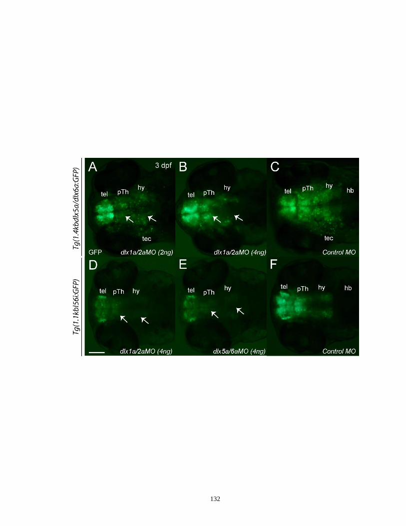
132
133
We also knocked down the function of both dlx5a and dlx6a and examined reporter
gene expression in the 1.1kbI56i:GFP embryos. As expected, an obvious loss of GFP
expression was seen in the diencephalon (Figure 3.6E, arrows) as well as in the
telecephalon of the dlx5a+dlx6a morphants (Figure 3.6E), resembling the changes seen
after systemic knockdown of both dlx1a and dlx2a. Our data support a viewpoint that
self- and cross-regulation of different dlx genes may be conserved in the developing
zebrafish forebrain.
3.4. Discussion
3.4.1. Conserved activities of dlx regulatory elements between zebrafish and
mouse
Teleost fish are one of the most diverse groups of vertebrates and are composed of
more than 23000 different species including zebrafish (Powers, 1991). Teleost fish have
been proposed to undergo divergence from mammals about 430 millions years ago
(Powers, 1991). Although the basic mode of telencephalic development varies greatly
between teleosts (e.g. zebrafish) and mammals (e.g. mouse) (Nieuwenhuys and Meek,
1990), comparative studies of the expression patterns of a group of developmental genes
including Dlx genes as well as neurotransmitters have revealed many regional
homologies between the developing mouse and zebrafish forebrain (Wullimann and
Rink, 2002; Mueller et al., 2006; Mueller et al., 2008; Moreno et al., 2009; Wullimann,
2009). Dlx forebrain expression patterns of mouse and zebrafish appear to be well
conserved, suggesting that the potential mechanisms underlying Dlx regulation might be
also conserved over evolutionary time (Akimenko et al., 1994; Ellies et al., 1997;
134
Panganiban and Rubenstein, 2002; MacDonald et al., 2010a). One potential factor
contributing to this conservation may be the conservation of various Dlx CREs within
with their respective Dlx loci. Indeed, orthologous Dlx CREs exhibit a high degree of
sequence similarity among distantly related vertebrate species (Ghanem et al., 2003). The
I56i and I12a enhancers also belong to a group of 481 ultraconserved DNA fragments,
whose sequences are above 200 bp and are completely identical between the human,
mouse and rat genomes (Bejerano et al., 2004). While there exists a debate concerning
whether high sequence conservation of noncoding regulatory elements among distinct
vertebrates does faithfully reflect their conserved activities (Elgar and Vavouri, 2008;
McEwen et al., 2009), our present study showed that the 1.4kb dlx5a/dlx6a intergenic
region and a 1.1kb zebrafish I56i fragment both specifically drive GFP expression to
GABAergic neurons in the ventral telencephalon and diencephalon, mimicking the
activities of their mouse orthologs when tested in transgenic mice (Ghanem et al., 2007;
Ghanem et al., 2008). Consistent with these results, other dlx CREs, such as URE2 and
I12b, also showed activities similar to that of their mouse orthologous sequences as
reflected by GFP reporter gene expression in the developing zebrafish forebrain and
pharyngeal arches (Ghanem et al., 2003; Ghanem et al., 2007; MacDonald et al., 2010a;
MacDonald et al., 2010b). Furthermore, heterologous transgenic assays showed that
zebrafish I56i/I56ii and URE2 CREs are able to activate reporter gene expression in the
mouse subpallial telencephalon in a pattern resembling that of endogenous Dlx forebrain
expression (Zerucha et al., 2000; Stuhmer et al. 2002a; MacDonald et al., 2010b). The
genetic pathway involving Mash1 and Dlx that is required for proper GABAergic neuron
development is also likely to be conserved between mouse and zebrafish. The Mash1

135
ortholog, ascl1a, is not only co-expressed with dlx1a, dlx5a and gad1 in the early
zebrafish forebrain, but also acts as a upstream regulator mediating dlx expression
(MacDonald et al., unpublished data), which is consistent with previous observations
made in mice (Yun et al., 2002; Poitras et al., 2007). These data suggest that highly
conserved sequences and activities of various Dlx CREs may be crucial for maintaining
proper Dlx functioning and the overall genetic cascade modulating GABAergic neuron
development through vertebrate evolution.
3.4.2. Differential activity and contribution of zebrafish I56i and I56ii CREs
Modified gene expressions and gene-gene interactions due to altered regulatory
elements and/or changes in binding of trans-acting factors to these elements have been
long hypothesized as key molecular events that can induce novel morphological
structures and genetic networks over evolutionary time (Tvrdik and Capecchi, 2006;
Papatsenko and Levine, 2007; Kleinjan et al., 2008). Expression patterns of the
1.4kbdlx5a/dlx6a:GFP and 1.1kbI56i:GFP transgenes overlap extensively in most areas
of the ventral forebrain. Nevertheless, differences were observed in the mesencephalic
tectum most probably as a result of the absence of I56ii activity in the tg(1.1kbI56i:GFP)
construct. Despite the unknown roles of dlx genes in this area, this finding indicates that
zebrafish I56i and I56ii CREs may exert differential regulatory roles, at least in certain
domains in the developing zebrafish brain.
The apparent absence of specific activity for I56ii, when tested individually in
zebrafish, contrasts markedly with what we previously observed in mice. In mice, I56ii is
unique in its ability to specifically target expression to a distinct subpopulation of

136
postmitotic striatal projection neurons, termed “corridor cells”, which express two
homeoproteins Islet1 and Meis2 and is essential for the growth and guidance of
thalamocortical projections during midgestation (Lopez-Bendito et al., 2006; Ghanem et
al., 2007; Ghanem et al., 2008). One potential implication of this inter-species difference
would be the absence of zebrafish forebrain cells corresponding to the mouse corridor
cells. Moreover, our former results in transgenic mice revealed that zebrafish I56ii is
much less efficient at targeting reporter gene expression to the forebrain (0 of 2
transgenic lines; 1 of 10 primary transgenic embryos) compared to the full-length
dlx5a/dlx6a intergenic region as well as the individual zebrafish I56i enhancer (Zerucha
et al., 2000). Nevertheless, I56ii sequence conservation between mammals and teleosts is
hard to reconcile with its apparent inability to drive reporter transgene expression, at least
spatially. We cannot at present rule out a specific role for I56ii in driving expression in
later stage larvae or in adults. An essential cooperative effect between I56i and I56ii in
the zebrafish forebrain could also explain the conservation in sequence. In order to
function properly and efficiently, the zebrafish I56ii may require the presence of
additional regulatory sequences, either from I56i or from the promoter of one or both
flanking dlx genes. Similarly, I56ii could improve the efficiency of additional
dlx5a/dlx6a CREs, such as I56i. The I56i and I56ii enhancer differ markedly in sequence.
We have demonstrated that, compared to the mouse I56i CRE, the putative Dlx binding
sites and the surrounding nucleotides appear to be less conserved in I56ii (Zerucha et al.,
2000; Ghanem et al., 2003). Furthermore, the I56ii enhancer is less responsive to the
activation of Dlx proteins in in vitro transfection assays (Zerucha et al., 2000; Ghanem et
al., 2003). Finally, both Dlx1 and Dlx2 proteins can directly bind to I56i CRE in vivo but

137
not to I56ii as revealed by ChIP analysis (Zhou et al., 2004). In light of this evidence, we
propose that zebrafish I56ii CRE may be only bound by a particular set of transcription
factors, which are probably distinct from those acting through the I56i enhancer. As such,
I56ii may be necessary for the proper development of a particular subpopulation of
GABAergic neurons, for instance, those located in the midbrain tectum.
It is important to emphasize here that the two transgenic lines used in this study
were generated in very different ways: the 1.1kb line carries a β-globin minimal
promoter, whereas the 1.4kb line is flanked by a dlx6a 5’-flanking region. This difference
may potentially impact our results. Therefore, in order to provide more direct
comparisons, we are in the process of generating a novel transgenic line that contains the
1.4kb intergenic fragment as well as the same β-globin minimal promoter as the 1.1kb
line. The establishment and analysis of this stable transgenic line will help us understand
whether the dlx6a 5’-flanking region contribute to some extent the differential GFP
expression patterns seen in the two transgenic lines shown in the present study.
3.4.3. Functional divergence of dlx forebrain CREs between zebrafish and
mouse
Spatio-temporal expression of a given gene in specific tissue types is usually an
overall effect of multiple transcription factors which can exert their regulatory functions
in a correct combination by acting on one or more regulatory elements (Strahle and
Rastegar, 2008; Panne, 2008). On the other hand, if such a combination cannot be
accomplished under certain cellular microenvironments or if any of these transcription
factors are prevented from binding to their targeting CREs due to certain reasons, the

138
CREs will stay in an inactive status (Smale and Kadonga, 2003; Arnosti and Kulkarni,
2005). Although the sample size of our knockdown experiments was relatively small and
no rescue assays were conducted, we were able to show simultaneous inhibition of
dlx1a/dlx2a or dlx5a/dlx6a function in our transgenic lines resulted in a significant loss of
transgene expression in the diencephalon and the midbrain tectum as well as a lesser
extent of reduction in the telencephalon, suggesting that functional redundancy also exists
between zebrafish dlx genes. The decreased transgene activity in the morphants is most
likely due to the loss of dlx protein binding to I56i and/or I56ii enhancers, suggesting that
zebrafish dlx genes from distinct bigene clusters are also under auto- and cross-regulation
via their corresponding CREs, consistent with previous observations made for their
mouse orthologs (Zerucha et al., 2000; Zhou et al., 2004; Poitras et al., 2007; Potter et al.,
2009). For example, previous studies have shown that the Dlx1 and Dlx2 proteins may
bind to the I56i enhancer and regulate Dlx5 and Dlx6 expression in the developing mouse
forebrain (Anderson et al. 1997b; Zerucha et al., 2000; Stuhmer et al., 2002b). Recently,
both in vitro and in vivo evidence has also been provided for the auto-regulation of the
Dlx genes as both URE2 and I12b CREs have been found to be direct targets of Dlx1 and
Dlx2 proteins (Poitras et al., 2007; Potter et al., 2009). Moreover, Dlx1/Dlx2 expression
plays essential roles in regulating the activities of these CREs (Poitras et al., 2007; Potter
et al., 2009). Interestingly, telencephalic expression of the 1.4kbdlx5a/dlx6a:GFP and
1.1kbI56i:GFP transgenes was less affected by the absence of Dlx proteins when
compared to the diencephalic expression, which contrasts sharply with the situations seen
in mice (Anderson et al., 1997a; Anderson et al., 1997b; Wang et al., 2010). These
findings suggest the existence of parallel and possibly redundant genetic pathways

139
responsible for dlx expression in different contexts in the developing zebrafish forebrain
via the interactions with the I56i and/or I56ii enhancers.
Although the overall activities of orthologous Dlx CREs appear well conserved in
distinct vertebrate species, they may also serve slightly different functions. The mouse
I56i sequence targets lacZ reporter gene expression to the pharyngeal arches of transgenic
mice (Zerucha et al., 2000; Park et al., 2004), whereas its zebrafish counterpart is not able
to do the same as shown in the current study. Likewise, orthologous URE2 sequences
from zebrafish, mouse and elephant shark were shown to have differential roles in
regulating the mandibular arch development (MacDonald et al., 2010b). Functional
conservation and divergence of the cis-regulatory networks of the orthologous Hoxa2
gene and the HoxD cluster have also been observed in many distantly related vertebrates
(Tumpel et al., 2002; Spitz et al., 2003). It has been proposed that the regulatory elements
in the same gene locus in different species may have gradually acquired certain
differential mechanisms over evolution to modify their protein-binding activity (Tautz,
2000; Ludwig, 2002; Gibert and Simpson, 2003). The modifications that are specific to
certain vertebrate lineages, such as the mammal-specific loss or teleost-specific gain of
some upstream signaling, may also contribute to differential activities of orthologous
regulatory elements, at least in certain tissue types (Wittkopp, 2005). These modifications
perhaps have emerged following the teleost-specific genome duplication event after they
undergo divergence from mammals, which may produce many paralogous key regulatory
genes that could subsequently act as novel modulators interacting with enhancer elements
(Wittkopp, 2005).

140
3.4.4. Functional implication of dlx genes in zebrafish GABAergic neuron
differentiation
In the developing mouse forebrain, Dlx genes amongst other key regulatory genes
(e.g. Mash1, Necdin, Arx and Smad) play essential roles in regulating the production and
differentiation of GABAergic neurons (Cobos et al., 2005b; Kuwajima et al., 2006; Maira
et al., 2010; Long et al., 2009a). Various Dlx CREs have been shown to mark
GABAergic neuron progenitors in the ganglionic eminences in the subpallial
telecephalon as well as diverse subtypes of GABAergic interneurons in the adult cortex
(Ghanem et al., 2007). While the distribution of GABAergic neurons has been well
described in the developing and adult zebrafish brain (Doldan et al., 1999; Kim et al.,
2004; Mueller et al., 2006), very little is known about their differentiation process as well
as the underlying mechanisms. Due to the lack of antibodies specifically recognizing
zebrafish dlx proteins, we examined GFP expression driven by individual dlx CREs in
immunohistochemical analyses of developing GABAergic neurons. Based on
GABAergic interneuron markers that were mainly used in mice, we found that the
dlx5a/dlx6a regulatory elements are inactive in a large population of differentiated
GABAergic neurons in the early developing forebrain. However, colocalization of GFP
and interneuron marker expression was observed in some subpopulations of cells in the
ventral telencephalon and diencephalon as well as in the optic tectum. These findings are
in contrast with previous observations in mice and lead us to propose that the
differentiation of zebrafish GABAergic neurons might be also regulated by other dlx
forebrain CREs (e.g. URE2 and I12b) or the genetic pathways that are not identical with
141
those in mice, likely due to different microenvironment between the developing zebrafish
and mouse forebrain. Indeed, fgf19 and her6 genes were reported to participate in
regulating differentiation and survival of zebrafish GABAergic neurons in the developing
telencephalon and diencephalons (Miyake et al., 2005; Scholpp et al., 2009). The hairy-
related gene her6 has been found to serve as an important regulatory gene responsible for
maintaining the neurogenic gradient in the caudal thalamus and it also mediates the cell
fate switch between glutamatergic and GABAergic neurons (Scholpp et al., 2009). In
addition, sequence divergence of orthologous I56i/I56ii elements in mice and zebrafish
(Ghanem et al., 2003) may also possibly contribute to their ability to target distinct
populations of GABAergic neurons in these two species. It is also worth noting a
challenge for studying GABAergic neuron differentiation in zebrafish, because of the
differential cross-reactivity of commercially available antibodies used to detect the
different GABAergic neuron subtypes. In our case, even though the efficiency of the
antibodies we used as interneuron markers have been tested in other species including
mouse and human, they may be less effective or ineffective in labeling all isoforms of the
studied protein when tested in zebrafish, especially considering the fact that the zebrafish
genome contains a large number of duplicated genes. Liu et al. (2010) showed that there
are four and six SOM genes functionally expressed in the zebrafish telecephalon and
hypothalamus, respectively. Using an analysis of existing genomic information,
Friedberg (2005) identified a total of nine isoforms that encode PV in zebrafish. By
carrying out similar studies in transgenic lines made with other dlx CREs (MacDonald et
al., 2010b; MacDonald et al., 2010c), by using additional GABAergic interneuron
markers (e.g. SOM and NPY), and by extending our observations to adult fish, we should
142
better understand the roles of dlx genes and their CREs in zebrafish GABAergic
interneuron differentiation.
In summary, our data demonstrate that zebrafish dlx5a/dlx6a regulatory elements
exhibit partially conserved activity when compared to their mouse orthologs and these
CREs may be involved in the genetic pathways responsible for the production of some
populations of GABAergic neurons. I56i and I56ii CREs are likely to exert differential
function in modulating this process in distinct populations of cells. Similar to the mouse
Dlx genes, auto- or cross-regulatory mechanisms may also exist between different
zebrafish dlx genes, even though dlx genes may be regulated in a different way between
the developing telencephalon and diencephalon. Besides the dlx5a/dlx6a regulatory
elements, other dlx CREs or regulatory factors may also contribute to the differentiation
of GABAergic interneuron subtypes in the developing zebrafish forebrain. Further
examination, throughout development and in adult animals, of the activities of various
individual dlx CREs will provide us with additional clues with respect to the evolution of
regulatory mechanisms responsible for dlx forebrain expression and further improve our
understanding about the implications of dlx genes during vertebrate forebrain
development.
Acknowledgements
We would like to thank Vishal Saxena for assistance with zebrafish husbandry and
excellent technical support for maintaining transgenic zebrafish colonies. We
acknowledge Dr. Marie-Andrée Akimenko and Jared Talbot for insightful advice and
143
discussion. We are also grateful to Dr. Mélanie Debiais-Thibaud for critical reading of
the manuscript. This work was supported by grants from the Natural Sciences and
Engineering Research Council of Canada (NSERC) and the Canadian Institutes of Health
Research (CIHR #MOP14460). M.Y. was supported by a University of Ottawa
Excellence Scholarship and an Ontario Graduate Scholarship (OGS). M.E. holds a
Research Chair in Evolutionary Developmental Biology at the University of Ottawa.

144
4. Targeted deletion of a Dlx intergenic enhancer I12b reduces
Dlx1/Dlx2 expression and inhibits cell proliferation in the developing
mouse forebrain
Man Yu a,b
, Luc Poitras a, Lisa Tran
a, Marc Ekker
a,b,*
a Centre for Advanced Research in Environmental Genomics (CAREG), Department of
Biology, University of Ottawa, 20 Marie Curie, Ottawa, ON, Canada K1N 6N5
b Department of Cellular and Molecular Medicine, University of Ottawa, 451 Smyth
Road, Ottawa, ON, Canada K1H 8M5
* Corresponding author. Department of Biology, University of Ottawa, 30 Marie Curie,
Ottawa, ON, Canada K1N 6N5. Tel.: +1-613-5625800, ext. 2605; Fax: +1-613-5625486;
E-mail address: [email protected]
Keywords: cis-regulatory elements; cell proliferation; Dlx genes; forebrain; GABAergic
neurons; gene regulation; mice; neuron migration

145
Abstract
Dlx homeobox genes encode a family of transcription factors that are necessary for the
proper differentiation, migration and survival of GABAergic interneurons. So far, at least
four well conserved cis-regulatory elements (CREs) have been identified to be important
for regulating Dlx transcriptional control in the developing telencephalon and
diencephalon: I12b and URE2 in the Dlx1/Dlx2 bigene cluster, and, I56i and I56ii in the
Dlx5/Dlx6 bigene cluster. However, how Dlx regulation is linked to these various
individual CREs and how these CREs maintain correct Dlx forebrain expression are still
not fully characterized. Towards exploring the specific contribution of I12b CRE to
overall Dlx function as well as directly addressing its functional role in vivo, we have
used homologous recombination in mouse embryonic stem (ES) cells to generate mutant
mice in which I12b is deleted from its endogenous genomic locus. Here, we report that
mice completely lacking I12b CRE are viable and fertile and that I12b enhancer loss fails
to result in notable abnormalities when assayed for a variety of phenotypes including
brain size and forebrain morphology. Upon visual observation, tangential migration of
GABAergic neurons from the subpallial telencephalon to the neocortex in I12b-/-
mutant
embryos generally appear to be comparable to that in the wild-type littermates; instead,
our initial observations suggested a possibly reduced cell proliferation in the basal
telencephalon, particularly in the ventricular and subventricular zones (VZ and SVZ) of
the lateral/medial ganglionic eminences (LGE/MGE). Although expression patterns of
Dlx1, Dlx2 and Dlx5 revealed by in situ hybridization assay at E13.5 displayed no
significant difference between I12b homozygous mutants and their wild-type littermates,
our real-time RT-PCR quantification assays at this time point identified a moderate
146
reduction (~15-25%) of both Dlx1 and Dlx2 transcripts (Dlx5/Dlx6 mRNA levels remain
intact) in the forebrain of I12b-/-
mutants. Interestingly, this alteration coincides with a
~10% increase in the mRNA levels of Mash1, a direct upstream regulator of Dlx1 and
Dlx2. Taken together, our findings, while not inclusive of all possible phenotypic impacts
of I12b deletion, indicate that I12b enhancer may play an important role in controlling
Dlx expression as well as in GABAergic neurogenesis in the developing forebrain. These
data will lay the basis for future in vivo studies on the genetic networks involving Dlx
genes and their various CREs during vertebrate forebrain development.
4.1. Introduction
The forebrain is composed of a large diversity of neuronal cells with distinct
functions and morphologies and is thought to be one of the most complicated anatomical
structures in mammals. GABAergic interneurons play key roles in modulating and
maintaining the balanced activity of many forebrain areas, such as the cortex and
hippocampus, by directly exerting their inhibitory impacts upon excitatory glutamate-
releasing projection neurons (Owens and Kriegstein, 2002; Whittington and Traub,
2003). Improper balance between inhibitory and excitatory signaling has been proposed
to be relevant to many common human neurological disorders, such as autism and
epilepsy (Di Cristo, 2007). GABAergic interneurons are originally born in various
progenitor domains in the developing subpallium, specifically in the ganglionic
eminences, anterior entopeduncular area (AEP) and preoptic area (POA), and tangentially
migrate and then populate different layers in the developing cortex (Marin et al., 2000;
Xu et al., 2004; Wonders and Anderson, 2006). A number of transcription factors (e.g.

147
Mash1, Nkx2.1 and Gsh1/Gsh2) and signaling molecules, including retinoid acid (RA),
Sonic hedgehog (Shh) and fibroblast growth factors (FGFs), are responsible for
establishing regionalization of the developing subpallium (Medina et al., 2009; Moreno et
al., 2009; Nobrega-Pereira and Marin, 2009). Some of these molecules are also involved
in the genetic pathways important for GABAergic neuron generation. For instance,
Nkx2.1-/-
mutants have a significantly decreased number (~50%) of cortical interneurons
when compared to wild-type mice (Sussel et al., 1999). The sequences of Dlx homeobox
genes are related to that of the distal-less gene in Drosophila (Cohen et al., 1989). Four
Dlx genes (Dlx1, Dlx2, Dlx5 and then Dlx6) are sequentially expressed in the developing
mouse forebrain, where they mainly promote GABAergic interneuron progenitors to
proliferate and differentiate into different subtypes (Panganiban and Rubenstein, 2002).
While single Dlx gene knockouts exhibit relatively milder abnormal phenotypes when
compared to that of double Dlx null mutants (e.g. Dlx1/2-/-
and Dlx5/6-/-
) (Qiu et al.,
1997; Acampora et al., 1999; Cobos et al., 2005a), inactivation of both Dlx1 and Dlx2
genes causes severe deficiencies in the generation, differentiation and migration of
GABAergic neurons starting at early embryonic stages (Anderson et al., 1997a; Anderson
et al., 1997b; Marin et al., 2000; Pleasure et al., 2000; Long et al., 2007). Simultaneous
inactivation of Dlx5 and Dlx6 function leads to exencephaly of the anterior portion of the
embryonic forebrain and the mutants have a preferentially decreased number of PV-
expressing cortical interneurons with increased dendrite branching (Wang et al., 2010).
Vertebrate Dlx genes are generally organized as convergently transcribed bigene
clusters on different chromosomes and the two Dlx genes consisting of a bigene pair are
separated by a short intergenic sequence (~3.5 to 16 kb) (Zerucha and Ekker, 2000;
148
Sumiyama et al., 2003). The two Dlx genes from the same bigene cluster are expressed
with highly overlapping patterns in many tissue types possibly because they are under the
same regulatory mechanisms (Liu et al., 1997; Eisenstat et al., 1999). Through
phylogenetic footprinting in five distantly related vertebrate species and through
transgenic animal analyses, our laboratory has previously identified and characterized
four highly conserved cis-regulatory elements (CREs) that act as enhancers responsible
for regulating Dlx forebrain expression: I56i and I56ii within the Dlx5/Dlx6 loci (Zerucha
et al., 2000), and, URE2 and I12b within the Dlx1/Dlx2 loci (Ghanem et al., 2003;
Ghanem et al., 2007). Although their sequences vary greatly, these CREs all target
reporter transgene expression to the ventral telencephalon and diencephalon in a pattern
that faithfully mimics that of endogenous Dlx forebrain expression (Zerucha et al., 2000;
Ghanem et al., 2003; Ghanem et al., 2007). Further analyses of these CREs have
provided evidence that URE2, I12b and I56i are active in GABA-expressing progenitor
cells in the ganglionic eminences in the subpallial telecephalon as well as in diverse
subtypes of GABAergic interneurons in the adult cortex (Ghanem et al., 2007). I56ii is
specifically active in a subpopulation of GABAergic striatal projection neurons between
E11.5 and E13.5 during early embryogenesis (Ghanem et al., 2008). Moreover, more
recent studies demonstrated that in addition to being as a direct target of Dlx proteins, the
~400 bp I12b enhancer is also directly targeted by Mash1, a proneural bHLH
transcription factor (Poitras et al., 2007; Potter et al., 2009). Mash1 may regulate
Dlx1/Dlx2 expression by acting through I12b enhancer (Poitras et al., 2007; Potter et al.,
2009). Together, these data indicate that the genetic pathway involving Mash1 and Dlx

149
genes may control GABAergic neuron development through their dynamic interactions
with various Dlx CREs.
To date, our understanding of how Dlx forebrain expression is precisely regulated
and maintained through their various individual CREs is still very limited. In order to
investigate the relative contribution of I12b CRE to overall Dlx expression and to
evaluate its full functional potential in vivo, we applied an embryonic stem (ES) cell-
based approach to remove this enhancer from its native chromosomal context and
generated a mutant mouse model carrying a targeted I12b deletion. Here, we first
determine whether I12b enhancer deletion affects the survival of the animals as well as
the size and morphology of the forebrain, with a particular focus on the regions of the
olfactory bulb, neocortex and hippocampus, where I12b is highly active. Subsequently, to
define the potential implication of I12b CRE in GABAergic neurogenesis, we examined
cell proliferation and tangential migration streams of GABAergic neurons in I12b-/-
homozygous mutants using a set of immunohistochemical analyses. In addition, the
effects of I12b enhancer deletion on the transcript levels of Dlx genes and other
regulatory genes (e.g. Mash1, Nkx2.1 and Gsh2) essential for GABAergic neuron
production and differentiation were also evaluated by quantitative real-time RT-PCR
assays and/or in situ hybridization at various developmental stages.
4.2. Materials and Methods
4.2.1. Animal maintenance
Mouse colonies maintained on a C57BL/6 background were housed with free
access to standard chow and water in a pathogen-free environment under a 12h light/dark
150
cycle, according to the guidelines of the Canadian Council on Animal Care (CCAC). For
staging of embryos, the day on which a vaginal plug was detected was calculated as
embryonic day 0.5 (E0.5). All experimental procedures were approved by the University
of Ottawa Animal Care Ethics Committee. Wild-type littermates were used as controls
for all comparative studies.
4.2.2. Construction of I12b CRE targeting vector
In brief, a Bacterial Artificial Chromosome (BAC) clone (#510G1) containing a
~200 kb Dlx1/Dlx2 locus was obtained by screening a BAC library (BACPAC Resources
Center) made from the liver tissue of strain 129/Sv mice. To generate the I12b targeting
vector (ΔI12b-510G1), a modified LoxP-flanked neomycin (neo) selection cassette
driven by a murine PGK promoter was inserted into the place of the wild-type I12b
enhancer present on the isolated BAC clone, which was accomplished by homologous
recombination in E. coli EL250 at 32°C as described previously (Lee et al., 2001). The
recombined BAC clones were then verified for sequence accuracy at the site of
recombination prior to electroporation into ES cells. The organization of the BAC
targeting plasmid is shown in Figure 4.1A and details of the vector construction are
available upon request.
4.2.3. Production of I12b null mice
The circular targeting construct was electroporated into mouse R1 ES cells (129/Sv
strain background, Transgenic Mouse Core Facility, McGill Cancer Center), which were
then allowed to grow on mitomycin-treated neo-resistant STO fibroblast feeder cells.
151
Two days following electroporation, ES cells were selected for neo-resistance in 300
µg/ml of G418 (Gibco) for one week. Recombinant ES clones were selected, expanded,
trypsinized and divided into two separate 96-well plates for further culture: one was used
for genomic DNA extraction and genotyping, while the other one was frozen for later
use. Genotyping of correctly targeted clones that have undergone homologous
recombination was completed using a quantitative real-time PCR assay as previously
described by Valenzuela et al. (2003). The following two primer-probe pairs were used:
I12b CRE forward: 5’-GGC CCA TCA AAC ACA ACA-3’, I12b CRE reverse: 5’-GGC
TTT CAG TGC GTT ATC TCT T-3’, probe: FAM-GTT TCC CCA TCT CTC T-
TAMRA; I56i CRE (internal control) forward: 5’-TAC AGC GTT TTT TAC CGT CAA
AG-3’, I56i CRE reverse: 5’-AGG TCC TAC GTC TCT GCA AT-3’, probe: Yakima
Yellow-GAA CAG GAA AGG CGA AA-TAMRA. The real-time PCR conditions are an
initial “Hot Start” activation step at 95°C for 15 min, followed by 30 cycles of 15 s at
95°C, 30 s at 56°C and 30 s at 72°C. Two independent positive ES clones (#43 and #86)
containing a neo-resistant mutant allele in place of I12b CRE (Figure 4.1A) were
microinjected into blastocysts isolated from C57BL/6 mice, which were subsequently
reimplanted into the uteri of pseudo-pregnant CD1 mice. The eight resultant chimeras
were then bred with C57BL/6 mice and successful germline transmission of the I12b
disrupted allele was assessed by the presence of the agouti coat color and PCR
verification. Agouti F1 offspring heterozygous for I12b CRE deletion were intercrossed
to generate I12b-/-
homozygous mutant progenies. Genotyping of I12b mutant mice were
performed using a standard PCR assay described in details below.

152
4.2.4. Genotyping of embryos and mice
I12b mutant embryos and newborn progeny were screened by regular PCR assays
using genomic DNA isolated from tail biopsies. Briefly, tissue samples were lysed in
appropriate volumes of digestion solution containing 100 mM NaCl pH 7.4, 10 mM Tris-
HCl, 5 mM EDTA, 0.5% SDS and 0.2 mg/ml proteinase K (Invitrogen) O/N at 55°C,
followed by phenol/chloroform extraction and an ethanol precipitation at -20°C according
to standard protocols. The final DNA pellets were dissolved in sterile Milli-Q water,
quantified on a Nanodrop spectrophotometer and kept at 4°C. Genotyping analyses were
performed by PCR using two specific primer pairs: I12b-forward: 5’-CCA AGA GGG
TCA GCA TCA TT-3’, I12b-reverse: 5’-CAG AGA GAT GGG GAA ACT GC-3’; neo-
forward: 5’-AGA CAA TCG GCT GCT CTG AT-3’, neo-reverse: 5’-CTC GTC CTG
CAG TTC ATT CA-3’. The PCR reaction yielded a 122 bp band for the wild-type allele
and a 110 bp product when the floxed PGK-neo cassette was present in place of the I12b
CRE. The PCR reaction was carried out in a total volume of 50 µl mixture consisting of
100 ng DNA template, 200 µM of each dNTP, 20 pmol of each primer, 5 µl of 10×PCR
buffer and 1 U Taq polymerase using the following cycling conditions: an initial
denaturation step at 95°C for 10 min, followed by 30 cycles of 30 s at 95°C, 30 s at 58°C
and 1 min at 72°C, and a final extension step at 72°C for 10 min. Amplified PCR
fragments were checked by 0.8% agarose gel electrophoresis and a negative control was
included in each experiment to ensure the specificity of PCR amplification.
4.2.5. Tissue preparation and sectioning
153
E13.5 and E15.5 embryos were anesthetized by cooling, dissected, and fixed O/N
in cold 4% paraformaldehyde (PFA)/1×PBS at 4°C. Neonatal pups were anesthetized
with an exposure to CO2 in a chamber and sacrificed by decapitation. P0 brains were
dissected and immersed in cold 4% PFA/1×PBS O/N at 4°C. Fixed brain tissues were
then cryoprotected by sequentially immersing in 12%, 16% and 22% sucrose in 1×PBS
(E13.5 and E15.5) or by overnight immersion in 30% sucrose/1×PBS (P0) at 4°C,
followed by embedding in Tissue Freezing Medium (Triangle Biomedical Sciences) and
snap-frozen at −80°C. Frozen blocks were transversely sectioned at the thickness of 20
µm (E13.5 and E15.5) or 40 µm (P0) onto Superfrost Plus slides (Fisher) using a Leica
CM1850 standard cryostat (Leica Microsystems) at -20°C and stored at -80°C until
further use.
4.2.6. Nissl staining
To compare the general morphology between P0 I12b-/-
mutant and wild-type
forebrain, cresyl violet staining (Nissl staining) was performed as per a standard protocol.
Briefly, sections were air-dried at RT for 30 min, dehydrated in serial ethanol dilutions
with descending concentration (70–0%) for 1 min each, and immersed in a staining
solution (5g/l cresyl violet acetate in 0.3% acetic acid, Sigma) for 3 min. After rinsing
with water for 2 min, stained sections were subsequently dehydrated in ethanol dilutions
with increasing concentration (30–100%) for 1 min each, cleared in xylene and mounted.
Images were taken on a Nikon SMZ 1500 dissecting microscope by using a Nikon DXM
1200C digital camera.

154
4.2.7. Immunohistochemistry
Immunohistochemistry on cryostat sections was carried out based on standard
methods described in Ghanem et al. (2007). In brief, frozen sections were dried for 1h at
RT and washed 3×10 min in 1×PBS to eliminate residues from tissue cryoprotectant.
Sections were then incubated with a blocking solution containing 1% bovine serum
albumin (BSA), 10% newborn calf serum and 0.1% Tween 20 for 2h at RT. The solution
was subsequently replaced with a fresh aliquot containing the primary antibody and
incubation was processed in a humid chamber O/N at 4°C. Following 3×15 min washes
in 1×PBS, sections were reacted with the appropriate secondary fluorescence-coupled
antibody for 2h at RT in the dark. For fluorescent imaging, histological sections were
cover-slipped with a Vectashield mounting medium (Vector Labs), analyzed with a Zeiss
Axiophot fluorescence microscope using the appropriate filter sets and photographed
with a Zeiss Axiocam digital camera. The following primary antibodies were used in this
study: rabbit polyclonal anti-GABA (1:1000, Sigma), rabbit polyclonal anti-calbindin
(anti-CB, 1:1000, Swant), rabbit polyclonal anti-phospho-histone H3 (anti-PH3, 1:100,
Upstate), rabbit polyclonal anti-active caspase 3 (1:500, BD Pharmingen). Alexa Fluor
488®
goat anti-rabbit IgG (H+L) (1:300, Invitrogen) was applied as the second antibody
in all the experiments.
4.2.8. In situ RNA hybridization on sections
Antisense RNA probes for Dlx1, Dlx2 and Dlx5 were prepared as previously
described (Cobos et al., 2005a; Long et al., 2007). Briefly, the probes were synthesized
by in vitro transcription with T7 or Sp6 RNA polymerase (Promega) and labeled with

155
digoxigenin (DIG)-UTP using DIG RNA Labeling Mix (Roche). In situ hybridization
experiments on 20 µm frozen sections were performed according to the following
procedure (Smith et al., 2008). After defrosting at RT for 1h, the slides were transferred
to a sealed humid chamber and incubated for 1h at 65°C with 500 µl of a pre-
hybridization solution consisting of 50% formamide, 10% dextran sulfate, 1 mg/ml yeast
tRNA (Invitrogen), 1×Denhardts solution (Sigma), and 1×salt solution (0.2 M NaCl, 10
mM Tris-HCl, 5 mM NaH2PO4, 5 mM Na2HPO4, 1 mM Tris-base, 5 mM EDTA pH 7.5).
DIG-labeled RNA probes were denatured at 70°C for 10 min, cooled in ice, and added
into the prewarmed (65°C) prehybridization buffer at a final concentration of 200–400
ng/ml. The prehybridization solution was then replaced with ~300 µl of the probe mix,
covered with a coverslip and incubated O/N at 65°C. In the following day, after removal
of the coverslips, slides were washed 3×30 min in solution A (1×SSC, 50% formamide,
and 0.1% Tween 20) at 65°C, followed by 2×30 min washes in 1×TBST at RT. Slides
were then blocked in 10% heat-inactivated calf serum in 1×TBST (blocking solution) for
2h at RT, followed by incubation with a fresh aliquot of block solution containing the
anti-DIG-AP Fab fragment antibody (1:5000 dilution) O/N at 4°C. After washing 5×20
min in 1×TBST at RT, slides were incubated for 2×10 min in NTMT staining buffer (100
mM NaCl, 100 mM Tris HCL pH 9.5, 50 mM MgCl2, and 0.1% Tween 20). For staining,
slides were transferred into large Petri dishes protected from light. The anti-DIG-AP on
each slide was visualized by incubating with 400 µl of NTMT containing 1.4 µl of BCIP
(5-bromo-4-chloro-3-indolyl-phosphate) and 2.7 µl of NBT (nitrobluetetrazolium
chloride). Slides were checked periodically for signal detection and the reaction was
stopped by 2×10 min washes in distilled water, followed by fixation in 4% PFA O/N at

156
4°C. Slides were finally washed by distilled water and allowed to dry before cover-
slipping with Aqua-MountTM
aqueous mounting medium (Fisher). Staining was examined
under a Nikon SMZ 1500 dissecting microscope and images were taken using a Nikon
DXM 1200C digital camera.
4.2.9. Quantitative real-time RT-PCR assay
To detect changes in gene expression levels, we dissected the telencephalon from
E13.5 I12b homozygous (n = 10) and heterozygous mutants (n = 9) as well as eight wild-
type littermates. Total RNA was then extracted and purified using TRIzol reagent
(Invitrogen) according to the manufacturer’s protocol. RNA concentration was quantified
at A260/280 nm on a Nanodrop spectrophotometer (Thermo Scientific) and 3 µg of total
RNA were reverse transcribed for the synthesis of single-strand cDNA by using
Oligo(dT)15 primer and SuperScript II Reverse Transcriptase (Invitrogen). All solutions
were prepared with diethylpyrocarbonate (DEPC)-treated water. RNase-free instruments
and RNaseZap (Ambion) were applied during all the procedures. Quantitative real-time
RT-PCR was conducted on an ABI Prism 7700 Sequence Detection System (Applied
Biosystems) in a 50 μl reaction mixture containing 4 μl of cDNA template, 25 μl
QuantiTect SYBR Green PCR Master Mix (Qiagen) and 20 pmol of each primer. The
following primer sets were used: Dlx1-forward: 5′-CAG TTG CAG GCT TTG AAC C-
3′, Dlx1-reverse: 5′-ACT TGG AGC GTT TGT TCT GG-3′; Dlx2-forward: 5’-GCC TCA
CCC AAA CTC AGG-3’, Dlx2-reverse: 5’-GCC GCT TTT CCA CAT CTT C-3’; Dlx5-
forward: 5’-CGA CTT CCA AGC TCC GTT C-3’, Dlx5-reverse: 5’-TTC TTT CTC
TGG CTG GCT G-3’; Dlx6-forward: 5’-CGG ACC ATT TAT TCC AGC C-3’, Dlx6-
157
reverse: 5’-CGC TTA TTC TGA AAC CAT ATC-3’; Gsh2-forward: 5’-CTT CCA ATA
TGT ACC TGT CCC-3’, Gsh2-reverse: 5’-GCT TGT GTG ATT GTT CCT CG-3’;
Mash1-forward: 5’-TCT CCT GGG AAT GGA CTT TG-3’, Mash1-reverse: 5’-AGG
TTG GCT GTC TGG TTT G-3’; Nkx2.1-forward: 5’-AGC ACA CGA CTC CGT TCT
CA-3’; Nkx2.1-reverse: 5’-CCC TCC ATG CCC ACT TTC TT-3’. To quantify the
transcript levels of Dlx genes and other regulators, we detected and used the mRNA
levels of glyceraldehyde-3-phosphate dehydrogenase (GAPDH) as an internal control.
The primers for GAPDH amplification were: forward, 5′-TGC ACC ACC AAC TGC
TTA GC-3′; reverse, 5′-GGC ATG GAC TGT GGT CAT GAG-3′. The transcripts of the
target genes and GAPDH were amplified in the same tubes using the following protocol:
initial “Hot Start” activation step for 10 min at 95°C and 40 cycles of 30 s at 95°C, 1 min
at 55°C, and 1 min at 72°C. For each PCR reaction, standard curves for the target genes
and GAPDH were established to determine quantitative mRNA levels by using five serial
dilutions of cDNA as templates. The transcript levels of the target genes were normalized
against that of GAPDH to give the relative values of their expression levels. Real-time
RT-PCR assays were repeated at least twice in triplicate for each sample and a non-
template control was included in each experiment. All data quantification and statistical
analysis was performed with Microsoft Excel 2010. Data were calculated in relative units
and presented as the mean ± SEM. The differences in gene expression levels between
I12b homozygous mutant, heterozygous mutants and wild-type littermates were evaluated
by using the Student t-test. P values < 0.05 were considered as statistically significant.
158
4.3. Results
4.3.1. Production of mutant mice carrying a targeted deletion of I12b CRE
In an effort to precisely define the role of I12b CRE in vivo, an ES cell-based gene
targeting technique was used to establish a strain of mice in which the individual I12b
enhancer was deleted from its genomic locus. Specifically, we first built a mutant allele
where a ~400 bp region of DNA containing the entire I12b sequence was replaced with a
LoxP-flanked PGK-neo-resistance cassette conferring resistance to the selection drug
G418. The general strategy for removal of the I12b enhancer from the Dlx1/Dlx2 locus
by homologous recombination in ES cells is depicted in Figure 4.1A. The neo-resistant
ES cells harboring an appropriate recombination event were identified among a total of
96 clones by a quantitative real-time PCR screening. Two independent positive ES clones
were injected into host blastocysts and the resulting eight highly chimeric mice were
subsequently bred to generate heterozygous progenies (I12b+/-
) that carry the proper I12b
deletion in the germline. Intercrosses between heterozygotes gave birth to homozygous
mutants (I12b-/-
) at the predicted Mendelian segregation ratios (more than 200 embryos
genotyped), demonstrating that disruption of I12b CRE does not cause embryonic
lethality either in utero or shortly after birth. Regular PCR assessment of different
genotypic categories is shown in Figure 4.1B. When housed under standard conditions,
I12b null mice are viable and proved to be fertile, in contrast to Dlx1 and Dlx2 single
mutants and Dlx1/Dlx2 double knockouts, which all die perinatally from a spectrum of
severe central nervous system and/or craniofacial defects (Anderson et al., 1997a;
Anderson et al., 1997b; Qiu et al., 1995; Qiu et al., 1997; Acampora et al., 1999; Cobos et
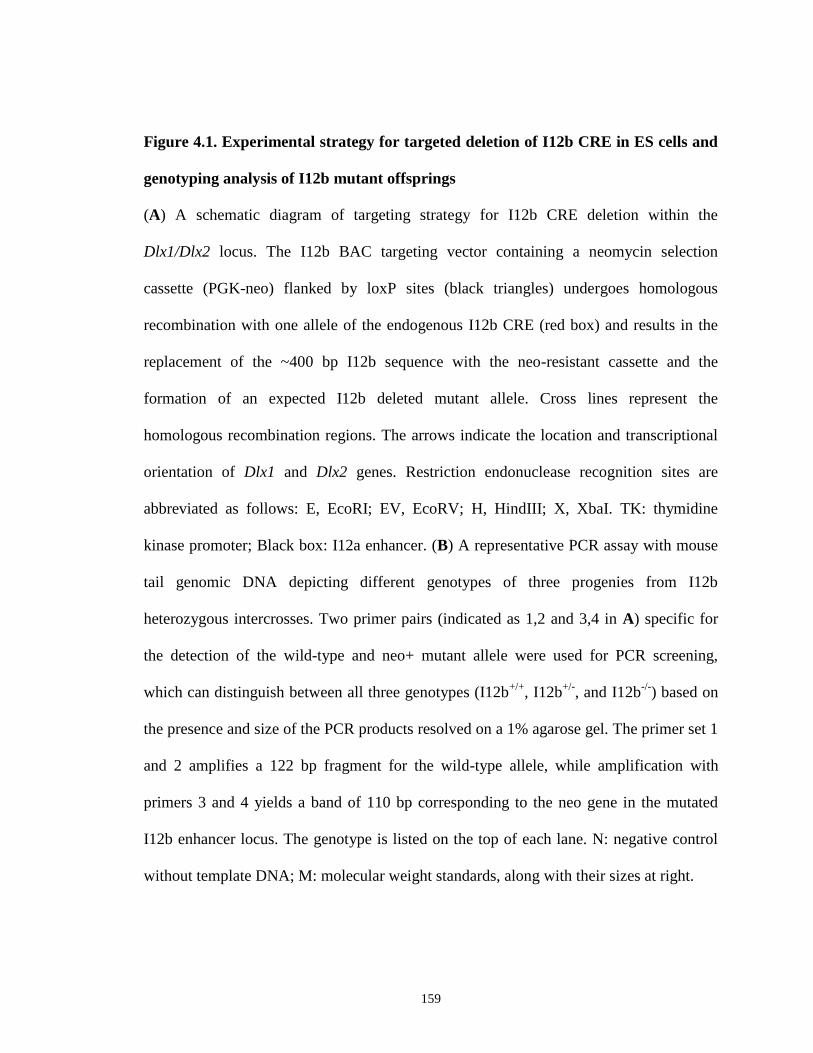
159
Figure 4.1. Experimental strategy for targeted deletion of I12b CRE in ES cells and
genotyping analysis of I12b mutant offsprings
(A) A schematic diagram of targeting strategy for I12b CRE deletion within the
Dlx1/Dlx2 locus. The I12b BAC targeting vector containing a neomycin selection
cassette (PGK-neo) flanked by loxP sites (black triangles) undergoes homologous
recombination with one allele of the endogenous I12b CRE (red box) and results in the
replacement of the ~400 bp I12b sequence with the neo-resistant cassette and the
formation of an expected I12b deleted mutant allele. Cross lines represent the
homologous recombination regions. The arrows indicate the location and transcriptional
orientation of Dlx1 and Dlx2 genes. Restriction endonuclease recognition sites are
abbreviated as follows: E, EcoRI; EV, EcoRV; H, HindIII; X, XbaI. TK: thymidine
kinase promoter; Black box: I12a enhancer. (B) A representative PCR assay with mouse
tail genomic DNA depicting different genotypes of three progenies from I12b
heterozygous intercrosses. Two primer pairs (indicated as 1,2 and 3,4 in A) specific for
the detection of the wild-type and neo+ mutant allele were used for PCR screening,
which can distinguish between all three genotypes (I12b+/+
, I12b+/-
, and I12b-/-
) based on
the presence and size of the PCR products resolved on a 1% agarose gel. The primer set 1
and 2 amplifies a 122 bp fragment for the wild-type allele, while amplification with
primers 3 and 4 yields a band of 110 bp corresponding to the neo gene in the mutated
I12b enhancer locus. The genotype is listed on the top of each lane. N: negative control
without template DNA; M: molecular weight standards, along with their sizes at right.
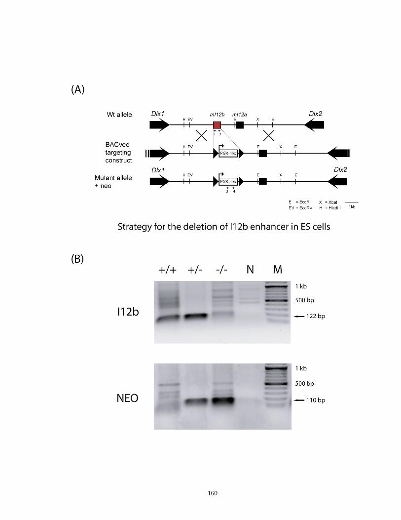
160
161
al., 2005a). In addition, mice homozygous for I12b loss develop normally and have body
weight and size (n > 20) that are comparable to that of heterozygotes and wild-type mice
and their gross appearance are phenotypically indistinguishable from wild-type and
heterozygous littermates during growth and maturation (Figure 4.2B,E and data not
shown). These initial findings suggest that I12b deletion does not markedly affect overall
growth and survival of the animals.
4.3.2. Morphological analyses of brain structures in I12b mutants
We next attempted to phenotype I12b homozygous mutants for potential
morphological abnormalities in the brain. Upon visual inspection, we failed to detect any
notable morphological deficits in the I12b null brain at the various time points examined
(E13.5, E15.5 and P0, Figure 4.2A,C,F,G). In addition, visual observation showed that
the size of I12b-/-
mutant brains are comparable to that of their wild-type littermates and
appear grossly normal (Figure 4.2A,C,F,G). We also performed a histological analysis of
Nissl-stained sections from the forebrain of animals sacrificed on the day of birth (P0).
As shown in Figure 4.3, morphological structures of the olfactory bulb, neocortex and
hippocampus, where I12b CRE is highy active (Ghanem et al., 2007; Ghanem and Ekker,
unpublished data), are generally comparable between I12b-/-
mutants and wild-type
controls, despite that the thickness of the neocortex in a few I12b null mice is slightly
smaller as compared to that of wild-type littermates (Figure 4.3C, C’, arrow and data not
shown).
4.3.3. I12b CRE deletion impacts normal cell proliferation in the basal ganglia
162
Figure 4.2. Histological analysis of I12b-/-
mutants at various developmental stages
(E13.5, E15.5 and P0)
(A,C,F,G) At various time points examined, the dissected I12b null brains are of almost
the same size as that of their wild-type littermate controls and appear grossly normal. The
images are shown in dorsal views, anterior upward. (B,D,E) Sagittal views of E13.5 and
E15.5 I12b homozygous mutants also reveal normal morphology as compared to the
corresponding wild-type embryos. Scales (1mm) are show on the left in all the panels.

163

164
Figure 4.3. Coronal sections through the brains of newborn I12b null and wild-type
mice stained with cresyl violet
Nissl staining shows that morphological structures and organization of the olfactory bulb
(A,A’), neocortex (B,B’) and hippocampus (C,C’) are generally comparable between P0
I12b-/-
mutants and wild-type littermate controls. Interestingly, the thickness of the
neocortex in a few I12b-/-
mice is slightly smaller (C’, arrow) in comparison with that of
the wild-type mice. Representative pictures from five independent experiments are shown
here. Scale bars: 200µm (A,A’,B,B’) and 500µm (C,C’).
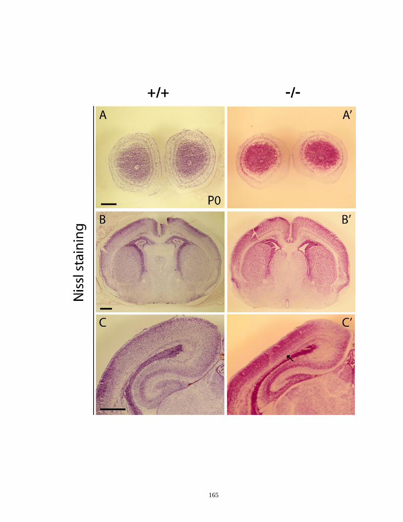
165
166
To investigate whether I12b elimination affects tangential migration of early-born
GABAegic neurons in the developing forebrain similar to the phenotypes seen in
compound Dlx mutants (Anderson et al., 1997a; Wang et al., 2010), we observed the
migration streams of GABA- and calbindin (CB)-expressing cells from the subpallial
telencephalon to the neocortex at E13.5 and E15.5, respectively. Although a detailed
quantitative analysis was not conducted, immunohistochemical staining for GABA
showed that the migration streams of GABAergic neurons in E13.5 I12b null mice are
generally comparable to that of the wild-type littermates (Figure 4.4). In parallel, we used
CB as a molecular marker to trace the tangential migration of a subpopulation of
GABAergic interneurons primarily moving from the subpallium to the developing
neocortex via the intermediate zone (Jimenez et al., 2002). Upon visual observation, we
found that the streams of CB-expressing tangential migratory cells in E15.5 I12b mutants
generally appear to be comparable to that in the wild-type littermates (Figure 4.5). To
further check whether I12b deletion influences normal proliferation of neural progenitor
cells in the developing telencephalon, we immunostained proliferating cells that have
entered the G2/M phase using a mitotic marker, anti-phospho-histone H3 (PH3) antibody.
Upon visual inspection, we noticed that the number of cycling cells in the subpallium of
E13.5 I12b-/-
telencephalon seems to be decreased compared to that in the wild-type
controls, especially along the regions of ventricular and subventricular zones (VZ and
SVZ) in the lateral/medial ganglionic eminences (LGE/MGE) (Figure 4.6A-H), a mutant
phenotype resembling that reported in Dlx1/Dlx2 double knockouts (Anderson et al.,
1997b). However, the retarded cell growth was not accompanied by an increased
expression of active caspase 3 in LGE, MGE and CGE cells (Figure 4.6I-P), suggesting
167
Figure 4.4. Comparison of tangential migration streams of GABA-expressing cells
between E13.5 I12b-/-
and wild-type telencephalon
Transverse telencephalic sections from E13.5 I12b-/-
mutant embryos (E-H) and their
corresponding wild-type littermates (A-D) were immunoreacted with an antibody against
GABA in order to evaluate the tangential migration streams of GABAergic progenitors.
The migration streams of GABAergic neurons at the rostral/medial (F, arrow) and caudal
levels (H, arrows) in E13.5 I12b null mice are generally comparable to that of the wild-
type littermates (B,D). Dorsal is upward in all the panels. The boxed regions in A,C,E,G
are shown at higher magnification in B,D,F,H. Scale bars: 300 μm (A,C,E,G) and 200
μm (B,D,F,H).
168
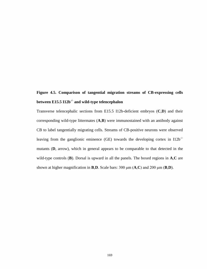
169
Figure 4.5. Comparison of tangential migration streams of CB-expressing cells
between E15.5 I12b-/-
and wild-type telencephalon
Transverse telencephalic sections from E15.5 I12b-deficient embryos (C,D) and their
corresponding wild-type littermates (A,B) were immunostained with an antibody against
CB to label tangentially migrating cells. Streams of CB-positive neurons were observed
leaving from the ganglionic eminence (GE) towards the developing cortex in I12b-/-
mutants (D, arrow), which in general appears to be comparable to that detected in the
wild-type controls (B). Dorsal is upward in all the panels. The boxed regions in A,C are
shown at higher magnification in B,D. Scale bars: 300 μm (A,C) and 200 μm (B,D).

170

171
Figure 4.6. I12b CRE deletion may potentially affect cell proliferation in the
subpallium
Immunolocalization of PH3 reveals that the number of cycling cells may be decreased in
the subpallium of E13.5 I12b-/-
telencephalon (E,F,G,H) when compared to the wild-type
littermate controls (A,B,C,D), particularly along the areas of ventricular and
subventricular zones in the LGE/MGE (E,F, arrows). However, the decreased cell
proliferation in I12b-/-
mutants is not accompanied with an upregulation of caspase 3
expression at the levels of LGE/MGE and CGE (I-P). Dorsal is upward in all the panels.
The boxed regions in A,C,E,G,I,K,M,O are shown at higher magnification in
B,D,F,H,J,L,N,P. Scale bars: 300 μm (A,C,E,G,I,K,M,O) and 200 μm
(B,D,F,H,J,L,N,P). Abbreviations: CGE, caudal ganglionic eminence; LGE, lateral
ganglionic eminence; MGE: medial ganglionic eminence.
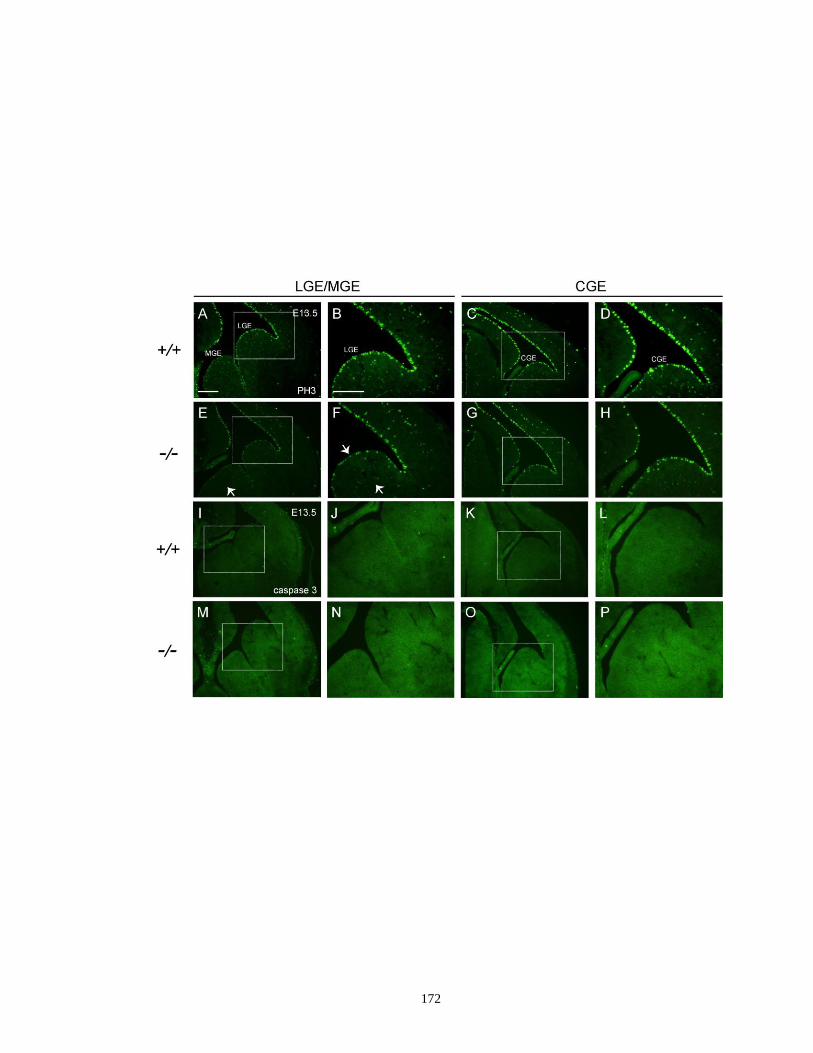
172
173
that this phenotype is not resultant from apoptotic death of subsets of subpallial
progenitor cells.
4.3.4. Removal of I12b leads to a reduction of Dlx1/Dlx2 mRNA levels in the
forebrain
In an effort to ascertain the necessity of I12b CRE for overall Dlx regulation in
vivo, we sought to examine the expression pattern and transcript levels of four Dlx genes
(Dlx1, Dlx2, Dlx5 and/or Dlx6) in E13.5 I12b null telencephalon. Our in situ
hybridization assays demonstrated that knockout of I12b element did not make
significant impact upon the expression pattern of Dlx1 and Dlx2, two genes in close
proximity to I12b sequence, in the developing forebrain (Figure 4.7A-F’). As seen with
Dlx1/Dlx2, I12b deletion did not delay the onset of Dlx5 expression and the spatial
distribution of transcripts appeared to be highly similar when homozygous I12b mutants
were compared with wild-type littermates (Figure 4.7G-I’). Normal Dlx expression
pattern identified in the I12b-/-
forebrain prompted us to carry out a real-time RT-PCR
analysis in order to directly address the potential changes in expression levels.
Interestingly, we found a ~25% reduction in relative levels of Dlx1 mRNA (0.74 ± 0.02,
P < 0.01) and a ~15% decrease in relative levels of Dlx2 mRNA (0.86 ± 0.03, P < 0.01)
in the telencephalon of I12b-/-
mutants after normalization with that in wild-type controls
(set as 1, Figure 4.8). We then determined what happens to the expression level of Dlx5
and Dlx6 mRNA since these two genes were previously shown to be down-regulated in
the ventral telencephalon of mice lacking Dlx1 and Dlx2 function (Anderson et al.,
1997b; Zerucha et al., 2000). Dlx5 and Dlx6 transcript levels appeared to be slightly

174
elevated to different degrees in E13.5 I12b null telencephalon as compared to that in the
corresponding wild-type animals (Figure 4.8). In addition to Dlx putative binding sites,
the I12b enhancer also contains a number of consensus-binding motifs for several other
transcription factors required for telencephalic development, such as Meis/Msx/Nkx
homeodomain proteins and Mash1 (Poitras et al., 2007). Hence, we looked at whether
there is any change in the quantitative levels of some of these key regulators as a
consequence of I12b deletion. Although the levels of Nkx2.1 and Gsh2 mRNA remained
intact, the normalized amount of Mash1 transcript, a direct upstream regulator of
Dlx1/Dlx2, increased by ~10% (1.09 ± 0.02, P < 0.01) in E13.5 I12b-/-
mutants relative to
wild-type controls (set as 1, Figure 4.8). These findings provide the first evidence
suggesting that I12b CRE deletion possibly does not lead to changes of Dlx expression
pattern; however, it is sufficient to alter the basal levels of some genes (such as
Dlx1/Dlx2 and Mash1) involved in the Dlx-mediated regulatory pathway.
4.4. Discussion
4.4.1. Functional redundancy between Dlx forebrain CREs
The sequences of I12b enhancer and of their orthologs in distantly related
vertebrate species are highly conserved with a sequence similarity above ~80% (Ghanem
et al., 2003). In transgenic mice, I12b specifically targets lacZ reporter gene expression to
the ventral telencephalon and diencephalon in a pattern that faithfully resembles
endogenous Dlx forebrain expression (Ghanem et al., 2003; Ghanem et al., 2007),
indicating that it may be a key regulatory element responsible for Dlx regulation. It has
been proposed that highly conserved or ultraconserved regulatory elements, which show
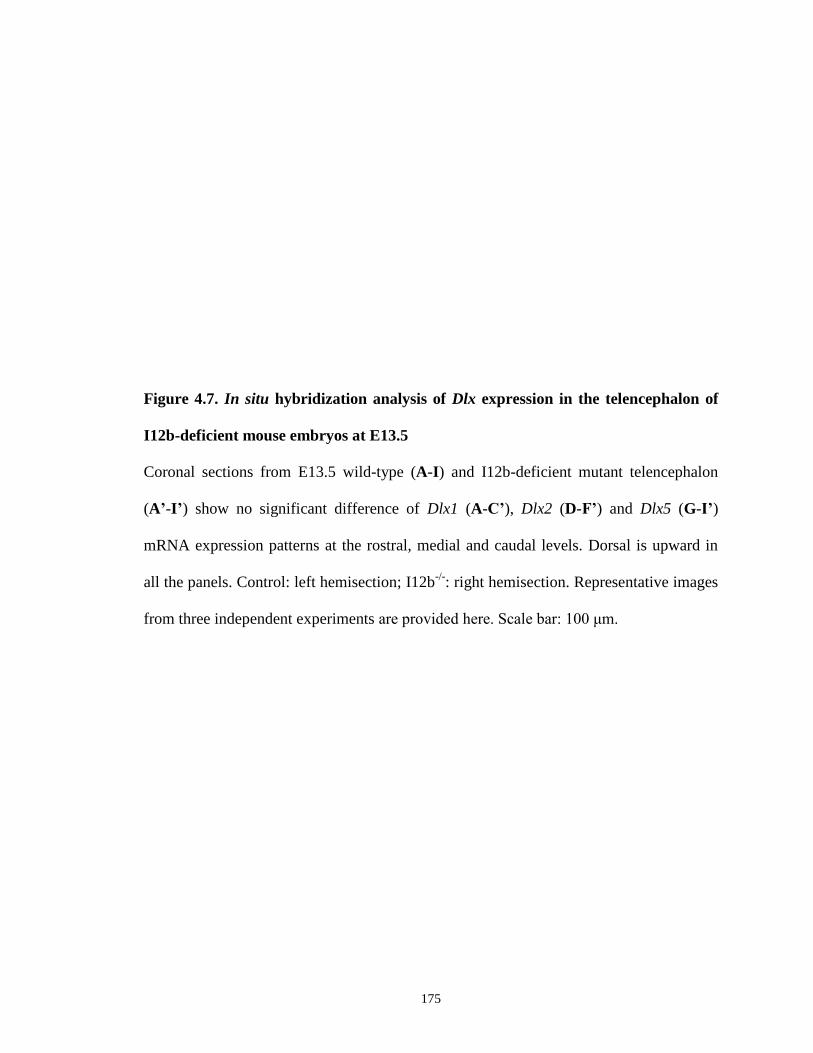
175
Figure 4.7. In situ hybridization analysis of Dlx expression in the telencephalon of
I12b-deficient mouse embryos at E13.5
Coronal sections from E13.5 wild-type (A-I) and I12b-deficient mutant telencephalon
(A’-I’) show no significant difference of Dlx1 (A-C’), Dlx2 (D-F’) and Dlx5 (G-I’)
mRNA expression patterns at the rostral, medial and caudal levels. Dorsal is upward in
all the panels. Control: left hemisection; I12b-/-
: right hemisection. Representative images
from three independent experiments are provided here. Scale bar: 100 μm.
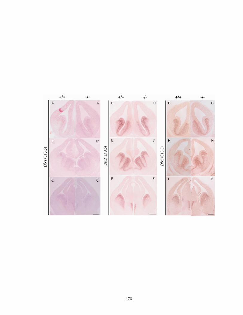
176
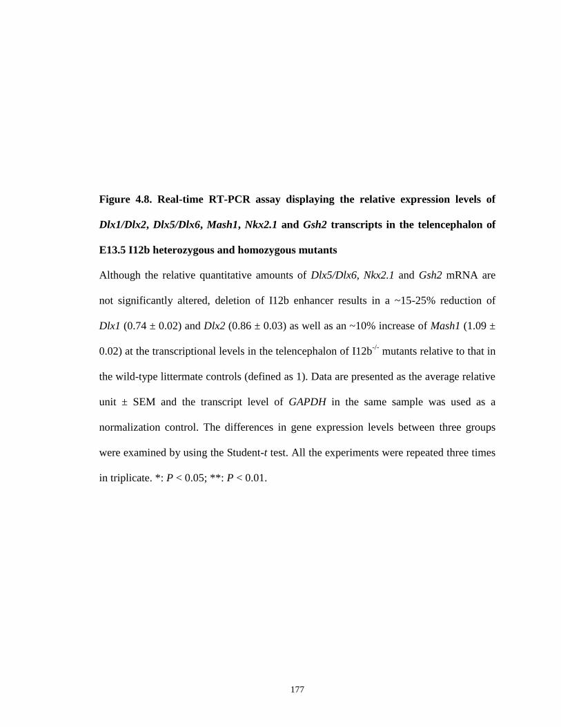
177
Figure 4.8. Real-time RT-PCR assay displaying the relative expression levels of
Dlx1/Dlx2, Dlx5/Dlx6, Mash1, Nkx2.1 and Gsh2 transcripts in the telencephalon of
E13.5 I12b heterozygous and homozygous mutants
Although the relative quantitative amounts of Dlx5/Dlx6, Nkx2.1 and Gsh2 mRNA are
not significantly altered, deletion of I12b enhancer results in a ~15-25% reduction of
Dlx1 (0.74 ± 0.02) and Dlx2 (0.86 ± 0.03) as well as an ~10% increase of Mash1 (1.09 ±
0.02) at the transcriptional levels in the telencephalon of I12b-/-
mutants relative to that in
the wild-type littermate controls (defined as 1). Data are presented as the average relative
unit ± SEM and the transcript level of GAPDH in the same sample was used as a
normalization control. The differences in gene expression levels between three groups
were examined by using the Student-t test. All the experiments were repeated three times
in triplicate. *: P < 0.05; **: P < 0.01.
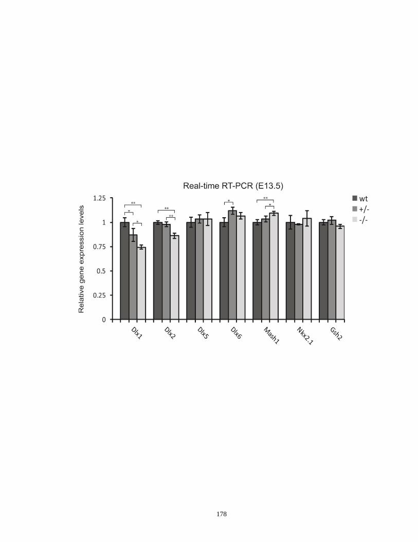
178

179
little or even no sequence variations across different species, are preserved during the
evolutionary process due to their essential biological activities (Meireles-Filho and Stark,
2009; Paixao and Azevedo, 2010). If the function of these regulatory elements is truly
important as hypothesized, deleting these elements from their endogenous genomic loci
would cause significant phenotypic effects on the host organisms, such as severe
morphological defects leading to embryonic lethality. For example, deleting an
evolutionarily conserved enhancer element which regulates Pax3 expression in the neural
crest expression leads to exencephaly and spina bifida during early embryogenesis
(Degenhardt et al., 2010). Similarly, targeted deletion of an enhancer responsible for
dHAND branchial arch expression results in many severe defects in the craniofacial
structures and mutant mice die soon after birth (Yanagisawa et al., 2003). In contrast, in
our case, I12b+/-
and I12b-/-
mutant mice all survive and reproduce as anticipated. The
viability and litter size of their offspring exhibit no significant differences when
compared to that of their wild-type littermates. Despite that the thickness of the neocortex
in I12b null mice seems to be a bit reduced compared to wild-type controls, no apparent
morphological and histological defects were found in the I12b-/-
forebrain. These
observations indicate that while the I12b sequence is well conserved during evolution and
its enhancer activity is detectable in our transgenic assays, targeted removal of this
enhancer in vivo does not necessarily result in pronounced effects on survival, fertility
and brain morphology, which is consistent with other enhancer deletion studies (Chen et
al., 2002; Chen and Goldhamer, 2004; Ahituv et al., 2007). One possible reason for this
phenomenon could be a functional redundancy among various Dlx CREs. Redundant
activities among different regulatory elements have been suggested as one of the
180
protective mechanisms in host organisms when dealing with random mutations occurring
within the critical sequences that otherwise might trigger detrimental phenotypic
consequences (Piano et al., 1999; Frankel et al., 2010; Paixao and Azevedo, 2010). As
such, when I12b is absent, the remaining Dlx forebrain enhancers, such as URE2
upstream of Dlx1, may step in and exert their activities to maintain proper Dlx gene
regulation and avoid the pathologies in I12b mutants. This point may be particularly true
for I56i CRE within the Dlx5/Dlx6 intergenic sequence. Our previous transgenic assays
have demonstrated that I56i and I12b are active in almost identical populations of
GABAergic neurons in vivo (Ghanem et al., 2007). In addition, I56i was found to play a
role in the cross-regulation between Dlx1/Dlx2 and Dlx5/Dlx6 genes (Zerucha et al.,
2000). It should be emphasized here that the various individual Dlx CREs contain
different putative binding motifs for distinct trans-acting factors, which may contribute to
at least some of their differential functional activities.
4.4.2. Enhancer sharing between the Dlx genes
One typical feature of the Dlx gene family is the highly overlapping expression
patterns between different members and this property of Dlx genes appears well
conserved across various vertebrate species (Panganiban and Rubenstein, 2002;
Sumiyama et al., 2003). Thus, the same cells may show expression of several Dlx genes
at the same time. To date, it remains to be determined whether the two Dlx genes on the
same cluster are simultaneously under the control of each individual enhancer (enhancer
sharing) or if one of them is specifically linked to one Dlx gene, possibly through an
insulator mechanism which blocks its interaction with the other Dlx gene. The question

181
could become more important when taking into consideration that enhancer sharing
between the two Dlx genes from the same cluster may be at least partially responsible for
their overlapping expression pattern (Krumlauf, 1994; Gould et al., 1997). It could be
also one contributing factor maintaining the conserved organization of Dlx bigene
clusters during the evolutionary process (Krumlauf, 1994; Gould et al., 1997). The
relative levels of Dlx1 and Dlx2 transcripts are significantly reduced by ~3-13% and ~15-
25% in I12b heterozygous and homozygous mutants respectively, providing some
evidence that these two neighboring genes may be regulated in the same I12b-dependent
manner in vivo. The sharing of regulatory elements between clustered genes has been
previously documented in chicken β–globin genes and in the Hox gene complex (Choi
and Engel, 1988; Gerard et al., 1996; Zakany et al., 1997). Specifically, Zakany et al.
(1997) identified an evolutionarily conserved enhancer named RVIII within the mouse
Hoxd10/Hoxd11 loci and this element can be shared by the two genes for regulating and
maintaining their proper expression in the trunk. In the future, it will be interesting to
further explore whether other Dlx CREs also interact with both Dlx genes from the same
cluster in their surrounding genome by employing our other enhancer deletion mice (e.g.
I56i-/-
and I56ii-/-
mutants).
4.4.3. Contribution of I12b to the regulation of the genetic pathway involving
Mash1 and Dlx1/Dlx2
To date, how different lineages of GABAergic neurons are generated and
differentiate in the developing subpallium still remains largely obscure. During early
embryogenesis, Dlx1 and Dlx2 gene expression begins in the VZ and SVZ of the

182
ganglionic eminences at ~E9.5 and these two genes are thought to play a role in
regulating the proliferation and differentiation of early progenitor cells in these regions
(Bulfone et al., 1993a; Anderson et al., 1997b). Indeed, Dlx1/Dlx2 double knockouts
exhibit significantly decreased cell proliferation and deficient neurogenesis in the
progenitor domains of the LGE and MGE (Anderson et al., 1997b). Consistent with these
findings, we noticed that cell growth in the E13.5 I12b-/-
telencephalon may be affected in
the VZ/SVZ regions of the LGE and MGE, likely due to reduced Dlx1 and Dlx2
transcript levels in these areas or to yet to be determined alterations in the expression of
some key regulatory genes involved in the Dlx1/Dlx2-mediated genetic pathway. In the
developing mouse forebrain and, very likely, in other tissues, expression of Dlx genes is
thought to be modulated by both auto- and cross-regulation through their dynamic
interactions with the CREs at their own genomic loci or those from other Dlx bigene
clusters (Zerucha et al., 2000; Zhou et al., 2004; Bond et al., 2009; Potter et al., 2009).
Mutations of the putative Dlx binding sites in the Dlx CREs (e.g. I12b and I56i) result in
markedly reduced reporter gene expression in the developing forebrain of transgenic
animals and also impact on the binding of Dlx proteins to these enhancers (Zerucha et al.,
2000; Poitras et al., 2007). Of interest, expression patterns Dlx5 and Dlx6 remain largely
unchanged and their mRNA levels are only slightly affected in the embryonic forebrain
of I12b-/-
mutants, suggesting that Dlx5 and Dlx6 expression may be independent on the
altered levels of Dlx1 and Dlx2 due to I12b deletion. Moreover, the transcript levels of
Mash1, a direct upstream modulator of Dlx1/Dlx2, are slightly elevated following I12b
enhancer deletion probably as a result of a compensation effect, although the molecular
mechanisms underlying this event needs to be further investigated. These data support a
183
notion that I12b enhancer might be functionally involved in regulating and maintaining
the genetic network in which Mash1 and Dlx1/Dlx2 genes are involved in the developing
mouse forebrain.
4.4.4. Possible implication of I12b in cell-fate determination in the developing
forebrain
Molecular mechanisms of cell-fate determination have been extensively studied in
the developing mammalian forebrain, where Dlx genes as well as a number of bHLH
transcription factors including Mash1, Ngn2, and Olig1/Olig2 have been shown to exert
important regulatory functions (Kondo and Raff, 2000; Bertrand et al, 2002). In the
progenitor domains of the developing subpallium and in the SVZ of the postnatal mouse
brain, Mash1 plays key roles in promoting the commitment of multipotent neural
progenitor cells to a neuronal fate through inhibition of astrocyte proliferation and
differentiation (Parras et al., 2004; Parras et al., 2007). Previous studies also showed that
Mash1 is responsible for regulating the production of oligodendrocyte precursor cells
(OPCs) (Parras et al., 2004; Parras et al., 2007). Both Mash1 single and Mash1;Ngn2
double knockouts have been found to have significantly decreased number of precursors
that are committed to an astrocytic fate in the developing subpallium when compared to
that of the wild-type littermates (Casarosa et al, 1999; Horton et al, 1999; Nieto et al,
2001; Yun et al., 2002). Reduced numbers of OPCs were also observed in Mash1 mutant
embryos as well as in Mash1 mutant cells cocultured with wild-type cells, further
supporting a role of Mash1 in oligodendrogliogenesis (Bertrand and Guillemot,
unpublished data). Interestingly, Dlx1/Dlx2, the downstream targets of Mash1, were
184
found to be expressed in the subpallial-originated OPCs, indicating that the genetic
pathway involving Mash1 and Dlx1/Dlx2 may be also implicated in regulating the
production and/or differentiation of oligodendrocytes, in addition to GABAergic neurons
(He et al., 2001; Marshall et al., 2003). Petryniak et al. (2007) recently reported that
Dlx1/Dlx2 can negatively regulate the expression of Olig2, a key factor regulating OPC
specification and maturation (Lu et al., 2002), in progenitor cells in order to promote the
generation of GABAergic neurons, while Mash1 can inhibit the process of neurogenesis
and induce the production of oligodendrocytes by downregulating Dlx1/Dlx2 expression
(Supplementary Figure 4.1). Because the expression levels of Dlx1/Dlx2 and Mash1 are
significantly altered in the I12b-/-
forebrain, we are presently studying the expression of
Olig1 and Olig2 in I12b mutants in order to test if this enhancer is involved in the
regulatory network modulating neuronal and oligodendrocytic cell fate specification.
4.4.5. Potential importance of I12b to GABAergic interneuron differentiation
Dlx genes are essential players in the molecular and genetic pathways responsible
for the production of neuronal diversity, particularly for GABAergic interneurons, during
mouse forebrain development (Panganiban and Rubenstein, 2002). As noted, disruption
of both Dlx1 and Dlx2 function leads to major defects in the generation and
differentiation of GABAergic interneuron progenitors born in the embryonic ganglionic
eminences (Anderson et al., 1997a; Bulfone et al., 1998; Pleasure et al., 2000). These
cells accumulate in the basal ganglia and fail to migrate to the neocortex, olfactory bulb
and hippocampus through different migratory routes (Anderson et al., 1997a; Bulfone et
al., 1998; Pleasure et al., 2000). However, our initial phenotype analysis showed the

185
Supplementary Figure 4.1. A proposed model showing the Dlx1&2/Mash1/Olig2-
mediated pathway that control the switch between neurogenesis and
oligodendrogliogenesis in the developing telencephalon
Dlx1 and Dlx2 repress the expression of Olig2 in progenitor cells in order to promote the
generation of GABAergic neurons. Mash1 can inhibit the process of neurogenesis and
induce the production of oligodendrocytes via downregulating Dlx1/Dlx2 expression.
Adapted from Petryniak et al., 2007.
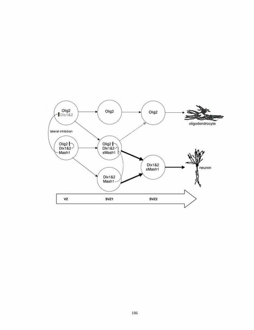
186
187
tangential migration streams of GABAergic neurons in the forebrain of I12b-/-
mutant
embryos are generally comparable to those seen in wild-type mice. The underlying
reason(s) for this observation is still unknown at this moment but might be linked to
functional redundancy among Dlx proteins or different Dlx CREs as discussed earlier.
Alternatively, there may be other undiscovered mechanisms, such as those underlying
cell movement or extracellular environment changes, which can compensate for the
potential effects induced by I12b enhancer deletion (Rakic and Komuro, 1995). A
detailed quantitative analysis is in progress to confirm this observation. Although we
have not observed obvious impacts of I12b deletion on GABAergic interneuron
development at early embryonic stages, its potential effects may become more evident at
later time points throughout development or in adult animals. Adult Dlx1-/-
mutants
maintained in a C57BL/6J×CD1 background display a specific loss of several
GABAergic interneurons subtypes (CR-, SOM- and NPY-expressing cells) in the cortex
and hippocampus in a time-dependent manner (Cobos et al., 2005a). These mutant mice
also exhibit an epileptic phenotype and have a reduced response to fear conditioning
(Cobos et al., 2005a; Mao et al., 2009). In addition, Dlx5/Dlx6-/-
mutants have a
selectively reduced number of PV-expressing cortical interneurons and adult Dlx5/Dlx6+/-
mice exhibit spontaneous electrographic seizures probably due to improper function of
PV-positive neurons (Wang et al., 2010). These findings suggest that various individual
Dlx genes may play specific roles in regulating the development and/or maturation of
distinct subtypes of GABAergic interneurons starting from the early embryonic stages
until adulthood. Hence, it is reasonable to infer that while certain alterations in Dlx
regulation/function as a result of aberrant regulatory activities of their CREs may only
188
have subtle effects at early embryonic stages, they may induce a severe later-onset
phenotype in adult animals. To test this possibility, we are presently exploring whether
the somatosensory cortex and hippocampus of 1-month-old I12b-/-
mice have loss of
some molecular markers for GABAergic neurons (Gad1/Gad2, CB, CR, PV, SOM, NPY,
reelin). Given that I12b deletion leads to ~20% decrease in Dlx1/Dlx2 expression and
adult Dlx1-/-
mutants have marked loss of CR-, SOM- and NPY-expressing interneurons
(Cobos et al., 2005a), we expect that I12b enhancer deletion will make an impact at least
on the development of these three interneuron subtypes. In the long term, it will be of
important significance to study whether I12b-/-
mutants harbor some behavioral
abnormalities associated with human neurological disorders by employing the tests
previously described in (Cobos et al., 2005a; Mao et al., 2009).
In summary, our results demonstrate that mice homozygous for I12b enhancer
deletion are viable, develop normally, and display no apparent morphological
abnormalities in the forebrain. Targeted deletion of I12b leads to a significant reduction
of Dlx1 and Dlx2 expression levels and may potentially interfere with normal cell
proliferation in the developing subpallium. These data provide the first evidence
regarding the specific contribution of an individual CRE to overall Dlx expression in vivo
and indicates a complex transcriptional regulation of Dlx genes during forebrain
development. Further studies on the underlying molecular mechanisms regulating Dlx
expression by probing the function of their various CREs will ultimately contribute to our
understanding of the genetic pathways involved in controlling GABAergic neuron
development. This novel mouse model may also set a stage for future investigations of

189
potential functional redundancy or their dynamic interactions among different individual
Dlx CREs when mediating Dlx forebrain expression.
Acknowledgments
We would like to thank Gary Hatch and Sylvie Emond for their excellent technical
assistance. We are grateful to Dr. John Rubenstein for providing us with the plasmids for
Dlx1/Dlx2/Dlx5 cRNA probes, Dr. Andras Nagy for providing us with the R1 ES cell
line, and Janice Penney at Transgenic Mouse Core Facility, McGill Cancer Research
Center for supporting the production of enhancer deletion mice. This work was supported
by grants-in-aid from the Canadian Institutes of Health Research (MOP14460). M.Y. was
supported by a University of Ottawa Excellence Scholarship and Ontario Graduate
Scholarship (OGS). M.E. holds a Research Chair in Evolutionary Developmental Biology
at the University of Ottawa.
190
5. Conclusions
The Dlx genes encode a group of homeobox-containing transcription factors, which
are responsible for proper development of various embryonic structures in many
tissues/organs during the period of mid to late gestation. In the developing mouse
forebrain, Dlx genes are expressed in a highly overlapping but distinct pattern and are
critically involved amongst other master regulatory genes in a genetic cascade necessary
for the normal differentiation, migration and survival of GABAergic neurons. These
neurons produce inhibitory signals in order to maintain the balance of neuronal activities
in many important regions of the forebrain, such as the cerebral cortex and hippocampus.
Spatio-temporal expression of Dlx genes, at least in the developing forebrain, is
modulated by a number of highly conserved regulatory elements that have been identified
within the Dlx loci. In my PhD thesis, I have investigated the potential implications of
some of these regulatory elements in the developing mouse and zebrafish forebrain.
Specifically, I have characterized in detail the activity of the I56ii enhancer in the
developing telencephalon through a comparative analysis of the activities of the other
three forebrain-specific Dlx CREs (I12b, URE2 and I56i) using transgenic mice and have
thus contributed to the understanding of the roles played by I56ii in the overall Dlx
regulation during mouse telencephalic development. I have also provided evidence that
zebrafish I56i and I56ii CREs may have conserved activity in populations of GABAergic
neurons within the zebrafish telencephalon and diencephalon which mimics the situation
we have seen in mice and knockdown of dlx genes significantly interferes with enhancer
activity, strongly indicating a possible involvement of dlx genes in controlling zebrafish
GABAergic neuron production potentially through an evolutionarily conserved
191
regulatory mechanism, at least in certain domains of the developing forebrain. Finally, I
have successfully established a mouse model harboring a targeted deletion of I12b CRE
and have initiated the preliminary phenotypic analyses. This study will provide more
insight into the specific contribution of individual Dlx CREs to overall Dlx regulation
during mouse forebrain development.
Although some aspects of Dlx regulation and function in the developing forebrain
have been addressed in my PhD projects (see Figure 5.1 for a model of Dlx-mediated
genetic network), there are many interesting issues that deserve further investigation. In
the following text, I will discuss some of the pertinent questions that still remain
unanswered and may be the goals of future studies.
5.1. Investigation of specific contribution of I56ii CRE to overall Dlx regulation
Although the expression patterns of reporter transgenes under the control of
various CREs in the developing mouse forebrain faithfully recapitulate that of
endogenous Dlx genes, our previous findings indicate that these distinct CREs are most
likely involved in regulating Dlx expression in overlapping but different subpopulations
of subpallium-derived GABAergic interneuron progenitor cells as well as in different
subsets of cortical GABAergic interneurons in adult animals (Ghanem et al., 2007). This
possibility is greatly strengthened by the fact that the activity of I56ii CRE is restricted to
a subpopulation of Dlx-expressing GABAergic projection neurons lining the superficial
mantle of the ganglionic eminences between E10.5 and E13.5 and does not overlap with
the other forebrain enhancers (Ghanem et al., 2008). Despite these interesting findings,
the specific effects of each individual CRE on overall regulation of Dlx genes during
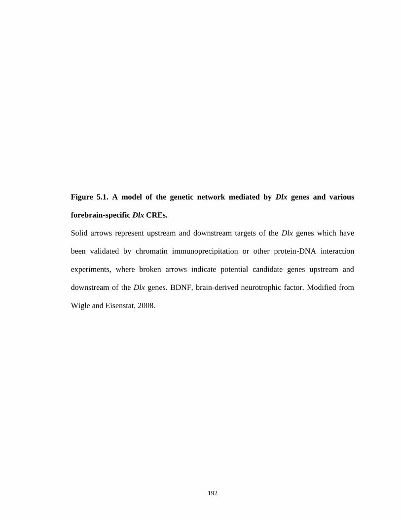
192
Figure 5.1. A model of the genetic network mediated by Dlx genes and various
forebrain-specific Dlx CREs.
Solid arrows represent upstream and downstream targets of the Dlx genes which have
been validated by chromatin immunoprecipitation or other protein-DNA interaction
experiments, where broken arrows indicate potential candidate genes upstream and
downstream of the Dlx genes. BDNF, brain-derived neurotrophic factor. Modified from
Wigle and Eisenstat, 2008.

193

194
forebrain development as well as their specific involvement in generating GABAergic
interneuron diversity remains largely unknown. This issue can be directly demonstrated
by targeted deletion of one enhancer or simultaneous knockout of many enhancers in
mice. Using the same technique as I describe in Chapter IV for generating I12b-/-
mutant
mice, our laboratory has recently generated a mutant mouse model carrying a targeted
deletion of I56ii CRE. This mutant mouse model differs from Dlx5/Dlx6 single or double
knockout mice (Beverdam et al., 2002; Depew et al., 2002; Wang et al., 2010), since in
this case, Dlx5 and/or Dlx6 function is only inactivated in a subset of cells in the ventral
telencephalon and thus it will result in a phenotype specifically linked to improper
function of these cells. The expression patterns and quantitative levels of Dlx1/Dlx2 and
Dlx5/Dlx6 in the I56ii-/-
mutants are currently being examined by using both in situ
hybridization and real-time RT-PCR assays since it has been shown that Dlx family
members can cross-regulate each other (Zerucha et al., 2000). Hence, altered expression
patterns and/or levels of specific Dlx genes as a result of enhancer deletion may influence
the expression of other family members. Given that I56ii CRE is specifically active in a
group of GABAergic projection neurons, the expression of several striatal projection
neuron markers (Islet1, Meis2, Ebf1 and DARPP-32) will also be examined in I56ii
heterozygous and homozygous mutants.
As discussed in Chapter II, the position of I56ii-positive cells in the ventral
telencephalon corresponds to that of a group of cells, termed “corridor cells”, which
contribute to the establishement of a permissive route necessary for the guidance and
pathfinding of thalamocortical axons (Lopez-Bendito et al., 2006). Perhaps, I56ii is
involved in a genetic cascade important for maintaining proper Dlx5/Dlx6 expression that
195
play key roles in establishing the “corridor” cells during forebrain formation. To answer
this question, I56ii-/-
embryos will be examined for formation of thalamocortical
projections by using a series of molecular markers, such as CR. Moreover, because
thalamocortical projection system is one of the most essential processing connections in
the mammalian brain that conveys multiple important sensory and motor signals to the
cortex (Dickson 2002; Dufour et al., 2003), we also expect to observe some behavioral
changes in I56ii-/-
adult mice.
5.2. Identification of upstream activators regulating Dlx CRE activities
Interestingly, the sequences of various Dlx forebrain-specific CREs only share
limited similarity, despite the fact that three out of the four CREs (I12b, I56i and URE2)
display high overlapping activities when targeting reporter transgene to populations of
progenitors in the basal ganglia as well as when marking adult cortical GABAergic
interneurons (Ghanem et al., 2003; Ghanem et al., 2007). The length of these three Dlx
CREs, the degree of conservation among different vertebrate species for each individual
CRE, and their sequence divergence suggest that Dlx expression in intermingled
populations of GABAergic neurons or in their progenitors may be obtained and
maintained through distinct regulatory mechanisms, such as the binding of distinct
combinations of trans-acting factors to the various CREs. However, our current
knowledge regarding direct upstream modulators or signaling molecules influencing Dlx
expression is very poor. Using a combination of DNase I footprinting, transfection
assays, electrophoretic mobility shift assay (EMSA) and ChIP, Poitras et al. (2007)
provided evidence that the proneural transcription factor Mash1 directly interacts with

196
I12b and regulates Dlx expression, further supporting a notion that there exists a
Mash1/Dlx/Gad genetic cascade regulating GABAergic neuron development. In addition
to the putative Dlx binding sites (A/C/G)TAATT(G/A)(C/G), comparisons between
sequences of Dlx CREs and transcription factor binding site databases have revealed a list
of other potential binding motifs within I56i (6-8 sites identified thus far) and URE2 (10
sites) CREs. Ongoing work in the laboratory are aimed at further determining whether
some of the candidate transcription factors can physically bind to these CREs and
mediate Dlx expression, using the molecular approaches previously adopted in (Poitras et
al., 2007). As noted in Chapter II, Meis2 and Islet1 may be potential upstream regulators
of Dlx genes in corridor cells by acting upon I56ii enhancer during early embryogenesis.
One weakness of this study is that physical interactions between I56ii and these
transcription factors have not yet been directly proved. It would be important in further
experiments to study the affinity of Meis2 and Islet1 putative binding sites in I56ii using
competition EMSA and whether Meis2 and Islet1 can directly bind to I56ii in vivo by
employing ChIP. It would also be interesting to investigate possible impact of loss or
disruption of Meis2 and Islet1 binding sites on the I56ii enhancer activity by establishing
transgenic mice with reporter constructs containing mutations in the putative Meis2 and
Islet1 binding sites. Such experiments will definitely help us better understand the key
upstream factors in the regulatory pathways involving Dlx genes and shed more light on
their specific interaction with various individual Dlx CREs.
5.3. Conserved activity of Dlx CREs in the forebrain of other vertebrate species
197
Dlx forebrain expression patterns between mouse and zebrafish appear to be well
conserved, suggesting that the potential mechanisms underlying Dlx regulation might be
also conserved over evolutionary time (Akimenko et al., 1994; Ellies et al., 1997;
Panganiban and Rubenstein, 2002; MacDonald et al., 2010a). Indeed, orthologous Dlx
CREs exhibit a high degree of sequence similarity among distantly related vertebrate
species (Ghanem et al., 2003). In Chapter III, I demonstrated that in addition to the
sequence conservation, zebrafish dlx5a/dlx6a regulatory elements display a well
conserved activity by targeting GFP transgene expression to GABAergic neurons in the
developing telencephalon and diencephalon, highly mimicking the regulatory activities of
their mouse orthologs when tested in transgenic mice (Ghanem et al., 2007; Ghanem et
al., 2008). In addition, zebrafish dlx5a/dlx6a regulatory elements may participate in
regulating the differentiation of at least some subsets of GABAergic interneurons in
certain specific regions in the developing forebrain. These data are interesting in terms of
evolutionary biology since the teleost lineage has been proposed to undergo divergence
from mammals about 430 million years ago (Powers, 1991). Considering the entire or
partial genome of increasing number of vertebrates have been sequenced so far, one
resultant question would be whether some of these CREs, if not all, are also present
within the Dlx locus in these vertebrate species. If this is the case, it will be also
important to investigate whether the activity of the CREs is functionally conserved as
compared to their respective mouse and zebrafish orthologs. Our laboratory has identified
a URE2-like regulatory element in the genome of the elephant shark (Callorhinchus
milii), which is a cartilaginous fish and has a key phylogenetic position essential for
studying the evolution of jawed vertebrates (Venkatesh et al. 2006; Venkatesh et al.
198
2007). The sequence of elephant shark URE2 shares remarkably high degree of identity
with that of its mouse (~85% identity) and zebrafish orthologs (~73% identity)
(MacDonald et al., 2010b). When tested in transgenic animals, the elephant shark URE2
targets reporter transgene to the developing forebrain in an expression pattern that is
extensively overlapping with that of mouse and zebrafish URE2 CREs, even though the
activities of the three orthologous enhancers slightly vary in the diencephalon
(MacDonald et al., 2010b). Additionally, large-scale comparative studies done by
Venkatesh and colleagues (Venkatesh et al. 2006; Venkatesh et al. 2007) has shown that
the number of conserved non-coding regions shared between the human and elephant
shark genomes is significantly higher than that between the human and zebrafish
genomes, suggesting that the elephant shark may be a novel and appropriate model
organism for studying the gene regulatory mechanism during vertebrate evolution. Along
this line, it will be interesting to extend our current findings to other Dlx CREs (I12b, I56i
and I56ii) to test if they are also present in the elephant shark and then compare their
activity with that of their corresponding mouse and zebrafish orthologs. From an
evolutionary perspective, understanding the function of Dlx CREs in a more diverse set
of vertebrate species will hopefully shed more light on how the regulatory mechanisms
responsible for Dlx forebrain expression are established and maintained as well as how
distinct function of orthologous Dlx genes in forebrain development is acquired over
evolutionary time since the divergence of modern vertebrates.
5.4. Investigation of molecular mechanisms underlying enhancer sharing
199
The bigene organization of Dlx genes has found to be a common feature in many
vertebrate species (Zerucha and Ekker, 2000). In addition to vertebrates, this typical
arrangement of the Dlx gene family has also been documented in the tunicate Ciona
intestinalis as well as in the lampreys (Di Gregorio et al., 1995; Kuraku et al., 2009). Yet,
the molecular basis underlying this conserved genomic arrangement through evolution
has not been precisely explored so far. Our identification of multiple CREs in the
intergenic sequences between Dlx bigene pairs may provide a plausible explanation for
this phenomenon, as these regulatory sequences are spatially located in close proximity to
both genes constituting a bigene pair and are thus probably shared between the two
genes. The highly overlapping expression patterns of the two Dlx genes from the same
bigene cluster lend supports to this hypothesis (Ellies et al., 1997a; Liu et al., 1997).
Indeed, sharing an intergenic enhancer as well as competition between the promoter
regions of adjacent genes for an intergenic regulatory element has been described for the
regulation of the Hoxb genes. It was shown that Hoxb3 and Hoxb4 are able to equally
share an intergenic r6/7 neural enhancer and Hoxb4 and Hoxb5 can share an intergenic
mesodermal enhancer (Gould et al., 1997; Sharpe et al., 1998). On the contrary, another
enhancer within Hoxb4 and Hoxb5 intergenic region that targets reporter gene expression
to the anterior part of neural tube was found to be capable to activate both genes, but its
preferential activity depends on the competition between these genes and also can vary
with the different local genomic context (Sharpe et al., 1998). In Chapter IV, I showed
evidence that the levels of Dlx1 and Dlx2 transcripts are significantly reduced by ~15-
25% in I12b-/-
mutants, suggesting that I12b enhancer may be shared by these two genes
in vivo. To further investigate whether each intergenic enhancer or possibly other
200
enhancers at the locus (e.g. URE2) can activate and regulate expression of one or both
genes from a Dlx bigene pair, a large transgene construct containing about 45kb of the
mouse Dlx1/Dlx2 loci (encompassing all the CREs) has been generated in the laboratory.
Two fluorescent reporter genes, GFP and mCherry, have been fused independently in
frame with the first exon of Dlx1 and Dlx2, respectively. Currently, this construct is being
injected for the generation of transgenic mice. Once stable transgenic lines are
established, the corresponding regulatory element (I12b, URE2, or both) can be deleted
and qualitative and quantitative impacts of each of these elements on the expression of
Dlx1/GFP and Dlx2/mCherry will be systematically analyzed. Data obtained from these
experiments may also supplement, at least in some aspects, our ongoing enhancer
deletion projects. Moreover, a chromosome conformation capture technique has been
shown to successfully characterize the physical interactions among different genomic
sequences, including those between transcriptional regulatory elements and the sequences
of their target genes (Splinter et al., 2004; Simonis et al., 2007). Thus, this methodology
may be applied in the future to better understand the specific interplay between Dlx
promoter regions and the various individual CREs.
5.5. Functional redundancy between different forebrain CREs and existence of
additional CREs in the Dlx locus
As shown in Chapter IV, we have not identified any severe phenotypes in the I12b-
/- mice, except that I12b deletion results in a decrease in Dlx1/Dlx2 transcript levels,
together with an elevated Mash1 expression and may possibly reduce the proliferation of
progenitor cells in the VZ/SVZ of the LGE and MGE in the mutants. These observations
201
are at least partially consistent with some forebrain phenotypes seen in Dlx1/Dlx2 double
knockouts, such as a significantly decreased cell proliferation and deficient neurogenesis
in the progenitor domains of the LGE and MGE (Anderson et al., 1997b). Given that
adult Dlx1-/-
knockouts show a subtype-dependent loss of both cortical and hippocampus
GABAergic interneurons and also harbor some behavioral alterations (e.g. epilepsy and
reduced fear response) due to the loss of interneurons (Cobos et al., 2005a; Mao et al.,
2009), we are presently testing these possibilities in our adult I12b-/-
mutant mice by
using immunohistochemistry and designing behavioral assays. We expect to see a subset
of the phenotypes seen in the adult Dlx1-/-
mutants. As mentioned in Chapter IV, sharing
of CREs between the two Dlx genes on the same cluster may contribute to their
overlapping expression patterns in the developing forebrain. In addition, the forebrain
expression patterns of the reporter transgene under the control of various individual
various Dlx CREs also largely overlap (Ghanem et al., 2007, 2008). Thus, functional
redundancy among various individual Dlx forebrain CREs may be a possible reason for
the lack of apparent forebrain phenotypes in I12b-/-
mutants (at least at this moment).
Since URE2 activity in the forebrain overlaps in part with that of I12b (Ghanem et al.,
2007), it is highly possible that the remaining URE2 CRE may compensate for the loss of
I12b function in the forebrain as a protective mechanism, at least in certain specific
populations of cells. This scenario may be particularly true for the I56i CRE, as we have
previously demonstrated that I12b and I56i CREs are active in almost identical subsets of
GABAergic interneuron progenitors (adult interneurons as well) and could be possibly
activated by the same or similar regulatory signals (Ghanem et al., 2007). In support of
this idea, recent evidence has suggested that the various enhancers within the zebrafish
202
hoxb4a locus have overlapping activities to ensure the correct hoxb4a dosage and
expression pattern in the hindbrain (Punnamoottil et al., 2010). Similarly, Rastegar et al.
(2008) reported that the combined activities of two regulatory elements are required to
maintain the proper notochord expression of sox9a. Therefore, generation of mice with
simultaneous deletion of two Dlx CREs would be a good option to better address this
question. In addition to the four forebrain-specific CREs identified thus far within the
Dlx1/Dlx2 and Dlx5/Dlx6 loci, there is evidence that additional conserved regulatory
elements controlling Dlx forebrain expression may exist and have not yet been fully
characterized. Two such elements have been found in the 5’ flanking region of mouse
Dlx6 gene (Jarinova and Ekker, unpublished observations) and in the 3’ flanking
sequence of zebrafish dlx2b gene (MacDonald et al., 2010c). Moreover, regulatory
elements that have relatively shorter length (<100 bp) and contain fewer conserved motifs
may have been missed in the original phylogenetic analysis (Ghanem et al., 2003).
Recent development of more sophisticated and powerful bioinformatics tools may
provide us a good opportunity to refine the original search with the purpose of identifying
more potential regulatory elements in the Dlx loci.
5.6. Potential association of Dlx CRE alterations with human diseases
In the future, it will be of pivotal importance to extend our findings in mouse and
zebrafish to the studies of the function of Dlx genes during human GABAergic neuron
production. In fact, it has been previously documented that up to ~50% of GABAergic
interneurons in the cerebral cortex of human brain originate from the supallium-derived
progenitor cells (Letinic et al., 2002), which is very similar to the case in mice.
203
Considering that genetic aberrations and abnormal expression of DLX genes have been
increasingly detected in many human neurological disorders (e.g. epilepsy, schizophrenia
and autism) associated with the lack/reduction of inhibitory control over signaling
activities (Liu et al., 2009; Nakashima et al., 2009), it would be meaningful to learn what
exact functions DLX genes may exert in controlling human GABAergic interneuron
generation as well as the implications of the highly conserved forebrain-specific CREs in
human DLX gene regulation and in these diseases. Indeed, aberrant alterations in
regulatory elements have been found as one of the potential causes for many human
genetic diseases. For example, several single base mutations identified in a SHH enhancer
and a duplication of an enhancer of BMP2 gene have been showed to cause human limb
malformations (Vandermeer and Ahituv, 2011). As noted in the introduction, we have
recently found that abnormal I56i enhancer activity due to a SNP is able to induce Dlx
dysfunction and then affect the development of several GABAergic interneuron subtypes
(Poitras et al., 2010). Clearly, more comprehensive investigations are warranted to further
reveal the potential links between altered activity of various DLX enhancers and DLX
regulation as well as the occurrence of human neurological diseases.
In conclusion, understanding the regulation and function of Dlx genes via distinct
individual CREs during vertebrate forebrain development will provide more clues about
how diverse types of neuronal cells, particularly GABAergic neurons, in the developing
forebrain are generated and differentiated from their common and/or respective
progenitors and then migrate and populate different specific areas. In the long run,
studying these underlying mechanisms will gradually bulid a pivotal molecular basis for a

204
better knowledge of many common human neurological disorders linked to abnormal
production and specification of GABAergic interneurons. Comparison of orthologous Dlx
function and the interaction with their CREs among distantly related vertebrates will
provide more insight into how Dlx genes have evolved and gained differential functional
properties through the evolutionary process and may also contribute to the establishement
of the molecular mechanisms underlying differential morphologies of the forebrain
structures between different vertebrate species.

205
References
Acampora D, Merlo GR, Paleari L, Zerega B, Postiglione MP, Mantero S, Bober E,
Barbieri O, Simeone A, Levi G. Craniofacial, vestibular and bone defects in mice lacking
the Distal-less-related gene Dlx5. Development 1999; 126(17): 3795-3809.
Ahituv N, Zhu Y, Visel A, Holt A, Afzal V, Pennacchio LA, Rubin EM. Deletion of
ultraconserved elements yields viable mice. PLoS Biol 2007; 5: e234.
Akimenko MA, Ekker M, Wegner J, Lin W, Westerfield M. Combinatorial expression of
three zebrafish genes related to distal-less: part of a homeobox gene code for the head. J
Neurosci 1994; 14(6): 3475-3486.
Amores A, Force A, Yan YL, Joly L, Amemiya C, Fritz A, Ho RK, Langeland J, Prince
V, Wang YL, Westerfield M, Ekker M, Postlethwait JH. Zebrafish hox clusters and
vertebrate genome evolution. Science 1998; 282(5394):1711-1714.
Ang ES Jr, Haydar TF, Gluncic V, Rakic P. Four-dimensional migratory coordinates of
GABAergic interneurons in the developing mouse cortex. J Neurosci 2003; 23(13): 5805-
5815.
Anderson SA, Eisenstat DD, Shi L, Rubenstein JL. Interneuron migration from basal
forebrain to neocortex: dependence on Dlx genes. Science 1997a; 278(5337): 474-476.
Anderson SA, Qiu M, Bulfone A, Eisenstat DD, Meneses J, Pedersen R, Rubenstein JL.
Mutations of the homeobox genes Dlx-1 and Dlx-2 disrupt the striatal subventricular
zone and differentiation of late born striatal neurons. Neuron 1997b; 19(1): 27-37.
Anderson S, Mione M, Yun K, Rubenstein JL. Differential origins of neocortical
projection and local circuit neurons: role of Dlx genes in neocortical interneuronogenesis.
Cereb Cortex 1999; 9(6):646-654.
206
Anderson SA, Marin O, Horn C, Jennings K, Rubenstein JL. Distinct cortical migrations
from the medial and lateral ganglionic eminences. Development 2001; 128(3):353-363.
Arnosti DN, Kulkarni MM. Transcriptional enhancers: Intelligent enhanceosomes or
flexible billboards? J Cell Biochem 2005; 94(5):890-898.
Bejerano G, Pheasant M, Makunin I, Stephen S, Kent WJ, Mattick JS, Haussler D.
Ultraconserved elements in the human genome. Science 2004; 304(5675):1321-1325.
Bertrand N, Castro DS, Guillemot F. Proneural genes and the specification of neural cell
types. Nat Rev Neurosci 2002;3(7):517-530.
Beverdam A, Merlo GR, Paleari L, Mantero S, Genova F, Barbieri O, Janvier P, Levi G.
Jaw transformation with gain of symmetry after Dlx5/Dlx6 inactivation: mirror of the
past? Genesis. 2002;34(4):221-227.
Bond AM, Vangompel MJ, Sametsky EA, Clark MF, Savage JC, Disterhoft JF, Kohtz
JD. Balanced gene regulation by an embryonic brain ncRNA is critical for adult
hippocampal GABA circuitry. Nat Neurosci 2009;12(8):1020-1027.
Braford MR. Stalking the everted telencephalon: comparisons of forebrain organization
in basal ray-finned fishes and teleosts. Brain Behav Evol 2009; 74(1): 56-76.
Bulfone A, Puelles L, Porteus MH, Frohman MA, Martin GR, Rubenstein JL. Spatially
restricted expression of Dlx-1, Dlx-2 (Tes-1), Gbx-2, and Wnt-3 in the embryonic day
12.5 mouse forebrain defines potential transverse and longitudinal segmental boundaries.
J Neurosci 1993a; 13(7):3155-3172.
Bulfone A, Kim HJ, Puelles L, Porteus MH, Grippo JF, Rubenstein JL. The mouse Dlx-2
(Tes-1) gene is expressed in spatially restricted domains of the forebrain, face and limbs
in midgestation mouse embryos. Mech Dev 1993b; 40(3):129-140.
207
Bulfone A, Wang F, Hevner R, Anderson S, Cutforth T, Chen S, Meneses J, Pedersen R,
Axel R, Rubenstein JL. An olfactory sensory map develops in the absence of normal
projection neurons or GABAergic interneurons. Neuron 1998; 21(6): 1273-1282
Caracciolo A, Di Gregorio A, Aniello F, Di Lauro R, Branno M. Identification and
developmental expression of three Distal-less homeobox containing genes in the ascidian
Ciona intestinalis. Mech Dev 2000; 99(1-2):173-176.
Casarosa S, Fode C, Guillemot F. Mash1 regulates neurogenesis in the ventral
telencephalon. Development 1999;126(3): 525-534.
Cauli B, Audinat E, Lambolez B, Angulo MC, Ropert N, Tsuzuki K, Hestrin S, Rossier J.
Molecular and physiological diversity of cortical nonpyramidal cells. J Neurosci
1997;17(10): 3894-3906.
Chen JC, Ramachandran R, Goldhamer DJ. Essential and redundant functions of the
MyoD distal regulatory region revealed by targeted mutagenesis. Dev Biol
2002;245(1):213-223.
Chen JC, Goldhamer DJ. The core enhancer is essential for proper timing of MyoD
activation in limb buds and branchial arches. Dev Biol 2004; 265(2):502-512.
Chiang C, Litingtung Y, Lee E, Young KE, Corden JL, Westphal H, Beachy PA.
Cyclopia and defective axial patterning in mice lacking Sonic hedgehog gene function.
Nature 1996; 383(6599): 407-413.
Choi OR, Engel JD. Developmental regulation of beta-globin gene switching. Cell 1988;
55(1):17-26.

208
Cobos I, Shimamura K, Rubenstein JL, Martinez S, Puelles L. Fate map of the avian
anterior forebrain at the four-somite stage, based on the analysis of quail-chick chimeras.
Dev Biol 2001; 239(1): 46-67.
Cobos I, Calcagnotto ME, Vilaythong AJ, Thwin MT, Noebels JL, Baraban SC,
Rubenstein JL. Mice lacking Dlx1 show subtype-specific loss of interneurons, reduced
inhibition and epilepsy. Nat Neurosci 2005a; 8(8): 1059-1068.
Cobos I, Broccoli V, Rubenstein JL. The vertebrate ortholog of Aristaless is regulated by
Dlx genes in the developing forebrain. J Comp Neurol 2005b;483(3):292-303.
Cobos I, Long JE, Thwin MT, Rubenstein JL. Cellular patterns of transcription factor
expression in developing cortical interneurons. Cereb Cortex 2006;16 Suppl 1:i82-8.
Cobos I, Borello U, Rubenstein JL. Dlx transcription factors promote migration through
repression of axon and dendrite growth. Neuron 2007; 54(6): 873-888.
Cohen SM, Bronner G, Kuttner F, Jurgens G, Jackle H. Distal-less encodes a
homoeodomain protein required for limb development in Drosophila. Nature 1989;
338(6214): 432-434.
Colasante G, Collombat P, Raimondi V, Bonanomi D, Ferrai C, Maira M, Yoshikawa K,
Mansouri A, Valtorta F, Rubenstein JL, Broccoli V. Arx is a direct target of Dlx2 and
thereby contributes to the tangential migration of GABAergic interneurons. J Neurosci
2008; 28(42): 10674-10686.
Colas JF, Schoenwolf GC. Towards a cellular and molecular understanding of
neurulation. Dev Dyn 2001; 221(2):117-145.
Colombo E, Collombat P, Colasante G, Bianchi M, Long J, Mansouri A, Rubenstein JL,
Broccoli V. Inactivation of Arx, the murine ortholog of the X-linked lissencephaly with

209
ambiguous genitalia gene, leads to severe disorganization of the ventral telencephalon
with impaired neuronal migration and differentiation. J Neurosci 2007;27(17):4786-4798.
Corbin JG, Gaiano N, Machold RP, Langston A, Fishell G. The Gsh2 homeodomain gene
controls multiple aspects of telencephalic development. Development 2000; 127(23):
5007-5020.
Corbin JG, Nery S, Fishell G. Telencephalic cells take a tangent: non-radial migration in
the mammalian forebrain. Nat Neurosci 2001; 4 Suppl: 1177-1182.
De Carlos JA, Lopez-Mascaraque L, Valverde F. Dynamics of cell migration from the
lateral ganglionic eminence in the rat. J Neurosci 1996; 16(19): 6146-6156.
Degenhardt KR, Milewski RC, Padmanabhan A, Miller M, Singh MK, Lang D, Engleka
KA, Wu M, Li J, Zhou D, Antonucci N, Li L, Epstein JA. Distinct enhancers at the Pax3
locus can function redundantly to regulate neural tube and neural crest expressions. Dev
Biol 2010; 339(2):519-527.
Dellovade TL, Pfaff DW, Schwanzel-Fukuda M. Olfactory bulb development is altered in
small-eye (Sey) mice. J Comp Neurol 1998; 402(3): 402-418.
De Felipe J, Farinas I. The pyramidal neuron of the cerebral cortex: morphological and
chemical characteristics of the synaptic inputs. Prog Neurobiol 1992; 39(6): 563-607.
Depew MJ, Lufkin T, Rubenstein JL. Specification of jaw subdivisions by Dlx genes.
Science 2002; 298(5592): 381-385.
Detrich HM, Westerfield M, Zon LI. The zebrafish: genetics and genomics, Academic
Press, San Diego, 1999.
Di Cristo G. Development of cortical GABAergic circuits and its implications for
neurodevelopmental disorders. Clin Genet 2007; 72(10):1-8.
210
Dickson BJ. Molecular mechanisms of axon guidance. Science 2002; 298(5600):1959-
1964.
Di Gregorio A, Spagnuolo A, Ristoratore F, Pischetola M, Aniello F, Branno M, Cariello
L, Di Lauro R. Cloning of ascidian homeobox genes provides evidence for a primordial
chordate cluster. Gene 1995;156(2):253-257.
Dodd A, Curtis PM, Williams LC, Love DR. Zebrafish: bridging the gap between
development and disease. Hum Mol Genet 2000; 9(16): 2443-2449.
Doldan MJ, Prego B, Holmqvist BI, de Miguel E. Distribution of GABA-
immunolabeling in the early zebrafish (Danio rerio) brain. Eur J Morphol 1999; 37(2-
3):126-129.
Dufour A, Seibt J, Passante L, Depaepe V, Ciossek T, Frisen J, Kullander K, Flanagan
JG, Polleux F, Vanderhaeghen P. Area specificity and topography of thalamocortical
projections are controlled by ephrin/Eph genes. Neuron 2003;39(3):453-465.
Eisen JS. Zebrafish make a big splash. Cell 1996; 87(6): 969-977.
Eisenstat DD, Liu JK, Mione M, Zhong W, Yu G, Anderson SA, Ghattas I, Puelles L,
Rubenstein JL. DLX-1, DLX-2, and DLX-5 expression define distinct stages of basal
forebrain differentiation. J Comp Neurol 1999; 414(2): 217-237.
Ekker M, Akimenko MA, Bremiller R, Westerfield M. Regional expression of three
homeobox transcripts in the inner ear of zebrafish embryos. Neuron 1992; 9(1):27-35.
Elgar G, Vavouri T. Tuning in to the signals: noncoding sequence conservation in
vertebrate genomes. Trends Genet 2008; 24(7):344-352

211
Ellies DL, Stock DW, Hatch G, Giroux G, Weiss KM, Ekker M. Relationship between
the genomic organization and the overlapping embryonic expression patterns of the
zebrafish dlx genes. Genomics 1997a; 45(3): 580-590.
Ellies DL, Langille RM, Martin CC, Akimenko MA, Ekker M. Specific craniofacial
cartilage dysmorphogenesis coincides with a loss of dlx gene expression in retinoic acid-
treated zebrafish embryos. Mech Dev 1997b; 61(1-2): 23-36.
Feledy JA, Beanan MJ, Sandoval JJ, Goodrich JS, Lim JH, Matsuo-Takasaki M, Sato
SM, Sargent TD. Inhibitory patterning of the anterior neural plate in Xenopus by
homeodomain factors Dlx3 and Msx1. Dev Biol 1999; 212(2): 455-464.
Feng J, Bi C, Clark BS, Mady R, Shah P, Kohtz JD. The Evf-2 noncoding RNA is
transcribed from the Dlx-5/6 ultraconserved region and functions as a Dlx-2
transcriptional coactivator. Genes Dev 2006; 20(11): 1470-84.
Fernandez AS, Pieau C, Reperant J, Boncinelli E, Wassef M. Expression of the Emx-1
and Dlx-1 homeobox genes define three molecularly distinct domains in the
telencephalon of mouse, chick, turtle and frog embryos: implications for the evolution of
telencephalic subdivisions in amniotes. Development 1998; 125(11): 2099-2111.
Fisher S, Grice EA, Vinton RM, Bessling SL, Urasaki A, Kawakami K, McCallion AS.
Evaluating the biological relevance of putative enhancers using Tol2 transposon-
mediated transgenesis in zebrafish. Nat Protoc 2006; 1(3):1297-1305.
Flames N, Marin O. Developmental mechanisms underlying the generation of cortical
interneuron diversity. Neuron 2005; 46(3): 377-381.
212
Fode C, Ma Q, Casarosa S, Ang SL, Anderson DJ, Guillemot F. A role for neural
determination genes in specifying the dorsoventral identity of telencephalic neurons.
Genes Dev 2000; 14(1): 67-80.
Frankel N, Davis GK, Vargas D, Wang S, Payre F, Stern DL. Phenotypic robustness
conferred by apparently redundant transcriptional enhancers. Nature 2010;
466(7305):490-493.
Friedberg F. Parvalbumin isoforms in zebrafish. Mol Biol Rep 2005; 32(3):167-175.
Friocourt G, Liu JS, Antypa M, Rakic S, Walsh CA, Parnavelas JG. Both doublecortin
and doublecortin-like kinase play a role in cortical interneuron migration. J Neurosci
2007; 27(14): 3875-3883.
Gaiano N, Kohtz JD, Turnbull DH, Fishell G. A method for rapid gain-of-function
studies in the mouse embryonic nervous system. Nat Neurosci 1999; 2(9):812-819.
Gall CM, Hendry SH, Seroogy KB, Jones EG, Haycock JW. Evidence for coexistence of
GABA and dopamine in neurons of the rat olfactory bulb. J Comp Neurol 1987; 266(3):
307-318.
Gehring WJ, Qian YQ, Billeter M, Furukubo-Tokunaga K, Schier AF, Resendez-Perez
D, Affolter M, Otting G, Wuthrich K. Homeodomain-DNA recognition. Cell 1994;
78(2):211-223.
Gelman DM, Marin O. Generation of interneuron diversity in the mouse cerebral cortex.
Eur J Neurosci 2010;31(12):2136-2141.
Gerard M, Chen JY, Gronemeyer H, Chambon P, Duboule D, Zakany J. In vivo targeted
mutagenesis of a regulatory element required for positioning the Hoxd-11 and Hoxd-10
expression boundaries. Genes Dev 1996;10(18):2326-2334.
213
Gerfen CR. The neostriatal mosaic: multiple levels of compartmental organization in the
basal ganglia. Annu Rev Neurosci 1992; 15:285-320.
Ghanem N, Jarinova O, Amores A, Long Q, Hatch G, Park BK, Rubenstein JL, Ekker M.
Regulatory roles of conserved intergenic domains in vertebrate Dlx bigene clusters.
Genome Res 2003; 13(4): 533-543.
Ghanem N, Yu M, Long J, Hatch G, Rubenstein JL, Ekker M. Distinct cis-regulatory
elements from the Dlx1/Dlx2 locus mark different progenitor cell populations in the
ganglionic eminences and different subtypes of adult cortical interneurons. J Neurosci
2007; 27(19): 5012-5022.
Ghanem N, Yu M, Poitras L, Rubenstein JL, Ekker M. Characterization of a distinct
subpopulation of striatal projection neurons expressing the Dlx genes in the basal ganglia
through the activity of the I56ii enhancer. Dev Biol 2008; 322(2): 415-424.
Ghysen A. The developmental biology of neural connectivity. Int J Dev Biol 1992;
36(1):47-58.
Gibert JM, Simpson P. Evolution of cis-regulation of the proneural genes. Int J Dev Biol
2003; 47(7-8):643-651.
Gilbert SF. Developmental Biology, eighth edition. Sinauer Associates, Sunderland,
2006.
Gorski JA, Talley T, Qiu M, Puelles L, Rubenstein JL, Jones KR. Cortical excitatory
neurons and glia, but not GABAergic neurons, are produced in the Emx1-expressing
lineage. J Neurosci 2002;22(15):6309-6314.
214
Gould A, Morrison A, Sproat G, White RA, Krumlauf R. Positive cross-regulation and
enhancer sharing: two mechanisms for specifying overlapping Hox expression patterns.
Genes Dev 1997;11(7):900-913.
Guillemot F, Joyner AL. Dynamic expression of the murine Achaete-Scute homologue
Mash-1 in the developing nervous system. Mech Dev 1993; 42(3):171-185.
Guillemot F. Spatial and temporal specification of neural fates by transcription factor
codes. Development 2007;134(21):3771-3780.
Gupta A, Tsai LH, Wynshaw-Boris A. Life is a journey: a genetic look at neocortical
development. Nat Rev Genet 2002; 3(5): 342-355.
Halliday AL, Cepko CL. Generation and migration of cells in the developing striatum.
Neuron 1992; 9(1): 15-26.
Hallonet M, Hollemann T, Wehr R, Jenkins NA, Copeland NG, Pieler T, Gruss P. Vax1
is a novel homeobox-containing gene expressed in the developing anterior ventral
forebrain. Development 1998; 125(14): 2599-2610.
Hamasaki T, Goto S, Nishikawa S, Ushio Y. Neuronal cell migration for the
developmental formation of the mammalian striatum. Brain Res Brain Res Rev 2003;
41(1):1-12.
Hamilton SP, Woo JM, Carlson EJ, Ghanem N, Ekker M, Rubenstein JL. Analysis of
four DLX homeobox genes in autistic probands. BMC Genet 2005; 6:52.
He W, Ingraham C, Rising L, Goderie S, Temple S. Multipotent stem cells from the
mouse basal forebrain contribute GABAergic neurons and oligodendrocytes to the
cerebral cortex during embryogenesis. J Neurosci 2001;21(22):8854-8862.
215
Hevner RF, Daza RA, Englund C, Kohtz J, Fink A. Postnatal shifts of interneuron
position in the neocortex of normal and reeler mice: evidence for inward radial migration.
Neuroscience 2004;124(3): 605-618.
Hoch RV, Rubenstein JL, Pleasure S. Genes and signaling events that establish regional
patterning of the mammalian forebrain. Semin Cell Dev Biol 2009;20(4):378-386.
Holland ND, Panganiban G, Henyey EL, Holland LZ. Sequence and developmental
expression of AmphiDll, an amphioxus Distal-less gene transcribed in the ectoderm,
epidermis and nervous system: insights into evolution of craniate forebrain and neural
crest. Development 1996; 122(9): 2911-2920.
Horton S, Meredith A, Richardson JA, Johnson JE. Correct coordination of neuronal
differentiation events in ventral forebrain requires the bHLH factor MASH1. Mol Cell
Neurosci 1999; 14(4-5): 355-369.
Hsieh-Li HM, Witte DP, Szucsik JC, Weinstein M, Li H, Potter SS. Gsh-2, a murine
homeobox gene expressed in the developing brain. Mech Dev 1995; 50(2-3): 177-186.
Ingham PW. The power of the zebrafish for disease analysis. Hum Mol Genet
2009;18(R1): R107-112.
Irvine SQ, Cangiano MC, Millette BJ, Gutter ES. Non-overlapping expression patterns of
the clustered Dll-A/B genes in the ascidian Ciona intestinalis. J Exp Zool B Mol Dev Evol
2007; 308(4): 428-441.
Jimenez D, Lopez-Mascaraque LM, Valverde F, De Carlos JA. Tangential migration in
neocortical development. Dev Biol 2002;244(1):155-169.
Kallen B. Embryonic studies on the nuclei and their homologization in the vertebrate
forebrain. Kgl Fysiogr Sallsk Lund Handl NF 1951; 62: 3-34.

216
Kaphingst K, Kunes S. Pattern formation in the visual centers of the Drosophila brain:
wingless acts via decapentaplegic to specify the dorsoventral axis. Cell 1994; 78(3): 437-
448.
Kappeler C, Saillour Y, Baudoin JP, Tuy FP, Alvarez C, Houbron C, Gaspar P, Hamard
G, Chelly J, Métin C, Francis F. Branching and nucleokinesis defects in migrating
interneurons derived from doublecortin knockout mice. Hum Mol Genet 2006;
15(9):1387-1400.
Kandel ER, Schwartz JH, Jessell TM. Principles of Neural Science. McGraw-Hill Press,
New York, 2000.
Kawaguchi Y, Wilson CJ, Augood SJ, Emson PC. Striatal interneurones: chemical,
physiological and morphological characterization. Trends Neurosci 1995; 18(12): 527-
535.
Kawaguchi Y. Neostriatal cell subtypes and their functional roles. Neurosci Res 1997a;
27(1): 1-8.
Kawaguchi Y, Kubota Y. GABAergic cell subtypes and their synaptic connections in rat
frontal cortex. Cereb Cortex 1997b; 7(6):476-486.
Kawakami K. Transgenesis and gene trap methods in zebrafish by using the Tol2
transposable element. Methods Cell Biol 2004; 77: 201-222.
Kim YJ, Nam RH, Yoo YM, Lee CJ. Identification and functional evidence of
GABAergic neurons in parts of the brain of adult zebrafish (Danio rerio). Neurosci Lett
2004; 355(1-2):29-32.
Kimmel CB, Ballard WW, Kimmel SR, Ullmann B, Schilling TF. Stages of embryonic
development of the zebrafish. Dev Dyn 1995; 203(3):253-310.
217
Kimura S, Hara Y, Pineau T, Fernandez-Salguero P, Fox CH, Ward JM, Gonzalez FJ.
The T/ebp null mouse: thyroid-specific enhancer-binding protein is essential for the
organogenesis of the thyroid, lung, ventral forebrain, and pituitary. Genes Dev 1996;
10(1): 60-69.
Kleinjan DA, Bancewicz RM, Gautier P, Dahm R, Schonthaler HB, Damante G,
Seawright A, Hever AM, Yeyati PL, van Heyningen V, Coutinho P. Subfunctionalization
of duplicated zebrafish pax6 genes by cis-regulatory divergence. PLoS Genet 2008;
4(2):e29.
Krumlauf R. Hox genes in vertebrate development. Cell 1994;78(2):191-201.
Kohtz JD, Baker DP, Corte G, Fishell G. Regionalization within the mammalian
telencephalon is mediated by changes in responsiveness to Sonic Hedgehog.
Development 1998; 125(24): 5079-5089.
Kondo T, Raff M. Basic helix-loop-helix proteins and the timing of oligodendrocyte
differentiation. Development 2000;127(14): 2989-2998.
Kubota Y, Hattori R, Yui Y. Three distinct subpopulations of GABAergic neurons in rat
frontal agranular cortex. Brain Res 1994; 649(1-2):159-173.
Kuraku S, Meyer A, Kuratani S. Timing of genome duplications relative to the origin of
the vertebrates: did cyclostomes diverge before or after? Mol Biol Evol 2009; 26(1):47-
59.
Kuschel S, Ruther U, Theil T. A disrupted balance between Bmp/Wnt and Fgf signaling
underlies the ventralization of the Gli3 mutant telencephalon. Dev Biol 2003; 260(2):
484-495.

218
Kuwajima T, Nishimura I, Yoshikawa K. Necdin promotes GABAergic neuron
differentiation in cooperation with Dlx homeodomain proteins. J Neurosci 2006;
26(20):5383-5392.
LaMantia AS, Colbert MC, Linney E. Retinoic acid induction and regional differentiation
prefigure olfactory pathway formation in the mammalian forebrain. Neuron 1993; 10(6):
1035-1048.
Lavdas AA, Grigoriou M, Pachnis V, Parnavelas JG. The medial ganglionic eminence
gives rise to a population of early neurons in the developing cerebral cortex. J Neurosci
1999;19(18):7881-7888.
Le TN, Du G, Fonseca M, Zhou QP, Wigle JT, Eisenstat DD. Dlx homeobox genes
promote cortical interneuron migration from the basal forebrain by direct repression of
the semaphorin receptor neuropilin-2. J Biol Chem 2007; 282(26):19071-19081.
Lee EC, Yu D, Martinez de Velasco J, Tessarollo L, Swing DA, Court DL, Jenkins NA,
Copeland NG. A highly efficient Escherichia coli-based chromosome engineering system
adapted for recombinogenic targeting and subcloning of BAC DNA. Genomics
2001;73(1):56-65.
Letinic K, Zoncu R, Rakic P. Origin of GABAergic neurons in the human neocortex.
Nature 2002; 417(6889): 645-649.
Lindsay S, Sarma S, Martinez-de-la-Torre M, Kerwin J, Scott M, Luis Ferran J, Baldock
R, Puelles L. Anatomical and gene expression mapping of the ventral pallium in a three-
dimensional model of developing human brain. Neuroscience 2005;136(3):625-632.

219
Liodis P, Denaxa M, Grigoriou M, Akufo-Addo C, Yanagawa Y, Pachnis V. Lhx6
activity is required for the normal migration and specification of cortical interneuron
subtypes. J Neurosci 2007; 27(12):3078-3089.
Liu JK, Ghattas I, Liu S, Chen S, Rubenstein JL. Dlx genes encode DNA-binding
proteins that are expressed in an overlapping and sequential pattern during basal ganglia
differentiation. Dev Dyn 1997; 210(4):498-512.
Liu X, Novosedlik N, Wang A, Hudson ML, Cohen IL, Chudley AE, Forster-Gibson CJ,
Lewis SM, Holden JJ. The DLX1 and DLX2 genes and susceptibility to autism spectrum
disorders. Eur J Hum Genet 2009;17(2):228-235.
Liu Y, Lu D, Zhang Y, Li S, Liu X, Lin H. The evolution of somatostatin in vertebrates.
Gene 2010;463(1-2):21-28.
Lois C, Alvarez-Buylla A. Long-distance neuronal migration in the adult mammalian
brain. Science 1994; 264(5162): 1145-1148
Long JE, Garel S, Depew MJ, Tobet S, Rubenstein JL. DLX5 regulates development of
peripheral and central components of the olfactory system. J Neurosci 2003;23(2):568-
578.
Long JE, Garel S, Alvarez-Dolado M, Yoshikawa K, Osumi N, Alvarez-Buylla A,
Rubenstein JL. Dlx-dependent and -independent regulation of olfactory bulb interneuron
differentiation. J Neurosci 2007; 27(12):3230-3243.
Long JE, Cobos I, Potter GB, Rubenstein JL. Dlx1&2 and Mash1 transcription factors
control MGE and CGE patterning and differentiation through parallel and overlapping
pathways. Cereb Cortex 2009a; 19 Suppl 1:i96-106.
220
Long JE, Swan C, Liang WS, Cobos I, Potter GB, Rubenstein JL. Dlx1&2 and Mash1
transcription factors control striatal patterning and differentiation through parallel and
overlapping pathways. J Comp Neurol 2009b;512(4):556-572.
Lopez-Bendito G, Cautinat A, Sanchez JA, Bielle F, Flames N, Garratt AN, Talmage
DA, Role LW, Charnay P, Marin O, Garel S. Tangential neuronal migration controls
axon guidance: a role for neuregulin-1 in thalamocortical axon navigation. Cell 2006;
125(1):127-142.
Lu QR, Sun T, Zhu Z, Ma N, Garcia M, Stiles CD, Rowitch DH. Common
developmental requirement for Olig function indicates a motor neuron/oligodendrocyte
connection. Cell 2002; 109(1): 75-86.
Ludwig MZ. Functional evolution of noncoding DNA. Curr Opin Genet Dev 2002;
12(6):634-639.
Lumsden A, Krumlauf R. Patterning the vertebrate neuraxis. Science 1996;
274(5290):1109-1115.
Lupo G, Harris WA, Lewis KE. Mechanisms of ventral patterning in the vertebrate
nervous system. Nat Rev Neurosci 2006; 7(2):103-114.
Luskin MB. Restricted proliferation and migration of postnatally generated neurons
derived from the forebrain subventricular zone. Neuron 1993; 11(1): 173-189.
MacDonald RB, Debiais-Thibaud M, Ekker M. Regulation of Dlx gene expression in the
zebrafish pharyngeal arches: from conserved enhancer sequences to conserved activity. J
Appl Ichthyol 2010a; 26(2): 187–191.
221
MacDonald RB, Debiais-Thibaud M, Martin K, Poitras L, Tay BH, Venkatesh B, Ekker
M. Functional conservation of a forebrain enhancer from the elephant shark
(Callorhinchus milii) in zebrafish and mice. BMC Evol Biol 2010b;10:157.
MacDonald RB, Debiais-Thibaud M, Talbot JC, Ekker M. The relationship between dlx
and gad1 expression indicates highly conserved genetic pathways in the zebrafish
forebrain. Dev Dyn 2010c; 239(8):2298-2306.
Maira M, Long JE, Lee AY, Rubenstein JL, Stifani S. Role for TGF-beta superfamily
signaling in telencephalic GABAergic neuron development. J Neurodev Disord 2010;
2(1):48-60.
Mao R, Page DT, Merzlyak I, Kim C, Tecott LH, Janak PH, Rubenstein JL, Sur M.
Reduced conditioned fear response in mice that lack Dlx1 and show subtype-specific loss
of interneurons. J Neurodev Disord 2009; 1(3): 224-236.
Marin O, Anderson SA, Rubenstein JL. Origin and molecular specification of striatal
interneurons. J Neurosci 2000; 20(16): 6063-6076.
Marin O, Rubenstein JL. A long, remarkable journey: tangential migration in the
telencephalon. Nat Rev Neurosci 2001; 2(11):780-790.
Marin O, Yaron A, Bagri A, Tessier-Lavigne M, Rubenstein JL. Sorting of striatal and
cortical interneurons regulated by semaphorin-neuropilin interactions. Science 2001;
293(5531): 872-875.
Marin O, Rubenstein JL. Cell migration in the forebrain. Annu Rev Neurosci 2003; 26:
441-483.
Marin O, Plump AS, Flames N, Sanchez-Camacho C, Tessier-Lavigne M, Rubenstein JL.
Directional guidance of interneuron migration to the cerebral cortex relies on subcortical

222
Slit1/2-independent repulsion and cortical attraction. Development 2003;130(9):1889-
1901.
Marklund M, Sjodal M, Beehler BC, Jessell TM, Edlund T, Gunhaga L. Retinoic acid
signalling specifies intermediate character in the developing telencephalon. Development
2004; 131(17):4323-4332.
Markram H, Toledo-Rodriguez M, Wang Y, Gupta A, Silberberg G, Wu C. Interneurons
of the neocortical inhibitory system. Nat Rev Neurosci 2004; 5(10):793-807.
Marshall CA, Suzuki SO, Goldman JE. Gliogenic and neurogenic progenitors of the
subventricular zone: who are they, where did they come from, and where are they going?
Glia 2003; 43(1): 52-61.
Martin SC, Heinrich G, Sandell JH. Sequence and expression of glutamic acid
decarboxylase isoforms in the developing zebrafish. J Comp Neurol 1998; 396(2):253-
266.
McEwen GK, Goode DK, Parker HJ, Woolfe A, Callaway H, Elgar G. Early evolution of
conserved regulatory sequences associated with development in vertebrates. PLoS Genet
2009; 5(12):e1000762.
McGuinness T, Porteus MH, Smiga S, Bulfone A, Kingsley C, Qiu M, Liu JK, Long JE,
Xu D, Rubenstein JL. Sequence, organization, and transcription of the Dlx-1 and Dlx-2
locus. Genomics. 1996; 35(3):473-485.
Medina L, Abellan A. Development and evolution of the pallium. Semin Cell Dev Biol
2009; 20(6): 698-711.
Meinecke DL, Peters A. GABA immunoreactive neurons in rat visual cortex. J Comp
Neurol 1987; 261(3):388-404.
223
Meireles-Filho AC, Stark A. Comparative genomics of gene regulation-conservation and
divergence of cis-regulatory information. Curr Opin Genet Dev 2009; 19(6):565-570.
Metin C, Baudoin JP, Rakic S, Parnavelas JG. Cell and molecular mechanisms involved
in the migration of cortical interneurons. Eur J Neurosci 2006; 23(4):894-900.
Mione M, Baldessari D, Deflorian G, Nappo G, Santoriello C. How neuronal migration
contributes to the morphogenesis of the CNS: insights from the zebrafish. Dev Neurosci
2008; 30(1-3): 65-81.
Miyake A, Nakayama Y, Konishi M, Itoh N. Fgf19 regulated by Hh signaling is required
for zebrafish forebrain development. Dev Biol 2005; 288(1):259-275.
Moore KL. Before we are born: essentials of embryology and birth defects. W.B.Sauders,
Philadelphia, 1993.
Moreno N, Gonzalez A, Retaux S. Development and evolution of the subpallium. Semin
Cell Dev Biol 2009; 20(6):735-743.
Mueller T, Wullimann MF. Anatomy of neurogenesis in the early zebrafish brain. Brain
Res Dev Brain Res 2003; 140(1): 137-55.
Mueller T, Vernier P, Wullimann MF. A phylotypic stage in vertebrate brain
development: GABA cell patterns in zebrafish compared with mouse. J Comp Neurol
2006; 494(4): 620-634.
Mueller T, Wullimann MF, Guo S. Early teleostean basal ganglia development visualized
by zebrafish Dlx2a, Lhx6, Lhx7, Tbr2 (eomesa), and GAD67 gene expression. J Comp
Neurol 2008; 507(2):1245-1257.
Mueller T, Wullimann MF. An evolutionary interpretation of teleostean forebrain
anatomy. Brain Behav Evol 2009;74(1):30-42.
224
Muenke M, Beachy PA. Genetics of ventral forebrain development and
holoprosencephaly. Curr Opin Genet Dev 2000; 10(3): 262-269.
Myojin M, Ueki T, Sugahara F, Murakami Y, Shigetani Y, Aizawa S, Hirano S, Kuratani
S. Isolation of Dlx and Emx gene cognates in an agnathan species, Lampetra japonica,
and their expression patterns during embryonic and larval development: conserved and
diversified regulatory patterns of homeobox genes in vertebrate head evolution. J Exp
Zool 2001; 291(1): 68-84.
Nadarajah B, Parnavelas JG. Modes of neuronal migration in the developing cerebral
cortex. Nat Rev Neurosci 2002; 3(6): 423-432.
Nakamura S, Stock DW, Wydner KL, Bollekens JA, Takeshita K, Nagai BM, Chiba S,
Kitamura T, Freeland TM, Zhao Z, Minowada J, Lawrence JB, Weiss KM, Ruddle FH.
Genomic analysis of a new mammalian distal-less gene: Dlx7. Genomics 1996; 38(3):
314-324.
Nakashima N, Yamagata T, Mori M, Kuwajima M, Suwa K, Momoi MY. Expression
analysis and mutation detection of DLX5 and DLX6 in autism. Brain Dev 2010;
32(2):98-104.
Neidert AH, Virupannavar V, Hooker GW, Langeland JA. Lamprey Dlx genes and early
vertebrate evolution. Proc Natl Acad Sci USA 2001; 98(4): 1665-1670.
Nery S, Fishell G, Corbin JG. The caudal ganglionic eminence is a source of distinct
cortical and subcortical cell populations. Nat Neurosci 2002; 5(12):1279-1287.
Nieto M, Schuurmans C, Britz O, Guillemot F. Neural bHLH genes control the neuronal
versus glial fate decision in cortical progenitors. Neuron 2001; 29(2): 401-413.

225
Nieuwenhuys R. The morphological pattern of the vertebrate brain. Eur J Morphol 1999;
37(2-3): 81-84.
Nieuwenhuys R, Meek J. The telencephalon of actinopterygian fishes. Plenum Press,
New York, 1990.
Nobrega-Pereira S, Kessaris N, Du T, Kimura S, Anderson SA, Marin O. Postmitotic
Nkx2-1 controls the migration of telencephalic interneurons by direct repression of
guidance receptors. Neuron 2008; 59(5): 733-745.
Nobrega-Pereira S, Marin O. Transcriptional control of neuronal migration in the
developing mouse brain. Cereb Cortex 2009;19 Suppl 1:i107-13.
Owens DF, Kriegstein AR. Is there more to GABA than synaptic inhibition? Nat Rev
Neurosci 2002;3(9):715-727.
Paixao T, Azevedo RB. Redundancy and the evolution of cis-regulatory element
multiplicity. PLoS Comput Biol 2010;6(7):e1000848.
Panganiban G, Rubenstein JL. Developmental functions of the Distal-less/Dlx homeobox
genes. Development 2002; 129(19): 4371-4386.
Pankratova EV, Polanovsky OL. Oct-1 promoter region contains octamer sites and
TAAT motifs recognized by Oct proteins. FEBS Lett 1998;426(1):81-85.
Panne D. The enhanceosome. Curr Opin Struct Biol 2008;18(2):236-242.
Papatsenko D, Levine M. A rationale for the enhanceosome and other evolutionarily
constrained enhancers. Curr Biol 2007; 17(22):R955-R957.
Park BK, Sperber SM, Choudhury A, Ghanem N, Hatch GT, Sharpe PT, Thomas BL,
Ekker M. Intergenic enhancers with distinct activities regulate Dlx gene expression in the
mesenchyme of the branchial arches. Dev Biol 2004; 268(2): 532-545.

226
Parnavelas JG, Lieberman AR, Webster KE. Organization of neurons in the visual cortex,
area 17, of the rat. J Anat 1977;124(2): 305-322.
Parras CM, Galli R, Britz O, Soares S, Galichet C, Battiste J, Johnson JE, Nakafuku M,
Vescovi A, Guillemot F. Mash1 specifies neurons and oligodendrocytes in the postnatal
brain. EMBO J 2004;23(22):4495-4505.
Parras CM, Hunt C, Sugimori M, Nakafuku M, Rowitch D, Guillemot F. The proneural
gene Mash1 specifies an early population of telencephalic oligodendrocytes. J Neurosci
2007; 27(16): 4233-4242.
Petryniak MA, Potter GB, Rowitch DH, Rubenstein JL. Dlx1 and Dlx2 control neuronal
versus oligodendroglial cell fate acquisition in the developing forebrain. Neuron 2007;
55(3): 417-433.
Piano F, Parisi MJ, Karess R, Kambysellis MP. Evidence for redundancy but not trans
factor-cis element coevolution in the regulation of Drosophila Yp genes. Genetics
1999;152(2):605-616.
Pierani A, Brenner-Morton S, Chiang C, Jessell TM. A sonic hedgehog-independent,
retinoid-activated pathway of neurogenesis in the ventral spinal cord. Cell 1999; 97(7):
903-915.
Pleasure SJ, Anderson S, Hevner R, Bagri A, Marin O, Lowenstein DH, Rubenstein JL.
Cell migration from the ganglionic eminences is required for the development of
hippocampal GABAergic interneurons. Neuron 2000; 28(3): 727-740.
Poitras L, Ghanem N, Hatch G, Ekker M. The proneural determinant MASH1 regulates
forebrain Dlx1/2 expression through the I12b intergenic enhancer. Development 2007;
134(9): 1755-1765.

227
Poitras L, Yu M, Lesage-Pelletier C, Macdonald RB, Gagne JP, Hatch G, Kelly I,
Hamilton SP, Rubenstein JL, Poirier GG, Ekker M. An SNP in an ultraconserved
regulatory element affects Dlx5/Dlx6 regulation in the forebrain. Development 2010;
137(18): 3089-3097.
Price M, Lemaistre M, Pischetola M, Di Lauro R, Duboule D. A mouse gene related to
Distal-less shows a restricted expression in the developing forebrain. Nature
1991;351(6329):748-751.
Porteus MH, Bulfone A, Liu JK, Puelles L, Lo LC, Rubenstein JL. DLX-2, MASH-1, and
MAP-2 expression and bromodeoxyuridine incorporation define molecularly distinct cell
populations in the embryonic mouse forebrain. J Neurosci 1994;14(11): 6370-6383.
Potter GB, Petryniak MA, Shevchenko E, McKinsey GL, Ekker M, Rubenstein JL.
Generation of Cre-transgenic mice using Dlx1/Dlx2 enhancers and their characterization
in GABAergic interneurons. Mol Cell Neurosci 2009; 40(2):167-186.
Powell EM, Mars WM, Levitt P. Hepatocyte growth factor/scatter factor is a motogen for
interneurons migrating from the ventral to dorsal telencephalon. Neuron 2001; 30(1): 79-
89.
Price M, Lemaistre M, Pischetola M, Di Lauro R, Duboule D. A mouse gene related to
Distal-less shows a restricted expression in the developing forebrain. Nature 1991;
351(6329): 748-751.
Puelles L, Rubenstein JL. Expression patterns of homeobox and other putative regulatory
genes in the embryonic mouse forebrain suggest a neuromeric organization. Trends
Neurosci 1993; 16(11): 472-479.
228
Puelles L, Kuwana E, Puelles E, Rubenstein JL. Comparison of the mammalian and avian
telencephalon from the perspective of gene expression data. Eur J Morphol 1999; 37(2-
3): 139-150.
Puelles L, Kuwana E, Puelles E, Bulfone A, Shimamura K, Keleher J, Smiga S,
Rubenstein JL. Pallial and subpallial derivatives in the embryonic chick and mouse
telencephalon, traced by the expression of the genes Dlx-2, Emx-1, Nkx-2.1, Pax-6, and
Tbr-1. J Comp Neurol 2000; 424(3): 409-438.
Puelles L, Rubenstein JL. Forebrain gene expression domains and the evolving
prosomeric model. Trends Neurosci 2003; 26(9):469-476.
Punnamoottil B, Herrmann C, Pascual-Anaya J, D'Aniello S, Garcia-Fernàndez J, Akalin
A, Becker TS, Rinkwitz S. Cis-regulatory characterization of sequence conservation
surrounding the Hox4 genes. Dev Biol 2010;340(2):269-282.
Pyati UJ, Look AT, Hammerschmidt M. Zebrafish as a powerful vertebrate model system
for in vivo studies of cell death. Semin Cancer Biol 2007; 17(2): 154-165.
Qiu M, Bulfone A, Martinez S, Meneses JJ, Shimamura K, Pedersen RA, Rubenstein JL.
Null mutation of Dlx-2 results in abnormal morphogenesis of proximal first and second
branchial arch derivatives and abnormal differentiation in the forebrain. Genes Dev 1995;
9(20): 2523-2538.
Qiu M, Bulfone A, Ghattas I, Meneses JJ, Christensen L, Sharpe PT, Presley R, Pedersen
RA, Rubenstein JL. Role of the Dlx homeobox genes in proximodistal patterning of the
branchial arches: mutations of Dlx-1, Dlx-2, and Dlx-1 and -2 alter morphogenesis of
proximal skeletal and soft tissue structures derived from the first and second arches. Dev
Biol 1997; 185(2): 165-184.
229
Quint E, Zerucha T, Ekker M. Differential expression of orthologous Dlx genes in
zebrafish and mice: implications for the evolution of the Dlx homeobox gene family. J
Exp Zool 2000; 288(3): 235-241.
Rakic P, Komuro H. The role of receptor/channel activity in neuronal cell migration. J
Neurobiol 1995; 26(3):299-315
Rakic P, Lombroso PJ. Development of the cerebral cortex: I. Forming the cortical
structure. J Am Acad Child Adolesc Psychiatry 1998; 37(1):116-117.
Rallu M, Corbin JG, Fishell G. Parsing the prosencephalon. Nat Rev Neurosci 2002;
3(12): 943-951.
Rastegar S, Hess I, Dickmeis T, Nicod JC, Ertzer R, Hadzhiev Y, Thies WG, Scherer G,
Strahle U. The words of the regulatory code are arranged in a variable manner in highly
conserved enhancers. Dev Biol 2008;318(2):366-377.
Reichert H. Conserved genetic mechanisms for embryonic brain patterning. Int J Dev
Biol 2002; 46(1):81-87.
Robinson GW, Wray S, Mahon KA. Spatially restricted expression of a member of a new
family of murine Distal-less homeobox genes in the developing forebrain. New Biol
1991; 3(12): 1183-94.
Robinson-Rechavi M, Marchand O, Escriva H, Bardet PL, Zelus D, Hughes S, Laudet V.
Euteleost fish genomes are characterized by expansion of gene families. Genome Res
2001; 11(5): 781-788.
Robledo RF, Rajan L, Li X, Lufkin T. The Dlx5 and Dlx6 homeobox genes are essential
for craniofacial, axial, and appendicular skeletal development. Genes Dev 2002; 16(9):
1089-1101.
230
Rohr KB, Barth KA, Varga ZM, Wilson SW. The nodal pathway acts upstream of
hedgehog signaling to specify ventral telencephalic identity. Neuron 2001; 29(2): 341-
351.
Rossant J and Tam PT. Mouse development: patterning, morphogenesis and
organogenesis. Academic Press, San Diego, 2002.
Rubenstein JL, Shimamura K, Martinez S, Puelles L. Regionalization of the
prosencephalic neural plate. Annu Rev Neurosci 1998; 21: 445-477.
Scholpp S, Delogu A, Gilthorpe J, Peukert D, Schindler S, Lumsden A. Her6 regulates
the neurogenetic gradient and neuronal identity in the thalamus. Proc Natl Acad Sci USA
2009; 106(47):19895-19900.
Schuurmans C, Guillemot F. Molecular mechanisms underlying cell fate specification in
the developing telencephalon. Curr Opin Neurobiol 2002; 12(1): 26-34.
Sharpe J, Nonchev S, Gould A, Whiting J, Krumlauf R. Selectivity, sharing and
competitive interactions in the regulation of Hoxb genes. EMBO J 1998;17(6):1788-
1798.
Simeone A, Acampora D, Pannese M, D'Esposito M, Stornaiuolo A, Gulisano M,
Mallamaci A, Kastury K, Druck T, Huebner K, et al. Cloning and characterization of two
members of the vertebrate Dlx gene family. Proc Natl Acad Sci USA 1994; 91(6): 2250-
2254.
Simonis M, Kooren J, de Laat W. An evaluation of 3C-based methods to capture DNA
interactions. Nat Methods 2007;4(11):895-901.
Smale ST, Kadonaga JT. The RNA polymerase II core promoter. Annu Rev Biochem
2003;72:449-479.

231
Smith A, Avaron F, Guay D, Padhi BK, Akimenko MA. Inhibition of BMP signaling
during zebrafish fin regeneration disrupts fin growth and scleroblasts differentiation and
function. Dev Biol 2006; 299(2):438-454.
Sperber SM, Saxena V, Hatch G, Ekker M. Zebrafish dlx2a contributes to hindbrain
neural crest survival, is necessary for differentiation of sensory ganglia and functions
with dlx1a in maturation of the arch cartilage elements. Dev Biol 2008; 314(1):59-70.
Spitz F, Gonzalez F, Duboule D. A global control region defines a chromosomal
regulatory landscape containing the HoxD cluster. Cell 2003;113(3):405-417.
Splinter E, Grosveld F, de Laat W. 3C technology: analyzing the spatial organization of
genomic loci in vivo. Methods Enzymol 2004;375:493-507.
Stern CD. Neural induction: old problem, new findings, yet more questions. Development
2005; 132(9): 2007-2021.
Stock DW, Ellies DL, Zhao Z, Ekker M, Ruddle FH, Weiss KM. The evolution of the
vertebrate Dlx gene family. Proc Natl Acad Sci USA 1996; 93(20): 10858-10863.
Storm EE, Garel S, Borello U, Hebert JM, Martinez S, McConnell SK, Martin GR,
Rubenstein JL. Dose-dependent functions of Fgf8 in regulating telencephalic patterning
centers. Development 2006; 133(9): 1831-1844.
Strahle U, Rastegar S. Conserved non-coding sequences and transcriptional regulation.
Brain Res Bull 2008; 75(2-4):225-230.
Striedter GF. The telencephalon of tetrapods in evolution. Brain Behav Evol 1997; 49(4):
179-213.
Stuhmer T, Puelles L, Ekker M, Rubenstein JL. Expression from a Dlx gene enhancer
marks adult mouse cortical GABAergic neurons. Cereb Cortex 2002a; 12(1): 75-85.

232
Stuhmer T, Anderson SA, Ekker M, Rubenstein JL. Ectopic expression of the Dlx genes
induces glutamic acid decarboxylase and Dlx expression. Development 2002b; 129(1):
245-252.
Sumiyama K, Irvine SQ, Stock DW, Weiss KM, Kawasaki K, Shimizu N, Shashikant CS,
Miller W, Ruddle FH. Genomic structure and functional control of the Dlx3-7 bigene
cluster. Proc Natl Acad Sci USA 2002; 99(2): 780-785.
Sumiyama K, Ruddle FH. Regulation of Dlx3 gene expression in visceral arches by
evolutionarily conserved enhancer elements. Proc Natl Acad Sci USA 2003; 100(7):4030-
4034.
Sumiyama K, Irvine SQ, Ruddle FH. The role of gene duplication in the evolution and
function of the vertebrate Dlx/distal-less bigene clusters. J Struct Funct Genomics
2003;3(1-4):151-159.
Sun X, Meyers EN, Lewandoski M, Martin GR. Targeted disruption of Fgf8 causes
failure of cell migration in the gastrulating mouse embryo. Genes Dev 1999; 13(14):
1834-1846.
Sussel L, Marin O, Kimura S, Rubenstein JL. Loss of Nkx2.1 homeobox gene function
results in a ventral to dorsal molecular respecification within the basal telencephalon:
evidence for a transformation of the pallidum into the striatum. Development 1999;
126(15): 3359-3370.
Szucsik JC, Witte DP, Li H, Pixley SK, Small KM, Potter SS. Altered forebrain and
hindbrain development in mice mutant for the Gsh-2 homeobox gene. Dev Biol 1997;
191(2): 230-242.

233
Tamamaki N, Fujimori KE, Takauji R. Origin and route of tangentially migrating
neurons in the developing neocortical intermediate zone. J Neurosci 1997;17(21):8313-
8323.
Tamamaki N, Yanagawa Y, Tomioka R, Miyazaki J, Obata K, Kaneko T. Green
fluorescent protein expression and colocalization with calretinin, parvalbumin, and
somatostatin in the GAD67-GFP knock-in mouse. J Comp Neurol 2003; 467(1): 60-79.
Tautz D. Evolution of transcriptional regulation. Curr Opin Genet Dev 2000;10(5):575-
579.
Tekki-Kessaris N, Woodruff R, Hall AC, Gaffield W, Kimura S, Stiles CD, Rowitch DH,
Richardson WD. Hedgehog-dependent oligodendrocyte lineage specification in the
telencephalon. Development 2001;128(13):2545-2554.
Thomas JL, Spassky N, Perez Villegas EM, Olivier C, Cobos I, Goujet-Zalc C, Martinez
S, Zalc B. Spatiotemporal development of oligodendrocytes in the embryonic brain. J
Neurosci Res 2000;59(4):471-476.
Toresson H, Potter SS, Campbell K. Genetic control of dorsal-ventral identity in the
telencephalon: opposing roles for Pax6 and Gsh2. Development 2000; 127(20): 4361-
4371.
Tumpel S, Maconochie M, Wiedemann LM, Krumlauf R. Conservation and diversity in
the cis-regulatory networks that integrate information controlling expression of Hoxa2 in
hindbrain and cranial neural crest cells in vertebrates. Dev Biol 2002; 246(1):45-56.
Tvrdik P, Capecchi MR. Reversal of Hox1 gene subfunctionalization in the mouse. Dev
Cell 2006;11(2):239-50.

234
Valcanis H, Tan SS. Layer specification of transplanted interneurons in developing
mouse neocortex. J Neurosci 2003; 23(12):5113-5122.
Valenzuela DM, Murphy AJ, Frendewey D, Gale NW, Economides AN, Auerbach W,
Poueymirou WT, Adams NC, Rojas J, Yasenchak J, Chernomorsky R, Boucher M,
Elsasser AL, Esau L, Zheng J, Griffiths JA, Wang X, Su H, Xue Y, Dominguez MG,
Noguera I, Torres R, Macdonald LE, Stewart AF, DeChiara TM, Yancopoulos GD. High-
throughput engineering of the mouse genome coupled with high-resolution expression
analysis. Nat Biotechnol 2003; 21(6):652-659.
Valerius MT, Li H, Stock JL, Weinstein M, Kaur S, Singh G, Potter SS. Gsh-1: a novel
murine homeobox gene expressed in the central nervous system. Dev Dyn 1995; 203(3):
337-351.
Vandermeer JE, Ahituv N. cis-regulatory mutations are a genetic cause of human limb
malformations. Dev Dyn 2011;240(5):920-930.
Venkatesh B, Kirkness EF, Loh YH, Halpern AL, Lee AP, Johnson J, Dandona N,
Viswanathan LD, Tay A, Venter JC, Strausberg RL, Brenner S. Ancient noncoding
elements conserved in the human genome. Science 2006;314(5807):1892.
Venkatesh B, Kirkness EF, Loh YH, Halpern AL, Lee AP, Johnson J, Dandona N,
Viswanathan LD, Tay A, Venter JC, Strausberg RL, Brenner S. Survey sequencing and
comparative analysis of the elephant shark (Callorhinchus milii) genome. PLoS Biol
2007;5(4):e101.
Vieux-Rochas M, Coen L, Sato T, Kurihara Y, Gitton Y, Barbieri O, Le Blay K, Merlo
G, Ekker M, Kurihara H, Janvier P, Levi G. Molecular dynamics of retinoic acid-induced
235
craniofacial malformations: implications for the origin of gnathostome jaws. PLoS One
2007; 2(6):e510.
Walker MB, Miller CT, Swartz ME, Eberhart JK, Kimmel CB. phospholipase C, beta 3 is
required for Endothelin1 regulation of pharyngeal arch patterning in zebrafish. Dev Biol
2007; 304(1):194-207.
Wallace VA, Raff MC. A role for Sonic hedgehog in axon-to-astrocyte signalling in the
rodent optic nerve. Development 1999;126(13):2901-2909.
Wang Y, Dye CA, Sohal V, Long JE, Estrada RC, Roztocil T, Lufkin T, Deisseroth K,
Baraban SC, Rubenstein JL. Dlx5 and Dlx6 regulate the development of parvalbumin-
expressing cortical interneurons. J Neurosci 2010; 30(15):5334-5345.
Weiss KM, Bollekens J, Ruddle FH, Takashita K. Distal-less and other homeobox genes
in the development of the dentition. J Exp Zool 1994; 270(3): 273-284.
Westerfield M. The zebrafish book: a guide for the laboratory use of zebrafish (Danio
rerio). University of Oregon Press, Eugene, 2000.
Whittington MA, Traub RD. Interneuron diversity series: inhibitory interneurons and
network oscillations in vitro. Trends Neurosci 2003; 26(12): 676-682.
Wichterle H, Turnbull DH, Nery S, Fishell G, Alvarez-Buylla A. In utero fate mapping
reveals distinct migratory pathways and fates of neurons born in the mammalian basal
forebrain. Development 2001;128(19):3759-3771.
Wichterle H, Alvarez-Dolado M, Erskine L, Alvarez-Buylla A. Permissive corridor and
diffusible gradients direct medial ganglionic eminence cell migration to the neocortex.
Proc Natl Acad Sci USA 2003;100(2):727-732.
236
Wigle JT, Eisenstat DD. Homeobox genes in vertebrate forebrain development and
disease. Clin Genet 2008;73(3):212-226.
Willaime-Morawek S, Seaberg RM, Batista C, Labbe E, Attisano L, Gorski JA, Jones
KR, Kam A, Morshead CM, van der Kooy D. Embryonic cortical neural stem cells
migrate ventrally and persist as postnatal striatal stem cells. J Cell Biol 2006;175(1):159-
168.
Wittkopp PJ. Genomic sources of regulatory variation in cis and in trans. Cell Mol Life
Sci 2005; 62(16):1779-1783.
Wonders CP, Anderson SA. The origin and specification of cortical interneurons. Nat
Rev Neurosci 2006; 7(9): 687-696.
Wu J, Cohen SM. Proximodistal axis formation in the Drosophila leg: subdivision into
proximal and distal domains by Homothorax and Distal-less. Development 1999; 126(1):
109-117.
Wullimann MF, Mueller T. Expression of Zash-1a in the postembryonic zebrafish brain
allows comparison to mouse Mash1 domains. Brain Res Gene Expr Patterns 2002; 1(3-
4):187-192.
Wullimann MF, Rink E. The teleostean forebrain: a comparative and developmental view
based on early proliferation, Pax6 activity and catecholaminergic organization. Brain Res
Bull 2002; 57(3-4): 363-370.
Wullimann MF, Mueller T. Teleostean and mammalian forebrains contrasted: Evidence
from genes to behavior. J Comp Neurol 2004; 475(2):143-162.
Wullimann MF. Secondary neurogenesis and telencephalic organization in zebrafish and
mice: a brief review. Integr Zool 2009; 4(1): 123-133.
237
Xu Q, Cobos I, De La Cruz E, Rubenstein JL, Anderson SA. Origins of cortical
interneuron subtypes. J Neurosci 2004; 24(11): 2612-2622.
Yamamoto N, Ishikawa Y, Yoshimoto M, Xue HG, Bahaxar N, Sawai N, Yang CY,
Ozawa H, Ito H. A new interpretation on the homology of the teleostean telencephalon
based on hodology and a new eversion model. Brain Behav Evol 2007;69(2):96-104.
Yanagisawa H, Clouthier DE, Richardson JA, Charite J, Olson EN. Targeted deletion of a
branchial arch-specific enhancer reveals a role of dHAND in craniofacial development.
Development 2003;130(6):1069-1078.
Yang L, Zhang H, Hu G, Wang H, Abate-Shen C, Shen MM. An early phase of
embryonic Dlx5 expression defines the rostral boundary of the neural plate. J Neurosci
1998; 18(20): 8322-8330.
Yang Z, Liu N, Lin S. A zebrafish forebrain-specific zinc finger gene can induce ectopic
dlx2 and dlx6 expression. Dev Biol 2001; 231(1):138-148.
Yee SP, Rigby PW. The regulation of myogenin gene expression during the embryonic
development of the mouse. Genes Dev 1993;7(7A):1277-1289.
Yun K, Potter S, Rubenstein JL. Gsh2 and Pax6 play complementary roles in
dorsoventral patterning of the mammalian telencephalon. Development 2001; 128(2):
193-205.
Yun K, Fischman S, Johnson J, Hrabe de Angelis M, Weinmaster G, Rubenstein JL.
Modulation of the notch signaling by Mash1 and Dlx1/2 regulates sequential
specification and differentiation of progenitor cell types in the subcortical telencephalon.
Development 2002; 129(21):5029-5040.

238
Zakany J, Gerard M, Favier B, Duboule D. Deletion of a HoxD enhancer induces
transcriptional heterochrony leading to transposition of the sacrum. EMBO J
1997;16(14):4393-4402.
Zaki PA, Quinn JC, Price DJ. Mouse models of telencephalic development. Curr Opin
Genet Dev 2003; 13(4): 423-437.
Zerucha T, Ekker M. Distal-less-related homeobox genes of vertebrates: evolution,
function, and regulation. Biochem Cell Biol 2000; 78(5): 593-601.
Zerucha T, Stuhmer T, Hatch G, Park BK, Long Q, Yu G, Gambarotta A, Schultz JR,
Rubenstein JL, Ekker M. A highly conserved enhancer in the Dlx5/Dlx6 intergenic
region is the site of cross-regulatory interactions between Dlx genes in the embryonic
forebrain. J Neurosci 2000; 20(2):709-21.
Zhao Y, Marin O, Hermesz E, Powell A, Flames N, Palkovits M, Rubenstein JL,
Westphal H. The LIM-homeobox gene Lhx8 is required for the development of many
cholinergic neurons in the mouse forebrain. Proc Natl Acad Sci USA 2003;
100(15):9005-9010.
Zhou QP, Le TN, Qiu X, Spencer V, de Melo J, Du G, Plews M, Fonseca M, Sun JM,
Davie JR, Eisenstat DD. Identification of a direct Dlx homeodomain target in the
developing mouse forebrain and retina by optimization of chromatin
immunoprecipitation. Nucleic Acids Res 2004; 32(3): 884-892.

239

240