function measurements of hla-ii transgenic pigs for ...cdn.intechweb.org/pdfs/27905.pdf · function...
TRANSCRIPT

4
Function Measurements of HLA-II Transgenic Pigs for Xenotransplantation
Hao-Chih Tai1, Ching-Fu Tu2,*, Tien-Shuh Yang2, Jang-Ming Lee1, San-Yuan Huang2,3 and Bao-Tyan Wang4
1Department of Surgery, National Taiwan University Hospital and College of Medicine 2Divisions of Biotechnology and Applied Biology, Animal Technology Institute Taiwan
3Department of Animal Science, National Chung Hsing University, Taichung 4Department of Genomic Medicine, Changhua Christian Hospital, Changhua
Taiwan
1. Introduction
Using xenograft from transgenic (Tg) pigs is a promising approach to lessen the organ shortage for transplantation. Transgenesis such as CD55 or CD46, and CD59 as well as alpha 1, 3-galactosyl transferase gene knockouts shall avoid rejections. Indeed, grafts obtained either from hDAF (CD55) Tg pigs or from alpha 1, 3-galactosyl transferase gene knock-out pigs, all can overcome hyperacute rejection in xenotransplantation since porcine hearts could survive heterotopically in non-human primates more than 6 months (Kuwaki et al., 2005; Tai et al., 2007;). Obstacles are still remaining however, as early inflammation, acute humoral and acute cellular xeno-rejections and thrombotic microangiopathy are the following problems yet to tackle (Tai et al., 2007).
The early inflammation involves up-regulation of pro-inflammatory mediators in the graft
and occurs before the T cell responses after engraftment. It is an innate response of NK cells
to tissue injury and independent of the adaptive immune system. The major
histocompatibility complex (MHC) molecules in human, including human leukocyte antigen
(HLA) E, G, and class I molecules, in theory, can inhibit human NK cells xeno-rejection
(Sasaki et al., 1999). This is proven by generating the HLA-E/human beta 2-microglobulin
Tg pigs that can express transgenes consistently in peripheral blood mononuclear cells and
on endothelial cells of organs, including heart and kidney, and these transgenes can provide
partial protection against human NK rejections (Weiss et al., 2009).
In acute cellular rejection, T-cells cytotoxicity is responsible for the major cell-mediated rejections. In human allotransplantation, the donor-recipient match of HLA-II improves graft survivals, especially in kidney transplantation (Sheldon et al., 1999; McKenna et al., 2000). Yet, the roles of HLA-II to attenuate acute cell-mediated xeno-rejections remain uncertain. We have successfully generated HLA-DP, DQ and DR Tg pigs and showed that human-to-pig xenogenic cellular responses could be significantly depressed by expressing
*Corresponding Author
www.intechopen.com
Xenotransplantation
56
HLA-DP, -DQ or -DR exogenes on porcine cells (Tu et al., 2000a, 1999, 2000b, and 2001; Wang et al., 2004; Tu et al., 2003). The purpose of this review is an attempt to elucidate the functions of HLA-II in xenotransplantation.
2. The expression of DP or DQ exogenes in HLA-DP or DQ transgenic pigs reduced human-to-pig cellular responses
In allotransplantation, HLA-II matching could improve graft survivals, especially in kidney
transplantation (Sheldon et al., 1999; McKenna et al., 2000), possibly due to better donor
lymphocyte survival in recipients, so-called microchimerism (Starzl et al., 1992). However,
the roles of HLA-II in porcine xenograft remain to be elucidated. In acute cell-mediated
xeno-rejections, both direct and indirect pathways in human T-cell rejections are involving.
The direct pathway, presumably the dominant one, engages in the early alloimmune
response initiated by direct contact of host T-cells with allo-HLA molecules. Initially, human
T cells recognize intact xeno-swine leukocyte antigens (SLA) on the surface of the antigen
presenting cells (APC) or endothelial cells (EC) of transplanted pig organs. Then, human T
cells identify xeno-SLA molecules bound xeno-peptide as being equivalent in shape to self-
HLA bound foreign peptide and, hence, treat the xeno-tissue as foreign. In the proposed
indirect pathway, human T cells recognize processed xeno-antigens presented as peptides
by human APCs.
The roles of the HLA-II antigen in iso-, allo-, and xenotransplantation have also been
studied in HLA-DQ and HLA-DP transgenic mice (Tsuji et al., 1994). The HLA-DP and DQ
transgenic pigs were further produced through the technique of microinjection. Genomic
DNA clones, including HLA-DP (including both A1 and B1 sequences) (Tu et al., 1999), and
HLA-DQA1 and HLA-DQB1 (Tu et al., 2000) were transferred into pronucleus of porcine
fresh fertilized eggs by microinjection. The successful integration of both HLA-DP and
HLA-DQ transgenes were proven by polymerase chain reaction (PCR), Southern blot (Tu et
al., 1999 and Tu et al., 2000, respectively), and FISH (Wang et al., 2004). The expression was
also revealed by reverse transcriptase-PCR (RT-PCR) and by flow cytometry in HLA-DP (Tu
et al., 1999 and 1998; Lee et al., 2000 and 2002) and HLA-DQ (Tu et al. 2003) transgenesis. To
elucidate the function of HLA-DP and DQ antigens, the proliferation of human peripheral
blood mononuclear cells (PBMC) to porcine xeno-antigen could be attenuated by primed or
direct xenogenic mixed lymphocyte culture (MLC) tests.
The PBMC of the HLA-DPw0401 transgenic (Tg) pigs induced a stronger cellular reaction
to HLA-DPw0401+-primed lymphocyte test reagents than their non-transgenic (NTg)
littermates. In direct xenogenic mixed lymphocyte culture (MLC) tests with responders
from HLA-DPw0401+ humans, the PBMCs from the HLA-DPw0401 Tg pigs, as compared
with those from the NTg littermates, induced low xenogenic cellular responses to human
PBMCs (Figure 1). Furthermore, after 7 days of stimulation, the human responders
(PBMC) without the HLA-DQw0601 allele displayed stimulating index (SI) of 1.37 (+
0.53), 1.85 (+ 0.19), and 1.76 (+ 0.14) upon stimulation by PBMC from NTg littermates,
wild pigs (WP), and third-party human (H) (HLADQ0601+) respectively. Human PBMC
responders bearing the HLA DQw0601 allele showed SI of 1.35 (+ 0.12), 1.42 (+ 0.09), and
1.10 (+ 0.16) upon stimulated by PBMC from NTg, WP, and H (HLADQ0601-) controls,
www.intechopen.com

Function Measurements of HLA-II Transgenic Pigs for Xenotransplantation
57
respectively (p < 0.05 for Tg versus WP, and Tg versus NTg). After 3 days of stimulation,
the human PBMC responder without the human DQw0601 allele produced a higher level
of INF-gamma when the stimulators came from the PBMC of WP, compared to Tg pigs
(WP versus Tg: 55 + 3.75 versus 24 + 7.92 pg/mL). A similar trend was observed when the
responders (PBMC) were obtained from the human DQw0601+ genotype (WP versus Tg:
153 + 21.2 versus 69 + 0 pg/mL). (Figure 2)
Fig. 1. Stimulating indexes (SI) of direct xenogenic mixed lymphocyte culture (MLC) test
with responder of HLA-DPw4+ human lymphocytes. In direct xenogenic MLC tests with
responders from HLA-DPw0401+ humans, the PBMCs from the HLA-DPw0401 transgenic
(Tg) pigs, as compared with those from the non-transgenic (NTg) pigs, induced a lower
degree of xenogenic cellular responses to human PBMCs. (n=4).
The cellular proliferation of human PBMC under stimulation by porcine PBMC was reduced
in the presence of HLA-DQ molecules expressed on the porcine cells, as compared to that in
the presence of the NTg littermate control. The human-to-porcine xenogenic Th1 response,
as represented by the production of INF-gamma, was also attenuated by stimulation with
HLA-DQ transgenic pig cells (Lee et al., 2003). These evidences were demonstrated in
human PBMCs with or without the HLADQw0601 allele. The studies on the HLA-DPw0401
and DQw0601 transgenic pigs supported the concept that increasing the similarity of MHC
class II determinants between HLA-II Tg pigs and human beings shall reduce the xenogenic
cellular responses.
www.intechopen.com
Xenotransplantation
58
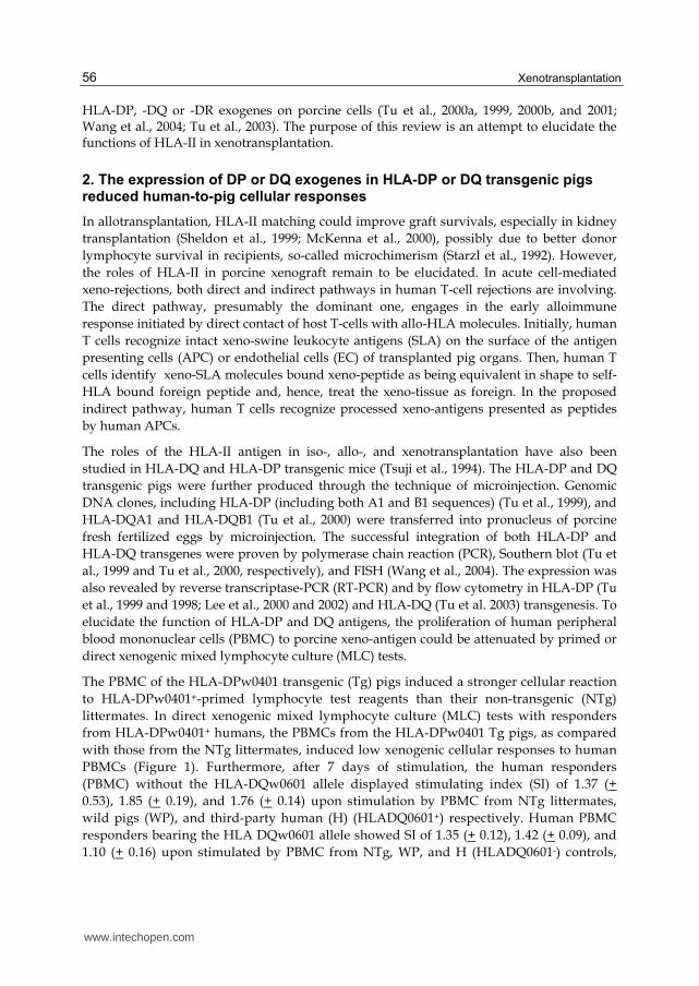
Fig. 2. Stimulating indices (SI) (A1 and A2) and TH1 response (INF production) (B1 and B2) of direct xenogenic mixed lymphocyte culture (MLC) tests with matched or mismatched responders of HLA DQw0601(+ or -) human PBMCs. A1 and A2: the cellular proliferation of human PBMC under stimulation by porcine PBMC could be reduced in the presence of HLA-DQ molecules expressed on the porcine cells, as compared to that in the presence of the NTg littermate control. B1 and B2: the human-to-porcine xenogenic Th1 response, as represented by the production of INF-gamma, was also attenuated by stimulation with HLA-DQ transgenic pig cells. Tg: HLA-DQ transgenic pig; NTg: non-transgenic littermate; WP: wild pigs; and each group n=3. * p < 0.05 as compared to Tg; # using the cpm of Tg as referent (SI=1).
3. The expression of DR exogenes in HLA-DR transgenic pigs enhanced cyclosporine effect
The integration of HLA-DR15+ transgenes in transgenic pigs (Tu et al., 2001) was directly revealed by FISH which has localized both DRA1 and DRB1 transgenes on pig chromosome 13 near the centromere (unpublished data). The expression of transgenes has been confirmed by flow cytometry and the immunohistochemical stain. Results (unpublished data) have also shown that about 39.2% of porcine peripheral blood mononuclear cells and the endothelial cells on the blood vessel of transgenic pig successfully expressed HLA-DR antigens.
Proteomic approach (Huang et al., 2006) revealed that the HLA-DR15+ transgenic pigs could express more proteins including triosephosphate isomerase, cyclophilin B (CyPB), proteaseme and RhoA than their non-transgenic littermates. It is of great interest to elucidate the association of HLA-DR with CyPB on transgenic pigs especially in xenotransplantation. Triosephosphate isomerase can stimulate lymphocyte proliferation
www.intechopen.com

Function Measurements of HLA-II Transgenic Pigs for Xenotransplantation
59
(Richter et al., 1993) and its minor structural change corresponds to substantially enhanced stimulation of a CD4+ tumor-infiltrating lymphocyte line (Sundberg et al., 2002). The proteasome is a multicatalytic complex of proteases involved in T-lymphocyte proliferation and activation and plays an important role in cell–cell interaction during T-lymphocyte activation (Kanaan et al., 2001). The GTP-binding protein RhoA is a member of the Ras GTPase superfamily and has been reported to be actively involved in the regulation of T-lymphocyte morphology and motility (Woodside et al., 2003). The Rho GTPases are molecular switches and are pivotal regulators of antigen-specific T-cell activation by antigen-presenting cells and immunological synapse formation (Deckert et al., 2005). The findings by Mzali et al. (Mzali et al., 2005) also suggested that Rho GTPases play crucial roles in T-lymphocyte functions and proliferation. These proteins are usually involved in T-cell activation or proliferation, except CyPB which belongs to a class of highly conserved proteins that accelerate the folding of proteins and being a cyclosporine A (CsA) binding protein (Schreiber, 1991). This protein is abundant in thymus cytoplasm and appears to be involved in the regulation of T-lymphocyte activation and proliferation (Harding et al., 1986), and inhibits the early T-cell activation (Liu et al., 1991) and prevents graft rejection (Schreiber and Crabtree, 1992). Furthermore, Denys et al. (1998) reported that plasma CyPB may enhance the immunosuppressive activity of CsA through a cell-mediated incorporation of CyPB–complexed CsA with in peripheral blood lymphocytes, and thus contributes to the acceptance and the good maintenance of organ transplantation. It is very interesting to elucidate the association of HLA-DR with CyPB on transgenic pigs especially in xenotransplantation.
By mixed lymphocyte culture, Tg and NTg pigs' lymphocytes (pLC, stimulator) were
compared to stimulate activation of human lymphocytes (hLC; effectors) so that the survival
of HLA-DR Tg pig endothelial cells (pETC) in contact with xenografting lymphocytosis
could be further evaluated. The hLC from HLA-DR15+ or HLA-DR15- healthy adults with O-
or B-type man or women, and from HLA-DR15+ Tg and NTg pLC were harvested from
peripheral blood samples by Ficoll-Paque™ Plus and separated from hematopoietic cells by
culturing in 35 mm dish for overnight. Porcine LC was inactivated by 10.0 g/mL of
mitomycin C for 30 min before mixed with hLC. These xeno-mixed LC reactions were
cultivated in RPMI1640 medium adding 0, 0.1 and 1 g/mL of CsA and for 48 hours. The
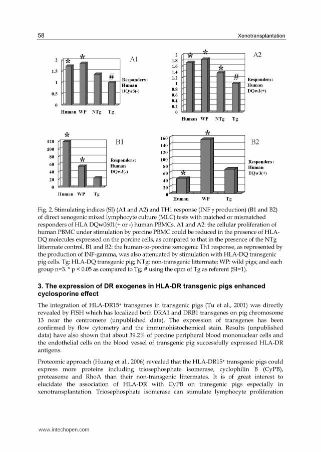
proliferation of hLC was evaluated by MTS assay. Results (Figure 3) thus obtained were:
without CsA, the pLC stimulated 1.2 ±2.0 ~ 47.0 ±4.0% of hLC proliferation; while after
addition of 0.1 and 1 g/mL CsA, the Tg vs. NTg pLC enhanced inhibition effects on hLC
proliferation was 23.8 ±2.5 ~ 23.8 ±2.9% vs. 14.7 ±2.5 ~ 14.9 ±2.5%, and at 47.9 ±2.4 ~ 48.1
±2.8% vs. 24.9 ±2.0 ~28.5 ±2.4%, respectively. Result showed that inactivation of hLC by CsA
could be enhanced by HLA-DR transgenesis, suggesting that an ameliorated effect has
occurred in acute xenograft rejection. The expression of CyPB in HLA-DR transgenic
lymphocytes (intracellular) (Huang et al., 2006) could depress hLC activation. However, the
improvement of survival rate of pETC (responder) varied highly in Tg pETC as compared to
NTg pETC (data not shown). Although CyPB was expressed on endothelial cells
(Carpentieret et al., 1999) and capable of enhancing T-cell adhesion to ETC extracellular
matrix, then it could significantly attenuate by CsA (Allain et al., 2002). It is worth noting
that HLA-DR transgenesis and the expression and secretion of CyPB on pETC will be firstly
attached by activated hLC.
www.intechopen.com
Xenotransplantation
60
Fig. 3. Human lymphocytes mixed reaction with HLA-DR15+ transgenic (Tg) or non-Tg (NTg) lymphocytes. The surface antigens on human lymphocytes were with DR15+ or DR15-. There
were 0, 0.1 and 1.0 g/mL immuosuppressor, CsA, added into the medium of mixed lymphocytes culture tests to evaluate the synergic effects of transgenes. N= 3; * p<0.05 and
** p<0.01.
4. Long-term survival of HLA-DR pig skin in SCID mice after reconstitution with human PBMC and under short-term immunosuppression
To test the role of donor-recipient HLA-II-match in xenotransplantation, the HLA-DR15+ porcine skins were transplanted to SCID mice which were thereafter reconstituted with HLA-DR 15+ or DR15- hPBMC. The studies were conducted under no immunosuppression (no CsA was given to hPBMC-SCID mice), or under immunosuppression (CsA was given intra-peritoneally to hPBMC-SCID mice for 12 days) to reveal the effectiveness of the graft.
In studies of HLA-DR15+ porcine skin grafts to hPBMC-SCID mice under no immunosuppression (Tu et al., 2008), human CD4+ and CD8+ cells were detected from days 7 to 29 after hPBMC reconstitution in hPBMC-SCID mice. Both CD4+ and CD8+ cells of HLA-DR15- hPBMC-SCID mice were significantly higher at day 29 post-grafting, compared with that of HLA-DR15+ hPBMC-SCID mice. In HLA-DR15+ hPBMC-SCID mice, the HLA-DR15+ Tg pig skin grafts survived and integrated into mice, and illustrated histopathologically less cellular rejections which showed intact dermis with little lymphocytic infiltration. However, in HLA-DR15- hPBMC-SCID mice, the HLA-DR15+ transgenic pig skin grafts illustrated more cellular rejections which showed disrupted collagen, as well as mild to moderate lymphocytic infiltration. The results suggested that HLA-DR matching attenuated xenogenic cellular rejection.
www.intechopen.com

Function Measurements of HLA-II Transgenic Pigs for Xenotransplantation
61
In studies of HLA-DR15+ porcine skin grafts to hPBMC-SCID mice under
immunosuppression with CsA (Tai et al., 2008), human CD4+ and CD8+ cells were found in
hPBMC-SCID mice after reconstitution. Tests of MLC showed more responses of HLA-
DR15- hPBMC against HLA-DR15+ porcine PBMC. HLA-DR15+ porcine skin grafts survived
more than 100 days in hPBMC-SCID mice which were reconstituted twice with HLA-DR15+
or HLA-DR15- hPBMC. In the negative control group, HLA-DR15+ porcine skins were
rejected in all non-SCID (Balb/c) mice (data not shown), and the gross pictures showed
disappeared porcine skin and growth of murine hair in non-SCID (Balb/c) mice.
Histological pictures of transplanted HLA-DR15+ porcine skin grafts showed survived
porcine epithelium in remodeling murine dermis (with organized collagen), and little
lymphocytes infiltration in murine dermis.
Fig. 4. Histological pictures (H&E staining) of transplanted HLA-DR15+ pig skin showed
survived porcine epithelium in remodeling murine dermis (with organized collagen), and
little lymphocytes infiltration in murine dermis. (DR+: reconstitution with HLA-DR 15+
hPBMC; DR-: reconstitution with HLA-DR 15- hPBMC.)
www.intechopen.com
Xenotransplantation
62
Although the results do not suggest that HLA-DR matching attenuated xenogenic cellular
rejection, it showed that HLA-DR15+ pig skin grafts could survive over a prolonged period
in hPBMC-SCID under a short period of immunosuppression with CsA. The long-term
survival of HLA-DR15+ pig skin grafts in either HLA-DR15+ or HLA-DR15- hPBMC-SCID
mice might be due to poor engraftment or function of reconstituted T cells, under
immunosuppression with CsA. Because of the gradually decreased number of reconstituted
T cells and suppression effect of CsA, HLA-DR15+ pig skin grafts were not rejected and
therefore survived more than 100 days. In studies of T cell proliferation responses to porcine
aortic endothelial cells (PAEC) either in the presence or absence of CsA, both allogeneic and
xenogeneic T cell responses could be inhibited by in vitro (Fig. 3) or by therapeutic levels of
CsA in vivo (Batten et al., 1999).
In the studies of Hagihara et al. (1996), in vitro MLC and in vivo skin grafting were
conducted by using HLA-DP Tg mice (B6-DP mice). Xenogenic iso-(B6-DP to B6 mice) MLC
showed positive but much less responses when compared to allo-MLC responses.
Nevertheless, B6-DP skin grafts were rejected in a similar time period as allo-skin grafts.
Further studies of in vitro cytotoxic lymphocyte responses and delayed-type
hypersensitivity reactions indicated that xenogeneic HLA-DP antigens could act as
significant transplantation antigens equivalent to alloantigens despite their less stimulative
activity in vitro. Results also support the interpretation that DP antigens act like a minor
histocompatibility antigen beyond the difference of species (Hagihara et al., 1996). In our
studies, xenogenic HLA-DR15+ antigens which act as minor histocompatibility antigens and
swine leukocyte antigens (SLA) contributed simultaneously to exert acute cellular rejection
of porcine skins in hPBMC-SCID mice.
During allogenic skin rejection, the destruction of critical dermal structures that
determine the ultimate viability of the skin graft is highly antigen-specific and is almost
certainly accomplished by cytotoxic T cells. Whereas destruction of non-critical epidermal
structures of the skin allograft is antigen-nonspecific and can be accomplished by
inflammatory cells or their secreted products. The MHC antigens are specific antigens for
skin rejection. Matched MHC antigens of donors and recipients may improve survivals of
allogenic skin graft. The in vivo skin-grafting in HLA-DR15+ hPBMC-SCID mice, matched
HLA-DR15+ transgenic pig skin grafts displayed less cellular rejections.
5. Summary and conclusion
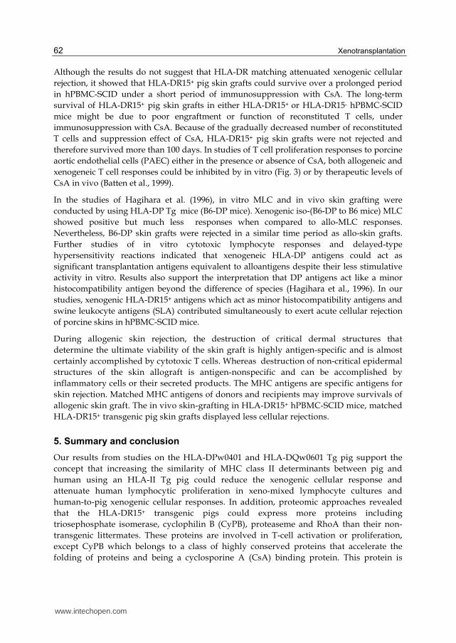
Our results from studies on the HLA-DPw0401 and HLA-DQw0601 Tg pig support the
concept that increasing the similarity of MHC class II determinants between pig and
human using an HLA-II Tg pig could reduce the xenogenic cellular response and
attenuate human lymphocytic proliferation in xeno-mixed lymphocyte cultures and
human-to-pig xenogenic cellular responses. In addition, proteomic approaches revealed
that the HLA-DR15+ transgenic pigs could express more proteins including
triosephosphate isomerase, cyclophilin B (CyPB), proteaseme and RhoA than their non-
transgenic littermates. These proteins are involved in T-cell activation or proliferation,
except CyPB which belongs to a class of highly conserved proteins that accelerate the
folding of proteins and being a cyclosporine A (CsA) binding protein. This protein is
www.intechopen.com
Function Measurements of HLA-II Transgenic Pigs for Xenotransplantation
63
abundant in thymus cytoplasm and appears to engage in the regulation of T-lymphocyte
activation and proliferation, and inhibits the early T-cell activation and prevents graft
rejection. Furthermore, the plasma CyPB may enhance the immunosuppressive activity of
CsA through a cell-mediated incorporation of CyPB–complexed CsA within peripheral
blood lymphocytes, and thus contributes to the acceptance and the good maintenance of
organ transplanted. By using in vitro mixed xeno-lymphocytes culture, the inactivation of
hLC by CsA could be enhanced by HLA-DR transgenesis, suggesting that an ameliorated
effect has occurred in acute xenograft rejection. Further studies using in vivo skin-grafting
in HLA-DR15+ hPBMC-SCID mice, HLA-DR15+ Tg pig skin grafts displayed less cellular
rejections due to additional histocompatibility factor, HLA-DR15+, especially at the
administration of CsA.
6. Acknowledgements
The project was jointly initiated by professor Kimiyoshi Tsuji of Tokai University of Japan
and late professor Chun-Jean Lee of National Taiwan University in 1994. Their inspirations
and encouragements are very much appreciated. Financial supports by National Science
Council, Executive Yuan, Taiwan, ROC, are gratefully acknowledged. Sincere thanks are
also due to Mr. Takayuki Sato for his expertise in preparation of injection DNA and to Ms
YH Chen, MS Liu, CP Wu, LL Ho, WT Lien, and WR Chang for their skillful assistances in
generation of transgenic animals, in vitro study, and skin-graft animal trials. The
suggestions on the data statistics by Dr. SF Guo are also appreciated.
7. References
[1] Allain F., C. Vanpouille, M. Carpentier, M.-C. Slomianny, S. Durieux, and G. Spik.
Interaction with glycosaminoglycans is required for cyclophilin B to trigger
integrin-mediated adhesion of peripheral blood T lymphocytes to extracellular
matrix. PNAS, 2002 ; 99: 2714-2719.
[2] Batten P, McCormack AM, Page CS, Yacoub MH, Rose ML. Human T cell responses to
human and porcine endothelial cells are highly sensitive to cyclosporin A and
FK506 in vitro. Transplantation. 1999; 68: 1552-1560.
[3] Carpentier, M., Descamps, L., Allain, F., Denys, A., Durieux, S., Fenart, L., Kieda, C.,
Cecchelli, R. and Spik, G. Receptor-mediated transcytosis of cyclophilin B
through the blood-brain barrier. J. Neurochem. 1999, 73: 260–270.
[4] Deckert M, Moon C, and Le Bras S. The Immunological Synapse and Rho GTPases.
Curr. Top. Microbiol. Immunol. 2005, 291, 61–90.
[5] Denys A, Allain F, Masy E, Dessaint JP, Spik G. Enhancing the effect of secreted
cyclophilin B on immunosuppressive activity of cyclosporine. Transplantation.
1998; 65: 1076-1084.
[6] Hagihara M, Shimura T, Takebe K, Munkhbat B, Sato T, Tsuchida F, Sato K, Tsuji K.
Xenogeneic iso-skin graft and mixed lymphocyte reaction studies using HLA-DP
transgenic mice. Transpl Immunol. 1996; 4: 220-226.
[7] Harding MW, Handschumacher RE, and Speicher DW. Isolation and amino acid
sequence of cyclophilin. J Biol Chem. 1986; 261: 8547-8555.
www.intechopen.com
Xenotransplantation
64
[8] Huang SY, Chen YH, Teng SH, Chen IC, Ho LL, Tu CF. Protein expression of
lymphocytes in HLA-DR transgenic pigs by a proteomic approach. Proteomics
2006; 6: 5815-5825.
[9] Kanaan N, Luo H, Wu J. Proteasome activity is required for T lymphocyte aggregation
after mitogen activation. J Cell Biochem. 2001; 81: 347-356.
[10] Kuwaki K, Tseng YL, Dor FJMF, Shimizu A, Houser SL, Sanderson TM, Lancos CJ,
Prabharasuth DD, Cheng J, Moran K, Hisashi Y, Mueller N, Yamada K,
Greenstein JL, Hawley RJ, Patience C, Awwad M, Fishman JA, Robson SC,
Schuurman HJ, Sachs DH, and Cooper DK. Heart transplantation in baboons
using α1,3-galactosyltransferase gene-knockout pigs as donors: initial experience.
Nat Med. 2005; 11: 29-31.
[11] Lee JM, Tu CF, Yang PW, Lee YC, Lee CJ. The effective antigen presentation of
human MHC on the lymphocytes of HLA DPW0401 transgenic pigs: examination
with xenogenic mixed lymphocyte culture and primed lymphocyte tests.
Transplant Proc. 2000; 32: 2503-2504.
[12] Lee JM, Tu CF, Yang PW, Lee KH, Tsuji K, Tsai MK, Chen RJ, Hu CY, Hsieh RP, Tai
HC, Chiang BL, Weng CN, Lee YC, Lee CJ. Reduction of human-to-pig cellular
response by alteration of porcine MHC with human HLA DPW0401 exogenes.
Transplantation. 2002; 73: 193-197.
[13] Lee JM, Tu CF, Huang SC, Tsuji K, Chen RJ, Hu CY, Hsieh RP, Tai HC, Weng CN, Lee
YC, Lee CJ. Attenuation of human-to-pig xenogenic cellular proliferation and Th1
response by expressing the human MHC II DQ exogenes on porcine cells.
Transplant Proc. 2003; 35: 527-528.
[14] Liu J, Farmer JD Jr, Lane WS, Friedman J, Weissman I, Schreiber SL. Calcineurin is a
common target of cyclophilin-cyclosporin A and FKBP-FK506 complexes. Cell.
1991; 66: 807-815.
[15] McKenna RM, Takemoto SK. Improving HLA matching for kidney transplantation by
use of CREGs. Lancet. 2000; 355: 1842-1843.
[16] Mzali R, Seguin L, Liot C, Auger A, Pacaud P, Loirand G, Thibault C, Pierre J,
Bertoglio J. Regulation of Rho signaling pathways in interleukin-2-stimulated
human T-lymphocytes. FASEB J. 2005; 19: 1911-1913.
[17] Richter D, Reynolds SR, Harn DA. Candidate vaccine antigens that stimulate the
cellular immune response of mice vaccinated with irradiated cercariae of
Schistosoma mansoni. J Immunol. 1993; 151: 256-265.
[18] Sasaki H, Xu X-C, and Mohanakumar T. HLA-E and HLA-G expression on porcine
endothelial cells inhibit xenoreactive human NK cells through CD94/NKG2-
dependent and –independent pathways. J. Immunol., 1999. 163: 6301–6305.
[19] Schreiber SL. Chemistry and biology of the immunophilins and their
immunosuppressive ligands. Science. 1991, 251: 283-287.
[20] Schreiber SL, Crabtree GR. The mechanism of action of cyclosporin A and FK506
Immunol Today. 1992; 13: 136-142.
[21] Sheldon S, Yonan NA, Aziz TN, Hasleton PS, Rahman AN, Deiraniya AK, Campbell
CS, Dyer PA. The influence of histocompatibility on graft rejection and graft
www.intechopen.com
Function Measurements of HLA-II Transgenic Pigs for Xenotransplantation
65
survival within a single center population of heart transplant recipients.
Transplantation. 1999; 68: 515-519.
[22] Starzl TE, Demetris AJ, Murase N, Ildstad S, Ricordi C, Trucco M. Cell migration,
chimerism, and graft acceptance. Lancet. 1992; 339: 1579-1582.
[23] Sundberg EJ, Sawicki MW, Southwood S, Andersen PS, Sette A, Mariuzza RA. Minor
structural changes in a mutated human melanoma antigen correspond to
dramatically enhanced stimulation of a CD4+ tumor-infiltrating lymphocyte line.
J .Mol. Biol. 2002, 319, 449–461.
[24] Tai HC, Ezzelarab M, Hara H, Ayares D, and Cooper DK. Progress in
xenotransplantation following the introduction of gene-knockout technology.
Transpl Int. 2007; 20:107-117.
[25] Tai HC, Tu CF, Lee JM, Ho LL, Tseng YL, Chou NK, Yang TS, Weng CN, Lee PH,
Chang KJ, Tang YB. Long-term survival of HLA-DR15+ pig skin in SCID mice
after reconstitution with human peripheral blood mononuclear cells and under
short-term immunosuppression. Transplant Proc. 2008; 40: 570-573.
[26] Tsuji K, Hagihara M, Sato T, Shimura T, Takebe K, Munkhbat B. The role of HLA
class II antigens/genes in xenogeneic iso, allo, and xeno transplantation.
Transplant Proc. 1994; 26: 2441-2443.
[27] Tu CF, Hsieh SL, Lee JM, Yang LL, Sato T, Lee KH, Weng CN, Mao SJ, Tsuji K, Lee CJ.
Successful generation of transgenic pigs for human decay-accelerating factor and
human leucocyte antigen DQ. Transplant Proc. 2000; 32: 913-915.
[28] Tu CF, Lee JM, Sato T, Tai HC, Chung YF, Lee FR, Yang PW, Tsuji K, Lee CJ.
Integration and expression of HLA-DR transgenic pigs. Xenoransplantation. 2001;
8 (Suppl. 1): 84.
[29] Tu CF, Lee JM, Sato T, Tai HC, Lee FR, Yang CK, Tsuji K, Lee CJ. Expression of HLA-
DQ genes in transgenic pigs. Transplant Proc. 2003; 35: 513-515.
[30] Tu CF, Sato T, Hagihara M, Lee KH, Lee YC, Weng CN, Chu RM, Tsuji K, Lee CJ.
Expression of HLA-DP antigen on peripheral blood mononuclear cells of HLA-
DP transgenic pigs. Transplant Proc. 1998; 30: 3502-3503.
[31] Tu CF, Tai HC, Chen CM, Huang TT, Lee JM, Yang TS, Chen CH, Tseng YL, Chou
NK, Lee PH. Human leukocyte antigen-DR matching improved skin graft
survival from transgenic pigs to accommodate SCID mice reconstituted with
human peripheral blood mononuclear cells. Transplant Proc. 2008; 40: 578-580.
[32] Tu CF, Tsuji K, Lee KH, Chu R, Sun TJ, Lee YC, Weng CN, Lee CJ. Generation of
HLA-DP transgenic pigs for the study of xenotransplantation. Int Surg. 1999;
84: 176-182.
[33] Wang BT, Tu CF, Hsieh LJ, Tai HC, Chiu YL, Lee JM, Kuo SJ, Tsuji K, Lee CJ. Rapid
detection of human HLA transgenes in pigs by fluorescence in situ hybridization
(FISH) for adjuvant study of human xenotransplantation. Xenotransplantation.
2004; 11: 471-475.
[34] Weiss EH, Lilienfeld BG, Müller S, Müller E, Herbach N, Kessler B, Wanke R,
Schwinzer R, Seebach JD, Wolf E, Brem G. HLA-E/human beta2-microglobulin
transgenic pigs: protection against xenogeneic human anti-pig natural killer cell
cytotoxicity. Transplantation. 2009; 87: 35-43.
www.intechopen.com

Xenotransplantation
66
[35] Woodside DG, Wooten DK, Teague TK, Miyamoto YJ, Caudell EG, Udagawa T,
Andruss BF, McIntyre BW. Control of T lymphocyte morphology by the GTPase
Rho. BMC Cell Biol. 2003; 4: 2.
www.intechopen.com
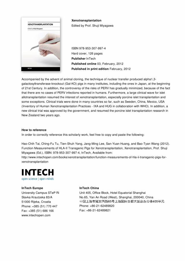
XenotransplantationEdited by Prof. Shuji Miyagawa
ISBN 978-953-307-997-4Hard cover, 126 pagesPublisher InTechPublished online 03, February, 2012Published in print edition February, 2012
InTech EuropeUniversity Campus STeP Ri Slavka Krautzeka 83/A 51000 Rijeka, Croatia Phone: +385 (51) 770 447 Fax: +385 (51) 686 166www.intechopen.com
InTech ChinaUnit 405, Office Block, Hotel Equatorial Shanghai No.65, Yan An Road (West), Shanghai, 200040, China
Phone: +86-21-62489820 Fax: +86-21-62489821
Accompanied by the advent of animal cloning, the technique of nuclear transfer produced alpha1,3-galactosyltransferase-knockout (Gal-KO) pigs in many institutes, including the ones in Japan, at the beginningof 21st Century. In addition, the controversy of the risks of PERV has gradually minimized, because of the factthat there are no cases of PERV infections reported in humans. Furthermore, a large clinical wave for isletallotransplantation resumed the interest of xenotransplantation, especially porcine islet transplantation andsome exceptions. Clinical trials were done in many countries so far, such as Sweden, China, Mexico, USA(Inventory of Human Xenotransplantation Practices - IXA and HUG in collaboration with WHO). In addition, anew clinical trial was approved by the government, and resumed the porcine islet transplantation research inNew Zealand two years ago.
How to referenceIn order to correctly reference this scholarly work, feel free to copy and paste the following:
Hao-Chih Tai, Ching-Fu Tu, Tien-Shuh Yang, Jang-Ming Lee, San-Yuan Huang, and Bao-Tyan Wang (2012).Function Measurements of HLA-II Transgenic Pigs for Xenotransplantation, Xenotransplantation, Prof. ShujiMiyagawa (Ed.), ISBN: 978-953-307-997-4, InTech, Available from:http://www.intechopen.com/books/xenotransplantation/function-measurements-of-hla-ii-transgenic-pigs-for-xenotransplantation