fully automated bone age assessment on large...
TRANSCRIPT

Research ArticleFully Automated Bone Age Assessment on Large-Scale HandX-Ray Dataset
Xiaoying Pan,1 Yizhe Zhao,1 Hao Chen ,1 De Wei,1 Chen Zhao ,2 and Zhi Wei3
1School of Computer Science and Technology, Xi’an University of Posts and Telecommunications, Xi’an, Shaanxi 710121, China2School of Computing Sciences and Computer Engineering, The University of Southern Mississippi, Hattiesburg,Mississippi 39406, USA3Department of Computer Science, New Jersey Institute of Technology, Newark, NJ 07102, USA
Correspondence should be addressed to Chen Zhao; [email protected]
Received 9 July 2019; Revised 23 September 2019; Accepted 26 November 2019; Published 3 March 2020
Academic Editor: Ahmed Soliman
Copyright © 2020 Xiaoying Pan et al. This is an open access article distributed under the Creative Commons Attribution License,which permits unrestricted use, distribution, and reproduction in any medium, provided the original work is properly cited.
Bone age assessment (BAA) is an essential topic in the clinical practice of evaluating the biological maturity of children. Because themanual method is time-consuming and prone to observer variability, it is attractive to develop computer-aided and automatedmethods for BAA. In this paper, we present a fully automatic BAA method. To eliminate noise in a raw X-ray image, we startwith using U-Net to precisely segment hand mask image from a raw X-ray image. Even though U-Net can perform thesegmentation with high precision, it needs a bigger annotated dataset. To alleviate the annotation burden, we propose to usedeep active learning (AL) to select unlabeled data samples with sufficient information intentionally. These samples are given toOracle for annotation. After that, they are then used for subsequential training. In the beginning, only 300 data are manuallyannotated and then the improved U-Net within the AL framework can robustly segment all the 12611 images in RSNA dataset.The AL segmentation model achieved a Dice score at 0.95 in the annotated testing set. To optimize the learning process, weemploy six off-the-shell deep Convolutional Neural Networks (CNNs) with pretrained weights on ImageNet. We use them toextract features of preprocessed hand images with a transfer learning technique. In the end, a variety of ensemble regressionalgorithms are applied to perform BAA. Besides, we choose a specific CNN to extract features and explain why we select thatCNN. Experimental results show that the proposed approach achieved discrepancy between manual and predicted bone age ofabout 6.96 and 7.35 months for male and female cohorts, respectively, on the RSNA dataset. These accuracies are comparable tostate-of-the-art performance.
1. Introduction
Bone age assessment (BAA) may provide important clinicalinformation for skeletal maturation estimation, especiallyfor the diagnosis of endocrinological problems and growthdisorders [1]. The discrepancy between bone age and chro-nological age indicates the abnormalities in skeletal develop-ment. In clinical practice, radiologists perform BAA throughexamining a left-hand-wrist X-ray image. Historically, twoBAA methods, Greulich and Pyle (GP) method [2] and Tan-ner Whitehouse method (TW2) [3], are widely used in clini-cal practice. The GP method compares the patient’s X-rayimage with a representative age atlas and determines the
bone age. In the TW2 method, twenty regions of interest(ROIs) located in the main hand bones are taken into consid-eration for the bone age evaluation. All these procedures aretedious, time-consuming, and prone to observer variability.
As we know, deep learning has been applied to computervision task and achieved drastic performance improvement.In this paper, we propose a method which learns real latentfeatures of hand X-ray images and facilitates the feature cap-ture to perform BAA. At the beginning of the method, wetrain the U-Net neural networks to precisely segment handimage from radiographs and eliminate insignificant infor-mation in raw X-ray images with an active learning tech-nique. Then, we use pretrained deep Convolutional Neural
HindawiInternational Journal of Biomedical ImagingVolume 2020, Article ID 8460493, 12 pageshttps://doi.org/10.1155/2020/8460493
Networks (CNNs) to extract high-level features with atransfer learning technique. After that, ensemble learningis employed to perform BAA with different base regressors.Finally, we evaluate the overall performance of our approachacross different models. The proposed method pipeline isshown in Figure 1. The experimental results demonstratethat the proposed method is more robust and achieves astate-of-the-art performance.
2. Review of BAA
The conventional BAA approaches could be categorized intoGP and TW methods. Some traditional machine learningmethods have been applied to a BAA approach, such as sup-port vector machine (SVM) [4], SVM with cross-correlation[5], and support vector regression [6]. Besides, the most prev-alent and widely used software for automatic BAA in Europeis BoneXpert [7]. This system works based on a shape-drivenactive appearance and TW RUS-based approach. However, itis sensitive to image quality and does not utilize the whole setof hand bones, although all of them are important for skeletalmaturity assessment. In summary, all of the aforementionedworks demonstrate a lack of accuracy.
Recently, motivated by the success of deep ConvolutionalNeural Networks (DCNN) in image classification, studies inmedical imaging have been exploring such methods. Rucciet al. uses an attention focuser and a bone classifier in a neu-ral network to extract features of carpal bones and performsBAA [8]. While the method presents the neural network asa useful technique for classification in the TW2 method, theapproach does not achieve satisfying results with an errorrate of 1.4 years. In 2012, Mansourvar et al. developed a fullyautomated BAA system that uses compression techniquesbase on histogram methods [9]. This approach works on animage repository to perform similarity measures and uses acontent-based image retrieval method for image processing.Because the proposed method does not perform precise handsegmentation, it is not reliable for images with poor qualityor abnormal bone structure. Deep learning-based methodsallow avoiding feature engineering by automatically learn-ing the hierarchy of discriminative features directly from aset training data. DCNN has been successfully applied inthe bone age estimation [10–12]. All of above methodsare end-to-end learning architecture to estimate bone ageusing DCNN.
Despite some methods yield very accurate results, mostexisting methods suffer from two main limitations:
(1) Most of the above methods operate on coarse seg-mentation of hand images, which might misleadBAA toward focusing on irrelevant ROIs
(2) Most of the proposed approaches use hand-craftedfeatures, such as HOG feature, LBP feature, and Harrfeature, thus constraining regressor or classifier to uselow-level X-ray image features rather than the higherand deeper latent features. This semantic gap alwayslimits the generalization capabilities of BAA systems
3. Active Learning for HandImage Segmentation
To perform BAA, we first extract a precise region of inter-est (ROI), a hand mask, from the raw X-ray image. Wethen remove all irrelevant objects which may misleadmodel training. It is necessary to establish a nonlinear map-ping from original X-ray images to hand ROIs for eliminat-ing noise in raw X-ray images. Recently, deep learningsolutions have been successfully used in a multitude ofmedical image semantic segmentation tasks [13]. Eventhough significant performance improvement has beenachieved, a bigger annotated dataset is essential for modeltraining. However, in practice, the task of annotating med-ical images is tedious, time-consuming, and may needexpert knowledge. To alleviate the annotation burden, weemploy a technique called active learning (AL) to activelyselect unlabeled hand radiograph during the training proce-dure and ask for human annotation. The proposed methodachieves high performance on hand segmentation with asfewer annotations as possible.
3.1. Active Learning with Query by Committee. In this paper,we propose a new framework for hand radiograph segmenta-tion using the AL strategy with a limited amount of labeledtraining data. The flowchart of the proposed framework isshown in Figure 2. Let labeled dataset, L = ffx1, y1g,⋯,fxn,yngg, be a collection of hand radiographs, fxig, and corre-sponding labeled hand binary masks fyig (background andhand mask are represented by 0 and 1, respectively). Wedefine U = fxn+1,⋯,xn+mg, be a collection of unlabeled data.The selection of samples inU is determined by the AL frame-work. During the training iteration, the most informativesamples which are beneficial for model training are selectedpreferentially. Our task is to train a nonlinear mapping func-tion, a deep neural network, with L and make use of U torefine the parameters in the network.
The main hypothesis in AL framework is that activelearning can judge which data contain the most abundantinformation for Oracle annotation. The process of AL is likea pupil learns a curriculum. Along the learning process, thepupil spontaneously determines which sample is hard andthen asks teachers which sample is well studied. In this set-ting, AL does not require human to annotate all training data,but only the most uncertain data in the training process.
In practice, query by committee (QBC) is a commonstrategy in the field of AL [14]. The fundamental prerequi-site behind QBC is to minimize the version space [15].QBC contains a committee C = fθ1, θ2,⋯, θkg of networkmodels, all of which are trained with the same labeleddataset L. In BAA problem, we demonstrate θi as a modelwhich learns nonlinear mapping from hand radiograph tohand binary mask. We define each model as a deep neuralnetwork. In QBC framework, every member cooperates todetermine which unlabeled data need to be annotated byOracle after each training epoch. Our goal is to train themand develop sound cooperative relations. After each trainingepoch, all members in C jointly determine the uncertainty ofeach unlabeled datum.
2 International Journal of Biomedical Imaging

Now, we define an uncertainty measure for the level ofdisagreement of a committee. Since we train a set of mem-bers in the committee, each member can extract features ofthe unlabeled data. We use a different random seed to gen-erate the initial model parameters for a different member sothat in each training iteration, the features extracted byeach member are different. In practice, we flatten the fea-ture to a vector, and the feature similarity of two membersare as follows:
cosinesim = vector1 ∗ vector2vector1j j vector2j j , ð1Þ
where the vectori stands for features extracted by θi in com-mittee C.
The unlabeled datum with the lowest feature similarityindicates the datum has the most significant informationwhich is supposed to be helpful for the model training.Therefore, the ground truth of such unlabeled datum shouldbe annotated by Oracle and then added into the labeled data-set for the following training epochs.
3.2. U-Net for Hand Segmentation. As demonstrated in [13],U-Net is robust in medical image segmentation and needs asmaller number of labeled data. U-Net uses convolution layerto automatically extracted features and uses skip connectionto remove the cropping operation and maintains the low-level image outline information in high-level feature maps[16]. We deepen the network and widen the receptive fieldto refine the structure of U-Net by increasing the numberof filters in each convolutional and upsampling layer. Therefined U-Net structure is shown in Figure 3.
In Figure 3, each blue block corresponds to a multi-channel feature map. The arrows with different colors sug-gest different data flow operations. The purple block(vector) represents the deep feature vector of the inputimage inferred by each member. The vector is used inQBC to calculate the similarity between each member inC and to determine whether the input image needs to beannotated or not.
In addition, we optimize the loss function in the processof training a member, i.e., a U-Net. In the field of image seg-mentation, a pixel-wise loss function is usually used to penal-ize the distance between the ground truth and the predictedprobability map. We define the pixel-wise loss function witha cross entropy formulation:
Lpixel‐wise =〠i
− yi log yið Þ − 1 − yið Þ log 1 − yið Þ, ð2Þ
where yi and yi stand for the ground truth and the pre-dicted probability map of pixel i, respectively. This lossexamines each pixel individually, and this helps in speed-ing up the training for neural networks in comparison tothe quadratic loss.
3.3. Algorithm Description. In this section, we summarize thealgorithm for hand image segmentation.
4. Bone Age Assessment Model
Even though CNNs are more commonly used in image clas-sification tasks, BAA is a regression task in fact. Indeed, theessential of CNN is to extract different level features with
U-N
et
Crop
RoI
Segm
ent
Tran
sfer l
earn
ing
Feat
ure v
ecto
r
Ense
mbl
e reg
ress
ion
Bone
age
Raw X-ray Hand mask
Active learning (AL) Normalization
Figure 1: Overview of the proposed automated BAA deep learning pipeline.
Segmentor
Segmentor
Appl
yse
gmen
tor
Train committee(image segmentor)
Sele
ctun
cert
ain
AL
Requ
est
anno
tatio
n
Oracle annotation
Add to annotated data
...
Annotated hand radiographs
Figure 2: Overview of proposed deep AL framework for hand radiograph segmentation.
3International Journal of Biomedical Imaging

various convolutional filters. The extracted features arealways fed into a softmax classifier followed by several FullyConnected (FC) layers to classify input images. Inspired bythe classification task model, we aim to use deep CNNs andtraditional regression algorithms to perform BAA.
4.1. Hand Bone Features Extracting. The key point in BAA isto extract distinct features from preprocessed hand images.Usually, a large training dataset is necessary to fine-tune adeep CNN. However, RSNA dataset only provides 12611images, which is a pittance amount of data compared withImageNet dataset, which contains nearly 15 million images.Consequently, training a high-level feature extractor is diffi-cult on RSNA BAA dataset.
In this situation, we use a transfer learning techniqueand a variety of models with pretrained weights to acquire
features. Transfer learning has been applied to datasets whichare similar to large-scale ImageNet dataset such as [17].Although medical data are different from natural image,transfer learning can be a possible solution for medical datafeature extraction. It uses weights trained on images in otherdomains and infers medical image high-level featuresthrough the network pipeline. Recent researches in [18] dem-onstrate that it is possible to transfer domain-specific knowl-edge from natural images to medical images and achievebrilliant performance.
Since CNN has been proposed, researchers have designedvarious deep CNN models. Several state-of-the-art examplesare VGG-16 [19], VGG-19 [19], ResNet-50 [20], Inception-V3 [21], Inception-ResNet-V2 [22], and Xception [23]. Inthis paper, we use the same preprocessing methods in theabove networks to preprocess segmented hand bone images.
21×
652×
65
256×
256×
1
128×
128×
64
64×
64×
128
256×
256×
32
16×
16×
512
16×
16×
512
256×
256×
1
256×
256×
1256×
256×
9612
8×12
8×64
128×
128×
192
32×
32×
256
32×
32×
768
32×
32×
256
64×
64×
384
64×
64×
128
UpsamplingMax pooling
ConcatenateData flow
Extracted feature
Figure 3: The overview of hand segmentation AL model.
Input:L = ffx1, y1g,⋯,fxn, yngg: initial labeled training data, composed of n samples.U = fxn+1,⋯,xn+mg: initial unlabeled training data, composed of m samples.Output:C = fθ1, θ2,⋯, θkg: trained committee of U-Nets.Repeat:1. Train C = fθ1, θ2,⋯, θkg with the loss function in EQ2 on L2. Calculating unlabeled data’s uncertainty between different U-Nets and select the data with the largest uncertainty3. Annotate the selected data by Oracle and add them into LUntil: hand segmentation is satisfied on U .
Algorithm 1. Hand image segmentation.
4 International Journal of Biomedical Imaging

We initialize the six above networks with pretrained weightson ImageNet then use them to extract features of handradiographs.
By using transfer learning, the high-level feature maps orhigh-level 3-dimensional tensor of hand radiographs can beacquired from the last CNN layer of the CNN network. Weuse Global Average Pooling (GAP) to flatten the featuremaps into a 1-dimensional vector, and the vector denotesthe high-level feature of the images.
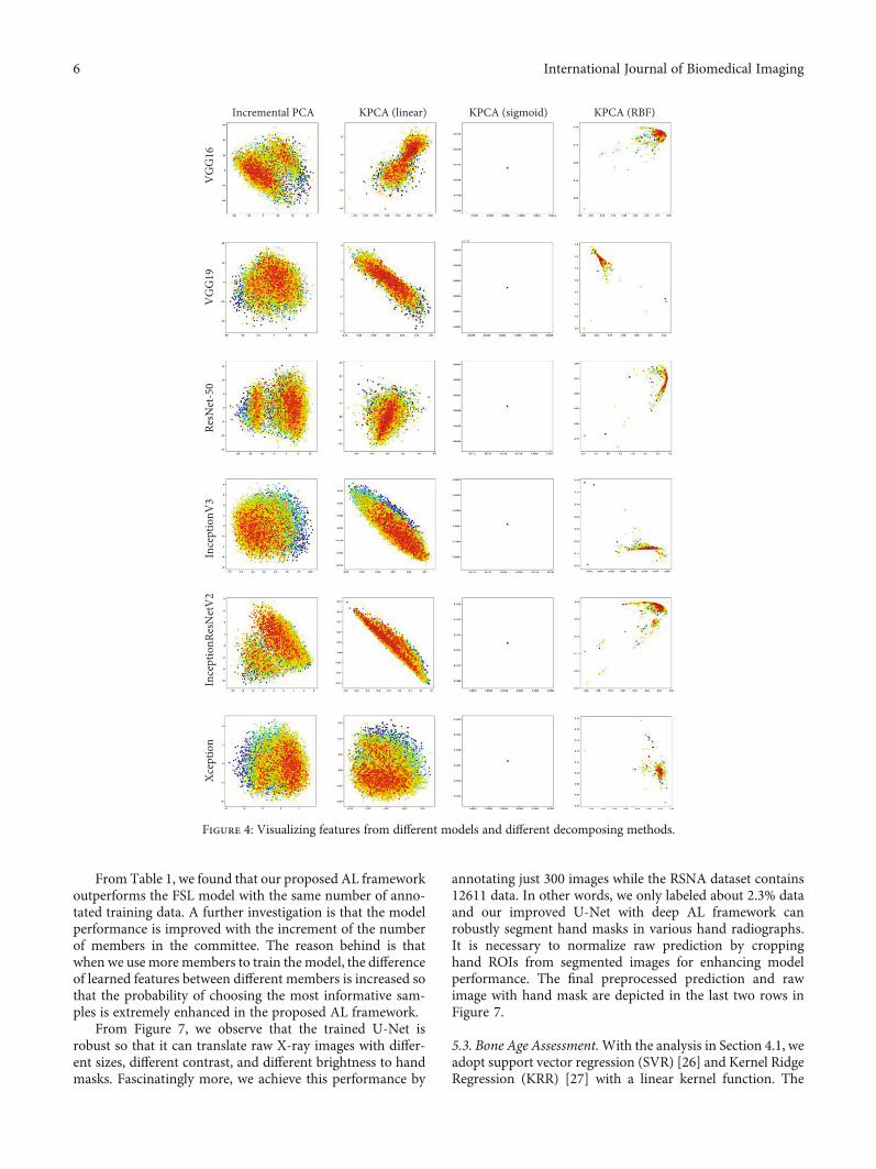
After extracting the features of hand radiographs, wedecompose them into 2-dimensional features by incremen-tal PCA [24] and kernel PCA [25] with different kernelfunctions. Then, we visualize the 2-dimensional featuredistribution. The visualization results are depicted inFigure 4.
In Figure 4, each row represents the features extractedfrom a specific model, and each column represents scatterplots processed by different PCA methods. Colors in differ-ent points represent the data with varying ages of bone. Thehorizational and vertical axes represent the decomposedfeature value. The numbers of features extracted by the lastlayer of VGG, ResNet-50, Xception, Inception-V3, andInception-ResNet-V2 are 4096, 1000, 2048, 2048, and2048, receptively. By applying the PCA algorithm, wedecompose the feature dimension to 2 so that we couldvisualize it in 2D plots. Hence, the meaning of Figure 4 isjust to demonstrate which model is suitable for performingBAA, rather than calculates or visualizes the value ofextracted features.
From the first column in Figure 4, we conclude that fea-tures extracted by Inception-V3, Inception-ResNet-V2, andXception are easy to distinguish since that data with thesame bone ages incline to aggregate into a cluster. In otherwords, features of similar labels are gathered and ordered.On their counterpart for VGG and ResNet, the data repre-sents with the color of red are prone to gather in two differ-ent clusters. This is because all of Inception-V3, Inception-ResNet-V2, and Xception are multiscale deep CNNs. Theyhave a powerful ability to process different size of handbone in preprocessed hand radiographs and generate dis-tinct features.
A further conclusion is that linear kernel function maybe better for differentiating data compared to RBF kernelfunction.
4.2. Bone Age Assessment.With the analysis in Section 4.1, weadopt support vector regression (SVR) [26] and Kernel RidgeRegression (KRR) [27] with a linear kernel function. Thepenalty parameter of SVR is 1.0, the kernel function of SVRis linear kernel, and the tolerance for stopping criterion is1e-3. The kernel function of KRR is linear, the coefficient ofKRR is 3 which leads to a cubic linear function, and theimprovement parameter of KRR is 1.0. Before doing theregression task, we scale bone ages from [0, 228] months toa uniform float value [0, 1]. At the inference stage, we projecttheir bone age back to the original range, i.e., 0 to 228months. Cross-validation is employed at the training stageto prevent overfitting and achieve better generalization per-formance. We set cross fold as 5.
5. Experiment and Discussion
5.1. Data Overview. We obtain hand bone radiograph fromthe 2017 Pediatric Bone Age Challenge organized by theRadiological Society of North America (RNSA) [28]. Theprovided dataset contains 12611 left-hand X-ray images withcorresponding bone age ranging from 0 to 228 months. Thebone age distribution for radiographs of all the dataset,female, and male is depicted, respectively, in Figures 5(a)–5(c). The horizational axis represents the bone age of monthswhich the vertical axis indicates the histogram value of rele-vant patients.
The X-ray data provided by RSNA vary considerably inintensity, contrast, and brightness. A part of the dataset ran-domly selected is shown in Figure 6. This variance increasesthe difficulty of training a robust and precise segment forhand image. Furthermore, it prevents algorithms from learn-ing unified and salient features from the radiographs. Theoptimization of parameters always traps in bad local minimato yield incorrect bone age prediction. In this circumstance, arobust preprocessing engine plays a vital role in data prepro-cessing, and the standardized images are essential for modelaccuracy. Performing BAA using the whole RSNA dataset isa challenging task. That is why all the previous works useonly selected part of the dataset.
5.2. Hand Segmentation. Taking our hardware computationability and memory space into consideration, we set the com-mittee size as k = ½3, 5, 7�. That is to say, we train 3 or 5 or 7U-Nets with the same architecture and initialize their modelparameters with different random seeds. At the initial stage,we randomly select 100 hand radiographs from RSNA data-set and manually annotate the hand masks. The training pro-cedure starts with the labeled 100 paired data. Before feedingthe data into U-Net, we normalize each pixel by using img= ðimg –meanðimgÞÞ/stdðimgÞ, where mean and std indi-cate the mean of pixels and standard variance of pixels,respectively. After each training iteration, we evaluate thesimilarity of all the unlabeled data between two U-Nets andselect the data with the lowest similarity. Finally, the designedinteractive training program will tell us which data need tobe annotated.
In practice, in addition to the initial 100 annotated handradiographs, we annotated another 200 images within thefirst 20 training epochs. After every training epoch, we anno-tated 10 radiographs and added them to the training dataset.Then, we trained the committee with another 80 epochs.The value of loss function convergence at a satisfying stageand we visually inspect all the predicted masks and keepall of them. The segmentation results are shown inFigure 7. As demonstrated in Section 2, RSNA hand radio-graphs vary considerably in intensity, contrast, and bright-ness. To enhance model performance, we normalize thedifferent grayscale bases by using Contrast Limited AdaptiveHistogram Equalization (CLAHE) [29]. Besides, we evalu-ated the Dice score, sensitivity, and specificity of the segmen-tation results, shown in Table 1. As a comparison, we useFully Supervised Learning (FSL) to train the hand segmenta-tion network.
5International Journal of Biomedical Imaging
From Table 1, we found that our proposed AL frameworkoutperforms the FSL model with the same number of anno-tated training data. A further investigation is that the modelperformance is improved with the increment of the numberof members in the committee. The reason behind is thatwhen we use more members to train the model, the differenceof learned features between different members is increased sothat the probability of choosing the most informative sam-ples is extremely enhanced in the proposed AL framework.
From Figure 7, we observe that the trained U-Net isrobust so that it can translate raw X-ray images with differ-ent sizes, different contrast, and different brightness to handmasks. Fascinatingly more, we achieve this performance by
annotating just 300 images while the RSNA dataset contains12611 data. In other words, we only labeled about 2.3% dataand our improved U-Net with deep AL framework canrobustly segment hand masks in various hand radiographs.It is necessary to normalize raw prediction by croppinghand ROIs from segmented images for enhancing modelperformance. The final preprocessed prediction and rawimage with hand mask are depicted in the last two rows inFigure 7.
5.3. Bone Age Assessment.With the analysis in Section 4.1, weadopt support vector regression (SVR) [26] and Kernel RidgeRegression (KRR) [27] with a linear kernel function. The
Incremental PCA
Xcep
tion
Ince
ptio
nRes
Net
V2
Ince
ptio
nV3
ResN
et-5
0V
GG
19V
GG
16
KPCA (linear) KPCA (sigmoid) KPCA (RBF)
Figure 4: Visualizing features from different models and different decomposing methods.
6 International Journal of Biomedical Imaging

penalty parameter of SVR is 1.0, the kernel function of SVR islinear kernel, and the tolerance for stopping criterion is 1e-3.The kernel function of KRR is linear, the coefficient of KRR is3 which leads to a cubic linear function, and the improve-ment parameter of KRR is 1.0. Before doing the regressiontask, we scale bone ages from [0, 228] months to uniformfloat value [0, 1]. At the inference stage, we project their boneage back to the original range, i.e., 0 to 228 months. Before we
trained our neural network, we balanced the data by sam-pling the same number of data with the same bone age. Whatis more important, not only we transferred the well-trainedparameters into our BAA model but also we fine-tuned theparameters in DCNN with SVR or KRR. Cross-validation isemployed at the training stage to prevent overfitting andachieve better generalization performance. We set the crossfold as 5.
00
250
500
750
1000
1250
1500
1750
2000
50 100 150Bone age
200
(a)
00
200
400
600
800
1000
1200
1400
50 100 150Bone age
200
(b)
00
100200300400500600700800
50 100 150Bone age
200
(c)
Figure 5: Bone age distribution of (a) full dataset, (b) male, and (c) female. The unit of the horizontal axis is the month.
Figure 6: A close-up of part of data in RSNA dataset. Different radiographs vary in size and height-width ratio.
7International Journal of Biomedical Imaging

We use Mean Average Error (MAE), Root Mean SquareError (RMSE), and Concordance Correlation Coefficient(CCC) to evaluate proposed methods. The MAE and RMSEintuitively represent the distance between real and predictionof bone age (lower is better). The CCC has better perfor-mance to evaluate the correlation between real bone ageand prediction (higher is better) than R2 score and explained
variance score [30]. The experimental results are shown inTable 2.
From Table 2, we observe that our models achieve thebest MAE, RMSE, and CCC by using KRR on data trans-ferred via Inception-ResNet-V2. A further crucial observa-tion is that all best measures are acquired in the samesetting. Separate regression models for male and femalecohorts demonstrate higher accuracy when compared tothose trained on a mixed population. With a single regressor,MAEs of the whole dataset, male, and female are 14.83months, 12.82 months, and 11.93 months, respectively. Thatsuggests that the loss error is about 1 year on average on asingle patient.
To enhance model performance, we employed ensemblelearning to lower the regression error further. Ensemblemodeling is a powerful way to improve the performance ofthe low generalized model by combining a diverse set oflearners and adjusting data weights in training stage. FromTable 1, KRR with Inception-ResNet-V2 achieved the bestperformance on all of the evaluations, so we employ KRRas a base estimator and ensemble them with the Bagging[31] method and AdaBoost. The performance of ensembleregression is shown in Figure 8.
In Figure 8, the horizontal and vertical axis represents thenumber of base estimators (KRR) used in ensemble learning
Raw
X-ra
yX
dedd
aP-r
aySe
gmen
tatio
nm
ask
Crop
ped
hand
mas
kPr
epro
cess
ed
Figure 7: Examples at each stage of preprocessing in the segmentation pipeline.
Table 1: Comparison of model performance for handsegmentation.
StrategyNumber of
annotated samplesSensitivity Specificity Dice
FSL 300 0.869 0.854 0.869
AL (k = 3) 150 0.864 0.845 0.863
AL (k = 3) 200 0.902 0.895 0.905
AL (k = 3) 300 0.903 0.942 0.939
AL (k = 5) 150 0.896 0.909 0.888
AL (k = 5) 200 0.904 0.925 0.916
AL (k = 5) 300 0.935 0.946 0.931
AL (k = 7) 150 0.879 0.902 0.899
AL (k = 7) 200 0.932 0.934 0.926
AL (k = 7) 300 0.948 0.960 0.952
8 International Journal of Biomedical Imaging

and the corresponding evaluations, respectively. FromFigure 8, we further enhance the model performance by Ada-Boost and Bagging ensemble learning. The final performanceof the proposed methods is listed in Table 3. In Table 3, thenumber in brackets indicates the number of base estimatorsused in ensemble learning. We lower the error rate about 3months on each part of dataset compared with a singleKRR estimator without Bagging. In our experiment, Baggingoutperforms AdaBoost. A further observation is that the bestCCC values are enhanced about 0.05 on the male and femaledataset. The experimental results suggest that by usingensemble learning, the correlation between real and pre-dicted bone ages is higher than using a single regressor.
Table 4 demonstrates the comparison of the model per-formance with several existing approaches on BAA tasks.By observing Table 4, it is clear that our proposed modeldominates over other methods in part of MAE evaluation.
6. Discussion and Conclusions
6.1. Discussion. Using our proposed BAA approach, weachieved a MAE of 8.59, 6.96, and 7.35 months on all, male,and female cohorts of the dataset.
Since AL queries unlabeled data and asks Oracle to anno-tate them, the number of training data is enlarged by the ALstrategy and more labeled data available can benefit in train-ing neural networks. More importantly, AL inclines to pickup the most uncertain and informative data for anothertraining epoch so that the active learner learns the most cru-cial data in training. Essentially, AL boosts the training pro-cess so that the trained model can get a better solution.
A further significant investigation is that we proposed aframework of medical image segmentation to relieve humanexpert annotation burden via deep active learning. Featurevector differences between different members in the commit-tee are taken into consideration. The members can workcooperatively to determine which datum is crucial in thetraining procedure and then ask oracle to annotate it. In thesegmentation stage, benefitting from deep active learning,we only annotated 300 images—about 2.3% of the wholedataset—to make precise hand segmentation.
With the annotated hand X-ray images, our results sup-port the finding by others demonstrating the effectivenessand applicability of transferring deep-learning weights todata from different domains [34]. Transfer learning isimportant in our framework since the pretrained network
is required for successful implementation of clinical deci-sion with a relative small medical image dataset. Further-more, ensemble learning significantly improves the modelperformance.
Although the proposed BAA approach achieved a state-of-the-art performance, there are also limitations and somevalues need to be discussed:
(1) Number of members in the committee. Although wefound the model performance of segmentation net-works are enhanced with the increment of the num-ber of members in the committee, the number ofmembers is hard to determine. Besides, we did notensemble the well-trained active learners to inferencethe segmentation results simultaneously
(2) The computational complexity of the proposedmodel. As demonstrated in [36], the total time com-plexity of all convolutional layers in a deep network isas follows:
O 〠d
l=1nl−1s
2l nlm
2l
!, ð3Þ
where l is the index of a convolutional layer and d is thedepth. nl is the number of filter in the lth layer. sl is the spatialsize of the filter, andml is the spatial size of the output featuremap. From (3), we know the time complexity of DCNN is thecombination of each convolutional layer. So the time com-plexity of the proposed BAA approach is Oðn6Þ6.2. Conclusions. In this paper, we have investigated theapplication of deep transfer learning on medical images,especially for automated bone age assessment using handradiographs. We tested several popular off-the-shell deepCNNs trained on the RSNA dataset with 12611 X-ray images.We proved that the transfer learning can cope effectively withbone age assessment task. By using an ensemble technique,our model achieved an MAE of 8.59, 6.96, and 7.35 monthson all, male, and female cohorts of the dataset, respectively,comparable to the state-of-the-art performance. Further-more, we explained which pretrained CNN is better to per-form BAA.
In summary, we have created a fully automated, deeplearning-based preprocessing pipeline to automatically detect
Table 2: Performance of different regression methods on different transferred data.
Model SexInception-V3 Xception Inception-ResNet-V2
MAE RMSE CCC MAE RMSE CCC MAE RMSE CCC
SVR (linear)
All 16.4688 21.1794 0.7139 15.6739 20.3728 0.7029 14.2175 18.0785 0.7143
Male 12.8732 17.7263 0.5987 11.9983 13.2222 0.6319 11.7378 14.8372 0.6417
Female 13.2739 17.9381 0.6163 13.6930 14.8271 0.6184 13.0116 17.3823 0.6371
KRR (linear)
All 15.1232 18.2813 0.7004 15.2830 17.7362 0.7793 13.9381 14.8373 0.7293
Male 13.0293 14.2521 0.6313 12.2321 14.9382 0.6098 11.9283 12.8231 0.6563
Female 14.7421 19.0855 0.6277 13.3361 17.3211 0.6176 12.7744 11.9321 0.6473
9International Journal of Biomedical Imaging

and segment the hand and wrist, standardize the images, andperform BAA with pretrained deep CNNs and high-efficiency regression model. In practice, our system can beeasily deployed in the clinical environment on a computerwith a single GPU.
7. Future Work
The investigation presented in this paper leaves many chal-lenges and issues for future research. We summarize thefuture work as follows:
(1) The proposed BAA framework, which containsimage segmentation, feature extraction, and ensem-ble modules, should be validated on other medicalimage decision problem
(2) To proof the effectiveness of AL framework theoreti-cally. Only if we proof it, could we find how manyactive learners is enough to form a committee
(3) Ensemble the well-trained active learners and gener-ate segmentation result simultaneously by AdaBoostor other ensemble learning algorithms
0
7
8
9
10
11
Mon
ths
AdaB
oost
12
13
14
2 4 6 8 10 12 14 16 18 20 22 24Number of base estimators
7.62 7.60
9.31
MAE
Mon
ths
0.94 0.940.95
0
0.7
0.6
0.8
0.9
1.0
1.1
2 4 6 8 10 12 14 16 18 20 22 24Number of base estimators
26
CCC
8.59
6.967.35
0
7
6
8
9
10
11
Mon
ths
Bagg
ing
12
13
14
2 4 6 8 10 12 14 16 18 20 22 24Number of base estimators
26
AllMaleFemale
Mon
ths
0.940.97 0.97
0
0.7
0.6
0.8
0.9
1.0
1.1
2 4 6 8 10 12 14 16 18 20 22 24Number of base estimators
26
Figure 8: Performance of different ensemble regression methods on data transferred by Inception-ResNet-V2.
Table 3: Performance of different ensemble regression methods.
Ensemble method Dataset MAE CCC
AdaBoost
All 9.31 (21) 0.94 (14)
Male 7.62 (14) 0.94 (17)
Female 7.60 (19) 0.95 (19)
Bagging
All 8.59 (21) 0.94 (14)
Male 6.96 (14) 0.97 (17)
Female 7.35 (19) 0.97 (19)
Table 4: Comparison of approaches in BAA in RSNA dataset.
Method MAE (m)
Iglovikov et al. [32] without ensemble 8.08
Iglovikov et al. [32] with ensemble 7.52
Wu et al. [33] 7.38
Han et al. [34] 8.40
Tajmir et al. [35] 7.93
Proposed 7.35
10 International Journal of Biomedical Imaging

Data Availability
The X-ray imaging data used to support the findings ofthis paper have been deposited in the RSNA repositoryat doi:10.1148/radiol.2018180736.
Conflicts of Interest
The authors declare no conflict of interest.
References
[1] R. Vanderwilde, L. T. Staheli, D. E. Chew, and V. Malagon,“Measurements on radiographs of the foot in normal infantsand children,” The Journal of Bone and Joint Surgery, vol. 70,no. 3, pp. 407–415, 1988.
[2] University S, “Radiographic atlas of skeletal development ofthe hand and wrist,” Journal of Anatomy, vol. 85, Part 1,p. 103, 1951.
[3] J. M. Tanner, R. H. Whitehouse, N. Cameron, W. A. Marshall,M. J. Healy, and H. Goldstein, Assessment of skeletal maturityand prediction of adult height (TW2 method), London: Aca-demic Press, 1975.
[4] S. W. Lin, K. C. Ying, S. C. Chen, and Z. J. Lee, “Particle swarmoptimization for parameter determination and feature selec-tion of support vector machines,” Expert Systems with Applica-tions, vol. 35, no. 4, pp. 1817–1824, 2008.
[5] M. Harmsen, B. Fischer, H. Schramm, T. Seidl, and T. M.Deserno, “Support vector machine classification based on cor-relation prototypes applied to bone age assessment,” IEEEJournal of Biomedical and Health Informatics, vol. 17, no. 1,pp. 190–197, 2013.
[6] K. Somkantha, N. Theera-Umpon, and S. Auephanwiriyakul,“Bone age assessment in young children using automatic car-pal bone feature extraction and support vector regression,”Journal of Digital Imaging, vol. 24, no. 6, pp. 1044–1058, 2011.
[7] H. H. Thodberg, S. Kreiborg, A. Juul, and K. D. Pedersen, “TheBoneXpert method for automated determination of skeletalmaturity,” IEEE Transactions on Medical Imaging, vol. 28,no. 1, pp. 52–66, 2009.
[8] M. Rucci, G. Coppini, I. Nicoletti, D. Cheli, and G. Valli,“Automatic analysis of hand radiographs for the assessmentof skeletal age: a subsymbolic approach,” Computers and Bio-medical Research, vol. 28, no. 3, pp. 239–256, 1995.
[9] M. Mansourvar, R. G. Raj, M. A. Ismail et al., “Automated webbased system for bone age assessment using histogram tech-nique,” Malaysian Journal of Computer Science, vol. 25,no. 3, pp. 107–121, 2012.
[10] C. Spampinato, S. Palazzo, D. Giordano, M. Aldinucci, andR. Leonardi, “Deep learning for automated skeletal bone ageassessment in X-ray images,” Medical Image Analysis, vol. 36,pp. 41–51, 2017.
[11] D. B. Larson, M. C. Chen, M. P. Lungren, S. S. Halabi, N. V.Stence, and C. P. Langlotz, “Performance of a deep-learningneural network model in assessing skeletal maturity on pediat-ric hand radiographs,” Radiology, vol. 287, no. 1, pp. 313–322,2018.
[12] H. Lee, S. Tajmir, J. Lee et al., “Fully automated deep learningsystem for bone age assessment,” Journal of Digital Imaging,vol. 30, no. 4, pp. 427–441, 2017.
[13] O. Ronneberger, P. Fischer, and T. Brox, “U-net: convolutionalnetworks for biomedical image segmentation,” in InternationalConference on Medical Image Computing and Computer-Assisted Intervention, N. Navab, J. Hornegger, W. Wells, andA. Frangi, Eds., pp. 234–241, Springer, 2015.
[14] H. S. Seung, M. Opper, and H. Sompolinsky, “Query bycommittee,” in Proceedings of the fifth annual workshop onComputational learning theory, pp. 287–294, Pittsburgh,Pennsylvania, USA, 1992.
[15] D. A. Cohn, Z. Ghahramani, andM. I. Jordan, “Active learningwith statistical models,” Journal of Artificial IntelligenceResearch, vol. 4, pp. 129–145, 1996.
[16] H. Fu, Y. Xu, D. W. K. Wong, and J. Liu, “Retinal vessel seg-mentation via deep learning network and fully-connected con-ditional random fields,” in 2016 IEEE 13th internationalsymposium on biomedical imaging (ISBI), pp. 698–701, Prague,Czech Republic, 2016.
[17] Z. Ding, N. M. Nasrabadi, and Y. Fu, “Task-driven deep trans-fer learning for image classification,” in 2016 IEEE Interna-tional Conference on Acoustics, Speech and Signal Processing(ICASSP), pp. 2414–2418, Shanghai, China, 2016.
[18] H. C. Shin, H. R. Roth, M. Gao et al., “Deep convolutional neu-ral networks for computer-aided detection: CNN architec-tures, dataset characteristics and transfer learning,” IEEETransactions on Medical Imaging, vol. 35, no. 5, pp. 1285–1298, 2016.
[19] K. Simonyan and A. Zisserman, “Very deep convolutionalnetworks for large-scale image recognition,” 2014, https://arxiv.org/abs/1409.1556.
[20] K. He, X. Zhang, S. Ren, and J. Sun, “Deep residual learning forimage recognition,” in 2016 IEEE Conference on ComputerVision and Pattern Recognition (CVPR), pp. 770–778, LasVegas, NV, USA, June 2016.
[21] C. Szegedy, V. Vanhoucke, S. Ioffe, J. Shlens, and Z. Wojna,“Rethinking the inception architecture for computer vision,”in 2016 IEEE Conference on Computer Vision and Pattern Rec-ognition (CVPR), pp. 2818–2826, Las Vegas, NV, USA, June2016.
[22] C. Szegedy, S. Ioffe, V. Vanhoucke, and A. A. Alemi, “Incep-tion-v4, inception-resnet and the impact of residual connec-tions on learning,” in Proceedings of the Thirty-First AAAIConference on Artificial Intelligence (AAAI’17), pp. 4278–4284, San Francisco, CA, USA, 2017.
[23] F. Chollet, “Xception: deep learning with depthwise separableconvolutions,” in 2017 IEEE Conference on Computer Visionand Pattern Recognition (CVPR), pp. 1251–1258, Honolulu,HI, USA, July 2017.
[24] I. Dagher, “Incremental pca-lda algorithm,” in 2010 IEEEInternational Conference on Computational Intelligence forMeasurement Systems and Applications, pp. 97–101, Taranto,Italy, 2010.
[25] B. Schölkopf, A. Smola, and K. R. Müller, “Kernel principalcomponent analysis,” in International conference on artificialneural networks, W. Gerstner, A. Germond, M. Hasler, and J.D. Nicoud, Eds., pp. 583–588, Springer, 1997.
[26] H. Drucker, C. J. Burges, L. Kaufman, A. J. Smola, andV. Vapnik, “Support vector regression machines,” Advancesin Neural Information Processing Systems, pp. 155–161, MITPress, 1997.
[27] V. Vovk, “Kernel ridge regression,” in Empirical inference,pp. 105–116, Springer, 2013.
11International Journal of Biomedical Imaging

[28] S. S. Halabi, L. M. Prevedello, J. Kalpathy-Cramer et al., “TheRSNA pediatric bone age machine learning challenge,” Radiol-ogy, vol. 290, no. 2, pp. 498–503, 2019.
[29] S. M. Pizer, R. E. Johnston, J. P. Ericksen, B. C. Yankaskas,and K. E. Muller, “Contrast-limited adaptive histogramequalization: speed and effectiveness,” in Proceedings of theFirst Conference on Visualization in Biomedical Computing,pp. 337–345, Atlanta, Georgia, 1990.
[30] I. Lawrence and K. Lin, “A concordance correlation coefficientto evaluate reproducibility,” Biometrics, vol. 45, no. 1, pp. 255–268, 1989.
[31] L. Breiman, “Bagging predictors,” Machine Learning, vol. 24,no. 2, pp. 123–140, 1996.
[32] V. I. Iglovikov, A. Rakhlin, A. A. Kalinin, and A. A. Shvets,“Paediatric bone age assessment using deep convolutionalneural networks,” in Deep Learning in Medical Image Analysisand Multimodal Learning for Clinical Decision Support, D.Stoyanov, Ed., pp. 300–308, Springer, 2018.
[33] E. Wu, B. Kong, X. Wang et al., “Residual attention basednetwork for hand bone age assessment,” in 2019 IEEE 16thInternational Symposium on Biomedical Imaging (ISBI 2019),pp. 1158–1161, Venice, Italy, Italy, 2019.
[34] J. Han, Y. Jia, C. Zhao, and F. Gou, “Automatic bone ageassessment combined with transfer learning and supportvector regression,” in 2018 9th International Conference onInformation Technology in Medicine and Education (ITME),pp. 61–66, Hangzhou, China, 2018.
[35] S. H. Tajmir, H. Lee, R. Shailam et al., “Artificial intelligence-assisted interpretation of bone age radiographs improvesaccuracy and decreases variability,” Skeletal Radiology, vol. 48,no. 2, pp. 275–283, 2019.
[36] K. He and J. Sun, “Convolutional neural networks at con-strained time cost,” in 2015 IEEE Conference on ComputerVision and Pattern Recognition (CVPR), pp. 5353–5360, Bos-ton, MA, USA, June 2015.
12 International Journal of Biomedical Imaging