full mouth rehabilitation of a hydrocephalus pediatric patientjdsor.com/2015vol6-2/a10.pdf ·...
TRANSCRIPT

Kaushal Jha et al
88
JDSOR
Full Mouth Rehabilitation of a Hydrocephalus Pediatric Patient1Kaushal Jha, 2Eva Pradhan, 3N Sathyajith Naik, 4V Vatsala, 5Swati Tripathi
ABSTRACTHydrocephalus is a clinical entity characterized by accumulation of cerebrospinal fluid in the lateral ventricles of brain, resulting in progressive ventricular dilatation. Children with hydrocephalus are at greater risk for caries and need both routine and emergency dental treatment. Dental practitioners must develop adequate treatment strategies to prevent risk factors occurring during their treatment. As far as in dental literature, there is lack of information about the management of hydrocephalus patients during dental treatment. Hence, this case report describes the chairside full mouth rehabilitation of a 12yearold female child with hydrocephalus.
Keywords: Dental caries, Full mouth rehabilitation, Hydrocephalus, Physically challenged.
How to cite this article: Jha K, Pradhan E, Naik NS, Vatsala V, Tripathi S. Full Mouth Rehabilitation of a Hydrocephalus Pediatric Patient. J Dent Sci Oral Rehab 2015;6(2):8891.
Source of support: Nil
Conflict of interest: None
INTRODUCTION
Hydrocephalus is considered a congenital disease affecting the central nervous system (CNS), which is characterized by accumulation of cerebrospinal fluid (CSF) in the lateral ventricles of the brain causing progressive ventri cular dilation. Hydrocephalus is diagnosed through clinical neurological evaluation and by using cranial imaging techniques, such as ultrasonography, computed tomography (CT), magnetic resonance imaging (MRI), or pressuremonitoring techniques. A physician selects the appropriate diagnostic tool based on an individual’s age, clinical presentation, and the presence of known or suspected abnormalities of the brain or spinal cord.
Currently, among various congenital malformations which involve central nervous system, hydrocephalus accounts for 0.4 to 0.8 cases per 1,000 births.1 Over the
CaSe RepORt
1Postgraduate Student (IIIrd Year), 2Postgraduate Student (IInd Year), 3Professor and Head, 4Senior Lecturer, 5Reader15Department of Pedodontics and Preventive Dentistry, Institute of Dental Sciences, Bareilly, Uttar Pradesh, India
Corresponding Author: Eva Pradhan, Postgraduate Student (IInd Year), Department of Pedodontics and Preventive Dentistry Institute of Dental Sciences, Pilibhit Bypass Road, Bareilly, Uttar Pradesh, India, email: [email protected]
10.5005/jp-journals-10039-1073
years, improvement in social status, awareness, better nutrition and better perinatal care have resulted in reduction in major anomalies and associated hydrocephalus. Prenatal diagnosis has become a reality in most centers.2 The postinfective hydrocephalus has seen a decline. The treatment of hydrocephalus comprises of: the shunt assembly with a proximal catheter located in the cerebral ventricles and a distal catheter draining into selected site of CSF absorption, connected by a valve and reservoir which is incorporated into the shunt system.
To keep the intracranial pressure within normal range, hydrocephalus patients are always on medication. Hence, they are very prone to dental decay. Therefore, pediatric dentists should be well equipped and have proper knowledge of adequate treatment strategies to prevent risk factors occurring during their treatment.3 In literature, there is lack of information about the manage ment of hydrocephalus patients during dental treatment. Hence, this case report describes the chairside full mouth rehabilitation of a 12yearold female child with hydrocephalus.
CASE REPORT
History
A 12yearold female reported to the department of pedodontics and preventive dentistry with the chief complaint of pain and swelling in lower right back region since 3 days. History revealed that the birth was uneventful. Patient was born to nonconsanguineous parents. All the signs and symptoms of hydrocephalus were evident, i.e. increased intracranial pressure due to excessive CSF accumulation along with abnormal head growth. Parents also gave history of patient’s seizures attacks, irritability and vomiting.
Extraoral Examination
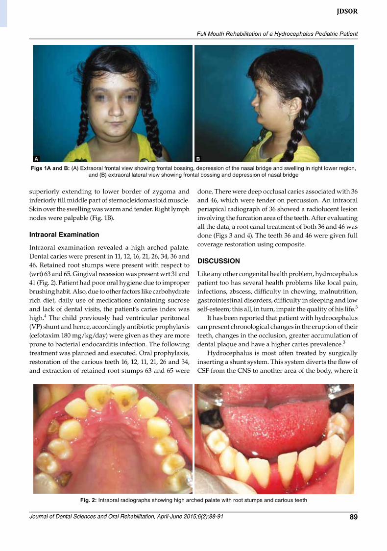
The patient presented with short stature, depression of the nasal bridge seen along with deviated nasal septum.This led to abnormal breathing habit due to which patient developed mouth breathing. Distended fontanels and frontal bossing with occipital bulge was evident. The orbits were deep sunken (Fig. 1A).
Patient had diffused extraoral swelling extending from the body of mandible till posterior border of ramus,
Full Mouth Rehabilitation of a Hydrocephalus Pediatric Patient
Journal of Dental Sciences and Oral Rehabilitation, April-June 2015;6(2):88-91 89
JDSOR
superiorly extending to lower border of zygoma and inferiorly till middle part of sternocleidomastoid muscle. Skin over the swelling was warm and tender. Right lymph nodes were palpable (Fig. 1B).
Intraoral Examination
Intraoral examination revealed a high arched palate. Dental caries were present in 11, 12, 16, 21, 26, 34, 36 and 46. Retained root stumps were present with respect to (wrt) 63 and 65. Gingival recession was present wrt 31 and 41 (Fig. 2). Patient had poor oral hygiene due to improper brushing habit. Also, due to other factors like carbohydrate rich diet, daily use of medications containing sucrose and lack of dental visits, the patient’s caries index was high.4 The child previously had ventricular peritoneal (VP) shunt and hence, accordingly antibiotic prophylaxis (cefotaxim 180 mg/kg/day) were given as they are more prone to bacterial endocarditis infection. The following treatment was planned and executed. Oral prophylaxis, restoration of the carious teeth 16, 12, 11, 21, 26 and 34, and extraction of retained root stumps 63 and 65 were
done. There were deep occlusal caries associated with 36 and 46, which were tender on percussion. An intraoral periapical radiograph of 36 showed a radiolucent lesion involving the furcation area of the teeth. After evaluating all the data, a root canal treatment of both 36 and 46 was done (Figs 3 and 4). The teeth 36 and 46 were given full coverage restoration using composite.
DISCUSSION
Like any other congenital health problem, hydrocephalus patient too has several health problems like local pain, infections, abscess, difficulty in chewing, malnutrition, gastrointestinal disorders, difficulty in sleeping and low selfesteem; this all, in turn, impair the quality of his life.3
It has been reported that patient with hydrocephalus can present chronological changes in the eruption of their teeth, changes in the occlusion, greater accumulation of dental plaque and have a higher caries prevalence.3
Hydrocephalus is most often treated by surgically inserting a shunt system. This system diverts the flow of CSF from the CNS to another area of the body, where it
Figs 1A and B: (A) Extraoral frontal view showing frontal bossing, depression of the nasal bridge and swelling in right lower region, and (B) extraoral lateral view showing frontal bossing and depression of nasal bridge
Fig. 2: Intraoral radiographs showing high arched palate with root stumps and carious teeth

Kaushal Jha et al
90
can be absorbed as part of the normal circulatory process, thereby reducing intracranial pressure and preventing brain damage through a system of small tubes known as catheters. A regulating device, also known as a valve, which may be more or less sophisticated, may be inserted into the pathway of the catheter. This technique allows the brain’s enlarged ventricles to return to a normal size in an effort to relieve the symptoms of hydrocephalus. Invasive dental procedures can produce a transient bacteremia. Patients with ventricular atrial (VA) shunt that drains into the right atrium are considered to be at risk for shunt infections during invasive dental procedures than patients with VP shunts. The child in this treatment was given antibiotic prophylaxis (cefotaxime 180 mg/kg/day), even though no such case reports of infection of any shunt as a result of invasive dental procedure in the literature.57
Children with hydrocephalus treated with VP shunts have craniofacial asymmetry that is associated with unilateral insertion of the device. This is clearly seen in those patients who always have shunt on same side. The mandibular plane is higher where shunt is placed,
possibly due to moderate growth restriction of that side.8 Only once, the VP shunt was placed in our patient. After 5 years, the patient got the shunt removed without undergoing the replacement with a new one (Fig. 5). Hence, our patient had characteristic huge head as seen in hydrocephalous patient. Postoperative intraoral view of maxillary arch is shown in Figure 6.
Another oral feature is maturation of their teeth, which seems to be advanced at initial stages of puberty and delayed during the final stages. The dental practi tioner should be careful not to put pressure on the reticularperitoneal. As there is always danger to com press the shunt during dental procedure, the use of pillows and cushions is advisable to make patient com fortable and give adequate posture to the patient while doing treatment.9 As our patient was epileptic, lower level of neuromuscular drug was used when the patient was undergoing treatment.
In hydrocephalus cases, we generally see early dental maturation with early pubertal maturation because of increased levels of anterior pituitary sex hormones.10
These findings were in tandem with our patient.
Fig. 3: Preoperative and postoperative intraoral radiographs of tooth 36
Fig. 4: Preoperative and postoperative intraoral radiographs of tooth 46

Full Mouth Rehabilitation of a Hydrocephalus Pediatric Patient
Journal of Dental Sciences and Oral Rehabilitation, April-June 2015;6(2):88-91 91
JDSOR
Hence, it is recommended to pediatric dentist to have thorough knowledge of antibiotic prophylaxis while treating patient with hydrocephalus with stunts. Routine dental care is advised for prevention of dental caries and control over gingival hyperplasia if the child is receiving treatment for seizure control.11
CONClUSION
Hydrocephalus in children is a neurological problem resulting in increase in head size from the normal growth pattern.Hydrocephalus child undergoes continuous medical treatment for the diseases. Due to regular intake of medication its been seen their is increase incidence of dental caries in such patient. Hence, as a pediatric dentist its our duty to properly treat the infected teeth and other oral health related issues so that child can have healthy dentition.
Fig. 5: Computed tomography scan revealing increased CSF volume resulting in hydrocephalus
Fig. 6: Postoperative intraoral view of maxillary arch
REFERENCES
1. De Morais Gallarreta FW, Bernardotti FP, de Freitas AC, deQueiroz AM, Faria G. Characteristics of individual with hydrocephalus and their dental needs. Spec Care Dentist 2010;30(2):7276.
2. Venkataramana NK. Hydrocephalus Indian scenario: a review. J Pediatr Neurosci 2011;6(1 Suppl):S11S22.
3. Pidamale R, Jose T, Ritesh KB, Kumar NN. Dental considerations for treating early childhood caries in a hydrocephalus patient: a case report. Ind J Stomatol 2013;4(2):109111.
4. Desai M, Messer LB, Calache H. A study of the dental treatment needs of children with disabilities in Melbourne, Australia. Aust Dent J 2001;46(1):4150.
5. Roberts GJ, Jaffray EC, Spratt DA, Petrie A, Greville C, Wilson M, Lucas VS. Duration, prevalence and intensity of bacteraemia after dental extractions in children. Heart 2006;92(9):12741277.
6. Greenwood M, Meechan JG, Welbury RR. General medicine and surgery for dental practitioners—Part 10: the paediatric patient. Br Dent J 2003;195(7):367372.
7. Lockhart PB, Loven B, Brennan MT, Fox PC. The evidence base for the efficacy of antibiotic prophyaxis in dental practice. J Am Dent Assoc 2007;138(4):458474.
8. Pirttiniemi PM, Huggare JA, Kantomaa TJ, Serlo WS. Craniofacial asymmetries in shunttreated hydrocephalic children. Cleft Palate Craniofac J 1991;28(4):369372.
9. Lockhart CH, Jenkins JJ. Ketamineinduced apnea in patients with increased intracranial pressure. Anesthesiol 1972;37(1):9293.
10. Kaufman B, Sandstrom PH, Young HF. Alteration in size and configuration of the sella turcica as the result of prolonged cerebrospinal fluid shunting. Radiol 1970;97(3):537542.
11. Mallikarjun K, Vatsala V, Bhayya DP. DandyWalker syndrome: a rare case report. J Adv Dent Res 2010;1(1):6770.