full-field and single-shot quantitative phase microscopy using dynamic speckle illumination
TRANSCRIPT
Full-field and single-shot quantitativephase microscopy using
dynamic speckle illuminationYoungwoon Choi, Taeseok Daniel Yang, Kyoung Jin Lee, and Wonshik Choi*
Department of Physics, Korea University, Seoul 136-701, Korea*Corresponding author: [email protected]
Received April 8, 2011; revised June 2, 2011; accepted June 3, 2011;posted June 3, 2011 (Doc. ID 145548); published June 22, 2011
We developed an off-axis quantitative phasemicroscopy that works for a light sourcewith an extremely short spatialcoherence length in order to reduce the diffraction noise and enhance the spatial resolution. A dynamic specklewavewhose coherence length is 440nmwas used as an illumination source. To implement an off-axis interferometryfor a source of low spatial coherence, a diffraction grating was inserted in the reference beam path. In doing so, anoblique illumination was generated without rotation of the wavefront, which leads to a full-field and single-shotphase recording with improved phase sensitivity of more than a factor of 10 in comparison with coherent illumina-tion. The spatial resolution, both laterally and axially, and the depth selectivity are significantly enhanced due to thewide angular spectrum of the speckle wave. We applied our method to image the dynamics of small intracellularparticles in live biological cells. With enhanced phase sensitivity and speed, the proposed method will serve as auseful tool to study the dynamics of biological specimens. © 2011 Optical Society of AmericaOCIS codes: 110.6150, 180.3170, 100.3175.
The speckle wave generated by the illumination of a co-herent wave on random media is an interesting subjectdue to its intriguing optical properties and its advantagesin holographic imaging for metrology purposes [1,2]. Ingeneral, the speckle wave has been considered an obsta-cle for imaging in astronomy and life sciences [3,4], butmany studies have proved that the speckle wave can beused to improve imaging performance. In [5], a uniquepropagation property of the speckle wave was used toretrieve the phase of the wave . In coherent imaging, suchas digital holographic microscopy and quantitative phasemicroscopy, either static or dynamic speckles have beenused to reduce diffraction noise [6–9]. The short spatialcoherence length of dynamic speckle waves has enabledrejection of the interference caused by unwanted reflec-tions [8,9]. In addition, short autocorrelation length, bothaxially and laterally, has provided confocal equivalentsectioning and enhanced spatial resolution [7]. How-ever, some care has to be taken to use a partially coher-ent source for interferometry. The sample and referencepaths need to be carefully matched to form a high-contrast interference image, which is a prerequisite forphase imaging. In addition, phase modulation should beapplied either in time (on-axis) or in space (off-axis) toreconstruct a complex electric field image from the inter-ference image. Conventional off-axis interferometry, inwhich a mirror in the reference beam path is tilted, will,however, fail to generate interference of uniform con-trast since physical rotation of the wavefront degradesthe cross correlation of the reference beam to the samplebeam [7]. In this sense, so-called phase-shifting interfero-metry [10] is a straightforward solution and, thus, hasbeen used in most of studies. However, the requirementof multiple recordings in time inevitably slows down thedata acquisition speed and thereby limits its applicationto dynamical systems.In this Letter, we present off-axis dynamic speckle
interferometric microscopy (ODSIM), which featuresfull-field and single-shot quantitative phase imaging with
improved phase sensitivity, spatial resolution, and depthselectivity. The key idea in this development is the use ofa diffraction grating in the reference beam path thatmakes it possible to generate fine interference fringes ofhigh contrast across the entire field of view for a lightsource of extremely low spatial coherence. We demon-strate imaging of live cell dynamics in which detailedmotions of intracellular particles are visualized. Theimproved sectioning ability is also demonstrated in com-parison with the coherent illumination method.
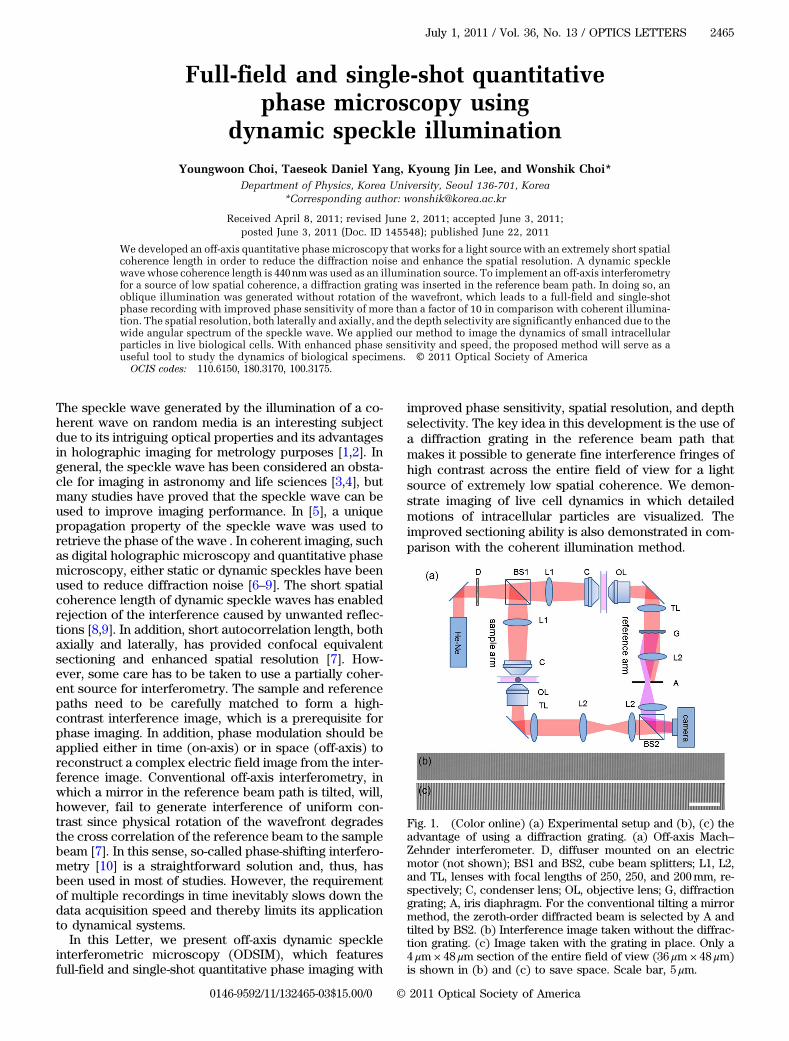
Fig. 1. (Color online) (a) Experimental setup and (b), (c) theadvantage of using a diffraction grating. (a) Off-axis Mach–Zehnder interferometer. D, diffuser mounted on an electricmotor (not shown); BS1 and BS2, cube beam splitters; L1, L2,and TL, lenses with focal lengths of 250, 250, and 200mm, re-spectively; C, condenser lens; OL, objective lens; G, diffractiongrating; A, iris diaphragm. For the conventional tilting a mirrormethod, the zeroth-order diffracted beam is selected by A andtilted by BS2. (b) Interference image taken without the diffrac-tion grating. (c) Image taken with the grating in place. Only a4 μm × 48 μm section of the entire field of view (36 μm × 48 μm)is shown in (b) and (c) to save space. Scale bar, 5 μm.
July 1, 2011 / Vol. 36, No. 13 / OPTICS LETTERS 2465
0146-9592/11/132465-03$15.00/0 © 2011 Optical Society of America

Our experimental setup [Fig. 1(a)] is an off-axis inter-ferometric microscope that uses a dynamic speckle fieldas an illumination source. An He–Ne laser illuminates arotating diffuser (DG20-1500-H2, Thorlabs, Inc.) to gener-ate a speckle field, which is then split by a beam splitter(BS1) into sample and reference beams. In ordinaryMach–Zehnder interferometry, it is difficult to form ahigh-contrast interference image due to the low spatial co-herence of the speckle waves. In our setup, the opticalconfigurations for the sample and reference arms arematched to deliver almost the same speckle waves to thedetector plane. Specifically, each arm has a condenserlens (Nikon 1.4 NA oil) for illumination, an objective lens(Olympus 1.4 NA oil), and a tube lens to deliver thesample image to the camera (Dalsa, Genie M1024) witha magnification of 100. The difference between the twoarms is that a sample in amedium is positioned in the sam-ple beam path while only the blank medium is placed inthe reference arm to match the path length. For the align-ment purpose, the diffuser is initially set to be stationaryand the speckle patterns coming through the two arms arematched by carefully positioning various optical elementsin the reference arm. Then the diffuser, mounted on a dcelectric motor, is rotated and the resulting interferenceimage is recorded with the camera, whose exposure timeis set to be longer than the period of the rotation. As a re-sult, wide intensity variation in the static speckle field iseliminated and the intensity distribution is made uniform.In order to obtain a quantitative phase image from a
single interference image, an off-axis interferometry isimplemented in which fine interference fringes have tobe generated by oblique illumination of a reference beamwith respect to the sample beam. When the spatial periodof the fringes is comparable to or smaller than the diffrac-tion-limited spot, the electric field of a sample beam canbe reconstructed by means of Hilbert transform up to thediffraction-limited resolution [11]. As mentioned earlier,the conventional approach of tilting a mirror in the refer-ence beam path results in an uneven contrast of interfer-ence across the field of view [Fig. 1(b)]. The physical tiltof the wavefront makes the reference speckle wave dif-ferent from that of the sample beam except at the axisof the tilt. As a result, the cross correlation between thetwo beams is reduced and the interference contrastdecreases away from the center where the axis of the tiltis positioned. There is about 25% contrast reductionper 10 μm on average. To maintain cross correlation, adiffraction grating (Edmund Optics, Ronchi ruling,40 lines=mm) is inserted in the reference beam path atthe conjugate plane to the camera and only the first-orderdiffracted beam is selected by an iris diaphragm (A).Then, the reference beam becomes oblique with respectto the optic axis without physical rotation of its wave-front. The spatial filtering process just adds a linearspatial phase ramp to the existing speckle fields, whichmakes interference fringes. The choice of the diffractiongrating is such that the period of the grating at the cameraplane is 25 μm, which is smaller than the diffraction-limited spot of 28 μm at the same plane. This conditionensures that the spatial resolution of the reconstructedimage is at least as good as that of the diffraction limit.As can be seen in Fig. 1(c), the fringe contrast is nowalmost uniform across the entire view field.
It is important to reduce the spatial coherence ofthe speckle illumination since it is strongly related to theattenuation of diffraction noise and enhancement in spa-tial resolution. The diffraction from dust particles, optics,and other parts of a sample located farther away from thefocal plane than the coherence length will not interferewith the signal of interest. The coherence length is deter-mined by the size of a speckle wave illuminating the con-denser lens. We set the opening of an aperture located atthe back of the condenser lens such that the condenserlens has an effective NA of 0.8. Then the coherencelength is measured to be 440 nm from the autocorrelationwidth of multiple static speckle fields, which is quiteclose to 483 nm as predicted by Van Cittert–Zernike the-orem. If the sample and reference arms are perfectlyidentical, the magnitude of the cross correlation is ex-pected to be the same as that of the autocorrelation.However, due to the aberration caused by the diffractiongrating, the cross correlation is reduced to 45% of theautocorrelation in our experiment, which is still suffi-cient to obtain a high-contrast image.
With improved interference images, quantitative phaseimaging of a live biological cell is performed. A microgliacell immersed in a culturemedium is placed in the sampleplane. Microglia cells extracted from a rat brain are incu-bated for seven days in a culture medium of Dulbecco’smodified eagle medium with 10% fetal bovine serumand then plated on a coverslip in a density of 50 cells=mm2 for observation. The interference image taken with-out the diffuser is shown in Fig. 2(a), where the diffractionnoise due to the floating particles in the culture medium
Fig. 2. (Color online) Quantitative phase imaging of a live cellusing ODSIM: (a) raw interference image, (c) processed quan-titative phase image, and (e) numerically simulated DIC imagefor the coherent illumination. (b), (d), and (f) Same as (a), (c),and (e) but with dynamic speckle illumination. The insets in (a)and (b) are zoom-in images at the background by three folds.Scale bar, 10 μm. Color bar, phase in radians. Media 1 shows thedynamics of the intracellular particles in the same cell. Imagesare taken with a frame rate of 2 fps.
2466 OPTICS LETTERS / Vol. 36, No. 13 / July 1, 2011

and other sources in the optics distorts the interferencefringes. With the ODSIM, the fringes are extremely cleandue to the significant reduction of noise [Fig. 2(b)]. Theinterference images are processed to acquire quantitativephase images using Hilbert transform. As can be seen inFig. 2(d), tiny vesicles inside the cell are clearly visible intheODSIM image. In the case of coherent illumination, thediffraction noise masks the particles with small phaseshifts [Fig. 2(c)]. The phase sensitivity is estimated by cal-culating the standard deviation of the phase variation inthe area of 8 μm × 8 μm void of the cell. With the use ofdynamic speckle illumination, the background phasenoise is reduced from 160 to 14mrad, which is equivalentto an optical path length of 1:3 nm. This is less than twicethe shot-noise-limited phase noise of 7:9mrad. Using thesingle-shot recording ability of the phase image, amovie ofthe same cell is recorded (Media 1). The data acquisitionspeed is limited only by the camera frame rate. As can beseen in Media 1, the vigorous motion of small particles isclearly observed, which was not possible with the coher-ent illumination. In addition, by numerically processingthe acquired phase image into a differential interferencecontrast (DIC) image to emphasize the field gradient, nu-clear membrane and internal structures of the cell areclearly visible in ODSIM [Fig. 2(f)]. For the coherent illu-mination case, diffraction noise is emphasized and maskscellular structures [Fig. 2(e)]. These results demonstratefull-field and single-shot phase recording of the ODSIMwith improved phase sensitivity. The ability of our techni-que in tracking intracellular particles paves the way forstudyingmicrorheology of biological cells without admin-istrating external agents [12,13].We now validate the sectioning ability of the ODSIM
originated from the short autocorrelation of the specklewave. Multiple 10-μm-sized polystyrene beads (Polybead,Microsphere) are prepared following the arrangementshown in Fig. 3(a). The medium is index-matching oilwith a refractive index of 1.56 (Cargille Labs). In conven-tional off-axis interferometry, the beads located awayfrom the objective focus are visible, with their bound-aries exhibiting diffraction rings [Figs. 3(b) and 3(c)].In contrast, when the ODSIM is used, each layer is visibleat the same time [Fig. 3(d) and 3(e)]. This clearly showsthe sectioning ability of the ODSIM. Only those beads lo-cated at the focus plane are visible, and each bead showsa clean boundary. In this recording, it is to be noted thatthe objective lens in the reference arm is scanned in con-junction with the scanning of the focus in the sample armin order to maintain the cross correlation. The pattern ofa speckle field varies as the focus plane is scanned.Therefore, constructive interference occurs only for thedepth matched to the reference arm, which is the originof the sectioning. In an additional experiment, the axialwidth of the point spread function is determined to be968� 38 nm by measuring the phase profile of a 350 nmbead along the axial direction. This is quite close to thetheoretically expected axial correlation length of thespeckle, which is λ=NA2 ¼ 980 nm. The lateral width ofthe same bead is also measured to be 343� 8nm. Thisis approximately the same as the diffraction limit of350 nm, but a little bit better than the correlation lengthof 440 nm. This will be due to the use of a higher NAobjective lens than the effective NA of a condenser lens.
Both axial and lateral resolutions are about twice betterthan those of the coherent illumination.
In conclusion, we developed an off-axis interfero-metric microscopy that works with extremely shortspatial coherence sources. Our method offers enhancedphase sensitivity, depth selectivity, and spatial resolutionin comparison with a conventional off-axis method thatuses coherent sources. We believe that the ODSIM willoffer opportunities to investigate nanoscale motion ofbiological specimens at the full speed of a camera.
This research was supported by the Basic Science Re-search Program through the National Research Founda-tion of Korea (NRF) funded by the Ministry of Education,Science and Technology (MEST, 2011-0005018 and 2011-0016568), the National R&D Program for Cancer Control,Ministry of Health & Welfare, South Korea (1120290), theKorea Science and Engineering Foundation (KOSEF,R17-2007-017-01000-0), and a Korea University grant.
References
1. J. W. Goodman, Speckle Phenomena in Optics: Theory and
Applications (Roberts, 2007).2. P. Jacquot and J. M. Fournier, in Interferometry in Speckle
Light: Theory and Applications: Proceedings of the Inter-
national Conference (Springer, 2000), pp. 25–28.3. J. E. Baldwin, C. A. Haniff, C. D. Mackay, and P. J. Warner,
Nature 320, 595 (1986).4. A. E. Desjardins, B. J. Vakoc, G. J. Tearney, and B. E.
Bouma, Opt. Express 14, 4736 (2006).5. P. F. Almoro, G. Pedrini, P. N. Gundu, W. Osten, and S. G.
Hanson, Opt. Lett. 35, 1028 (2010).6. Y. Park, W. Choi, Z. Yaqoob, R. Dasari, K. Badizadegan, and
M. S. Feld, Opt. Express 17, 12285 (2009).7. M. G. Somekh, C. W. See, and J. Goh, Opt. Commun. 174,
75 (2000).8. M. C. Pitter, C. W. See, and M. G. Somekh, Opt. Lett. 29,
1200 (2004).9. F. Dubois, M. L. N. Requena, C. Minetti, O. Monnom, and
E. Istasse, Appl. Opt. 43, 1131 (2004).10. K. Creath, Prog. Opt. 26, 349 (1988).11. T. Ikeda, G. Popescu, R. R. Dasari, and M. S. Feld, Opt. Lett.
30, 1165 (2005).12. D. Wirtz, Annu. Rev. Biophys. 38, 301 (2009).13. Z. Wang, L. Millet, M. Mir, H. F. Ding, S. Unarunotai, J.
Rogers, M. U. Gillette, and G. Popescu, Opt. Express 19,1016 (2011).
Fig. 3. (Color online) Depth selectivity of ODSIM. (a) Sche-matic diagram for beads in the sample plane. (b) and (c) Phaseimages taken without the diffuser with the objective foci lo-cated at the upper and lower dashed lines, respectively. (d) and(e) Same as (b) and (c) but with the use of the diffuser. Theobjective lens in the reference arm is also adjusted to matchthe speckle waves at each focus. Color bar, phase in radians.Scale bar, 10 μm.
July 1, 2011 / Vol. 36, No. 13 / OPTICS LETTERS 2467