fujifilm medical systems product profiles - home | fujifilm … · 2009-05-26 · 2 3 innovative...
TRANSCRIPT
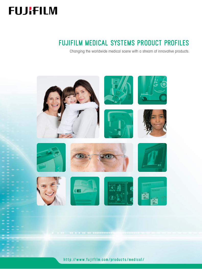
FUJIFILM MEDICAL SYSTEMS PRODUCT PROFILESChanging the worldwide medical scene with a stream of innovative products.
http://www.fujifilm.com/products/medical/Ref. No. XB-960E (SK·09·02·F1120·F9711) Printed in Japan ©2009 FUJIFILM Corporation

2 3
Innovative technology from Fujifilm is changing the medical scene all over the world.
Fujifilm helps to provide an efficient and precise diagnostic procedure by analyzing the image from an X-ray with techniques such as contrast and density automatic adjustment which enable parts of an image that were difficult to be seen to be displayed more clearly.
Image Intelligence™ also contributes to the field of medical imaging,
presenting only necessary information to the doctor.
Enhances FCR images. All diagnostic scopes will be enhanced except for noise.
Having pioneered the world’s first digital x-ray system Fuji Computed Radiography (FCR) in 1983, Fujifilm has maintained its focus on building technological innovations and offers yet another new solution to the medical field, Fujifilm Digital Radiography Systems (FUJIFILM DR). Utilizing Focused Phosphor Technology, FUJIFILM DR realizes simultaneously both high image quality and reduced x-ray exposure, and our product lineup has broadened as represented by our VELOCITY Ufp, VELOCITY Tfp, VELOCITY Unity fp, and the AMULET which is specifically designed for mammography. Fujifilm’s superb technology and diversified product lineup has gained recognition from medical institutions of all practices and over 60,000*1 digital imaging systems have been sold worldwide.
Just as with our FDR products, we are also firmly committed to expanding the array of our other product lines. Take a look at our FCR product lineup. We have introduced the FCR Go, Fujifilm’s first portable CR x-ray unit. Its flexible, compact design allows you to go just about everywhere within your facility to conduct exams and preview exposed images right on the spot all with speed and efficiency. And see the CAPSULA XL II which is evolving into a compact, multi-functional workstation compatible also for mammography exams.
As for our consoles, the FCRView has been released. Combining the capacity of an image viewer with operational and data administration capabilities, FCRView is the ultimate all-in-one viewer that gives you functionality from the start of initiating x-ray exposures to the end of archiving your data.
Then we have our DRYPIX series of dry imagers. Through technologies only available at Fujifilm, we provide the world’s fastest high quality images to any location at a medical institution.
And SYNAPSE, our image and information management system which has been implemented at over 2,000*2 medical institutions worldwide and alone proves its high evaluation for exceptional system stability and high image quality, can now be applied to cardiology and is constantly evolving.
Having gained its position as a leading company in medical imaging systems, Fujifilm is totally committed to bringing change to the medical field through its philosophy “Innovative Products through Continuous Progress.”
FujifilmMedical Imaging
Solutions
MFP Multi-Frequency Processing
Provides a non-grainy image by mainly isolating and suppressing the noise for the signal.
Removes the stationary grid patterns thus preventing Moiré from being generated resulting in easier diagnosis.
FNC Flexible Noise Control GPR Grid Pattern Removal
*1: As of 2008 1st half *2: As of 2009.1
Phosphor
Aluminium
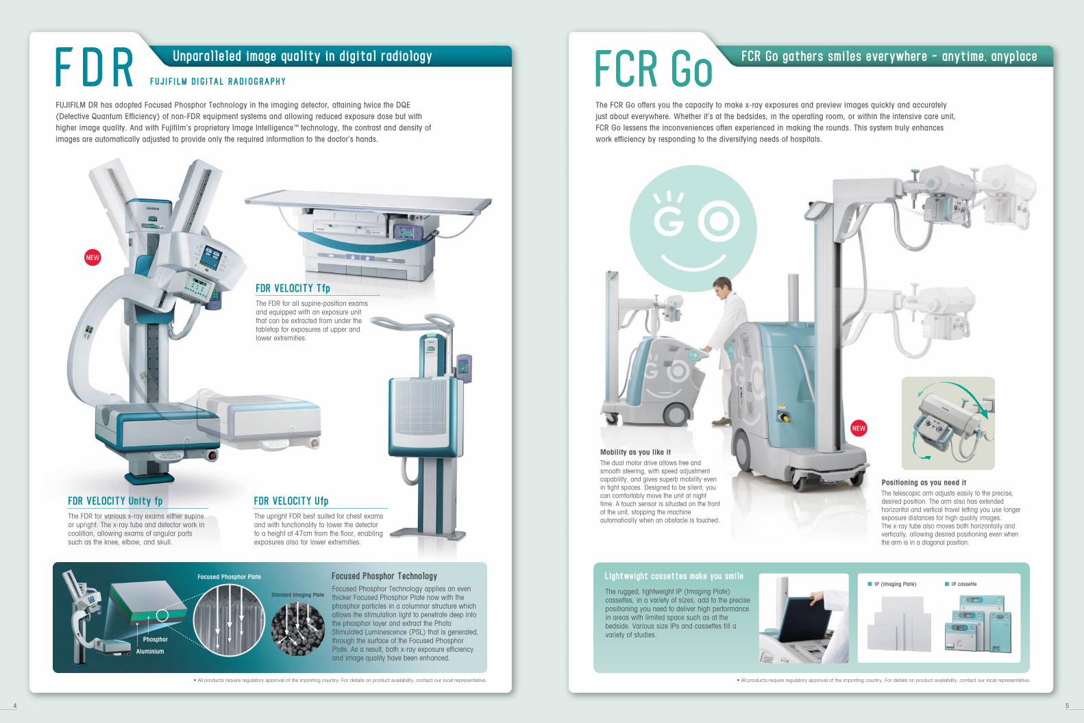
Focused Phosphor Technology applies an even thicker Focused Phosphor Plate now with the phosphor particles in a columnar structure which allows the stimulation light to penetrate deep into the phosphor layer and extract the Photo Stimulated Luminescence (PSL) that is generated, through the surface of the Focused Phosphor Plate. As a result, both x-ray exposure efficiency and image quality have been enhanced.
Focused Phosphor Technology
• All products require regulatory approval of the importing country. For details on product availability, contact our local representative.
FDR FUJ IF ILM DIGITAL RADIOGRAPHY
Unparalleled image quality in digital radiology
FUJIFILM DR has adopted Focused Phosphor Technology in the imaging detector, attaining twice the DQE (Detective Quantum Efficiency) of non-FDR equipment systems and allowing reduced exposure dose but with higher image quality. And with Fujifilm’s proprietary Image Intelligence™ technology, the contrast and density of images are automatically adjusted to provide only the required information to the doctor’s hands.
Standard Imaging Plate
Focused Phosphor Plate
FDR VELOCITY Unity fp
The upright FDR best suited for chest exams and with functionality to lower the detector to a height of 47cm from the floor, enabling exposures also for lower extremities.
The FDR for various x-ray exams either supine or upright. The x-ray tube and detector work in coalition, allowing exams of angular parts such as the knee, elbow, and skull.
FDR VELOCITY Ufp
The FDR for all supine-position exams and equipped with an exposure unit that can be extracted from under the tabletop for exposures of upper and lower extremities.
FDR VELOCITY Tfp
FCR Go gathers smiles everywhere - anytime, anyplace
The FCR Go offers you the capacity to make x-ray exposures and preview images quickly and accurately just about everywhere. Whether it’s at the bedsides, in the operating room, or within the intensive care unit, FCR Go lessens the inconveniences often experienced in making the rounds. This system truly enhances work efficiency by responding to the diversifying needs of hospitals.
FCR Go
The dual motor drive allows free and smooth steering, with speed adjustment capability, and gives superb mobility even in tight spaces. Designed to be silent, you can comfortably move the unit at night time. A touch sensor is situated on the front of the unit, stopping the machine automatically when an obstacle is touched.
The telescopic arm adjusts easily to the precise, desired position. The arm also has extended horizontal and vertical travel letting you use longer exposure distances for high quality images. The x-ray tube also moves both horizontally and vertically, allowing desired positioning even when the arm is in a diagonal position.
Mobility as you like it
Positioning as you need it
■ IP (Imaging Plate) ■ IP cassetteThe rugged, lightweight IP (Imaging Plate) cassettes, in a variety of sizes, add to the precise positioning you need to deliver high performance in areas with limited space such as at the bedside. Various size IPs and cassettes fill a variety of studies.
Lightweight cassettes make you smile
• All products require regulatory approval of the importing country. For details on product availability, contact our local representative.
4 5
FCR has remained the leader in the field for more than 20 years. FCR is a premium digital X-ray solution, offering the broadest product line to suit the requirements of nearly every imaging application. FCR’s leadership position is driven by uncompromised image quality, continued investment in technology innovation, development of systems with the highest productivity, and system implementation through the most experienced group of Professional Service individuals in the industry. FCR is the best possible solution for transition to digital at both large and small facilities.
Ideally suited for chest imaging with advanced scanning and image processing capabilities; features include HD LINESCAN Technology.
FCR VELOCITY U
Equipped with state-of-the-art functions including an optional 50-micron reading kit for Mammography applications.
High-quality and compact FCR for a broad range of diagnostic imaging. Small enough to fit almost anywhere – footprint 0.22 m2.
The high-efficiency FCR reader offering quality imaging and all-round versatility for superior diagnostic capability.
FCR CAPSULA XLII
High-resolution one-stacker FCR with 20 pixel/mm sampling pitch for digital mammography and pediatric imaging.
Superior image quality with 20 pixel/mm sampling pitch mammography and pediatric imaging with four-cassette stacker.
Flagship upright CR system with unique Energy Subtraction processing option.
Proven FCR technology for supine examinations with advanced scanning and image processing capabilities; features include HD LINESCAN Technology.
FCR CAPSULA X FCR XG5000 FCR PROFECT ONE*4 FCR PROFECT CS*4 FCR XU-D1
FCR VELOCITY T
*1: Not available in the US and Canada. In other countries, follow local regulations/guidelines*2: PMA (Premarket Approval) *3: FDA (U.S. Food and Drug Administration)*4: Image reader for Mammography
Imaging Plate and Cassette
FCR FUJ I COMPUTED RADIOGRAPHY
Pioneered over 20 years ago and still leading the way
6 7
• All products require regulatory approval of the importing country. For details on product availability, contact our local representative.
IP ST-VI IP HR-V IP Cassette Type CC IP Cassette Type CH
IP Cassette Type DSIP ST-BD IP HR-BD IP Cassette Type DM
IP Cassette Type LC
IP Cassette Type PC
FCR Imaging Plate for general purpose.
FCR Imaging Plate for single-sided mammography reading.
• 14" ✕ 17" (35.4 ✕ 43.2 cm)• 14" ✕ 14" (35.4 ✕ 35.4 cm)• 10" ✕ 12" (25.7 ✕ 30.5 cm)• 8" ✕ 10" (20.3 ✕ 25.4 cm)
• 24 ✕ 30 cm• 18 ✕ 24 cm
Standard Dual-Side Imaging Plate for Pediatric imaging.
FCR Imaging Plate for dual-sided mammography reading.
• 24 x 30 cm• 18 x 24 cm
• 24 ✕ 30 cm• 18 ✕ 24 cm
• 24 ✕ 30 cm• 18 ✕ 24 cm
FCR standard cassette with or without lead foil backside.
FCR special cassette for IP HR-V reading.
• 14" ✕ 17" (35.4 ✕ 43.2 cm)• 14" ✕ 14" (35.4 ✕ 35.4 cm)• 10" ✕ 12" (25.7 ✕ 30.5 cm)• 8" ✕ 10" (20.3 ✕ 25.4 cm)• 15 ✕ 30 cm
• 24 ✕ 30 cm• 18 ✕ 24 cm
FCR long view cassette for Scoliosis
• 35.4 ✕ 124.5 cm• 35.4 ✕ 101.7 cm• 35.4 ✕ 83.0 cm• 25.2 ✕ 58.0 cm• 24.0 ✕ 57.0 cm
FCR special cassette for Linac/Oncology
• 14" ✕ 17" (35.4 ✕ 43.2 cm)• 14" ✕ 14" (35.4 ✕ 35.4 cm)
FCR cassette for IP ST-BD. FCR cassette for dual-sided mammography reading.
• 24 ✕ 30 cm• 18 ✕ 24 cm
*1
Fujifilm Computed Radiography (CR), the world’s first CR that has acquired PMA*2 approval from FDA*3 for mammography.
Increases DQE (Detective Quantum Efficiency) by collecting the emissions from both sides of the IP with optimal, spatial frequency-dependent factors.
Dual-Side Reading Technology
a-Se used for both X-ray detection and switching
Fujifilm CR Digital Mammography Systems Bringing high quality with economy and reliability
Image reader for Mammography
Mammography QC Program
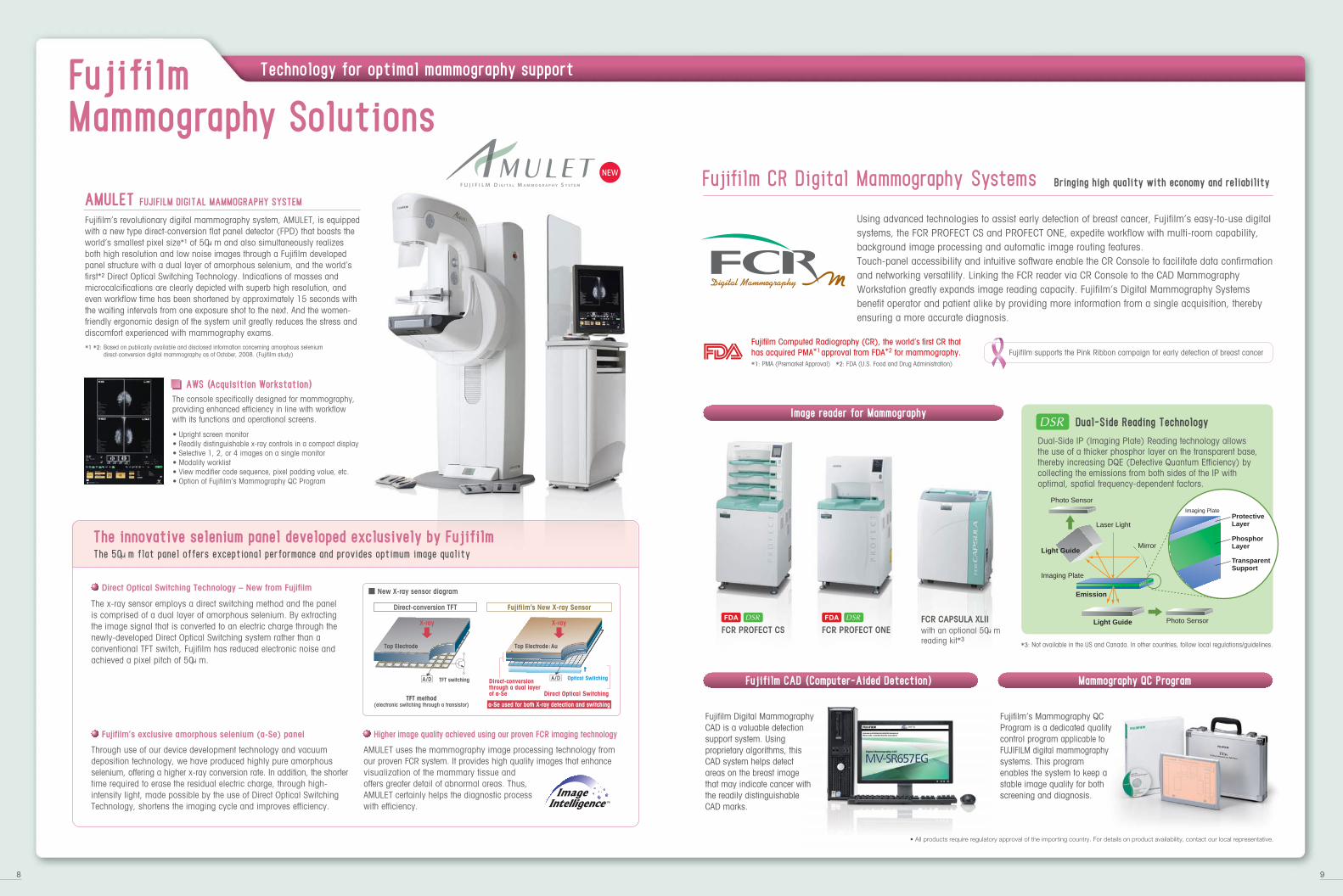
Using advanced technologies to assist early detection of breast cancer, Fujifilm’s easy-to-use digital systems, the FCR PROFECT CS and PROFECT ONE, expedite workflow with multi-room capability, background image processing and automatic image routing features. Touch-panel accessibility and intuitive software enable the CR Console to facilitate data confirmation and networking versatility. Linking the FCR reader via CR Console to the CAD Mammography Workstation greatly expands image reading capacity. Fujifilm’s Digital Mammography Systems benefit operator and patient alike by providing more information from a single acquisition, thereby ensuring a more accurate diagnosis.
Technology for optimal mammography support
8 9
FujifilmMammography Solutions
Fujifilm’s revolutionary digital mammography system, AMULET, is equipped with a new type direct-conversion flat panel detector (FPD) that boasts the world’s smallest pixel size*1 of 50μm and also simultaneously realizes both high resolution and low noise images through a Fujifilm developed panel structure with a dual layer of amorphous selenium, and the world’s first*2 Direct Optical Switching Technology. Indications of masses and microcalcifications are clearly depicted with superb high resolution, and even workflow time has been shortened by approximately 15 seconds with the waiting intervals from one exposure shot to the next. And the women-friendly ergonomic design of the system unit greatly reduces the stress and discomfort experienced with mammography exams.
AMULET FUJIFILM DIGITAL MAMMOGRAPHY SYSTEM
The console specifically designed for mammography, providing enhanced efficiency in line with workflow with its functions and operational screens.
• Upright screen monitor• Readily distinguishable x-ray controls in a compact display• Selective 1, 2, or 4 images on a single monitor• Modality worklist• View modifier code sequence, pixel padding value, etc.• Option of Fujifilm’s Mammography QC Program
Dual-Side IP (Imaging Plate) Reading technology allows the use of a thicker phosphor layer on the transparent base, thereby increasing DQE (Detective Quantum Efficiency) by collecting the emissions from both sides of the IP with optimal, spatial frequency-dependent factors.
Photo Sensor
Photo SensorLight Guide
Light Guide
Imaging Plate
Emission
Mirror
Laser LightProtectiveLayer
Transparent Support
Imaging Plate
PhosphorLayer
FCR CAPSULA XLIIFCR PROFECT CS FCR PROFECT ONE
Dual-Side Reading Technology
The 50μm flat panel offers exceptional performance and provides optimum image quality
Direct Optical Switching Technology – New from Fujifilm
AWS (Acquisition Workstation)
The innovative selenium panel developed exclusively by Fujifilm
The x-ray sensor employs a direct switching method and the panel is comprised of a dual layer of amorphous selenium. By extracting the image signal that is converted to an electric charge through the newly-developed Direct Optical Switching system rather than a conventional TFT switch, Fujifilm has reduced electronic noise and achieved a pixel pitch of 50μm.
Through use of our device development technology and vacuum deposition technology, we have produced highly pure amorphous selenium, offering a higher x-ray conversion rate. In addition, the shorter time required to erase the residual electric charge, through high-intensity light, made possible by the use of Direct Optical Switching Technology, shortens the imaging cycle and improves efficiency.
AMULET uses the mammography image processing technology from our proven FCR system. It provides high quality images that enhance visualization of the mammary tissue and offers greater detail of abnormal areas. Thus, AMULET certainly helps the diagnostic process with efficiency.
Fujifilm’s exclusive amorphous selenium (a-Se) panel Higher image quality achieved using our proven FCR imaging technology
*1 *2: Based on publically available and disclosed information concerning amorphous selenium direct-conversion digital mammography as of October, 2008. (Fujifilm study)
Fujifilm Digital Mammography CAD is a valuable detection support system. Using proprietary algorithms, this CAD system helps detect areas on the breast image that may indicate cancer with the readily distinguishable CAD marks.
Fujifilm CAD (Computer-Aided Detection)
with an optional 50μmreading kit*3
*3: Not available in the US and Canada. In other countries, follow local regulations/guidelines.
Direct-conversion TFT Fujifilm’s New X-ray Sensor
Direct Optical SwitchingTFT method
(electronic switching through a transistor)
Fujifilm’s Mammography QC Program is a dedicated quality control program applicable to FUJIFILM digital mammography systems. This program enables the system to keep a stable image quality for both screening and diagnosis.
• All products require regulatory approval of the importing country. For details on product availability, contact our local representative.
New X-ray sensor diagram
Top Electrode
A/D A/DTFT switching Optical SwitchingDirect-conversionthrough a dual layerof a-Se
Top Electrode:Au
X-ray X-ray
Fujifilm Computed Radiography (CR), the world’s first CR that has acquired PMA*1 approval from FDA*2 for mammography.*1: PMA (Premarket Approval) *2: FDA (U.S. Food and Drug Administration)
Fujifilm supports the Pink Ribbon campaign for early detection of breast cancer
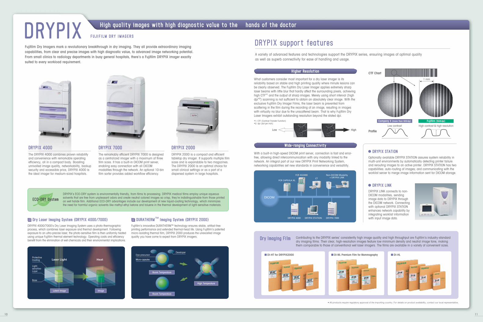
Fujifilm Dry Imagers mark a revolutionary breakthrough in dry imaging. They all provide extraordinary imaging capabilities, from clear and precise images with high diagnostic value, to advanced image networking potential. From small clinics to radiology departments in busy general hospitals, there’s a Fujifilm DRYPIX imager exactly suited to every workload requirement.
The DRYPIX 4000 combines proven reliability and convenience with remarkable operating efficiency, all in a compact body. Boasting unrivalled image quality, networkability, backup security and accessible price, DRYPIX 4000 is the ideal imager for medium-sized hospitals.
DRYPIX 4000
The remarkably efficient DRYPIX 7000 is designed as a centralized imager with a maximum of three film sizes. It has a built-in DICOM print server, enabling easy connection with all DICOM modalities through the network. An optional 10-bin film sorter provides added workflow efficiency.
DRYPIX 7000
DRYPIX 2000 is a compact and efficient tabletop dry imager. It supports multiple film sizes and is expandable to two magazines. The DRYPIX 2000 is an optimal choice for small clinical settings or as a part of a dispersed system in large hospitals.
DRYPIX 2000
DRYPIX support features
A variety of advanced features and technologies support the DRYPIX series, ensuring images of optimal quality as well as superb connectivity for ease of handling and usage.
What customers consider most important for a dry laser imager is its reliability based on stable and high printing quality where minute lesions can be clearly observed. The Fujifilm Dry Laser Imager applies extremely sharp laser beams with little blur that hardly affect the surrounding pixels, achieving high CTF*1 and the output of sharp images. Merely using short interval (high dpi*2) scanning is not sufficient to obtain an absolutely clear image. With the exclusive Fujifilm Dry Imager Films, the laser beam is prevented from scattering in the film during the recording of an image, resulting in images with virtually no blur due to the unscattered beam. That is why Fujifilm Dry Laser Imagers exhibit outstanding resolution beyond the stated dpi.
With a built-in high-speed DICOM print server, connection is fast and error-free, allowing direct intercommunication with any modality linked to the network. An integral part of our new DRYPIX Print Networking System, networking capabilities set new standards in convenience and versatility.
DRYPIX LINK
Optionally available DRYPIX STATION assures system reliability in multi-unit environments by automatically detecting printer failure and rerouting images to an active printer. DRYPIX STATION has two capabilities: auto-routing of images; and communicating with the worklist server to merge image information sent for DICOM storage.
DRYPIX STATION
DRYPIX LINK connects to non-DICOM modalities, sending image data to DRYPIX through the DICOM network. Connecting with optional DRYPIX STATION enhances network capability by integrating worklist information with input image data.
DRYPIX 4000/7000’s Dry Laser Imaging System uses a photo-thermographic process, which combines laser exposure and thermal development. Following exposure to an ultra-precise laser, the photo-sensitive film is then uniformly heated using unique Fujifilm thermal element technology. Operating costs and efficiency benefit from the elimination of wet chemicals and their environmental implications.
Dry Laser Imaging System (DRYPIX 4000/7000)Fujifilm’s innovative DURATHERM™ technology ensures stable, artifact-free printing performance and extended thermal-head life. Using Fujifilm’s patented micro-isolating thermal film, DRYPIX 2000 produces the unexcelled image quality you have come to expect from DRYPIX imagers.
Laser Light HeatProtectiveCoating
Light-sensitive-Layer
Base
Latent Image Image
Room Temperature
Room Temperature
High Temperature
Micro-capsules
DeveloperDye-precursor
DRYPIX’s ECO-DRY system is environmentally friendly, from films to processing. DRYPIX medical films employ unique aqueous solvents that are free from unpleasant odors and create neutral colored images so crisp, they’re indistinguishable from those printed on wet halide film. Additional ECO-DRY advantages include our development of new liquid-coating technology, which minimizes the need for harmful organic solvents like methyl-ethyl ketone and toluene in the thermal development of light-sensitive materials.
Dry Imaging Film Contributing to the DRYPIX series’ consistently high image quality and high throughput are Fujifilm’s industry-standard dry imaging films. Their clear, high-resolution images feature low minimum density and neutral image tone, making them comparable to those of conventional wet laser imagers. The films are available in a variety of convenient sizes.
■ DI-HL■ DI-HT for DRYPIX2000 ■ DI-ML Premium Film for Mammography
FCR CAPSULA XL
FCR XG5000
CT
Non-DICOM Modality+ DRYPIX LINK
DICOM
DRYPIX 4000 DRYPIX 7000DRYPIX STATION
Higher Resolution
Wide-ranging Connectivity
DRYPIX FUJ IF ILM DRY IMAGERS
Low High
CTF Chart
Profile
1 mm
Low contrast High contrast & high resolution
Company X (more than 600 dp) Fujifilm (508 dpi)
ECO-DRY System
DURATHERM™ Imaging System (DRYPIX 2000)
10 11
• All products require regulatory approval of the importing country. For details on product availability, contact our local representative.
High quality images with high diagnostic value to the hands of the doctor
*1: CTF (Contrast Transfer Function)*2: dpi (dot per inch)
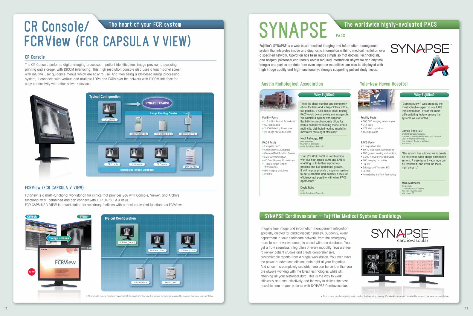
Fujifilm’s SYNAPSE is a web-based medical imaging and information management system that integrates image and diagnostic information within a medical institution over a specified network. Operation has been made simple so that doctors, technologists, and hospital personnel can readily obtain required information anywhere and anytime. Images and past exam data from even separate modalities can also be displayed with high image quality and high-functionality, strongly supporting patient study needs.
SYNAPSE PACS
The worldwide highly-evaluated PACS
Austin Radiological Association
“With the sheer number and complexity of our facilities and subspecialties within our practice, a rules-based (auto-routing) PACS would be completely unmanageable. We wanted a system with superior flexibility to simultaneously allow for both a centralized reading model and a multi-site, distributed reading model to maximize radiologist efficiency.”
Neal Rutledge, MD Neuroradiologist Chairman, IT Committee Austin Radiological Association
“Our SYNAPSE PACS in combination with our high speed WAN and SAN is enabling us to further expand our practice and fuel additional growth. It will help us provide a superior service to our customers and achieve a level of efficiency not possible with other PACS approaches.”
Doyle RabeCEOAustin Radiological Association
Facility Facts• 1.2 Million Annual Procedures• 62 Radiologists• 2,500 Referring Physicians• 27 Image Acquisition Sites
PACS Facts• Enterprise PACS• Clustered PACS Database• Clustered Multifunction Servers• EMC Symmetrix®SAN• 40 Dual Display Workstations• 100s of Single Display Workstations•160 Imaging Modalities• IDX RIS
Why Fujifilm?
Yale-New Haven Hospital
“CommonView™ was probably the most valuable aspect of our PACS implementation. It was the main differentiating feature among the systems we evaluated.”
James Brink, MDChief of Diagnostic Radiology Yale-New Haven Hospital Professor and Chairman Dept. of Diagnostic Radiology Yale University School of Medicine New Haven, CT
Facility Facts• 300,000 imaging exams a year • 944 beds • 471 staff physicians • 45 radiologists
PACS Facts• 9 acquisition sites • 60-70 diagnostic workstations • 700 general viewing workstations • 3,000-4,000 SYNAPSE®users • 160 imaging modalities • Fuji CR • Eclipsys and Tektronix HIS • GE RIS • PowerScribe and Talk Technology
“The system has allowed us to create an enterprise-wide image distribution system. A case from 7 years ago can be requested, and it will be there right away...”
Why Fujifilm?
Mike Matthews Administrator Clinical Information Systems Yale-New Haven Hospital New Haven, CT
Imagine true image and information management integration specially created for cardiovascular studies: Suddenly, every department in your healthcare network, from the emergency room to non-invasive areas, is united with one database. You get a truly seamless integration of every modality. You are free to review patient studies and create comprehensive, customizable reports from a single workstation. You even have the power of advanced clinical tools right at your fingertips. And since it is completely scalable, you can be certain that you are always working with the latest technologies while still retaining all your historical data. This is the way to work efficiently and cost-effectively and the way to deliver the best possible care to your patients with SYNAPSE Cardiovascular.
SYNAPSE Cardiovascular - Fujifilm Medical Systems Cardiology
12 13
• All products require regulatory approval of the importing country. For details on product availability, contact our local representative.
CR Console/FCRView (FCR CAPSULA V VIEW) The CR Console performs digital imaging processes – patient identification, image preview, processing, printing and storage, with DICOM interfacing. This high resolution console also uses a touch-panel screen with intuitive user guidance menus which are easy to use. And then being a PC-based image processing system, it connects with various and multiple FDRs and FCRs over the network with DICOM interface for easy connectivity with other network devices.
FCRView is a multi-functional workstation for clinics that provides you with Console, Viewer, and Archive functionality all combined and can connect with FCR CAPSULA X or XLII. FCR CAPSULA V VIEW is a workstation for veterinary facilities with almost equivalent functions as FCRView.
Typical Configuration
SYNAPSE (PACS)
Distributed Image Database
Image Reading Cluster
CR Console
DRYPIX 7000CR Console CR Console CR Console
FCR PROFECT CS FCR VELOCITY UFCR CAPSULA XLII
FCRView (FCR CAPSULA V VIEW)
CR Console
Change screens
Typical Configuration
FCRView (server)
FCRView (client) FCRView (client)
FCR CAPSULA X DRYPIX 2000
• All products require regulatory approval of the importing country. For details on product availability, contact our local representative.
The heart of your FCR system
Console Viewer
* Processing capacity is an assumed mixture of lumbar spine (40%), abdomen (20%) and extremities (40%).
Processing Capacity (IPs/hr)
Pixels
Pixel size
Preview image
W
D
H
Weight (kg)
More Details (Ref. No.)
VELOCITY T fp
140
4280 ✕ 4280
2350
810
650-850
471
XB-762E
VELOCITY U fp
240
4280 ✕ 4280
645
450
1835
220
XB-761E
100 microns
9 seconds
Exposure Unit
Image size
Pixels
Pixel size
Preview image
X-ray Generator Power rating
W
D
H
Weight (Universal Arm Stand: kg)
More Details (Ref. No.)
VELOCITY Unity
XB-866E XB-861ER
FPP200 HS100
17” ✕ 17”
4280 ✕ 4280
100 microns
Approx. 9 seconds
50/64/80 kW
2360
1465
2650
500
VELOCITY Unity fp
Dimensions(Universal Arm Stand: mm)
IP type
Cassette type
Long view
17” ✕ 17”
14” ✕ 14”
Power rating
kVp range
Traveling speed
Dimensions (mm) W ✕ D ✕ H
Weight (kg)
More Details (Ref. No.)
*1: ST-VI type processing *2: High speed mode
• QC,QA functions: supported • AWS: supported
FCR Go
ST-VI, ST-VN
62 (87)*2
70 (94)*2
15 kW
40-130 (1kV steps)
max 5 km/hr
620 ✕ 1335 ✕ 1925
540
XB-868E
Approx.15 seconds
SPECIFICATIONS
Pixel size
X-ray tube Target
Filter
More Details (Ref. No.)
50 microns
W/Mo
Mo/Rh
XB-961E
AMULET
IP size
FUJIFILM DR FUJIFILM DR
Modality Worklist, Modality Performed Procedure Step, Basic Grayscale Print, CR Image Storage, Storage Commitment
Electronic Shutter, Free Annotation, Image Composition, Auto-menu Selection, LUT Adjustment, FCR QC Program, Tiling QA, Multi-frequency Processing, Flexible Noise Control, Grid Pattern Removal,
Energy Subtraction, Pattern Enhancement Processing for Mammography
CAPSULA XLII
XB-764E
62
1464 ✕ 2964
1770 ✕ 2370
2364 ✕ 2964
—
—
—
ST-VI
n.a.
590
380
810
99
0.29
XB-564E
60
—
1770 ✕ 2370
2364 ✕ 2964
3540 ✕ 4740
4728 ✕ 5928
—
ST-VI, HR-V, ST-BD, HR-BD
yes (18✕24/24✕30)
655
740
1330
240
0.7
PROFECT ONE
15 ✕ 30 cm (10 pixels)
18 ✕ 24 cm (10 pixels)
24 ✕ 30 cm (10 pixels)
18 ✕ 24 cm (20 pixels)
24 ✕ 30 cm (20 pixels)
35 ✕ 35 cm (10 pixels)
35 ✕ 43 cm (10 pixels)
43 ✕ 43 cm (10 pixels)
20 ✕ 25 cm (10 pixels)
25 ✕ 30 cm (10 pixels)
Applicable IP Type
Dual Side Reading
W
D
H
Weight (kg)
Power Consumption (kW)
DICOM Compatibility
Other Options forCR Console
More Details (Ref. No.)
Matrix Size
CAPSULA X
XB-567E
43
1464 ✕ 2964
1770 ✕ 2370
2364 ✕ 2964
—
—
—
ST-VI
n.a.
590
380
810
99
0.2
XB-362E
103
—
1770 ✕ 2370
2364 ✕ 2964
—
—
—
ST-VI, HR-V
n.a.
655
740
1480
285
0.7
XG-5000
240
—
2000 ✕ 2510
2505 ✕ 3015
—
—
4280 ✕ 4280
Deviced IP
n.a.
645
450
1830
220
1.0
VELOCITY U
XB-364EXB-363E
103
—
1770 ✕ 2370
2364 ✕ 2964
3540 ✕ 4740
4728 ✕ 5928
—
ST-VI, HR-V, ST-BD, HR-BD
yes (18✕24/24✕30)
655
740
1480
285
0.7
PROFECT CS
140*
—
2000 ✕ 2510
2505 ✕ 3015
—
—
4280 ✕ 4280
Deviced IP
n.a.
2100
815
500 to 900
411
1.0
VELOCITY T
XB-465E XB-264E
122
—
2000 ✕ 2510
2505 ✕ 3015
—
—
4280 ✕ 4280
ST-55BD
Yes
1170
800
1800
525
1.5
XU-D1
2000 ✕ 2510
2505 ✕ 3015
3520 ✕ 3520
3520 ✕ 4280
DI-HT
(90)
n.a.
(75)
n.a.
(50)
No
530
400
43
max. 0.5
XB-662E
70002000 4000
Film Type
Film Base
20 ✕ 25 cm
25 ✕ 30 cm
26 ✕ 36 cm
35 ✕ 35 cm
35 ✕ 43 cm
Format (Portrait)
Format (Landscape)
Density Adjustment
Mammographic Applicability
W
D
H
Weight (kg)
Power Consumption (kW)
More Details (Ref. No.)
Available Film Size(per hour capacity)
Dimensions (mm)
Automatic
Blue
470 (with small magazine)
590 (with large magazine)
*1 DI-ML 35 ✕ 43 film size is not available.
FCR DRYPIX
Throughput *1(IPs/hour)
Type CC, Type LC (Long view)
17” ✕ 14”, 14” ✕ 14” 10” ✕12”, 8” ✕ 10” 15 ✕ 30 cm (ST-VI)
Processing Capacity (35 ✕ 43 IP per hour)
Time interval required between exposures
Microsoft, Windows, and Internet Explorer are trademarks, or registered trademarks of Microsoft Corporation in the United States and/or other countries. All other trademarks are the property of their respective holders.
59(with optional
sheet feeder unit)
1, 2, 3, 4, 6, 8, 9, 12, 15, 16, 18, 20, 24, 25, 28, 30, 32, 35, 36, 40, 42, 48, 49, 54, 56, 60, 63, 64, 70, Mix formats
1, 2, 3, 4, 6, 8, 9, 12, 15, 16, 18, 20, 24, 25, 28, 30, 32, 35, 36, 40, 42, 48, 49, 54, 56, 60, 63, 64, 70, Mix formats
Dimensions(Reader Unit: mm)
X-ray Generator
Dimensions(reader unit, mm)
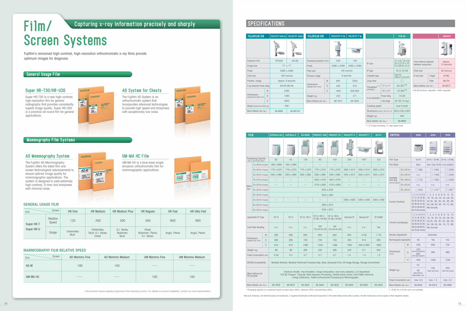
Super HR-T
Super HR-U
RelativeSpeed
Usage
HR FineScreenFilm HR Medium HR Medium Plus HR Regular HR Fast
120 200 300 400 600
HR Ultra Fast
800
ExtremitiesSkull
Extremities,Skull, G.I. Series,
Chest
G.I. Series,Abdomen,
Skull
Chest,Abdomen, Pelvis,
G.I. SeriesAngio, Pelvis Angio, Pelvis
100 140
AD Mammo Fine AD Mammo Medium UM Mammo Fine UM Mammo Medium
GENERAL USAGE FILM
MAMMOGRAPHY FILM RELATIVE SPEED
UM-MA HC
100 140AD-M
ScreenFilm
Super HR-T30/HR-U30
Super HR-T30 is a new high-contrast, high-resolution film for general radiography that provides consistently superb image quality. Super HR-U30 is a practical all-round film for general applications.
AD System for Chests
The Fujifilm AD System is an orthochromatic system that incorporates advanced technologies to provide high speed and sharpness with exceptionally low noise.
AD Mammography System
The Fujifilm AD Mammography System offers the latest film and screen technological advancements to ensure optimal image quality for mammographic applications. The system is designed to yield extremely high-contrast, D-max and sharpness with minimal noise.
UM-MA HC Film
UM-MA HC is a blue-base single-emulsion orthochromatic film for mammographic applications.
Fujifilm’s renowned high-contrast, high-resolution orthochromatic x-ray films provide optimum images for diagnosis.
14 15
Yes
600
585
1040
130 (with one tray)
max. 1.5
XB-561E
Yes
735
680
1240
203 (with two trays)
max. 2.2
XB-266E
DI-HL / DI-ML
(160)
(160)
(160)
n.a.
(110)*1
DI-HL / DI-ML
(200)
(230)
(240)
n.a.
(180)*1
Blue / Clear (DI-ML is not available)
1, 2, 3, 4, 6, 8, 9, 12, 15, 16, 18, 20, 24, 25, 28, 30, 32,35, 36, 40, 42, 48, 49, 54, 56, 63, 64, 70, 72, 80, Mix formats
1, 2, 3, 4, 6, 8, 9, 12, 15, 16, 18, 20, 24, 25, 28, 30, 32,35, 36, 40, 42, 48, 49, 54, 56, 63, 64, 70, 72, 80, Mix formats
General Usage Film
Mammography Film Systems
• All products require regulatory approval of the importing country. For details on product availability, contact our local representative.
Film/Screen Systems
Capturing x-ray information precisely and sharply
FUJIFILM MEDICAL SYSTEMS PRODUCT PROFILESChanging the worldwide medical scene with a stream of innovative products.
http://www.fujifilm.com/products/medical/Ref. No. XB-960E (SK·09·02·F1120·F9711) Printed in Japan ©2009 FUJIFILM Corporation