fujifilm endoscopy system · fujifilm endoscopy system product guide. enhancing the quality of life...
TRANSCRIPT
Specifications are subject to change without notice.10 New Industrial Road, FUJIFILM Building, Singapore 536201http://www.fujifilm.com.sg
FUJIFILM Asia Paci�c Pte. Ltd.
Electronic Video Endoscopy andEndoscopic Ultrasonography
FUJIFILMEndoscopy System
PRODUCT GUIDE
Enhancing the quality of life of people worldwide
FUJIFILM is known as the world’s largest photographic and imaging company and is pioneering in diagnostic
imaging and information systems for health-care facilities. The current endoscopic equipment provides high-definition
fo egnar lautca ehT .stsigolonomlup dna stsigoloretneortsag rof dnuosartlu cipocsodne dna ypocs odne oediv
endoscopes and the EPX-4450HD processor technology come with FICE Dual Mode and DICOM on-board.
We will use leading-edge, proprietary technologies to provide top-quality products and services that contribute
to the advancement of culture, science, technology and industry, as well as improved health and environmental
protection in society. Our overarching aim is to help enhance the quality of life of people worldwide.
Innovative solutions As one of the leading companies in the development
of endoscope technology, FUJIFILM regularly
sets new benchmarks in the industry, for example
with devices for double balloon endoscopy and
transnasal endoscopy. However, the focus at
FUJIFILM is very much on holistic patient care. Our
service portfolio therefore also includes competent
technical assistance, a comprehensive range of
hygiene products and individual consulting.
New opportunitiesWhether it is with the most advanced optical technology,
state-of-the-art digital image processing or new
examination methods, FUJIFILM is always creating
new opportunities in the world of endoscopy. In this
way, we are making a significant contribution to the
early detection of diseases and their successful
treatment.
Dedicated research, the continuous enhancement
of our technology, the highest quality demands and
close working relationships with international specialists
set the global standard in FUJIFILM endoscopy
and endosonography.
Index
600 series and EPX-4450HD Technology 4 – 12
600 series endoscopes
580 series endoscopes
Double Balloon Endoscope
590 series endoscopes
2500 system
530 series endoscopes
38 – 39
Therapeutic Accessories
Endoscopic ultrasonography
Peripherals
Processors
14 – 15
13
16 – 20
21 – 23
24 – 25
26 – 30
31 – 32
33 – 36
37
TherapeuticEndoscopy
EUS
Ultra-slim
TNEEndoscopy
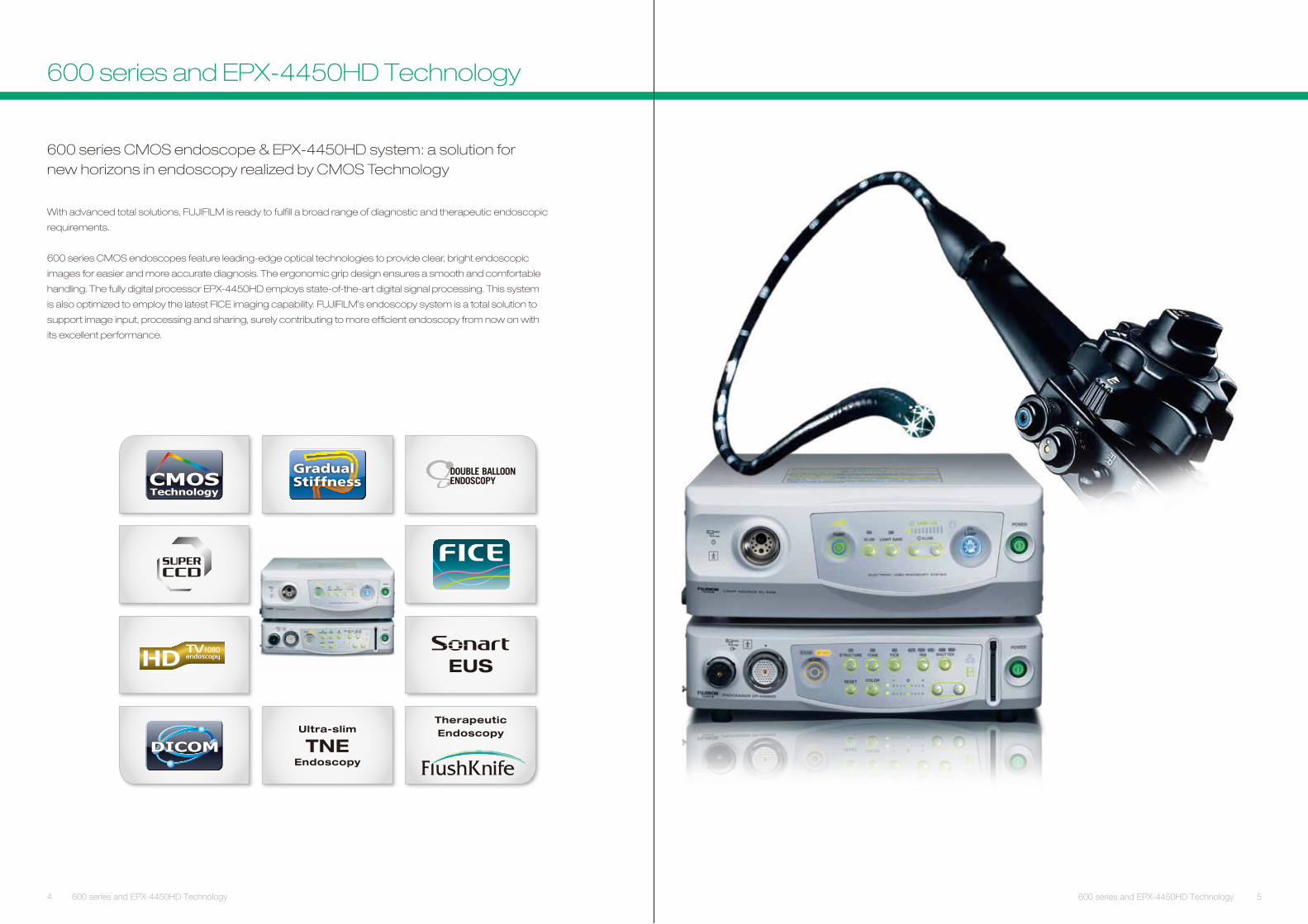
600 series CMOS endoscope & EPX-4450HD system: a solution fornew horizons in endoscopy realized by CMOS Technology
With advanced total solutions, FUJIFILM is ready to fulfill a broad range of diagnostic and therapeutic endoscopic
requirements.
600 series CMOS endoscopes feature leading-edge optical technologies to provide clear, bright endoscopic
images for easier and more accurate diagnosis. The ergonomic grip design ensures a smooth and comfortable
handling. The fully digital processor EPX-4450HD employs state-of-the-art digital signal processing. This system
is also optimized to employ the latest FICE imaging capability. FUJIFI LM’s endoscopy system is a total solution to
support image input, processing and sharing, surely contributing to more effi cient endoscopy from now on with
its excellent performance.
DICOM
CMOSTechnology
4 600 series and EPX-4450HD Technology 5600 series and EPX-4450HD Technology
GradualStiffness
600 series and EPX-4450HD Technology
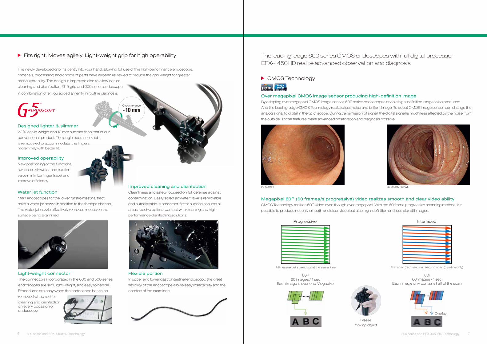
The leading-edge 600 series CMOS endoscopes with full digital processorEPX-4450HD realize advanced observation and diagnosis
CMOS Technology
Over megapixel CMOS image sensor producing high-definition imageBy adopting over megapixel CMOS image sensor, 600 series endoscopes enable high-definition image to be produced.
And the leading-edge CMOS Technology realizes less noise and brilliant image. To adopt CMOS image sensor can change the
analog signal to digital in the tip of scope. During transmission of signal, the digital signal is much less affected by the noise from
the outside. Those features make advanced observation and diagnosis possible.
EC-600WM/WI/WLEG-600WR
Megapixel 60P (60 frames/s progressive) video realizes smooth and clear video abilityCMOS Technology realizes 60P video even though over megapixel. With the 60 frame progressive scanning method, it is
possible to produce not only smooth and clear video but also high-definition and less blur still images.
InterlacedProgressive
All lines are being read out at the same time
60P60 images / 1 sec
Each image is over one Megapixel
First scan (red line only) , second scan (blue line only)
60i60 images / 1 sec
Each image only contains half of the scan
Freeze
moving object
CMOSTechnology 60P Video
MegaPixel
Overlay
6
Improved operabilityNew positioning of the functional
switches, air/water and suction
valve minimize finger travel and
improve efficiency.
7600 series and EPX-4450HD Technology 600 series and EPX-4450HD Technology
Circumference
- 10 mm
The newly developed grip fits gently into your hand, allowing full use of this high-performance endoscope.
Materials, processing and choice of parts have all been reviewed to reduce the grip weight for greater
maneuverability. The design is improved also to allow easier
cleaning and disinfection. G-5 grip and 600 series endoscope
in combination offer you added amenity in routine diagnosis.
. Light-weight grip for high operabilityyleliga sevoM .thgir stiF
Designed lighter & slimmer20 % less in weight and 10 mm slimmer than that of our
conventional product. The angle operation knob
is remodeled to accommodate the fingersmore firmly with better fit.
Light-weight connectorThe connectors incorporated in the 600 and 500 series
endoscopes are slim, light-weight, and easy to handle.
Procedures are easy when the endoscope has to be
removed/attached for
cleaning and disinfectionon every occasion ofendoscopy.
Flexible portionIn upper and lower gastrointestinal endoscopy, the great
flexibility of the endoscope allows easy insertability and the
comfort of the examinee.
Water jet functionMain endoscopes for the lower gastrointestinal tract
have a water jet nozzle in addition to the forceps channel.
The water jet nozzle effectively removes mucus on the
surface being examined.
Improved cleaning and disinfectionCleanliness and safety focused on full defense against
contamination. Easily soiled air/water valve is removable
and autoclavable. A smoother, flatter surface assures all
areas receive optimal contact with cleaning and high-
performance disinfecting solutions.
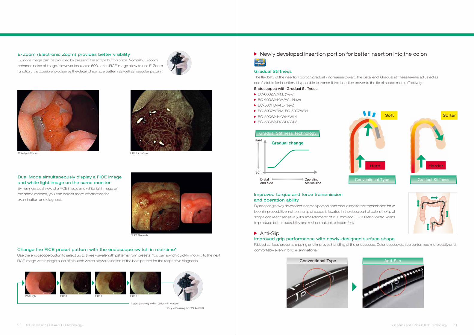
Tissue surface
Reconstructed image(FICE image)
Engine
Conventional imageWhite light
mn007mn004
FICE (Flexible spectral Imaging Color Enhancement)
FICE – “Flexible spectral Imaging Color Enhancement“ – in the new
EPX-4450HD yields diagnostic results without any need for tissue
staining. The procedure digitally limits the wavelengths of the light and
displays it in up to ten different color combinations. The endoscope
switch allows physicians to switch between the conventional image
and the FICE image in a split second, ensuring an uninterrupted
examination with the eyes always concentrated on the monitor.
FICE1 Esophagus FICE1 Stomach
FICE1 Colon FICE8 Colon
FICE with CMOS Technology
FICE combination with CMOS Technology provides advanced FICE imageThrough high-definition and improved noise reduction, FICE images are sharper and clearer than ever.
It enables easier differentiation between lesion-affected and non-affected tissue.
8
EC-600WM / WI / WL
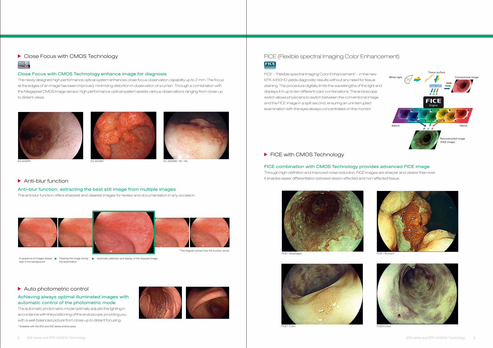
Close Focus with CMOS Technology
Auto photometric control
Close Focus with CMOS Technology enhance image for diagnosisThe newly designed high performance optical system enhances close focus observation capability up to 2 mm. The focus
at the edges of an image has been improved, minimizing distortion in observation of a lumen. Through a combination with
the Megapixel CMOS image sensor, high performance optical system assists various observations ranging from close-up
to distant views.
Achieving always optimal illuminated images with automatic control of the photometric modeThe automatic photometric mode optimally adjusts the lighting in
accordance with the positioning of the endoscope, providing you
with a well-balanced picture from close-up to distant focusing.
* Available with the 600 and 500 series endoscopes
Anti-blur function
Anti-blur function: extracting the best still image from multiple imagesThe anti-blur function offers sharpest and clearest images for review and documentation in any occasion.
CloseFocus
EG-600WR EG-600WR
* This diagram shows how the function works
Automatic selection and display of the sharpest imageFreezing the image during the examination
A sequence of images always kept in the background
9600 series and EPX-4450HD Technology 600 series and EPX-4450HD Technology
10 11
Change the FICE preset pattern with the endoscope switch in real-time*Use the endoscope button to select up to three wavelength patterns from presets. You can switch quickly, moving to the next
FICE image with a single push of a button which allows selection of the best pattern for the respective diagnosis.
White light FICE 0 FICE 1 FICE 8
Instant switching (switch patterns in rotation)
*Only when using the EPX-4450HD
Dual Mode simultaneously display a FICE image and white light image on the same monitorBy having a dual view of a FICE image and white light image on
the same monitor, you can collect more information for
examination and diagnosis.
FICE1 Stomach
E-Zoom (Electronic Zoom) provides better visibilityE-Zoom image can be provided by pressing the scope button once. Normally, E-Zoom
enhance noise of image. However less noise 600 series FICE image allow to use E-Zoom
function. It is possible to observe the detail of surface pattern as well as vascular pattern.
White light Stomach FICE0 + E-Zoom
600 series and EPX-4450HD Technology 600 series and EPX-4450HD Technology
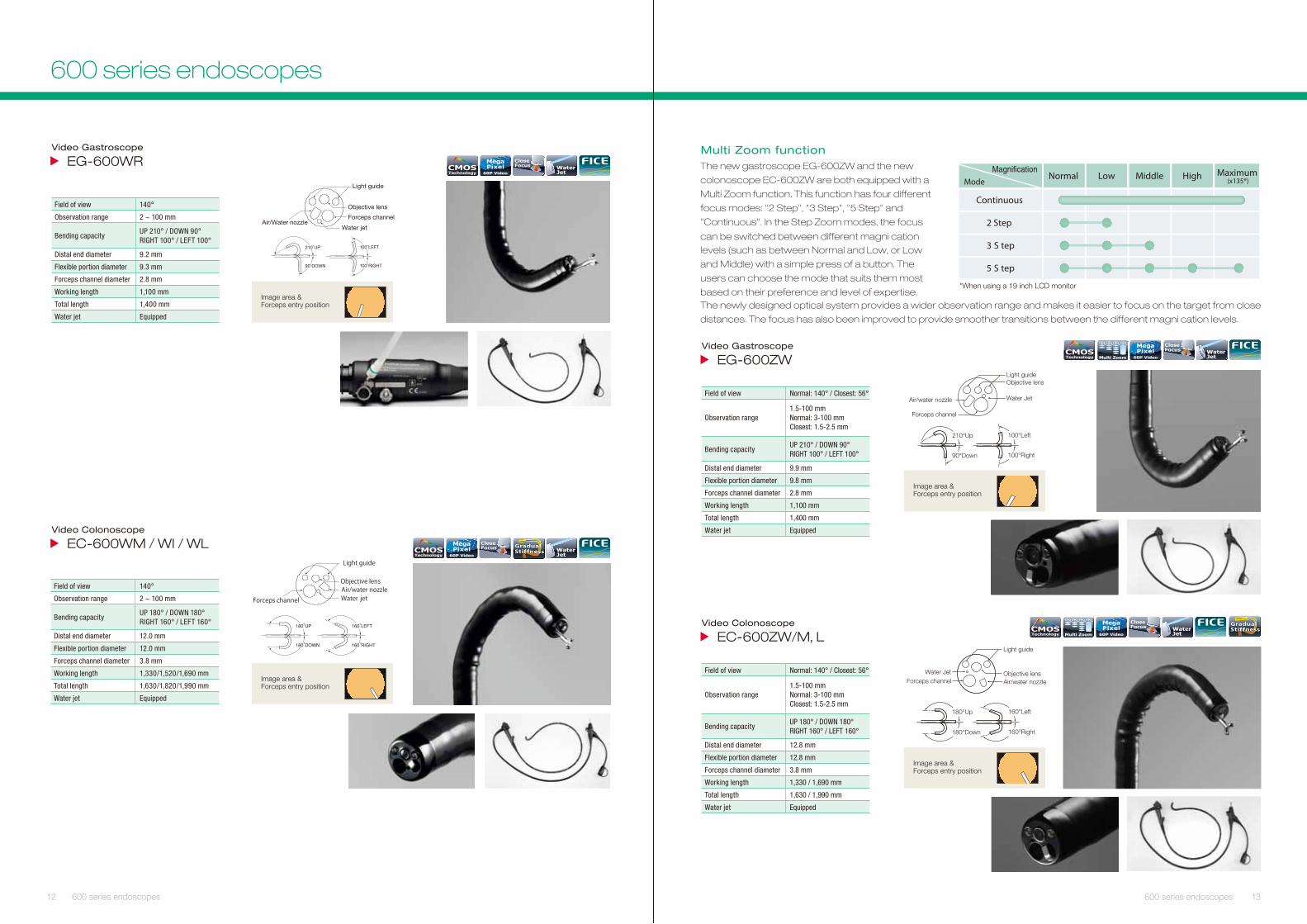
Newly developed insertion portion for better insertion into the colon
Gradual StiffnessThe flexibility of the insertion portion gradually increases toward the distal end. Gradual stiffness level is adjusted as
comfortable for insertion. It is possible to transmit the insertion power to the tip of scope more effectively.
Improved torque and force transmissionand operation abilityBy adopting newly developed insertion portion both torque and force transmission have
been improved. Even when the tip of scope is located in the deep part of colon, the tip of
scope can react sensitively. It’s small diameter of 12.0 mm (for EC-600WM/WI/WL) aims
to produce better operability and reduce patient’s discomfort.
Soft Softer
Hard Harder
Gradual StiffnessConventional Type
Endoscopes with Gradual Stiffness
EC-600WM/WI/WL (New)
EC-600ZW/M, L (New)
EC-580RD/M,L (New)
EC-590ZW3/M, EC-590ZW3/L
EC-590WM4/WI4/WL4
EC-530WM3/WI3/WL3
Gradual Stiffness Technology
Anti-Slip Improved grip performance with newly-designed surface shapeRibbed surface prevents slipping and improves handling of the endoscope. Colonoscopy can be performed more easily and
comfortably even in long examinations.
Anti-SlipConventional Type
GradualStiffness
12
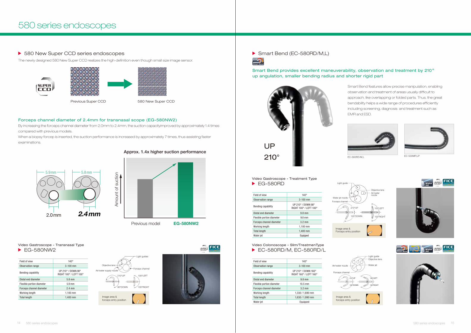
Video Gastroscope
EG-600WR
Field of view 140°
Observation range 2 ~ 100 mm
Bending capacityUP 210° / DOWN 90°RIGHT 100° / LEFT 100°
Distal end diameter 9.2 mm
Flexible portion diameter 9.3 mm
Forceps channel diameter 2.8 mm
Working length 1,100 mm
Total length 1,400 mm
Water jet Equipped
Image area & Forceps entry position
Water jet
Forceps channelAir/Water nozzle
Objective lens
Light guide
210˚UP
90˚DOWN
100˚LEFT
100˚RIGHT
CMOSTechnology
CloseFocus
60P Video
MegaPixel Water
Jet
13
Video Colonoscope
EC-600WM / WI / WL
Field of view 140°
Observation range 2 ~ 100 mm
Bending capacityUP 180° / DOWN 180°RIGHT 160° / LEFT 160°
Distal end diameter 12.0 mm
Flexible portion diameter 12.0 mm
Forceps channel diameter 3.8 mm
Working length 1,330/1,520/1,690 mm
Total length 1,630/1,820/1,990 mm
Water jet Equipped
CMOSTechnology
CloseFocus
60P Video
MegaPixel
Image area & Forceps entry position
WaterJet
GradualStiffness
600 series endoscopes 600 series endoscopes
600 series endoscopes
Multi Zoom function
NormalMode
Low Middle High Maximum (x135*)
2 Step
3 S tep
5 S tep
Continuous
*When using a 19 inch LCD monitor
The new gastroscope EG-600ZW and the new
colonoscope EC-600ZW are both equipped with a
Multi Zoom function. This function has four different
focus modes: "2 Step", "3 Step", "5 Step" and
"Continuous". In the Step Zoom modes, the focus
can be switched between different magni�cationlevels (such as between Normal and Low, or Low
and Middle) with a simple press of a button. The
users can choose the mode that suits them most
based on their preference and level of expertise.The newly designed optical system provides a wider observation range and makes it easier to focus on the target from close
distances. The focus has also been improved to provide smoother transitions between the different magni�cation levels.
Field of view Normal: 140° / Closest: 56°
Observation range1.5-100 mmNormal: 3-100 mmClosest: 1.5-2.5 mm
Bending capacityUP 210° / DOWN 90°RIGHT 100° / LEFT 100°
Distal end diameter 9.9 mm
Flexible portion diameter 9.8 mm
Forceps channel diameter 2.8 mm
Working length 1,100 mm
Total length 1,400 mm
Water jet Equipped
Video Gastroscope
EG-600ZWCloseFocus
60P Video
MegaPixelCMOS
TechnologyWaterJet
210°Up
90°Down
100°Left
100°Right
Objective lens
Water Jet
Forceps channel
Air/water nozzle
Light guide
Image area & Forceps entry position
Field of view Normal: 140° / Closest: 56°
Observation range1.5-100 mmNormal: 3-100 mmClosest: 1.5-2.5 mm
Bending capacityUP 180° / DOWN 180°RIGHT 160° / LEFT 160°
Distal end diameter 12.8 mm
Flexible portion diameter 12.8 mm
Forceps channel diameter 3.8 mm
Working length 1,330 / 1,690 mm
Total length 1,630 / 1,990 mm
Water jet Equipped
Video Colonoscope
EC-600ZW/M, LGradualStiffness
CloseFocus
60P Video
MegaPixelCMOS
TechnologyWaterJet
160°Left
160°Right
180°Up
180°Down
Objective lensAir/water nozzle
Light guide
Forceps channelWater Jet
Image area & Forceps entry position
Forceps channel diameter of 2.4mm for transnasal scope (EG-580NW2)By increasing the forceps channel diameter from 2.0mm to 2.4mm, the suction capacityimproved by approximately 1.4 times
compared with previous models.
When a biopsy forcep is inserted, the suction performance is increased by approximately 7 times, thus assisting faster
examinations.
Image area & forceps entry position
Video Gastroscope - Transnasal Type
EG-580NW2
Field of view 140°
Observation range 3~100 mm
Bending capabilityUP 210° / DOWN 90°
RIGHT 100° / LEFT 100°
Distal end diameter 5.8 mm
Flexible portion diameter 5.9 mm
Forceps channel diameter 2.4 mm
Working length 1,100 mm
Total length 1,400 mm
210°
Smart Bend
Smart Bend (EC-580RD/M,L)
EC-530MP,LP
Smart Bend features allow precise manipulation, enabling
observation and treatment of areas usually difficult to
approach, like overlapping or folded parts. Thus, the great
bendability helps a wide range of procedures efficiently
including screening, diagnosis and treatment such as
EMR and ESD.
UP
210° EC-580RD/M,L
Smart Bend provides excellent maneuverability, observation and treatment by 210 º up angulation, smaller bending radius and shorter rigid part
14 15580 series endoscopes 580 series endoscopes
580 New Super CCD series endoscopesThe newly designed 580 New Super CCD realizes the high-definition even though small size image sensor.
Previous Super CCD 580 New Super CCD
580 series endoscopes
WaterJet
Video Gastroscope - Treatment Type
EG-580RD
Field of view 140°
Observation range 3~100 mm
Bending capabilityUP 210° / DOWN 90°
RIGHT 100° / LEFT 100°
Distal end diameter 9.8 mm
Flexible portion diameter 9.8 mm
Forceps channel diameter 3.2 mm
Working length 1,100 mm
Total length 1,400 mm
Water jet Equipped
Light guideObjective lens
Water jet
Forceps channel
Air/water nozzle
Image area & forceps entry position
210°
Smart Bend
Video Colonoscope – Slim/TreatmenType
EC-580RD/M, EC-580RD/L WaterJet
Field of view 140°
Observation range 3~100 mm
Bending capabilityUP 210° / DOWN 160°
RIGHT 160° / LEFT 160°
Distal end diameter 9.8 mm
Flexible portion diameter 10.5 mm
Forceps channel diameter 3.2 mm
Working length 1,330 / 1,690 mm
Total length 1,630 / 1,990 mm
Water jet Equipped
GradualStiffness
100°LEFT
100°RIGHTN
Image area & Forceps entry position
210°UP
120°DOWN
Light guide
Forceps channel
Water jet nozzle
Objective lensAir/water nozzle
16 17Double Balloon Endoscope Double Balloon Endoscope
Double Balloon Endoscope
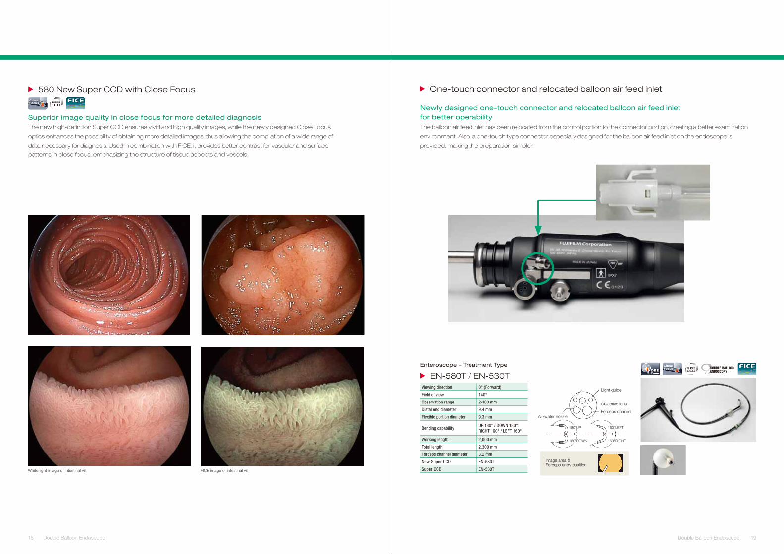
✴According to FUJIFI LM data
2.8mm 3.2mm
EN-580T EN-450T5
With Φ2.4 device
3.6 times
Suct
ion
volu
me
The 3.2 mm diameter forceps channel
providing greater suction performance
than that of conventional models.
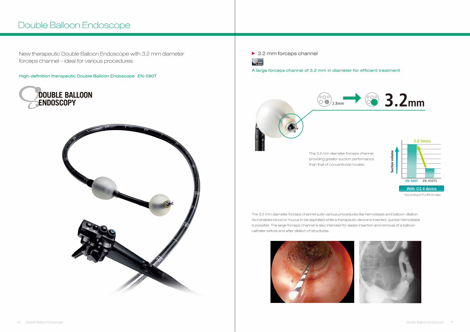
High-definition therapeutic Double Balloon Endoscope EN-580T
New therapeutic Double Balloon Endoscope with 3.2 mm diameterforceps channel – ideal for various procedures
The 3.2 mm diameter forceps channel suits various procedures like hemostasis and balloon dilation.
As it enables blood or mucus to be aspirated while a therapeutic device is inserted, quicker hemostasis
is possible. The large forceps channel is also intended for easier insertion and removal of a balloon
catheter before and after dilation of structures.
lennahc specrof mm 2.3
DBE3.2mm
A large forceps channel of 3.2 mm in diameter for effi cient treatment
18 19Double Balloon Endoscope Double Balloon Endoscope
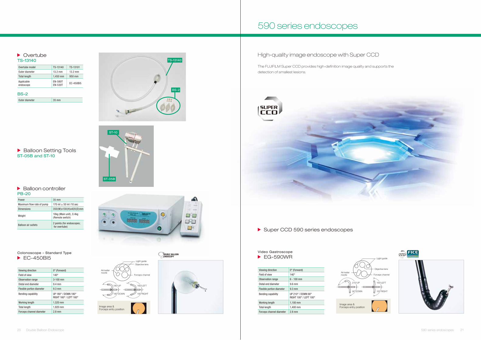
White light image of intestinal villi FICE image of intestinal villi
580 New Super CCD with Close Focus
Superior image quality in close focus for more detailed diagnosis The new high-definition Super CCD ensures vivid and high quality images, while the newly designed Close Focus
optics enhances the possibility of obtaining more detailed images, thus allowing the compilation of a wide range of
data necessary for diagnosis. Used in combination with FICE, it provides better contrast for vascular and surface
patterns in close focus, emphasizing the structure of tissue aspects and vessels.
CloseFocus
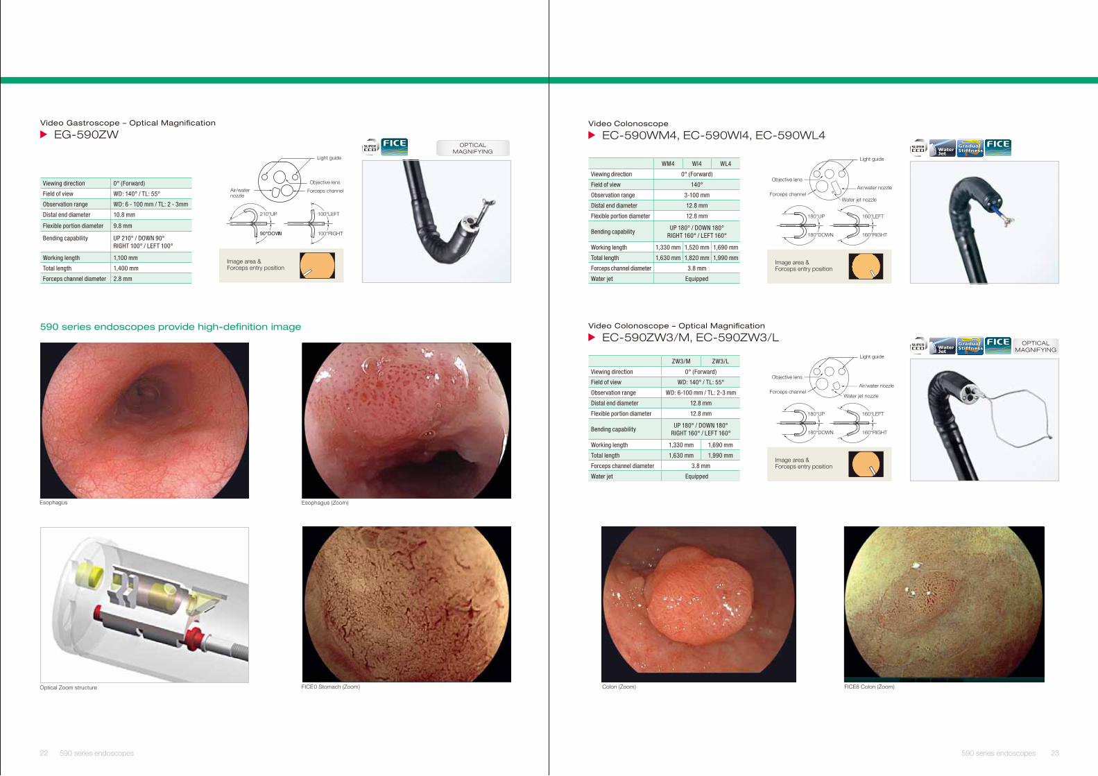
Newly designed one-touch connector and relocated balloon air feed inletfor better operability
The balloon air feed inlet has been relocated from the control portion to the connector portion, creating a better examination
environment. Also, a one-touch type connector especially designed for the balloon air feed inlet on the endoscope is
provided, making the preparation simpler.
One-touch connector and relocated balloon air feed inlet
Enteroscope – Treatment Type
EN-580T / EN-530TViewing direction 0° (Forward)
Field of view 140°
Observation range 2-100 mm
Distal end diameter 9.4 mm
Flexible portion diameter 9.3 mm
Bending capabilityUP 180° / DOWN 180°RIGHT 160° / LEFT 160°
Working length 2,000 mm
Total length 2,300 mm
Forceps channel diameter 3.2 mm
New Super CCD EN-580T
Super CCD EN-530T
Image area & Forceps entry position
160°LEFT
160°RIGHT
180°UP
180°DOWN
Light guide
Objective lens
Forceps channelAir/water nozzle
CloseFocusDBE
3.2mm
20 Double Balloon Endoscope 21590 series endoscopes
Super CCD 590 series endoscopes
High-quality image endoscope with Super CCD
The FUJIFILM Super CCD provides high-definition image quality and supports the
detection of smallest lesions.
Image area & Forceps entry position
Light guide
Objective lens
Forceps channelAir/water nozzle
100°LEFT
100°RIGHT
210°UP
90°DOWN
Video Gastroscope
RW095-GE
Viewing direction 0° (Forward)
Field of view 140°
Observation range 6 - 100 mm
Distal end diameter 9.6 mm
Flexible portion diameter 9.3 mm
Bending capability UP 210° / DOWN 90°RIGHT 100° / LEFT 100°
Working length 1,100 mm
Total length 1,400 mm
Forceps channel diameter 2.8 mm
590 series endoscopes
Image area & Forceps entry position
Light guideObjective lens
Forceps channel
Air/water nozzle
160°LEFT
160°RIGHT
180°UP
180°DOWN
PB-20 Balloon controller
Power 35 mm
Maximum fl ow rate of pump 170 ml ± 50 ml /10 sec
Dimensions 350(W)x130(H)x420(D)mm
Weight10kg (Main unit), 0.4kg (Remote switch)
Balloon air outlets2 points (for endoscopes; for overtube)
Viewing direction 0° (Forward)
Field of view 140°
Observation range 3-100 mm
Distal end diameter 9.4 mm
Flexible portion diameter 9.3 mm
Bending capability UP 180° / DOWN 180°RIGHT 160° / LEFT 160°
Working length 1,520 mm
Total length 1,820 mm
Forceps channel diameter 2.8 mm
Colonoscope – Standard Type
5IB054-CE
Balloon Setting ToolsST-05B and ST-10
ST-05B
ST-10
BS-2
TS-13140 Overtube
Overtube model TS-13140 TS-13101
Outer diameter 13.2 mm 13.2 mm
Total length 1,450 mm 950 mm
Applicable endoscope
EN-580TEN-530T
EC-450BI5
Outer diameter 35 mm
TS-13140
BS-2
FICE0 Stomach (Zoom)
Video Gastroscope – Optical Magnifi cation
WZ095-GE
Optical Zoom structure
Esophagus (Zoom)Esophagus
590 series endoscopes provide high-definition image
Light guide
Objective lens
Forceps channelAir/water nozzle
100°LEFT
100°RIGHT
210°UP
90°DOWN
Image area & Forceps entry position
Viewing direction 0° (Forward)
Field of view WD: 140° / TL: 55°
Observation range WD: 6 - 100 mm / TL: 2 - 3mm
Distal end diameter 10.8 mm
Flexible portion diameter 9.8 mm
Bending capability UP 210° / DOWN 90°RIGHT 100° / LEFT 100°
Working length 1,100 mm
Total length 1,400 mm
Forceps channel diameter 2.8 mm
OPTICALMAGNIFYING
Video Colonoscope
4LW095-CE ,4IW095-CE ,4MW095-CE
Colon (Zoom) FICE8 Colon (Zoom)
Image area & Forceps entry position
160°LEFT
160°RIGHT
180°UP
180°DOWN
Light guide
Objective lens
Forceps channelAir/water nozzle
Water jet nozzle
WM4 WI4 WL4
Viewing direction 0° (Forward)
Field of view 140°
Observation range 3-100 mm
Distal end diameter 12.8 mm
Flexible portion diameter 12.8 mm
Bending capabilityUP 180° / DOWN 180°
RIGHT 160° / LEFT 160°
Working length 1,330 mm 1,520 mm 1,690 mm
Total length 1,630 mm 1,820 mm 1,990 mm
Forceps channel diameter 3.8 mm
Water jet Equipped
WaterJet
GradualStiffness
Video Colonoscope – Optical Magnifi cation
L/3WZ095-CE ,M/3WZ095-CE
Image area & Forceps entry position
160°LEFT
160°RIGHT
180°UP
180°DOWN
Light guide
Objective lens
Forceps channelAir/water nozzle
Water jet nozzle
ZW3/M ZW3/L
Viewing direction 0° (Forward)
Field of view WD: 140° / TL: 55°
Observation range WD: 6-100 mm / TL: 2-3 mm
Distal end diameter 12.8 mm
Flexible portion diameter 12.8 mm
Bending capabilityUP 180° / DOWN 180°
RIGHT 160° / LEFT 160°
Working length 1,330 mm 1,690 mm
Total length 1,630 mm 1,990 mm
Forceps channel diameter 3.8 mm
Water jet Equipped
OPTICALMAGNIFYINGWater
Jet
GradualStiffness
22 23590 series endoscopes 590 series endoscopes
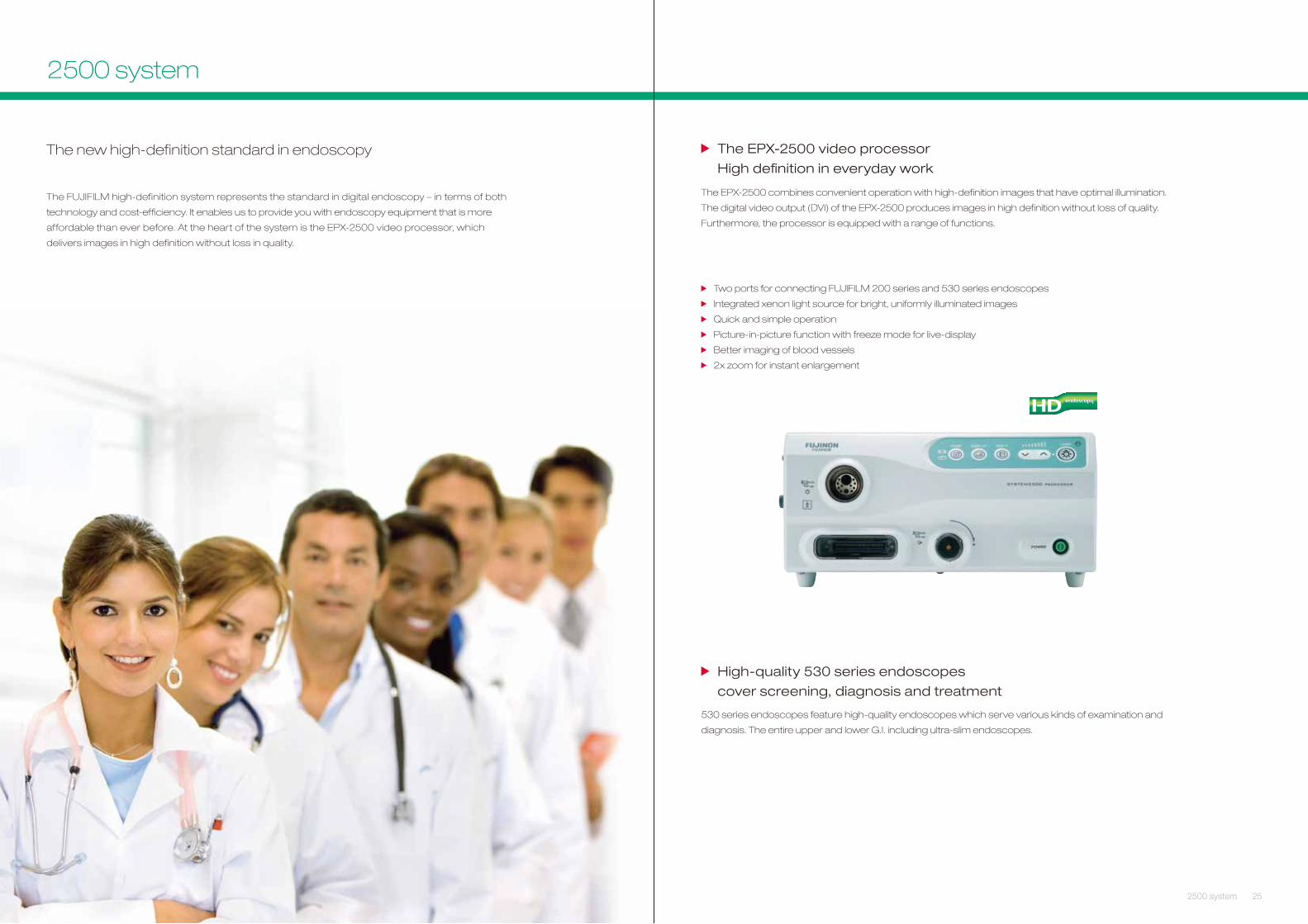
rossecorp oediv 0052-XPE ehT
High defi nition in everyday work
The EPX-2500 combines convenient operation with high-definition images that have optimal illumination.
The digital video output (DVI) of the EPX-2500 produces images in high definition without loss of quality.
Furthermore, the processor is equipped with a range of functions.
Two ports for connecting FUJIFILM 200 series and 530 series endoscopes
Integrated xenon light source for bright, uniformly illuminated images
Quick and simple operation
Picture-in-picture function with freeze mode for live-display
Better imaging of blood vessels
2x zoom for instant enlargement
High-quality 530 series endoscopes
cover screening, diagnosis and treatment
530 series endoscopes feature high-quality endoscopes which serve various kinds of examination and
diagnosis. The entire upper and lower G.I. including ultra-slim endoscopes.
252500 system
The new high-definition standard in endoscopy
The FUJIFILM high-definition system represents the standard in digital endoscopy – in terms of both
technology and cost-efficiency. It enables us to provide you with endoscopy equipment that is more
affordable than ever before. At the heart of the system is the EPX-2500 video processor, which
delivers images in high definition without loss in quality.
24 2500 system
2500 system
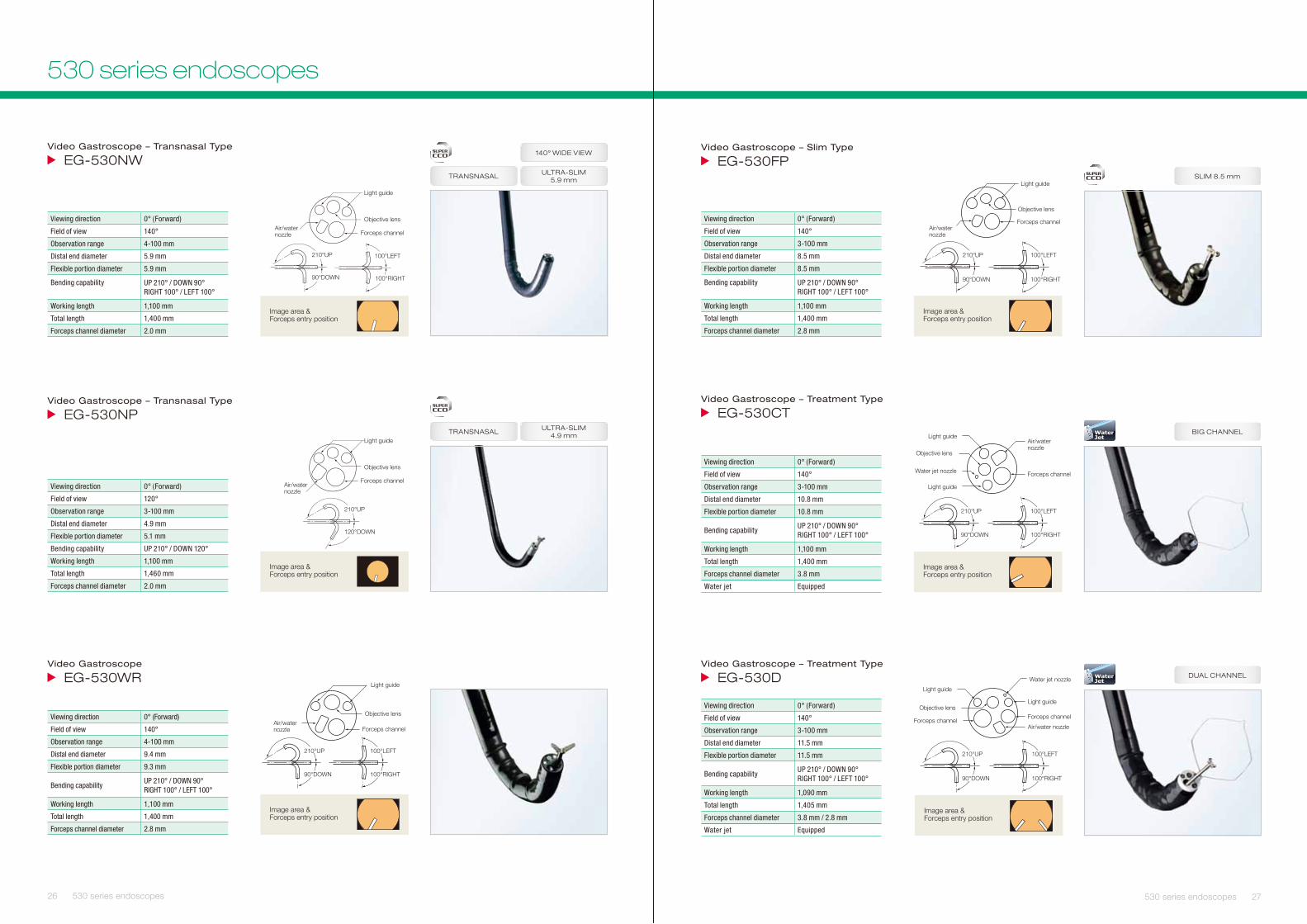
Image area & Forceps entry position
Light guide
Objective lens
Forceps channelAir/water nozzle
100°LEFT
100°RIGHT
210°UP
90°DOWN
Viewing direction 0° (Forward)
Field of view 140°
Observation range 4-100 mm
Distal end diameter 9.4 mm
Flexible portion diameter 9.3 mm
Bending capabilityUP 210° / DOWN 90°RIGHT 100° / LEFT 100°
Working length 1,100 mm
Total length 1,400 mm
Forceps channel diameter 2.8 mm
Video Gastroscope – Transnasal Type
WN035-GE
Video Gastroscope – Transnasal Type
PN035-GE
Video Gastroscope
RW035-GE
Image area & Forceps entry position
Light guide
Objective lens
Forceps channelAir/water nozzle
210°UP
90°DOWN
100°LEFT
100°RIGHT
Viewing direction 0° (Forward)
Field of view 140°
Observation range 4-100 mm
Distal end diameter 5.9 mm
Flexible portion diameter 5.9 mm
Bending capability UP 210° / DOWN 90°RIGHT 100° / LEFT 100°
Working length 1,100 mm
Total length 1,400 mm
Forceps channel diameter 2.0 mm
ULTRA-SLIM 5.9 mm
140° WIDE VIEW
TRANSNASAL
Image area & Forceps entry position
210°UP
120°DOWN
Light guide
Objective lens
Forceps channelAir/water nozzle
Viewing direction 0° (Forward)
Field of view 120°
Observation range 3-100 mm
Distal end diameter 4.9 mm
Flexible portion diameter 5.1 mm
Bending capability UP 210° / DOWN 120°
Working length 1,100 mm
Total length 1,460 mm
Forceps channel diameter 2.0 mm
ULTRA-SLIM 4.9 mmTRANSNASAL
26 530 series endoscopes
Video Gastroscope – Slim Type
PF035-GE
Video Gastroscope – Treatment Type
TC035-GE
Video Gastroscope – Treatment Type
D035-GE
Air/water nozzle
210°UP
90°DOWN
100°LEFT
100°RIGHT
Light guide
Objective lens
Forceps channel
Image area & Forceps entry position
Viewing direction 0° (Forward)
Field of view 140°
Observation range 3-100 mm
Distal end diameter 8.5 mm
Flexible portion diameter 8.5 mm
Bending capability UP 210° / DOWN 90°RIGHT 100° / LEFT 100°
Working length 1,100 mm
Total length 1,400 mm
Forceps channel diameter 2.8 mm
SLIM 8.5 mm
Light guide
Light guide
Objective lens
Forceps channel
Air/water nozzle
Water jet nozzle
Image area & Forceps entry position
100°LEFT
100°RIGHT
210°UP
90°DOWN
Viewing direction 0° (Forward)
Field of view 140°
Observation range 3-100 mm
Distal end diameter 10.8 mm
Flexible portion diameter 10.8 mm
Bending capabilityUP 210° / DOWN 90°RIGHT 100° / LEFT 100°
Working length 1,100 mm
Total length 1,400 mm
Forceps channel diameter 3.8 mm
Water jet Equipped
BIG CHANNELWaterJet
Objective lensForceps channel
Forceps channel
Light guide
Water jet nozzle
Light guide
Air/water nozzle
Image area & Forceps entry position
100°LEFT
100°RIGHT
210°UP
90°DOWN
Viewing direction 0° (Forward)
Field of view 140°
Observation range 3-100 mm
Distal end diameter 11.5 mm
Flexible portion diameter 11.5 mm
Bending capabilityUP 210° / DOWN 90°RIGHT 100° / LEFT 100°
Working length 1,090 mm
Total length 1,405 mm
Forceps channel diameter 3.8 mm / 2.8 mm
Water jet Equipped
DUAL CHANNELWaterJet
27530 series endoscopes
530 series endoscopes
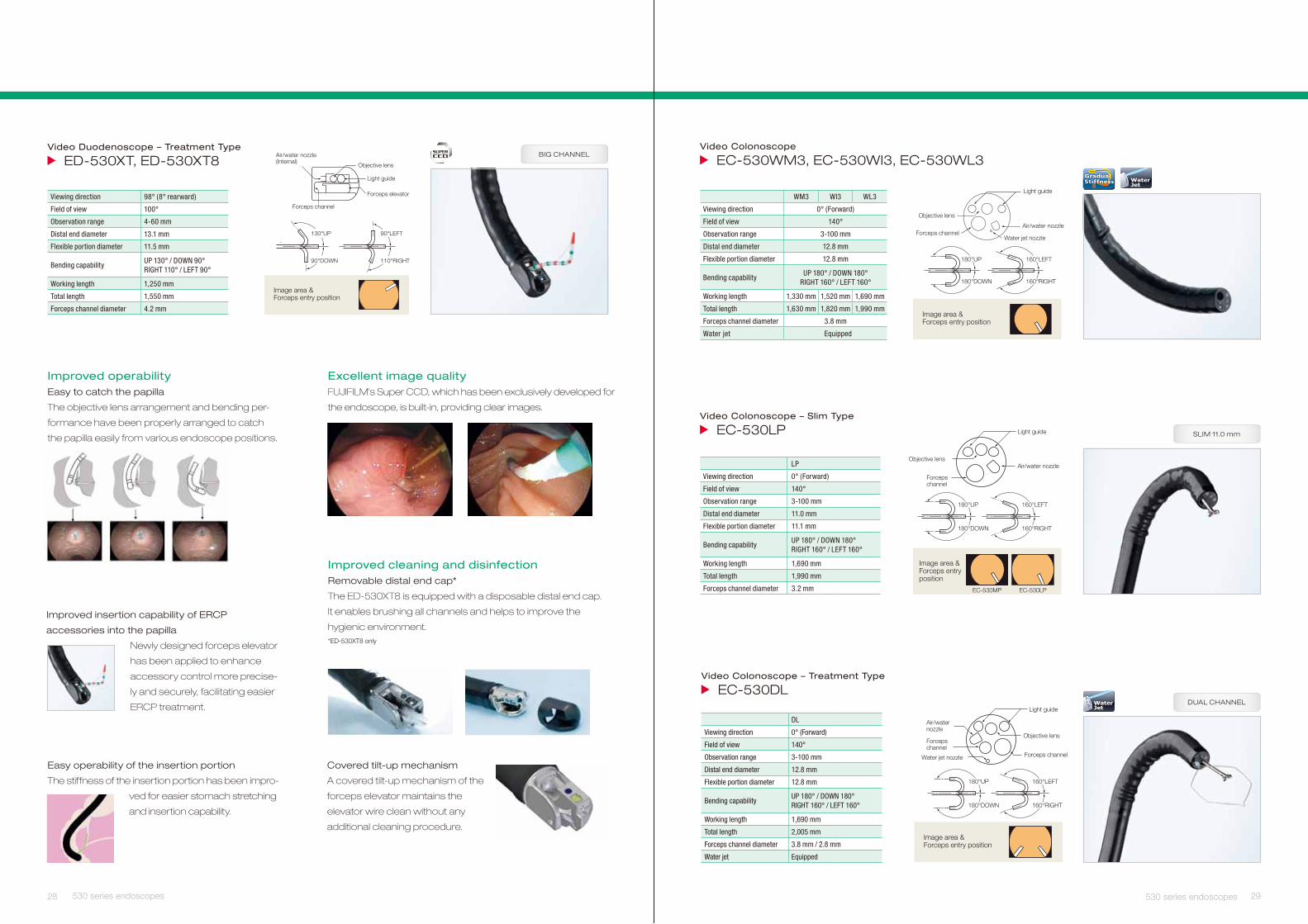
Easy operability of the insertion portion
The stiffness of the insertion portion has been impro-
ved for easier stomach stretching
and insertion capability.
Video Duodenoscope – Treatment Type
8TX035-DE ,TX035-DE
Improved operabilityEasy to catch the papilla
The objective lens arrangement and bending per-
formance have been properly arranged to catch
the papilla easily from various endoscope positions.
Improved insertion capability of ERCP
accessories into the papilla
Newly designed forceps elevator
has been applied to enhance
accessory control more precise-
ly and securely, facilitating easier
ERCP treatment.
Improved cleaning and disinfectionRemovable distal end cap*
The ED-530XT8 is equipped with a disposable distal end cap.
lt enables brushing all channels and helps to improve the
hygienic environment.*ED-530XT8 only
Covered tilt-up mechanism
A covered tilt-up mechanism of the
forceps elevator maintains the
elevator wire clean without any
additional cleaning procedure.
Excellent image qualityFUJIFILM’s Super CCD, which has been exclusively developed for
the endoscope, is built-in, providing clear images.
Air/water nozzle(Internal)
Forceps channel
Light guide
Forceps elevator
Objective lens
Image area & Forceps entry position
90°LEFT
110°RIGHT
130°UP
90°DOWN
Viewing direction 98° (8° rearward)
Field of view 100°
Observation range 4-60 mm
Distal end diameter 13.1 mm
Flexible portion diameter 11.5 mm
Bending capabilityUP 130° / DOWN 90°RIGHT 110° / LEFT 90°
Working length 1,250 mm
Total length 1,550 mm
Forceps channel diameter 4.2 mm
BIG CHANNEL
28
Video Colonoscope
3LW035-CE ,3IW035-CE ,3MW035-CE
Video Colonoscope – Slim Type
EC-530LP
180°UP
180°DOWN
160°LEFT
160°RIGHT
Objective lensAir/water nozzle
EC-530LPEC-530MP
Forceps channel
Light guide
Image area & Forceps entry position
LP
Viewing direction 0° (Forward)
Field of view 140°
Observation range 3-100 mm
Distal end diameter 11.0 mm
Flexible portion diameter 11.1 mm
Bending capabilityUP 180° / DOWN 180°RIGHT 160° / LEFT 160°
Working length 1,690 mm
Total length 1,990 mm
Forceps channel diameter 3.2 mm
SLIM 11.0 mm
Video Colonoscope – Treatment Type
EC-530DL
180°UP
180°DOWN
160°LEFT
160°RIGHT
Air/water nozzle
Light guide
Objective lens
Forceps channelWater jet nozzle
Forceps channel
Image area & Forceps entry position
DL
Viewing direction 0° (Forward)
Field of view 140°
Observation range 3-100 mm
Distal end diameter 12.8 mm
Flexible portion diameter 12.8 mm
Bending capabilityUP 180° / DOWN 180°RIGHT 160° / LEFT 160°
Working length 1,690 mm
Total length 2,005 mm
Forceps channel diameter 3.8 mm / 2.8 mm
Water jet Equipped
DUAL CHANNELWaterJet
29530 series endoscopes 530 series endoscopes
Image area & Forceps entry position
160°LEFT
160°RIGHT
180°UP
180°DOWN
Light guide
Objective lens
Forceps channelAir/water nozzle
Water jet nozzle
WM3 WI3 WL3
Viewing direction 0° (Forward)
Field of view 140°
Observation range 3-100 mm
Distal end diameter 12.8 mm
Flexible portion diameter 12.8 mm
Bending capabilityUP 180° / DOWN 180°
RIGHT 160° / LEFT 160°
Working length 1,330 mm 1,520 mm 1,690 mm
Total length 1,630 mm 1,820 mm 1,990 mm
Forceps channel diameter 3.8 mm
Water jet Equipped
WaterJet
GradualStiffness
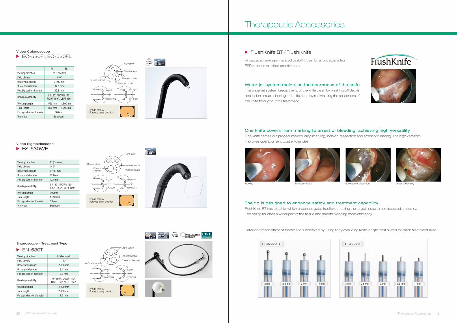
Video Colonoscope
LF035-CE ,IF035-CE
Video Sigmoidoscope
EW035-SE
Image area & Forceps entry position
Air/water nozzle
Water jet nozzle
180°UP
180°DOWN
160°LEFT
160°RIGHT
Light guide
Objective lens
Forceps channel
FI FL
Viewing direction 0° (Forward)
Field of view 140°
Observation range 3-100 mm
Distal end diameter 12.8 mm
Flexible portion diameter 12.8 mm
Bending capabilityUP 180° / DOWN 180°
RIGHT 160° / LEFT 160°
Working length 1,520 mm 1,690 mm
Total length 1,820 mm 1,990 mm
Forceps channel diameter 3.8 mm
Water jet Equipped
WaterJet
180°UP
180°DOWN
160°LEFT
160°RIGHT
Objective lensAir/water nozzle
Forceps channel
Light guide
Water jet nozzle
Image area & Forceps entry position
Viewing direction 0° (Forward)
Field of view 140°
Observation range 3-100 mm
Distal end diameter 12.8mm
Flexible portion diameter 12.8mm
Bending capabilityUP 180° / DOWN 180°RIGHT 160° / LEFT 160°
Working length 790mm
Total length 1,090mm
Forceps channel diameter 3.8mm
Water jet Equipped
WaterJet
30
FlushKnife BT
3 mm 2.5 mm 2 mm 1.5 mm
FlushKnife
3 mm 2.5 mm 2 mm 1.5 mm 1 mm
efinKhsulF / TB efinKhsulF
Aimed at achieving enhanced usability ideal for all physicians from
ESD trainees to skilled practitioners.
Water jet system maintains the sharpness of the knifeThe water jet system keeps the tip of the knife clean by washing off debris
and lesion tissue adhering to the tip, thereby maintaining the sharpness of
the knife throughout the treatment.
Safer and more efficient treatment is achieved by using the protruding knife length best suited for each treatment area.
The tip is designed to enhance safety and treatment capabilityFlushKnife BT has a ball tip, which produces good traction, enabling the target tissue to be dissected smoothly.
The ball tip touches a wider part of the tissue and arrests bleeding more efficiently.
improves operation and cost efficiencies.
One knife covers from marking to arrest of bleeding, achieving high versatilityOne knife carries out procedures including marking, incision, dissection and arrest of bleeding. The high versatility
Marking Mucosal incision Submucosal dissection Arrest of bleeding
31
Enteroscope – Treatment Type
EN-530TViewing direction 0° (Forward)
Field of view 140°
Observation range 2-100 mm
Distal end diameter 9.4 mm
Flexible portion diameter 9.3 mm
Bending capabilityUP 180° / DOWN 180°
RIGHT 160° / LEFT 160°
Working length 2,000 mm
Total length 2,300 mm
Forceps channel diameter 3.2 mm
CloseFocusDBE
3.2mm
Image area & Forceps entry position
160°LEFT
160°RIGHT
180°UP
180°DOWN
Light guide
Objective lens
Forceps channelAir/water nozzle
530 series endoscopes Therapeutic Accessories
Therapeutic Accessories
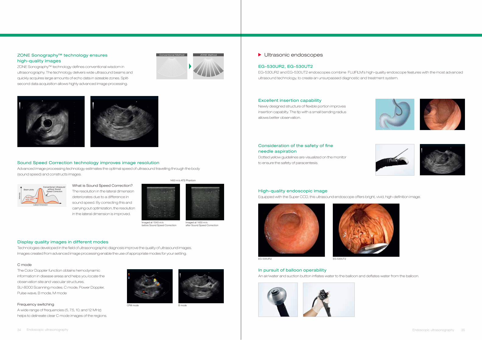
SU-8000
tpecnoc kcats eno-ni-lla – rewoT SUE
Years of research and development to reduce patient discomfort and
improve operator efficiency during endoscope examinations led to the
development of Sonart, the integration of ultrasonographic diagnosis and
endoscopy systems.
For a more accurate diagnosis, advanced image processing technology
integrates improved endoscope maneuverability and insertion capability.
The compact, one-cart system supports various applications.
Flexible image display and switchingKeyboard operation facilitates
smooth examinations and allows
switching among an ultrasound
image, an endoscopic image,
and a picture-in-picture image
with patient‘s history images.
Picture-in-picture image Patient’s history image
Ultrasound image
Endoscopic image
32 33Therapeutic Accessories Endoscopic ultrasonography
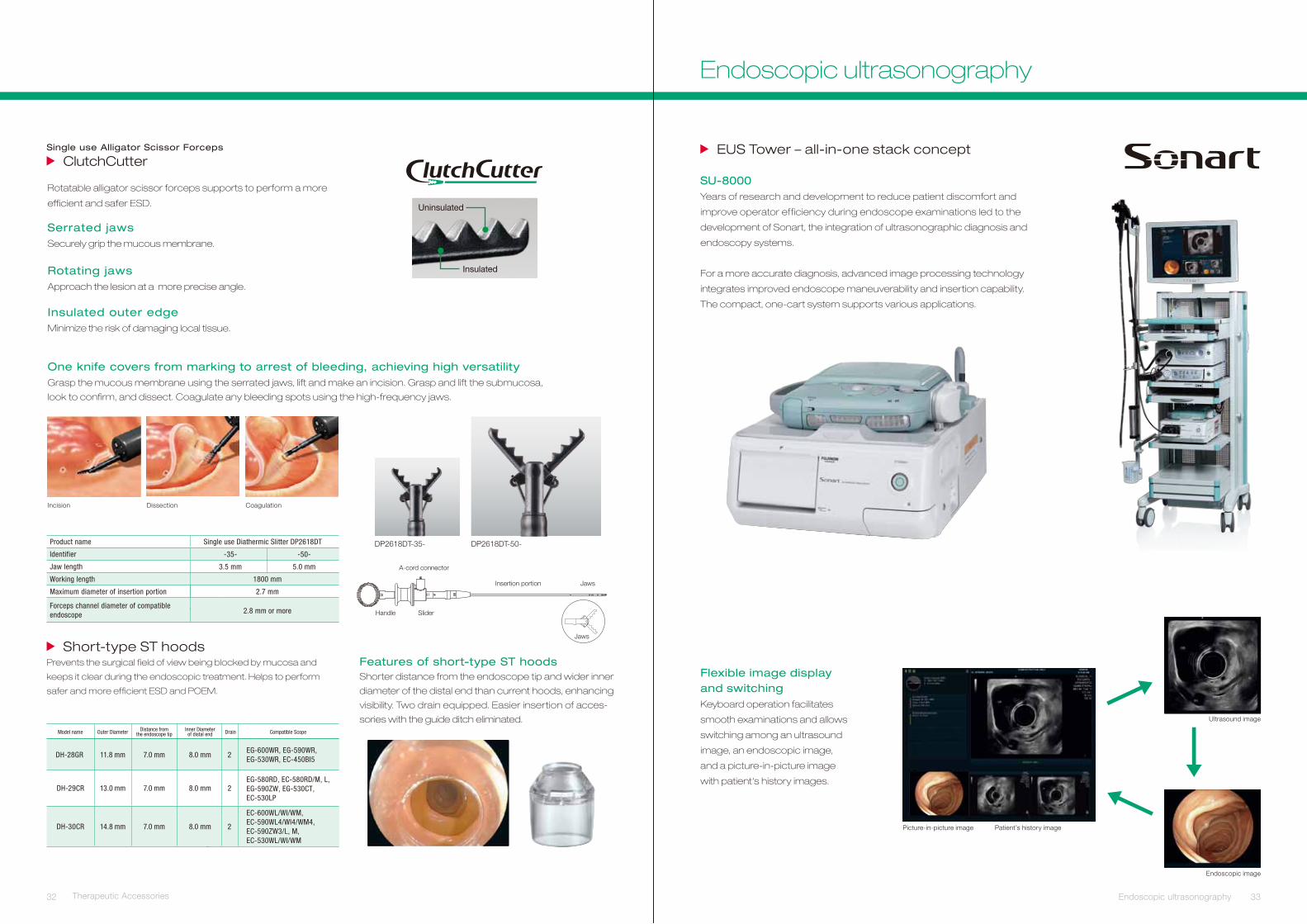
Single use Alligator Scissor Forceps
ClutchCutter
Rotatable alligator scissor forceps supports to perform a more
efficient and safer ESD. Uninsulated
Insulated
Serrated jawsSecurely grip the mucous membrane.
Rotating jawsApproach the lesion at a more precise angle.
Insulated outer edgeMinimize the risk of damaging local tissue.
One knife covers from marking to arrest of bleeding, achieving high versatilityGrasp the mucous membrane using the serrated jaws, lift and make an incision. Grasp and lift the submucosa,
look to confirm, and dissect. Coagulate any bleeding spots using the high-frequency jaws.
Short-type ST hoodsPrevents the surgical field of view being blocked by mucosa and
keeps it clear during the endoscopic treatment. Helps to perform
safer and more efficient ESD and POEM.
Features of short-type ST hoodsShorter distance from the endoscope tip and wider inner
diameter of the distal end than current hoods, enhancing
visibility. Two drain equipped. Easier insertion of acces-
sories with the guide ditch eliminated.
DH-28GR 11.8 mm 7.0 mm 8.0 mm 2EG-600WR, EG-590WR,EG-530WR, EC-450BI5
DH-29CR 13.0 mm 7.0 mm 8.0 mm 2EG-580RD, EC-580RD/M, L, EG-590ZW, EG-530CT,EC-530LP
DH-30CR 14.8 mm 7.0 mm 8.0 mm 2
EC-600WL/WI/WM, EC-590WL4/WI4/WM4,EC-590ZW3/L, M,EC-530WL/WI/WM
Model name Outer Diameter Distance fromthe endoscope tip
Inner Diameterof distal end Drain Compatible Scope
Maximum diameter of insertion portion
Forceps channel diameter of compatibleendoscope
2.7 mm
2.8 mm or more
-35-
3.5 mm
-50-
5.0 mm
Product name Single use Diathermic Slitter DP2618DT
Identifier
Jaw length
Working length 1800 mm
Incision Dissection Coagulation
DP2618DT-50-DP2618DT-35-
A-cord connector
Insertion portion Jaws
SliderHandle
Jaws
Endoscopic ultrasonography
Ultrasonic endoscopes
Excellent insertion capabilityNewly designed structure of flexible portion improves
insertion capabilty. The tip with a small bending radius
allows better observation.
EG-530UR2, EG-530UT2EG-530UR2 and EG-530UT2 endoscopes combine FUJIFILM’s high-quality endoscope features with the most advanced
ultrasound technology, to create an unsurpassed diagnostic and treatment system.
Consideration of the safety of fi ne needle aspirationDotted yellow guidelines are visualized on the monitor
to ensure the safety of paracentesis.
High-quality endoscopic imageEquipped with the Super CCD, this ultrasound endoscope offers bright, vivid, high-definition image.
In pursuit of balloon operabilityAn air/water and suction button inflates water to the balloon and deflates water from the balloon.
EG-530UR2 EG-530UT2
34 35Endoscopic ultrasonography Endoscopic ultrasonography
Conventional Method ZONE MethodZONE Sonography™ technology ensures high-quality imagesZONE Sonography™ technology defines conventional wisdom in
ultrasonography. The technology delivers wide ultrasound beams and
quickly acquires large amounts of echo data in sizeable zones. Split-
second data acquisition allows highly advanced image processing.
Sound Speed Correction technology improves image resolution Advanced image processing technology estimates the optimal speed of ultrasound travelling through the body
(sound speed) and constructs images.
1450 m/s ATS Phantom
What is Sound Speed Correction?
The resolution in the lateral dimension
deteriorates due to a difference in
sound speed. By correcting this and
carrying out optimization, the resolution
in the lateral dimension is improved.
Imaged at 1540 m/sbefore Sound Speed Correction
Imaged at 1450 m/safter Sound Speed Correction
C mode
The Color Doppler function obtains hemodynamic
information in disease areas and helps you locate the
observation site and vascular structures.
SU-8000 Scanning modes; C mode, Power Doppler,
Pulse wave, B mode, M mode
Frequency switching
A wide range of frequencies (5, 7.5, 10, and 12 MHz)
helps to delineate clear C mode images of the regions.
Display quality images in different modesTechnologies developed in the field of ultrasonographic diagnosis improve the quality of ultrasound images.
Images created from advanced image processing enable the use of appropriate modes for your setting.
CFM mode B mode
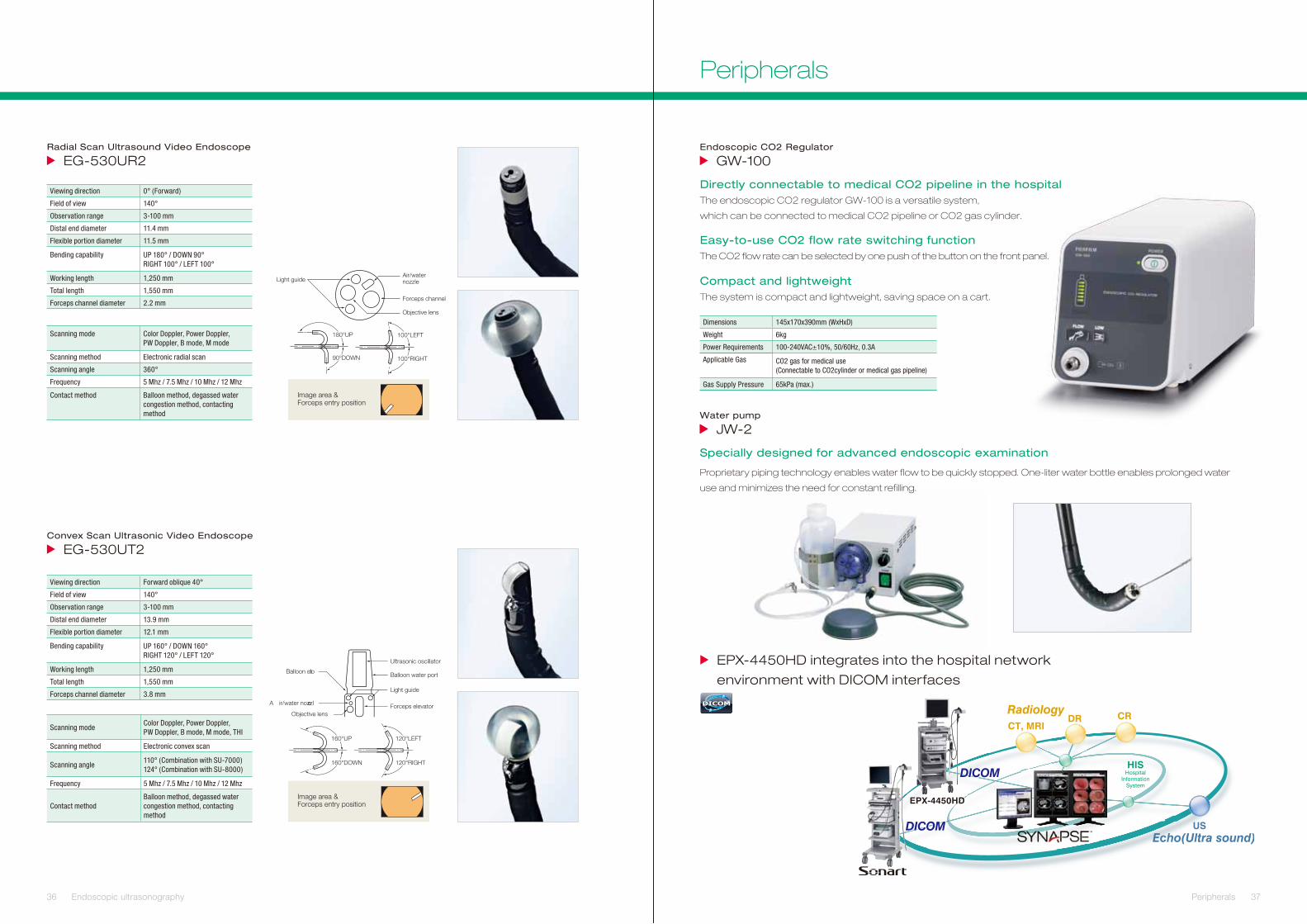
Image area & Forceps entry position
Image area & Forceps entry position
Proprietary piping technology enables water flow to be quickly stopped. One-liter water bottle enables prolonged water
use and minimizes the need for constant refilling.
36 Endoscopic ultrasonography
krowten latipsoh eht otni setargetni DH0544-XPE
environment with DICOM interfaces
DICOM
37 Peripherals
Endoscopic CO2 Regulator
GW-100
Dimensions 145x170x390mm (WxHxD)
Weight 6kg
Power Requirements 100-240VAC±10%, 50/60Hz, 0.3A
Applicable Gas
Gas Supply Pressure
CO2 gas for medical use(Connectable to CO2cylinder or medical gas pipeline)
65kPa (max.)
Directly connectable to medical CO2 pipeline in the hospital
Water pump
JW-2
Specially designed for advanced endoscopic examination
The endoscopic CO2 regulator GW-100 is a versatile system,
which can be connected to medical CO2 pipeline or CO2 gas cylinder.
Easy-to-use CO2 flow rate switching functionThe CO2 flow rate can be selected by one push of the button on the front panel.
Compact and lightweightThe system is compact and lightweight, saving space on a cart.
180°UP
Light guide
Objective lens
Forceps channel
100°LEFT
100°RIGHT
Air/water nozzle
90°DOWN
Radial Scan Ultrasound Video Endoscope
2RU035-GE
Viewing direction 0° (Forward)
Field of view 140°
Observation range 3-100 mm
Distal end diameter 11.4 mm
Flexible portion diameter 11.5 mm
Bending capability UP 180° / DOWN 90°RIGHT 100° / LEFT 100°
Working length 1,250 mm
Total length 1,550 mm
Forceps channel diameter 2.2 mm
Scanning mode Color Doppler, Power Doppler,PW Doppler, B mode, M mode
Scanning method Electronic radial scan
Scanning angle 360°
Frequency 5 Mhz / 7.5 Mhz / 10 Mhz / 12 Mhz
Contact method Balloon method, degassed water congestion method, contacting method
Convex Scan Ultrasonic Video Endoscope
2TU035-GE
Viewing direction Forward oblique 40°
Field of view 140°
Observation range 3-100 mm
Distal end diameter 13.9 mm
Flexible portion diameter 12.1 mm
Bending capability UP 160° / DOWN 160°RIGHT 120° / LEFT 120°
Working length 1,250 mm
Total length 1,550 mm
Forceps channel diameter 3.8 mm
Scanning modeColor Doppler, Power Doppler,PW Doppler, B mode, M mode, THI
Scanning method Electronic convex scan
Scanning angle110° (Combination with SU-7000)124° (Combination with SU-8000)
Frequency 5 Mhz / 7.5 Mhz / 10 Mhz / 12 Mhz
Contact methodBalloon method, degassed water congestion method, contacting method
Ultrasonic oscillator
Light guide
160°UP
160°DOWN
120°LEFT
120°RIGHT
Balloon water port
A ir/water nozzle
Objective lensForceps elevator
Balloon slot
Peripherals
Digital outputHD-SDI: HDTV 1080i (2ch)DVI (Digital Visual Interface): 1280 x 1024 pEthernet: 100 / 10 Base
Analog outputRGB: 1280 x 1024 pSDTV (120 V / NTSC, 230 V / PAL): RGB Y / C, Composite
Color adjustment Brightness, Red, Green, Blue, R-Hue, Chroma; 9 steps
Detail Hi, LO; 9 steps
Contrast (gamma) 3 steps
Hyper-Sharpness Hi, Mid, Lo, Off
Color emphasis Hi, Mid, Lo, Off
FICEFlexible spectral imagingColor Enhancement 10 presets
Iris Average / Peak / Auto
Image storage CF Card
Power rating 120 V 60 Hz 0.8 A 230 V 50 Hz 0.5 A
Dimensions (W x H x D) 390 x 105 x 460 mm
Weight 9.5 kg
DICOM MWL, Store
Lamp rated valueMain Lamp: 300 W Xenon lamp LMP-002Emergency Lamp: 75 W Halogen lamp
Light control Automatic light control
Lamp cooling method Forced air cooling
Air supply pump High, Mid, Lo, Off
Light save On, Off
Transmitted illumination On, Off
Power rating 120 V 60 Hz 3.3 A 230 V 50 Hz 1.7 A
Dimensions (W x H x D) 390 x 155 x 450 mm
Weight 15 kg
XL-4450 Light source
DH0544-XPE Video Processor
VP-4450 HD Processor
38 Processors
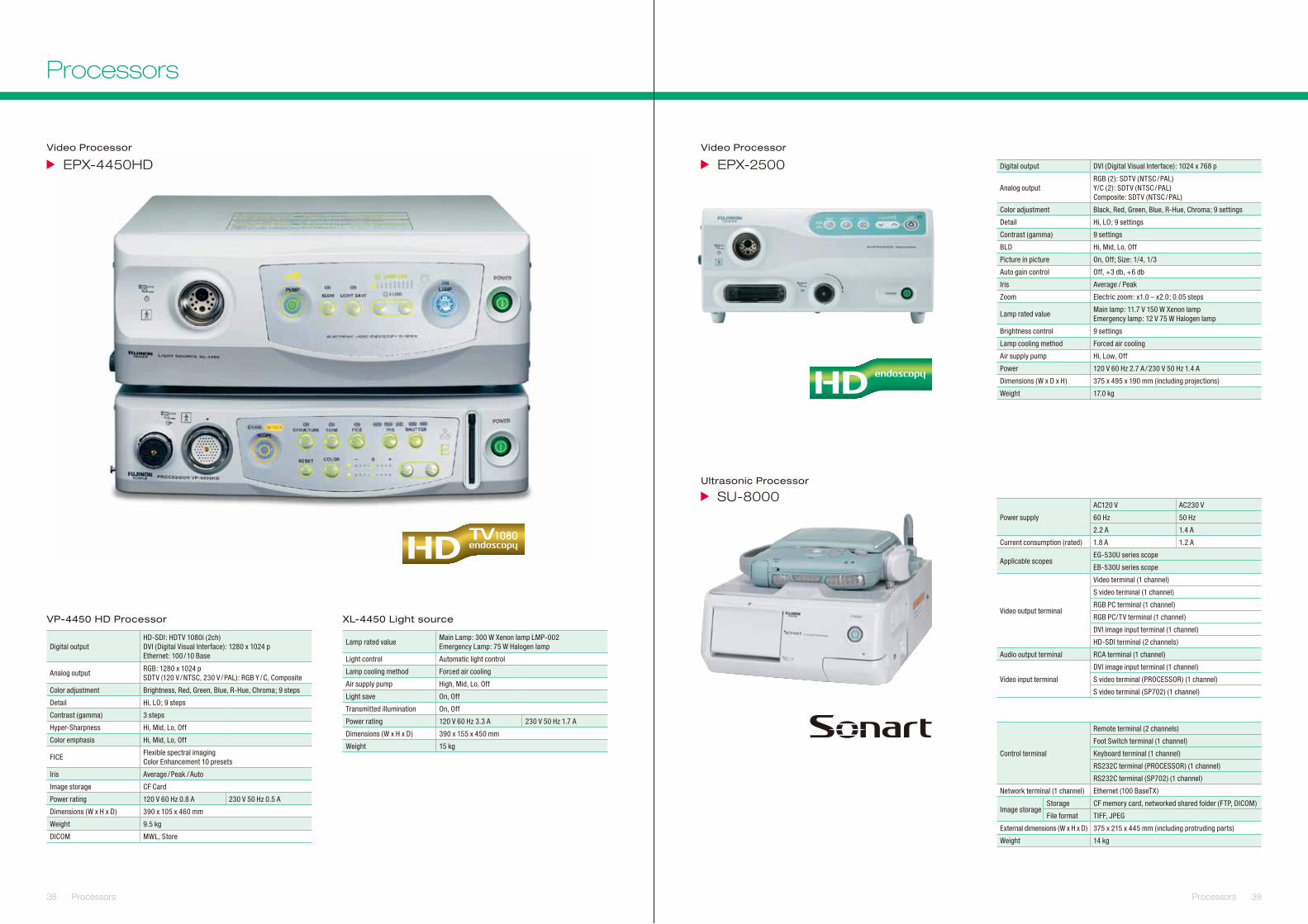
Power supply
AC120 V AC230 V
60 Hz 50 Hz
2.2 A 1.4 A
Current consumption (rated) 1.8 A 1.2 A
Applicable scopesEG-530U series scope
EB-530U series scope
Video output terminal
Video terminal (1 channel)
S video terminal (1 channel)
RGB PC terminal (1 channel)
RGB PC/TV terminal (1 channel)
DVI image input terminal (1 channel)
HD-SDI terminal (2 channels)
Audio output terminal RCA terminal (1 channel)
Video input terminal
DVI image input terminal (1 channel)
S video terminal (PROCESSOR) (1 channel)
S video terminal (SP702) (1 channel)
Control terminal
Remote terminal (2 channels)
Foot Switch terminal (1 channel)
Keyboard terminal (1 channel)
RS232C terminal (PROCESSOR) (1 channel)
RS232C terminal (SP702) (1 channel)
Network terminal (1 channel) Ethernet (100 BaseTX)
Image storageStorage CF memory card, networked shared folder (FTP, DICOM)
File format TIFF, JPEG
External dimensions (W x H x D) 375 x 215 x 445 mm (including protruding parts)
Weight 14 kg
0052-XPE
0008-US
Video Processor
Ultrasonic Processor
Digital output DVI (Digital Visual Interface): 1024 x 768 p
Analog outputRGB (2): SDTV (NTSC / PAL)Y/C (2): SDTV (NTSC / PAL)Composite: SDTV (NTSC / PAL)
Color adjustment Black, Red, Green, Blue, R-Hue, Chroma; 9 settings
Detail Hi, LO; 9 settings
Contrast (gamma) 9 settings
BLD Hi, Mid, Lo, Off
Picture in picture On, Off; Size: 1/4, 1/3
Auto gain control Off, +3 db, +6 db
Iris Average / Peak
Zoom Electric zoom: x1.0 – x2.0; 0.05 steps
Lamp rated valueMain lamp: 11.7 V 150 W Xenon lampEmergency lamp: 12 V 75 W Halogen lamp
Brightness control 9 settings
Lamp cooling method Forced air cooling
Air supply pump Hi, Low, Off
Power 120 V 60 Hz 2.7 A / 230 V 50 Hz 1.4 A
Dimensions (W x D x H) 375 x 495 x 190 mm (including projections)
Weight 17.0 kg
39Processors
Processors