fucosylation discriminates against mannose-derived gdp
TRANSCRIPT

Page 1/30
Insights into fucose metabolism: SLC35C1-independentfucosylation discriminates against mannose-derived GDP-fucoseEdyta Skurska
University of Wroclaw: Uniwersytet WroclawskiBożena Szulc
University of Wroclaw: Uniwersytet WroclawskiDorota Maszczak-Seneczko
University of Wroclaw: Uniwersytet WroclawskiMaciej Wiktor
University of Wroclaw: Uniwersytet WroclawskiWojciech Wiertelak
University of Wroclaw: Uniwersytet WroclawskiMariusz Olczak ( [email protected] )
University of Wroclaw: Uniwersytet Wroclawski https://orcid.org/0000-0001-8629-6364
Research Article
Keywords: SLC35C1, SLC35C2, GDP-fucose synthesis, fucose supplementation, LADII, nucleotide sugar transporter
Posted Date: November 23rd, 2021
DOI: https://doi.org/10.21203/rs.3.rs-1096518/v1
License: This work is licensed under a Creative Commons Attribution 4.0 International License. Read Full License
Page 2/30
AbstractMutations in the SLC35C1 gene, encoding the Golgi GDP-fucose transporter, cause leukocyte adhesion de�ciency II (LADII).Fucosylation improvement in LADII patients treated with fucose suggests the existence of an SLC35C1-independent route of GDP-fucose transport, which still remains a mystery. Here, we developed and characterized a human cell-based model de�cient in theSLC35C1 activity. The knockout cells displayed low but detectable levels of fucosylation. Strikingly, the fucosylation defect wasalmost completely reversed upon treatment with millimolar concentrations of fucose. Even if fucose was supplemented atnanomolar concentrations, it was still incorporated into glycans by the knockout cells. We also found that the SLC35C1-independenttransport preferred the salvage pathway over the de novo pathway as a source of GDP-fucose. Our results imply that the Golgisystems of GDP-fucose transport discriminate between the substrate pools obtained from different metabolic pathways, whichsuggests a functional connection between nucleotide sugar transporters and nucleotide sugar synthetases.
IntroductionFucose is an abundant component of many N- and O-glycans as well as some glycolipids. Except for O-fucosylation, where it is the�rst sugar in a sequence, fucose is always a terminal sugar in the structure of oligosaccharides. Fucose can be attached to othersugars or proteins via one of four types of glycosidic bonds: α-1,2, α-1,3, α-1,4 and α-1,6. In N-glycans, fucose is predominantly α-1,6-linked to the �rst (Asn-bound) N-acetylglucosamine residue. Such type of fucose is referred to as a core fucose and it is incorporatedinto the oligosaccharide structure only after the attachment of at least one N-acetylglucosamine residue to mannose. For a broaderoverview of the consecutive steps of N-glycan processing the reader is kindly referred to [1].
Fucosylated oligosaccharides have many biologically relevant functions. AB0 blood group antigens are among the best knownfucosylated glycans. O-linked fucose may affect certain ligand-receptor interactions involved in signal transduction. Fucosedecorates certain oligosaccharides that are exposed on the surface of leukocytes and serve as ligands for selectins. Binding of thelatter to the former initiates the leukocyte adhesion cascade, a multi-step process of leukocyte migration to the site of injury orinfection.
Incorporation of fucose into glycoconjugates is mediated by fucosyltransferases, which are predominantly Golgi-resident type IImembrane proteins. Fucosyltransferases use the active form of fucose, i.e. GDP-fucose, as a substrate. GDP-fucose is synthesized inthe cytoplasm by the two different pathways (shown in Fig. 1). The main source of this nucleotide sugar is the so-called de novopathway which is estimated to provide 90-95% of the total GDP-fucose pool in the cell [2, 3]. The primary substrate for this three-steppathway is mannose. First, this monosugar is converted into GDP-mannose, then GDP-D-mannose 4,6-dehydratase (GMDS) convertsGDP-mannose to GDP-4-keto-6-deoxymannose. This keto intermediate is then converted into GDP-fucose by an epimerase/reductaseenzyme complex termed the FX protein or GDP-L-fucose synthase (also known as GFUS, FCL, SDR4E1 or TSTA3) [4].
The second, alternative way of GDP-fucose synthesis is the so-called salvage pathway. This route uses cytoplasmic pool of freefucose, which on the one hand is recovered from glycoconjugates by lysosomal α-fucosidase activity, and on the other hand issupplied from the environment. Little is known about the fucose transport system from the extracellular space to the interior of thecell, but it appears to function through facilitated diffusion through speci�c channel(s) [5]. The �rst step of the salvage pathwayinvolves an ATP-dependent synthesis of fucose-1-phosphate by the fucokinase (FUK; also known as FCSK) enzyme. GDP-fucosepyrophosphorylase (FPGT), the second enzyme acting in the pathway, catalyzes the conversion of fucose-1-phosphate and GTP toGDP-fucose [6]. As already mentioned, it is estimated that only 5-10% of the total GDP-fucose pool is synthesized by this pathway [2,3].
To reach lumenally oriented catalytic centers of fucosyltransferases, GDP-fucose has to be transported across the Golgi membrane.This function is thought to be played by GDP-fucose-speci�c nucleotide sugar transporters (NSTs), which are hydrophobic type IIImembrane proteins with 6-10 transmembrane domains. NSTs are believed to act as antiporters; an imported nucleotide sugar isexchanged with the corresponding nucleoside monophosphate [7]. After reaching the Golgi lumen, the activated monosugarconstitutes a substrate for a respective glycosyltransferase, which attaches the monosugar to an acceptor, whereas the releasednucleoside diphosphate is broken down into a nucleoside monophosphate and an inorganic phosphate [8].
In mammals, the role of the main GDP-fucose transporter is attributed to the product of the SLC35C1 gene, which was identi�ed bytwo independent groups in 2001 [9, 10]. Although SLC35C1 is thought to be the major transporter in mammals, GDP-fucose has also

Page 3/30
been shown to translocate through its homolog, SLC35C2. SLC35C2 was shown to localize predominantly in the Golgi, but a smallsubset of it was found in the endoplasmic reticulum (ER) and ERGIC (ER-Golgi intermediate compartment). SLC35C2 is speci�callyrequired for O-fucosylation of certain proteins including the Notch receptor, which in turn does not require the SLC35C1 activity [11,12].
Leukocyte adhesion de�ciency II (LADII) is a rare autosomal recessive genetic disease characterized by an overall reduction infucosylation of glycoconjugates. This syndrome is caused by mutations in the SLC35C1 gene [13]. The characteristic symptoms ofthis disease include psychomotor retardation, facial dysmorphism, Bombay phenotype, short stature, immunode�ciency, leukocytosisas well as recurrent and frequent bacterial infections [14]. LADII was �rst diagnosed in 1992 [15]. To date, 19 individuals bearinginactivating mutations in the SLC35C1 gene have been reported [14–23] with a predominance of point mutations.
The concept of using exogenous fucose to improve fucosylation in LADII patients was �rst raised in 1998. Pioneering experimentswere performed with lymphoblastic cells obtained from one of the patients bearing the Arg147Cys mutation in the SLC35C1 aminoacid sequence. Supplementation of the cell culture with 10 mM fucose for 5 days triggered an increase in the number of fucosylatedstructures [24]. These results paved the way to the concept of using fucose supplementation in patients themselves. Since 1999,several LADII patients were treated with oral fucose. For some of them such treatment was successful, resulting in an improvementin psychomotor development and a reduction in neutrophil count [22, 25]. However, in the case of two LADII patients fucosesupplementation failed to improve their condition [20, 26]. The administered doses of fucose varied from 25 to 2000 mg/kg bodyweight [20, 25, 26]. An improvement in fucosylation was also achieved when the LADII patient-derived �broblasts were cultured in thepresence of 0.1-10 mM fucose [18].
To explain the effectiveness of the fucose treatment in responsive LADII patients, it was postulated that the mutant SLC35C1variants display some residual transport activity [9]. Therefore, a therapy causing an increase in the cytosolic concentration of thisnucleotide sugar could potentially allow to overcome the insu�cient transporting activity of the defective SLC35C1 variants.However, a direct proof for an increase in the cytosolic GDP-fucose concentration in the fucose-fed cells bearing mutations in theSLC35C1 gene has not been presented to date. An alternative hypothesis to explain the e�ciency of the oral fucose therapy could bethe existence of an accessory GDP-fucose transport system, however, no such route has been identi�ed so far.
In 2007, a knockout-based study conducted in mice revealed that the fucose treatment works even in the absence of SLC35C1 [27].These �ndings for the �rst time suggested the existence of an SLC35C1-independent GDP-fucose transport into the Golgi, as theobserved effect could no longer be explained by a residual activity of the SLC35C1 mutants. It was hypothesized that the SLC35C1de�ciency could be overcome by the SLC35C2 activity, but this has never been proved. In general, no studies that could possiblyexplain this phenomenon in bigger detail have been undertaken so far.
Since the responsiveness of the cells completely devoid of the SLC35C1 activity to fucose treatment has not been elucidated to date,here we have undertaken a more-in-depth investigation of this intriguing effect. Within our general interest in the metabolism offucose and protein fucosylation, speci�c goals included: (i) generation of single SLC35C1 and double SLC35C1/SLC35C2 knockouts,(ii) optimization of fucose feeding conditions, (iii) establishment of a method allowing quanti�cation of the extent of N-glycanfucosylation, (iv) structural characterization of N- and O-glycans produced by the wild-type and knockout cells, (v) investigation of N-glycan fucosylation in response to various ranges of exogenous fucose concentrations, (vi) single-point and time-course analyses ofintracellular GDP-fucose concentration in control and fucose-fed cells, (vii) determination of the contribution of de novo and salvageGDP-fucose biosynthetic pathways to fucose incorporation into N-glycans in the wild-type and knockout cells and (viii) assessmentof a potential cellular genetic response to fucose supplementation.
In this study we have developed an approach coupling exoglycosidase digestion with HPLC to quantify the percentage of the core-fucosylated N-glycans with a great precision. We showed that in the knockout cells the core fucosylation of N-glycans is greatlyreduced but can be nearly completely restored by supplementing the cells with millimolar concentrations of fucose. Supplementationwith 5 mM fucose caused a signi�cant increase in the intracellular GDP-fucose concentration, regardless of the presence/absence ofGDP-fucose transporters. Next, by feeding low (nanomolar) concentrations of radioactive fucose and mannose into the culturemedium we were able to follow the metabolic fate of the supplemented compounds and attribute radioactivity to fucosylatedstructures using our HPLC-based method. Surprisingly, the SLC35C1 knockouts showed a dramatic impairment of the ability toincorporate mannose-derived radioactivity into the fucosylated N-glycans. In sharp contrast, they were able to incorporate even higher

Page 4/30
quantities of radiolabeled fucose than the wild-type cells. To explain these observations we hypothesize that in the SLC35C1knockouts the salvage pathway is preferred over the de novo pathway as a source of GDP-fucose for fucosylation of N-glycans. Wehave also investigated potential genetic response of the SLC35C1 knockouts to fucose treatment. No major gene up/down-regulations were observed. Finally, we excluded SLC35C2 as an alternative supplier of GDP-fucose for N-glycan fucosylation.
ResultsGeneration of the single SLC35C1 and double SLC35C1/SLC35C2 knockouts
To develop a research model for our study, an SLC35C1 knockout (C1KO) was generated in two human cell lines, i.e. HEK293T andHepG2 using the CRISPR/Cas9 approach. Additionally, a double SLC35C1/SLC35C2 knockout (C1/C2KO) was developed in HEK293Tcells to evaluate the in�uence of SLC35C2 protein on the N- and O-glycosylation in the SLC35C1 knockout cells and to examine theeffect of fucose treatment in cells lacking both GDP-fucose transporters. The knockouts were con�rmed by RT-PCR performed ontotal RNA and PCR performed on genomic DNA (Fig. S1A and S1B). The SLC35C1 knockout was additionally veri�ed on the proteinlevel using an anti-SLC35C1 antibody by western blotting (Fig. S1C) as well as by double staining with the latter antibody and fucose-speci�c Aleuria aurantia lectin (AAL, Fig. S1D).
To show that the knockout phenotype was speci�cally associated only with the disruption of the SLC35C1 gene, we have alsodeveloped SLC35C1-de�cient clones stably over-expressing a recombinant, HA-tagged SLC35C1 (in both HEK293T and HepG2 celllines). The overexpression was con�rmed by western blotting performed on cell lysates using an anti-HA antibody (Fig. S1E). Theobtained stable transfectants were also subjected to immunostaining to con�rm the Golgi localization of the over-expressed protein(Fig. S1F).
Optimization of fucose supplementation conditions
In order to establish a system to study the effect of fucose supplementation on the restoration of fucosylation in the SLC35C1knockouts, feeding parameters, i.e. fucose concentration in the culture medium as well as duration of the treatment, had to beoptimized. In the �rst stage we employed a dot blot analysis using AAL lectin (Fig. 2A, 2B). Several concentrations of fucose weretested, i.e. 0.5, 1, 5 and 10 mM (concentration range inspired by [18]). We found that after 5 days, 5 mM concentration was su�cientto substantially restore fucosylation of glycoconjugates in the SLC35C1 knockouts. AAL stainings performed on the SLC35C1knockouts cultured in the presence of 5 mM fucose for 0-48 h demonstrated that the level of fucosylation gradually increased overtime and an optimum (i.e. fucosylation compared to the wild-type) was reached after 24 h. After another 12 h, however, the cellsbecame overloaded with aggregated fucose-containing structures (Fig. 2C).
Since the lectin-based analysis does not allow for quanti�cation of the observed fucosylation defects/rescue effects, we haveestablished a new, robust method, based on exoglycosidase digestions followed by HPLC separation, that allows quanti�cation ofthe degree of the α-1,6-fucosylation (core fucose). In our approach, we used speci�c glycosidases to reduce all multi-branchedcomplex-type N-glycan species into a simple biantennary (GlcNAc)2(Man)3 structure, either fucosylated or non-fucosylated (Fig. 2D,2E). Then by taking a ratio of the area of the fucosylated peak over the sum of areas of both peaks (fucosylated and non-fucosylated) we have determined the percentage of fucosylated structures for each sample. To con�rm that the fucosylated peakcontains only (GlcNAc)2(Man)3Fuc structure, the α- fucosidase digestion was performed (Fig. 2F)
By employing our method we tested supplementation of selected fucose concentrations (50 µM - 100 mM) for different time periods(1-5 days) in the wild-type and SLC35C1 knockout HEK293T cells. Feeding the cells with 50 µM or 1 mM fucose even for as long as 5days was not su�cient to restore the wild-type α-1,6-fucosylation level in the SLC35C1 knockout cells. Only supplementation of theSLC35C1 knockout cells with 5 mM fucose restored fucosylation phenotype to a level comparable to the wild-type, but at the sametime it did not affect core fucosylation in the wild-type cells. At the same time, supplementation with a very high fucose concentration(100 mM) did not increase the level of core fucosylation in the wild-type cells (Fig. 2G). Therefore, for the �nal experiments, theconditions of 5 mM fucose and 24 h were selected.
Supplementation of fucose in the knockout cells signi�cantly increases α-1,6-fucosylation of N-glycans

Page 5/30
We quanti�ed α-1,6-fucosylation of N-glycans in all modi�ed cell lines. From the analysis of the HEK293T cells we could concludethat: (i) the native level of core fucosylation in the non-fed wild-type cells oscillates around ~75% and is compromised to ~8-10% inthe single SLC35C1 and double SLC35C1/SLC35C2 knockouts, (ii) ectopic expression of the HA-tagged SLC35C1 in the singleSLC35C1 knockouts restores the wild-type phenotype, (iii) nearly complete α-1,6 fucosylation (~60%) can be achieved in both singleSLC35C1 and double SLC35C1/SLC35C2 knockouts upon administration of 5 mM fucose for 24 h (Fig. 3A). As anticipated, similarobservations were made for the HepG2 cells (Fig. 3B). MALDI-TOF analysis of N-glycan structures con�rmed those results, showingthe presence of a fucosylated structure in the SLC35C1 knockouts and restoration of fucosylated structures after fucose feeding (Fig.S2).
During the separation and puri�cation of N-glycans, there is a possibility of contamination with the oligosaccharides derived fromserum glycoproteins contained in the culture medium. To address this problem, the N-glycans derived from an engineered, ectopicallyexpressed and secreted reporter glycoprotein, Secreted Alkaline Phosphatase (SEAP), were also analyzed. The method for SEAPisolation e�ciently prevents contamination with serum glycoproteins [28]. From the analysis of α-1,6-fucosylated N-glycans derivedfrom the SEAP overexpressed in the wild-type and knockout HEK293T cells we concluded that in the knockouts α-1,6-fucosylation ofSEAP-derived N-glycans is still present (~3-5%) and supplementation of 5 mM fucose for 24 h restore α-1,6-fucosylation in thesecells. Interestingly, the level of α-1,6-fucosylation observed in the wild-type and knockout cells was higher than it was in the wild-typebefore supplementation, which could be a speci�c response of the overexpressed SEAP on the supplementation (Fig. 3C). Asanticipated, similar observations were made for the HepG2 cells (Fig. 3D).
Supplementation of fucose restores fucosylation of O-glycans in the knockout cells
Certain mucin-type O-glycans are also fucosylated. The effect of GDP-fucose transport de�ciency on this type of oligosaccharideswas only investigated using lectins [27]. Therefore, we have additionally analyzed cellular mucin-type O-glycans using the CORAstrategy [29].
From the analysis of the HEK293T cells we could conclude that: (i) in the wild-type cells 2 among 8 of the identi�ed glycans werefucosylated (m/z values of ~1218 and ~1578; Fig. 4A), (ii) both fucosylated species were absent in the knockouts in which only theirnon-fucosylated counter-partners were detected (m/z values of ~1044 and 1404), (iii) a new fucosylated species was observed in theknockouts (m/z~ 1013, Fig. 4B, C), (iv) supplementation with 5 mM fucose for 24 h led to a re-appearance of the fucosylated speciesin the knockouts. Surprisingly, after fucose supplementation additional fucosylated species were observed in the wild-type andSLC35C1 knockout cells, but not in the double SLC35C1/SLC35C2 knockout (Fig. 4D, E, F). In the case of HepG2 cells the fucosylatedO-glycan species were not produced by the SLC35C1 knockout. However, in contrast to HEK293T cells, fucose supplementation of thewild-type HepG2 cells did not have an in�uence on fucosylated O-glycans (Fig. 4G-J). The observed differences could be cell line-speci�c.
These results prove the role of SLC35C1 in the process of mucin-type O-glycan fucosylation and goes in line with the observationsmade for the N-glycans. Importantly, supplementation with 5 mM fucose for 24 h restored fucosylation of O-glycans in the singleSLC35C1 and double SLC35C1/SLC35C2 knockout cells.
Supplementation of fucose increases intracellular GDP-fucose concentration
Although it is anticipated that in the SLC35C1-de�cient cells the exogenous fucose e�ciently enters the cells and becomes convertedinto GDP-fucose, to our best knowledge this phenomenon was neither quanti�ed. For separation and quantitative analysis ofnucleotide sugars we employed a modi�ed ion-pair solid-phase extraction and HPLC strategy developed by Räbinä et al. [30]. Ourresults demonstrated that in the non-fed HEK293T cells the intracellular concentration of GDP-fucose oscillated around ~5 µM,regardless of the genotype (wild-type or single/double knockout). This shows that GDP-fucose transporter de�ciency of the SLC35C1knockout did not cause a GDP-fucose accumulation. This is in contrast to the research conducted on the SLC35A1 (CMP-sialic acidtransporter) knockout, for which such an effect was observed [31]. Importantly, upon fucose supplementation, GDP-fucoseconcentration increased ~40-50-fold in all HEK293T cell lines, up to ~200-250 µM (Fig. 5A). Similar experiment was performed forHepG2 cells where the basal concentrations of GDP-fucose were slightly higher (~10 µM) and the effect of supplementation wassomewhat less pronounced (~15-fold, up to ~150 µM) (Fig. 5B). Based on these �ndings we concluded that the externallysupplemented fucose e�ciently enters the cytoplasm and is being converted to GDP-fucose in all tested cell lines.
Page 6/30
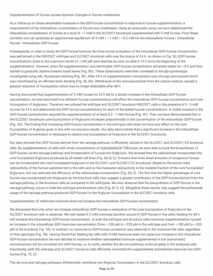
Supplementation of fucose causes dynamic changes in fucose metabolism
As a follow-up on these remarkable increases in the GDP-fucose concentration in response to fucose supplementation, ameasurement of the intracellular concentration of fucose was undertaken. Using an enzymatic assay we have determined theintracellular concentration of fucose at a level of ~1 mM in the SLC35C1-knockouts supplemented with 5 mM fucose. From thesenumbers one can postulate an approximate equilibrium of 5 mM / ~1 mM / ~0.2 mM for the extracellular fucose / intracellularfucose / intracellular GDP-fucose.
Subsequently, in order to study the GDP-fucose turnover, the time-course evaluation of the intracellular GDP-fucose concentrationwas performed in the HEK293T wild-type and SLC35C1 knockout cells over the course of 24 h. As shown in Fig. 5C, GDP-fucoseconcentrations close to the maximum levels of ~250 µM were reached as soon as after 6-12 h since the beginning of thesupplementation. However, when the supplementation was terminated, GDP-fucose concentration remained stable for ~8 h and thenstarted to gradually decline towards basal levels (Fig. 5D). These observations were then correlated to the glycophenotypeinvestigated using AAL �uorescent staining (Fig. 5E). After 24 h of supplementation fucosylation was strongly pronounced whichwas manifested by an e�cient lectin binding (Fig. 5E, 0h). Withdrawal of the monosaccharide from the culture medium caused agradual reduction of fucosylation which was no longer detectable after 48 h.
Having discovered that supplementation of 5 mM fucose for 24 h led to a drastic increase in the intracellular GDP-fucoseconcentration, we next examined how different fucose concentrations will affect the intracellular GDP-fucose concentration and corefucosylation of N-glycans. Therefore, we cultured the wild-type and SLC35C1 knockout HEK293T cells in the presence of 0 - 5 mMfucose for 24 h and determined the GDP-fucose concentrations for each of the tested fucose concentrations. A visible increase in theGDP-fucose concentration required the supplementation of at least 0.2 - 1 mM fucose (Fig. 5F). Then we have demonstrated that inthe SLC35C1 knockouts core fucosylation of N-glycans increases proportionally to the concentration of the intracellular GDP-fucose(Fig. 5G). The �nding that increasing GDP-fucose concentration in the wild-type cells does not have any effect on the corefucosylation of N-glycan goes in line with our previous results. Our data demonstrate that a signi�cant increase in the intracellularGDP-fucose concentration is necessary to restore core fucosylation of N-glycans in the SLC35C1 knockouts.
Our data showed that GDP-fucose derived from the salvage pathway is e�ciently utilized in the SLC35C1 and SLC35C1/C2 knockoutcells. By supplementation of cells with small concentration of radiolabeled [6-3H]fucose, we were able to track the biosynthesis ofGDP-fucose via the salvage pathway and incorporation of fucose into N-glycans. We showed that the radioisotope incorporated intocore fucosylated N-glycans produced by all tested cell lines (Fig. 6A, B, C). It means that even small amounts of exogenous fucosecan be incorporated into core fucosylated N-glycans in the SLC35C1 and SLC35C1/C2 knockouts. Based on the known totalradioactivity of the fucose substrate fed to the cells and on the measured radioactivity in the isolated fraction of the core fucosylatedN-glycans, one can estimate the e�ciency of the radioisotope incorporation (Fig. 6D, E). The fact that the higher percentage of corefucose was incorporated into N-glycans by the knockout cells may suggest a greater contribution of the GDP-fucose derived from thesalvage pathway in the knockout cells as compared to the wild-types. We also observed that the biosynthesis of GDP-fucose in thesalvage pathway occurs in both the wild-type and knockout cells (Fig. 6F, G, H). Altogether, these results may suggest the preferentialusage of the salvage pathway-produced GDP-fucose for the N-glycan fucosylation in the SLC35C1 knockout cells.
Supplementation of millimolar mannose does not increase the intracellular GDP-fucose concentration
We discovered that only when we increase intracellular GDP-fucose, a restoration of the core fucosylation of N-glycans in theSLC35C1 knockout cells is observed. We next tested if 5 mM mannose (another source of GDP-fucose in the cells) feeding for 48 hwill increase the intracellular GDP-fucose concentration. In both the wild-type and knockout cells mannose supplementation causedan increase in the intracellular GDP-mannose concentration (from ~100 µM to ~220 µM in the wild-type and from ~180 µM to ~330µM in the knockout; Fig. 7A). In contrast, no mannose to GDP-fucose conversion was observed in the mannose-fed cells, regardlessof their genotype (Fig. 7B). Having found that feeding the cells with 5 mM mannose does not cause any increase in the intracellularGDP-fucose concentration we next decided to examine whether radiolabeled mannose supplemented in low (nanomolar)concentrations will be converted into GDP-fucose, i.e. to verify, whether the de novo pathway works properly in the analyzed cells.Importantly, both the wild-type and SLC35C1 knockout cells e�ciently converted the supplemented radiolabeled mannose into GDP-fucose (Fig. 7C, D).
The de novo and salvage pathways differentially contribute into N-glycan fucosylation in the SLC35C1 knockout cells

Page 7/30
To compare the relative contribution of different GDP-fucose biosynthesis pathways into the N-glycan fucosylation, the wild-type andSLC35C1-de�cient HEK293T cells were cultured in the presence of nanomolar concentrations of either radioactive fucose (salvagepathway) or radioactive mannose (de novo pathway). However, apart from conversion into GDP-fucose, mannose can be alsoprocessed into other nucleotide sugars, e.g. GDP-mannose, and as such incorporated into the complex type N-glycans. To addressthis problem, we have established a new approach in which the N-glycans are �rst digested with α-fucosidase, then the post-reactionmixture is passed through a graphite column and �nally the radioactivity of the �ow-through is measured. This strategy allows us tospeci�cally assign the detected radioactivity to fucose present in N-glycans.
Our results showed a decrease in the incorporation of fucose derived from the GDP-fucose synthesized via the salvage pathway intoN-glycans in the SLC35C1 knockout cells, but still the extent of fucose incorporation was more than 23% of the wild-type (Fig. 8A).Surprisingly, incorporation of fucose derived from the GDP-fucose synthesized via the de novo pathway (i.e. from mannose) turnedout to be nearly completely abolished (6% of the wild-type) (Fig. 8B).
qPCR analysis of expression levels of selected glycosylation-related genes
There is a possibility that some fucosylation-related genes become up- or down-regulated by the exogenously added fucose.Therefore, to complement our metabolic studies, expression levels of selected glycosylation-related genes were analyzed in thecontrol and fucose-fed SLC35C1-de�cient cell lines. For the analysis of a larger subset of genes, a ULP probe-based assay wasdesigned (Fig S4). Here, 91 different genes, including NSTs, glycosyltransferases and biosynthetic enzymes involved in metabolismof different monosaccharides (galactose, N-acetylglucosamine, mannose, xylose and fucose) were investigated. No statisticallysigni�cant differences could be assigned for any of the tested genes.
To analyze differences in gene expression between the wild-type and SLC35C1 knockouts in HEK293T and HepG2 cells a SYBRGreen-based qPCR assay was performed for six selected genes, i.e. FCL, GMDS, FUK, FPGT1, FPGT2 and SLC35C2 (Sup tab.1, 2).Similarly to the previous �ndings, no statistically signi�cant differences between the wild-type and SLC35C1 knockout HEK293T andHepG2 cells (both control and supplemented with exogenous fucose) were observed.
DiscussionIn this study we have con�rmed the existence of SLC35C1-independent GDP-fucose transport routes in the Golgi complex anddetermined the intracellular fate of the exogenously supplied fucose. Moreover, we have shown that the SLC35C1-dependent routemainly transports GDP-fucose provided by the de novo biosynthetic pathway, whereas the SLC35C1-independent route nearlyexclusively translocates GDP-fucose derived from the salvage pathway.
The SLC35C1-de�cient mammalian model was for the �rst time developed and characterized by Hellbusch and coworkers in 2007[27]. In that study, treatment of cells from different mouse organs with exogenously supplied fucose partially restored glycoproteinfucosylation, which encouraged the authors to propose the existence of an alternative, SLC35C1-independent GDP-fucose transportmechanism. However, the improvement in fucosylation was demonstrated using only a single method, i.e. lectin-based �owcytometry, and no intermediate metabolic steps, e.g. conversion of fucose into GDP-fucose, have been investigated.
The study presented here in multiple aspects extends the previously conducted research. First of all, the improvement in fucosylationcaused by supplementation of the culture medium with millimolar concentrations of fucose was precisely determined to be ~77% byour self-developed quantitative HPLC-based approach. Moreover, we showed that also nanomolar concentrations of exogenousfucose are su�cient to observe its incorporation into N-glycans in the knockout cells. Furthermore, we determined the extent to whichexogenous fucose is transported into the cytoplasm and how it contributes to the increase of the concentration of the intracellularGDP-fucose (the extracellular fucose, intracellular fucose and intracellular GDP-fucose were shown to exist in a 5 mM / 1 mM / 0.2mM equilibrium). Finally, we showed that the SLC35C1-independent Golgi GDP-fucose transport systems preferentially use thenucleotide sugar pool derived from the salvage pathway, which we consider the main outcome of this study.
In this study we have developed a quantitative HPLC-based approach allowing for the very precise determination of the level of corefucosylation of N-glycans. This form of fucosylation, mediated by a single Fut8 enzyme, is present in many cell types includingHEK293T and HepG2 which were selected as our model. Moreover, it is easy to study due to the well-established procedures of N-glycan isolation and HPLC analysis. As shown by our data, the level of core-fucosylated N-glycans in the wild-type HEK293T and

Page 8/30
HepG2 cell lines is high (~80%) but does not reach 100%, giving a possibility to potentially observe a further increase in fucosylation,i.e. over-fucosylation, upon fucose treatment. On the other hand, in these cell lines the level of core-fucosylated structures is highenough to make a clear distinction between the wild-type and the SLC35C1 knockout phenotypes. To conclude, the choice of celllines was dictated by the high percentage of core-fucosylated N-glycans they produce, which in turn was demanded by thequantitative HPLC-based approached we employed.
Using our quantitative HPLC-based approach we showed that the core N-glycan fucosylation is signi�cantly compromised in theSLC35C1-de�cient cells. However, some residual fucosylation could still be detected in these cell lines. The SLC35C2 protein is alsospeci�c for GDP-fucose. Thus, its activity could potentially compensate for the lack of SLC35C1 in the single knockout cells. Here weshowed that this is not the case as knocking out the SLC35C2 gene in the SLC35C1-de�cient cells did not make the fucosylationdefect any more severe. There results suggest that there must be yet another system responsible for the SLC35C1-independent GDP-fucose import into the Golgi lumen.
Strikingly, supplementation of the culture medium with millimolar concentrations of fucose caused a substantial improvement ofcore N-glycan fucosylation in both single SLC35C1 and double SLC35C1/SLC35C2 knockout cells. In contrast, in the wild-type cellsfucose treatment did not cause any further improvement in fucosylation, which is in line with the results obtained by Moriwaki et al.[33] who did not observe any increase in AAL reactivity with glycoproteins produced by the wild-type Hep3B cells supplemented withup to 5 mM fucose. This �nding additionally supports our assumption that SLC35C1 mainly utilizes GDP-fucose produced by the denovo pathway, whereas GDP-fucose synthesized via the salvage pathway does not appear to be an optimal source of this nucleotidesugar in the wild-type cells, even if the latter is present in a large excess. We also hypothesize that the residual fucosylationdetectable in the knockout cells is derived from GDP-fucose produced by the salvage pathway, which is not the main source of thisnucleotide sugar in the wild-type cells. We believe that if the SLC35C1-independent GDP-fucose transport system used the nucleotidesugar substrate produced by the main biosynthetic pathway (i.e. de novo), the level of core fucosylated N-glycans in the knockoutcells would be much higher.
It should be emphasized that fucose treatment of the LADII patients and SLC35C1-de�cient cells can only be successful if thefollowing requirements are ful�lled: (i) fucose must e�ciently enter the cells, (ii) after entering the cell fucose must be readilyconverted into GDP-fucose via the salvage pathway and (iii) GDP-fucose must reach the Golgi lumen to become available for thecatalytic centers of fucosyltransferases. Importantly, quanti�tation of intracellular GDP-fucose concentration in fucose-fed SLC35C1knockout cells was not attempted in the previous studies. To the best of our knowledge, the changes in the GDP-fucose content inresponse to exogenously supplied fucose was examined only by Moriwaki et al. in the wild-type Hep3B cells [32] and, more recently,by Sosicka et al. in the wild-type HepG2, CHO and Huh7 cells [33]. In the �rst study, the range of analyzed fucose concentrations wasbetween 0 and 5000 µM. However, these results cannot be compared with the ones obtained in this study as both procedures differedin several aspects including the cell line used and the method for nucleotide sugar detection (Moriwaki et al. [33] used an indirect,enzyme-based detection and did not attempt to determine intracellular GDP-fucose concentration, whereas we performed a directdetection followed by estimation of intracellular GDP-fucose concentration). On the other hand, Sosicka et al. tested only oneconcentration of exogenous fucose (50 µM) [33]. Importantly, neither of these studies attempted to determine the intracellular GDP-fucose concentration in fucose-fed SLC35C1 knockout cells, which is crucial for our understanding of the responsiveness of thesecells to exogenous fucose.
Here, we determined GDP-fucose concentrations in the control and fucose-fed wild-type and knockout cells. We found that thebaseline concentrations of GDP-fucose were similar in the wild-type and knockout cells, whereas a remarkable increase could beobserved upon fucose treatment in all the cell lines analyzed. In native conditions, the salvage pathway was shown to play onlynegligible role in the overall GDP-fucose production. Here we show that the capacity of this pathway is very high, i.e., thecorresponding enzymes are able to process signi�cantly greater amounts of the primary substrate than are normally present in thecell. However, our data exclude the possibility that the exogenously added fucose triggers a strong up-regulation of expression of theenzymes acting in this pathway as we did not observe any signi�cant changes in the relative levels of the corresponding transcriptsbetween the control and fucose-fed SLC35C1 knockout cells.
Our results clearly show that the therapeutic approaches for CDGs associated with impaired NST activity should aim for a drasticincrease in the intracellular concentration of the corresponding nucleotide sugar substrates. It should be noted that treatment ofthese CDGs by oral monosaccharide supplementation can only be successful if the Km of an enzyme initiating the corresponding

Page 9/30
salvage pathway is su�ciently high. Otherwise, the treatment would simply be ineffective. Fucokinase is the �rst enzyme acting inthe salvage pathway of the GDP-fucose biosynthesis. Importantly, in the case of fucokinase puri�ed from pig kidney Km for fucosewas determined to be as high as 27 µM [32]. This explains why sub-millimolar and millimolar concentrations of exogenous fucoseare required to e�ciently stimulate GDP-fucose synthesis and rescue N-glycan fucosylation in SLC35C1-de�cient cells.
Given the fact that fucose treatment nearly completely rescued the fucosylation defect in the SLC35C1-de�cient cells, it appearssurprising that fucose-based therapy turned out to be ineffective for some of the LADII patients. There are two possible explanationsfor this phenomenon. First, the non-responsive LADII patients might bear additional mutations outside the SLC35C1 gene that affectthe performance of the fucosylation machinery. The other possible explanation is that the presence of certain mutant SLC35C1variants is somehow more deleterious to the cells than the absence of the protein.
Regardless of the improvement of fucosylation in the SLC35C1-de�cient cells treated with millimolar concentrations of fucose, wehave also demonstrated that these cells are able to incorporate fucose supplemented in nanomolar concentrations. This proves thatin the knockout cells the salvage pathway is able to fuel the SLC35C1-independent GDP-fucose transport system even at extremelylow fucose concentrations.
Since increasing the intracellular concentration of GDP-fucose via fucose treatment improved core fucosylation of N-glycans, wewondered if it would be possible to boost the production of this nucleotide sugar by feeding the cells with millimolar concentrationsof mannose given the fact that the de novo pathway of GDP-fucose synthesis utilizes GDP-mannose as a substrate. However,regardless of the cell line analyzed, mannose treatment did not cause any signi�cant changes in the GDP-fucose concentration. Thiscan be partially explained by the fact that mannose is converted to a variety of different metabolites. Although in mannose-fed cellsa statistically signi�cant increase in the GDP-mannose concentration was demonstrated, this did not translate into a similar increasein the GDP-fucose level. We therefore hypothesize that both in the wild-type and knockout cells the de novo pathway operates with anearly maximum e�ciency and it is impossible to improve its performance by increasing the amount of its primary substrate, i.e.,GDP-mannose. Based on these �ndings it can be concluded that treatment of LADII patients with mannose wouldn’t be an attractivealternative to fucose feeding.
Based on our results we propose the existence of three different GDP-fucose transport systems in the Golgi membrane. The �rst oneis SLC35C1-dependent and mainly utilizes the nucleotide sugar pool derived from the de novo pathway. The other two are notdependent on SLC35C1 and mainly use the nucleotide sugar pool synthesized in the salvage pathway. However, the �rst of themworks even under very low (nanomolar) concentrations of exogenous fucose that do not cause any increase in the GDP-fucoseconcentrations, whereas the other requires much higher concentrations of this nucleotide sugar that can only be obtained by feedingthe cells with sub-millimolar and millimolar concentrations of fucose. The existence of the latter SLC35C1-independent transportsystem is strongly supported by the non-linear dependence of the GDP-fucose content on the concentration of exogenous fucose. Wehypothesize that this route may not be physiological and is not exclusively speci�c for GDP-fucose.
The hypothesis raised above assumes the existence of distinct, independent, separate cytosolic pools of GDP-fucose. Such aphenomenon was recently proposed by Sosicka et al. [33] who elegantly demonstrated that different fucosyltransferases utilizedistinct GDP-fucose pools derived from distinct fucose sources. We believe that this effect could be executed by a selectivecooperation of the different GDP-fucose transport systems with distinct fucosyltransferases (similar to e.g. SLC35C2, which wasshown to speci�cally support O-fucosylation [11, 12]). But the question arises how different Golgi transport systems coulddiscriminate between the GDP-fucose pools derived from distinct metabolic pathways? This might require a physical proximity (oreven association) between the enzymes acting in the individual pathways and the corresponding transport systems. Although thepredominant Golgi localization of nucleotide sugar transporters is well established, little is known about the precise subcellulardistribution of nucleotide sugar synthases. The study performed by Coates et al. postulated cytoplasmic localization of the GDP-mannose and UDP-glucose pyrophosphorylases [35]. This appears to hold true for all the other nucleotide sugar synthases except forthe CMP-sialic acid synthetase (CMAS) whose nuclear localization was shown in the same study. However, it is widely believed thatcytoplasmic enzymes catalyzing consecutive reactions in the individual pathways are not randomly distributed. Instead, they arerather sequestered to speci�c cytoplasmic subcompartments where they form functional assemblies that support substratechanneling [36]. It cannot be excluded that the enzymes acting in the GDP-fucose biosynthetic pathways also display such atendency.

Page 10/30
Importantly, GDP-fucose was shown to inhibit the de novo pathway in humans [37] and bacteria [38]. At the same time, this pathwayis thought to be the main source of GDP-fucose in mammalian cells. Therefore, an immediate delivery of the GDP-fucose synthesizedby the de novo pathway to the site of its ultimate utilization (i.e., the Golgi lumen) would be highly bene�cial as it would ensure thee�cient course of the GDP-fucose biosynthesis by shifting the equilibrium of the reaction. Such a scenario could be possible if theenzymes acting in the de novo pathway and the main GDP-fucose transporter, i.e., SLC35C1, were located nearby. Although themammalian fucokinase was shown to be nearly completely inhibited by 60 µM GDP-fucose [34], we did not observe suchphenomenon, as we were able to increase the intracellular concentration of GDP-fucose up to ~200 µM by fucose treatment. Thismay suggest that the GDP-fucose formed in the salvage pathway does not accumulate in the site of its synthesis but instead isimmediately channeled to subcompartments in which it is subsequently utilized. Interestingly, the interactions between a plant UDP-glucose 4-epimerase and two UDP-galactose transporters were recently reported [39]. Therefore, it is highly likely that also theenzymes acting in the de novo pathway of GDP-fucose biosynthesis are near to the SLC35C1 protein in mammalian cells.
In this study we have con�rmed the existence of the alternative, SLC35C1-independent GDP-fucose transport systems in the Golgicomplex. We also have shown that SLC35C2 activity is dispensable for this transport to occur. We demonstrated that theexogenously supplemented fucose e�ciently enters the cells and is readily converted to GDP-fucose via the salvage pathway.Strikingly, the SLC35C1-de�cient cells were virtually unable to incorporate fucose derived from GDP-fucose produced by the de novopathway. There are several questions that still need to be answered, e.g. what is the molecular identity of the SLC35C1-independentGDP-fucose transport systems and how different Golgi transport systems discriminate between the nucleotide sugar pools derivedfrom different biosynthetic pathways. Nevertheless, we strongly believe that the �ndings obtained in this study initiate a brand newchapter in our perception of fucosylation.
Materials And MethodsCell culturing and gene inactivation
HEK293T cells purchased from ATCC (American Type Culture Collection, CRL-3216) and HepG2 cells purchased from the collectionof the Department of Cancer Immunology (Hirszfeld Institute of Immunology and Experimental Therapy, Polish Academy ofSciences) were cultured in Dulbecco’s Minimum Eagle Medium (DMEM High Glucose, Sigma-Aldrich) supplemented with 10% fetalbovine serum, 100 U/mL penicillin and 100 µg/mL streptomycin. Cells were kept at 37°C under 5% CO2.
Ready-to-use Santa Cruz Biotechnology CRISPR-Cas9 kit was used to inactivate SLC35C1 gene in HEK293T and HepG2 cell lines.Cells were transfected with a mixture of human SLC35C1 double nickase plasmids (sc-410008-NIC-2) using the FuGENE HDtransfection reagent (Promega) according to manufacturer's protocol. Next, cells were cultured in DMEM complete mediumsupplemented with 1 µg/mL of puromycin for three weeks to select transfected cells. After that time, clones were isolated andchecked for the presence of the SLC35C1 transcript using one-step reverse transcription PCR (RT-PCR) with the HEK293T and HepG2wild-type cells as the controls. Analysis of genomic DNA derived from the SLC35C1-de�cient cells was also performed. Primers usedin the reactions were listed in Table 1. Next step in con�rmation of the SLC35C1 gene knockout was western blotting.
Inactivation of the SLC35C2 gene in the SLC35C1 knockout HEK293T cells was performed in the same manner. Santa CruzBiotechnology CRISPR-Cas9 kit with mixture of human SLC35C2 double nickase plasmids (sc-409264-NIC-2) was applied.Con�rmation of the SLC35C2 gene knockout was performed at both RNA and genomic DNA levels with using the HEK293T wild-typecells as a control. Used primers are listed in Table 1.

Page 11/30
Table 1Primers used in this study.
No. Primer Name Primer Sequence 5’-3’ Product Length[bp]
Primers used in RT-PCR analysis with total RNA as a template
1 F_C1KO CTTCCCCAGCTTGCGCCTG 133
2 R_C1KO GCGGCCCACATTGTAGAAGGC 133
3 F_C1/C2KO GCTTCTCTACTACTGCTTCTC 233
4 R_C1/C2KO TCAAGCGCCGTCGCCAGAGCT 233
Primers used in PCR analysis with genomic DNA as a template
5 F_C1KO CTTCCCCAGCTTGCGCCTG 236
6 R_C1KO TTGTTCTTCACTGCTTCAGTC 236
7 F_C1/C2KO ATTTCCCCCTCTTCATGACG 165
8 R_C1/C2KO TTCTCATCCTATGGTTCCCCA
Primer used in gene ampli�cation and site directed mutagenesis
9 F_HAC1 AAAAAGTCGACATGGCATACCCATACGACGTACCAGACTACGCAATGAATAGGGCCCCTCTGAAGCGGT
-
10 R_HAC1 AAAAAGTCGACTCACACCCCCATGGCGCTCTT -
11 F_C1_420_PAM ACGTCGGTGTAGCCTTCTACA -
12 R_C1_420_PAM ACTTGAGGCAGAGGTTATTG -
Generation of cell lines expressing HA-tagged recombinant SLC35C1 protein
A cDNA encoding human SLC35C1 protein (NCBI accession number NM_018389.5) was generated from total RNA isolated fromHEK293T wild-type cells. Forward primer contained nucleotide sequence of the HA tag. The ampli�ed sequence was cloned intopSelect-zeo-mcs plasmid vector (InvivoGen) using SalI (New England Biolabs) restriction enzyme. Subsequently, HA-SLC35C1 mRNAwas modi�ed within protospacer adjacent motif region recognized by Cas9 protein by introducing a silent mutation using Q5 Site-Directed Mutagenesis Kit (New England Biolabs) according to manufacturer's protocol. This step was necessary to protect the HA-SLC35C1 mRNA from the activity of the Cas9 protein in the knockout cells. The primers used for mutagenesis were listed in Table 1.
The SLC35C1 knockout HEK293T and HepG2 cells were transfected with the obtained vector using the FuGENE HD transfectionreagent (Promega) according to manufacturer’s instructions. Cells were then cultured in DMEM complete medium with the additionof zeocin (400 µg/mL for HEK293T and 200 µg/mL for HepG2) until stable clones were isolated. Cells stably expressing the HA-tagged SLC35C1 were identi�ed by immuno�uorescent staining with an anti-HA primary antibody followed by a secondary antibodyconjugated with an Alexa Fluor dye. Additionally, con�rmation of the expression of the desired protein was done by western blotting.
Western blotting
Cell lysates were separated in 10% SDS-PAGE gels and transferred onto nitrocellulose membranes (Amersham). After the transfermembranes were blocked. Then, speci�c fragments of membranes were incubated with appropriate primary and secondaryantibodies (listed in Table 2). Steps of blocking, antibodies incubation, washing and signal detection were performed as describedpreviously [40].

Page 12/30
Table 2Antibodies used in western blotting analyses.
Antibody Cat # Origin Dilution Manufacturer
anti-SLC35C1 PA564146 rabbit 1:1000 Thermo Fisher Scienti�c
anti-HA ab9110 rabbit 1:1000 Abcam
anti-HSP60 sc-376261 mouse 1:50000 Santa Cruz Biotechnology
anti-GAPDH ab8245 mouse 1:10000 Abcam
anti-mouse HRP W402B goat 1:10000 Promega
anti-rabbit HRP A0545 goat 1:10000 Sigma-Aldrich
Fluorescence staining
Subcellular localization of the endogenous GDP-fucose transporter and HA-tagged recombinant SLC35C1 protein was determinedusing primary and secondary antibodies listed in Table 3. Cells were immunostained as described previously [41]. In the case ofdetermination of fucosylation level with biotinylated lectin in the wild-type and SLC35C1-de�cient cells, procedure was modi�ed. After�xation, cells were permeabilized for 5 min at RT using 0.1% Triton X-100 in TBS. Non-speci�c binding sites were blocked with 3%BSA in TBS for 1 h at RT. Then, biotinylated Aleuria aurantia lectin (AAL; Vector Laboratories cat # B-1395-1, 1:300) diluted inblocking solution containing 1 mM CaCl2 and 1 mM MnCl2 was added for 1 h at 37°C. Cells were washed with TBS. Slides were thenincubated with streptavidin-Cy3 (Sigma Aldrich, 1:500) solution for 1 h at 37°C. The rest of the protocol remained unchanged. Theresulting samples were analyzed using a Leica SP8 confocal microscope and the obtained images were processed using an ImageJsoftware.
Table 3Antibodies used in immuno�uorescence staining experiments.
Antibody Cat # Origin Dilution Manufacturer
anti-GM130 610823 mouse 1:100 BD Biosciences
anti-calnexin ab75801 rabbit 1:100 Abcam
anti-SLC35C1 PA564146 rabbit 1:100 Thermo Fisher Scienti�c
anti-HA ab9110 rabbit 1:100 Abcam
anti-HA 26183 mouse 1:100 Thermo Fisher Scienti�c
anti-rabbit Alexa Fluor 488 A21206 donkey 1:200 Life Technologies
anti-mouse Alexa Fluor 568 A10037 donkey 1:200 Life Technologies
anti-mouse Alexa Fluor 488 A21202 donkey 1:200 Life Technologies
anti-rabbit Alexa Fluor 568 A10042 donkey 1:200 Life Technologies
N-glycan analysis
Control and fucose-fed (5 mM, 24 h) cells were lysed and treated with acetone to concentrate proteins. Proteins were then dissolvedand enzymatically deglycosylated. Released glycans were puri�ed, labeled with 2-AB and analyzed as described previously [40].Obtained glycans were subjected to MALDI-TOF analysis.
O-glycan analysis
Control and fucose-fed (5 mM, 24 h) cells were cultured in medium with 5% fetal bovine serum with an addition of peracetylated O-glycan precursor (Ac3GalNAcBn) for 3 days. Subsequently, O-glycans contained in the culture medium were puri�ed according to anadopted method described by Kudelka et al [29]. The obtained glycans were subjected to MALDI-TOF analysis.

Page 13/30
MALDI-TOF mass spectroscopy analysis of N- and O-glycans
Prior to MALDI-TOF analysis, 2-AB-labeled N-glycans were desialylated using α2-3,6,8,9-neuraminidase A (New England Biolabs) andpuri�ed in the manner reported previously [40]. Both N- and O-glycans were analyzed in a positive-ion mode as described previously[42].
Analysis of SEAP-derived N-glycans
Cells were transiently transfected with 6xHis-SEAP construct using the FuGENE HD transfection reagent (Promega) according tomanufacturer's recommendations. Medium containing secreted protein was collected. The SEAP reporter glycoprotein was subjectedto further puri�cation following the protocol described by Olczak and Szulc [28]. Then, isolated N-glycans were labeled with 2-AB andanalyzed as described previously [40].
Preparation of RNA
Total RNA was extracted from ~3.2 x 10⁶ cells (HEK293T, HepG2) using the Total RNA Mini Kit (A&A Biotechnology, Gdynia, Poland).Isolated RNA was treated with DNase I (A&A Biotechnology) and puri�ed using the Clean-Up RNA Concentrator Kit (A&ABiotechnology). RNA integrity was veri�ed spectrophotometrically.
SYBR Green-based quantitative RT–PCR
Reverse transcription was carried out on 1 µg of total RNA using a SensiFAST cDNA Synthesis Kit (Bioline). PCR was carried outusing the Luna Universal qPCR Master Mix (New England Biolabs) and a LightCycler 96 instrument (Roche). The ampli�cationreaction comprised initial denaturation at 95°C for 1 min and followed by 45 ampli�cation steps (denaturation at 95 ℃ for 15 s;primer annealing and extension at 60 ℃ for 30 s). The melting curves were analyzed to monitor the quality of PCR products. Relativequanti�cation of expression of respective genes was determined by the ΔΔCq method using human glyceraldehyde-3-phosphatedehydrogenase (GAPDH; NM_002046.7) as a reference. Three independent experiments (biological replicates) were performed to testeach experimental condition and each sample was run in three technical replicates. Hence, in total 3x3=9 runs were executed per oneexperimental condition. No template controls were included on each reaction plate to check for contamination. Negative controlsconsisting of untranscribed RNA (no-RT controls) were performed to check for genomic DNA contamination. All primers used in thisstudy are listed in Table 4.
Table 4List of primers used in SYBR Green-based quantitative RT-PCR.
Gene NCBI Accession Forward Primer Reverse Primer
FX NM_003313.3 GACAAGACGACCTACCCGAT GTTCTGCACGTCGATCATCC
FUK NM_145059.2 GACTGTGGCAGGGCTTTCA CAGCCGATAGGTCATGATGG
FPGT1 NM_003838.4 GGGTGACATTGCCGATCTTA CCAAAGCCTGCAGAAAGTCA
FPGT2 NM_001199328.2 GGAGTCTGTTTCCTGTCATGC CAAACTGGGAAAATGCGTG
GAPDH NM_002046.7 AGGTCGGAGTCAACGGATTT TGACAAGCTTCCCGTTCTCA
GMDS NM_001253846.1 GCCATGCCAAGGACTATGTG TCTCGACAAATTCCCGGACA
Universal ProbeLibrary-based quantitative RT-PCR
Preparation of cDNA.
cDNA was produced using Transcriptor First Strand cDNA Synthesis Kit (Hoffmann-La Roche, Basel, Switzerland) according to themanufacturer's instructions. Total RNA (4 µg) extracted from HEK293T cells was mixed with 2 µL of 50 pmol/µL anchored-oligo(dT)18 primer and supplemented with PCR-grade water up to 26 µL �nal volume. The template-primer mixture was incubated ina thermal block cycler with a heated lid for 10 minutes at 65 ℃ to denature RNA secondary structures and cooled on ice.Subsequently, 5x concentrated Transcriptor Reverse Transcriptase Reaction Buffer (8 µL), 40 U/µL Protector RNase Inhibitor (1 µL)

Page 14/30
and Deoxynucleotide Mix (10 mM each; 4 µL) and 20 U/µL Transcriptor Reverse Transcriptase were added. Final reaction mixture (40µL) was incubated in a thermal block cycler with a heated lid for 30 minutes at 55 ℃ and inactivated for 5 minutes at 85 ℃.
Quantitative PCR procedure
qPCR reaction mixture was assembled at room temperature by mixing cDNA (40 µL) obtained in the previous step with PCR-gradewater (960 µL) and 2x concentrated LightCycler 480 Probes Master (Roche; 1 mL) and pipetted into a 96-well RealTime ready CustomPanel plate (Roche; 20 µL per well). The plate was sealed with LightCycler 480 Sealing Foil (Roche) and rotated in a horizontalposition for 5 minutes at 1000 rpm to dissolve primers and probes lyophilized on the bottom of the Custom Panel plate wells. Theplate was spun down in a centrifuge equipped with a 96-well plate swing-bucket rotor to remove any potential air bubbles andsubjected to the qPCR experiment using LightCycler 96 instrument (Roche). The PCR reaction was initiated by a 10-minute pre-incubation at 96 ℃ and followed by 45 ampli�cation steps (95 ℃ for 10 s; 60 ℃ for 30 s; 72 ℃ for 1 s).
Custom Panel qPCR plates
qPCR assays (primer pairs and probes) were purchased from Roche and supplied in a lyophilized form in 96-well plates. The exactcon�guration of the plate including assay positions on the plate, assay IDs and gene names, sequences of forward and reverseprimers and ULP probe numbers is provided in Table 5. Assays were veri�ed by the supplier to ful�ll a number of quality criteriaincluding: (i) PCR e�ciency 2.0 +/- 0.2 (equals 100 +/- 10%), (ii) Cq of highest cDNA concentration ≤34, (iii) Linear dynamic range ofat least 3 logs, (iv) High ampli�cation speci�city, no side products in gel analysis, (v) Sigmoidal ampli�cation curve, (vi) Fluorescenceintensity of ampli�cation curves between 5 and 50 �uorescence units.

Page 15/30
Table 5Con�guration of the qPCR plates.
Position AssayID
Gene Name Forward Primer Sequence Reverse Primer Sequence UPLProbeNumber
A1 130110 SLC35A1 AACCAGCCCAAGCTACAAAA GCTATAGCGCCAAACCCTAA 31
A2 124746 SLC35B3 CCCTCACTGGATATTTTGGAAT TGCTTTTCTTCCTGTTGTCACT 129
A3 136535 SLC35F1 GATGTGCTTGTGGGAAGACA AGACCAGAAGGTCCCCTACC 32
A4 130973 B4GALT2 CGTCTATGTCATCAACCAGCAT GCAGTCATAGGCGGCATC 161
A5 148704 GALK2 TCTGCAGGTGTCTGGGAAT CCCGCTGATAGAGTTTGAAGA 65
A6 148684 B3GNT7 CTTCGTCAACCCCACCAA ACGAACAGGTTTTCCTGTGG 129
A7 144336 MGAT4A AACGATTAGAAGATGGCTATTTCAG GGCTGAAATGGGATTGAGAC 138
A8 119576 POMGNT1 AGTGCTCATCTGCACTGTCAA GAGCCTTGGCTGTGTCCTT 12
A9 148697 FUT8 TCCCATGGAACCTGGAGATA CTTTAGAATAGCCATCCCAATGA 5
A10 148719 MAN2A2 ACACCGCAAGGGTTTTGAC CATCCAGGCCATGGAAAA 117
A11 114041 EXT2 GATCTTCCAGAGAAAGGACCAG GGATGGAGACCCACCTGAG 69
A12 101144 RPLP0 TCGACAATGGCAGCATCTAC GCCAATCTGCAGACAGACAC 6
B1 148521 SLC35A2_AG TGGGATTGATGAACTGTGGA CGCCCCGATACAGTTAAGG 16
B2 124726 SLC35B4 GCTGCAGTAACGTGATCTTCC AATGTCACAATGTTCCCACATC 128
B3 136504 SLC35F2 AGGCAAACTCTTCACCTGGA GCTGTCCCACATATACACAAGG 37
B4 148676 B4GALNT AAGGGCAATGAGGAAAATCC GTCCAGGAATTCTGGGTACG 79
B5 116669 GALM ACTGGCCTGATGCAGTCAAT GTGTGGTCATACTCCTCACCAG 146
B6 148685 B3GNT8 CTCACTAGTGGCCTGGGAGA TTTGAGCGTCTGGTTGAATG 95
B7 120178 MGAT4B ACCTTCCTGACGCTGCTG ACTCCCGCTGGTAAACGTC 29
B8 148683 UAP1 TCCAATGTGAAATCTCTCCTCTTA CGATGATTAGAGGTGCATGG 67
B9 148665 POFUT1 GGGTGGCATACTGCTTTGAG GCCAAAGGGGTTTCCTTC 26
B10 132110 MAN2B1 CTCGCTGGAGCTCATGGT TCCATTAGTGGCTCCGATACTC 66
B11 148696 EXTL1 GCCCCCTCTGAAGCTCAT TCATTGCTCCAGAGAACCAA 87
B12 102088 PPIA TTCATCTGCACTGCCAAGAC CACTTTGCCAAACACCACAT 158
C1 148562 SLC35A2_ER GCTCTCTCATGACCCACCTC TTTGCAAAAACTCAGCTAAGAGATAG 23
C2 148668 SLC35C1 TGCTCACCTGCGGTATCAT ACGACAGGGTGCCTTCTG 129
C3 132648 SLC35F6 CTCAGCGAAGTGATCACAGG CGAACTTCTCCTCTAGCACCA 158
C4 148701 B4GALT4 GTGAGGAGCATCCCAAGC CCCCAAAATATCCACTGTAACG 131
C5 149148 GALT GCCATGATGGGCTGTTCTA TGCTGACTCTTATAGGCCTGCT 61
C6 145085 B4GAT1(B3GNT1)
CCCCTGGGAGCCATTCTA TCAGGACCTCAAAATCAAACC 51
C7 144311 MGAT4C GAACGCTATGTTCATACTTTCAAGG AAGAAAGTCCAATTGTAAGATACCG 92
C8 148680 FPGT CCCTTGGAGTTCAATATCACG GTGTTGATCCTCCATTTCCAA 67
C9 139370 TSTA3 GCCTGTTCCGGAATATCAAA AGGACGTTGTCGTTCATGTG 65

Page 16/30
Position AssayID
Gene Name Forward Primer Sequence Reverse Primer Sequence UPLProbeNumber
C10 148702 GMDS CCTTTCTATCCCCGGTCAC ACGGAAGTTCACCACAATCC 43
C11 148672 EXTL2 AGCAAACAGGATGAGAAATCG GGAAATTGCATTGGTTTCCA 85
C12 102108 ALAS1 GAAATGAATGCCGTGAGGAA CCTCCATCGGTTTTCACACT 57
D1 134300 SLC35A3 CAGGGATATAACCGACTGACCT CAGCTATTACAAGGCCTCCAA 124
D2 136533 SLC35C2 TCCTGTATGTCACCGTCTCG TCCAGCTTGAAGATCAGAGAGA 73
D3 148669 SLC35G2 TGTTTTTGTCCTTGCTGGCTA TTTCCTGGTAGTCCTGCTTTCT 65
D4 129969 B4GALT5 CCATTCCTCATCACCATCG CCCTTGCCGTTCTTTTGA 73
D5 148721 A4GNT GGATGTTGAGGGTATGGTGTAAA GAAGGATATGTTCAGACACCTGAG 79
D6 116036 GCNT2 TGGAGTGACATGGAAGACAGA CCGTTTCCATAGATACAAATACCA 75
D7 143919 MGAT5 TGAAAAGAGAAAGCGGAAGAAA CTTAAATCCAGATTCCTTGGTCA 95
D8 148700 FUK AGCCGAAGGGAGTTGATTG GCCGCACTTCCAGTTCTCT 85
D9 122110 MAN1A1 TTGTCATGAATCATATAATCGAACATT GCTTCAACACCACCATCAAA 93
D10 126626 XYLT1 GACTTCCACCGCTTCCAG ATTCACCACGGCTTCAAACT 140
D11 148694 EXTL3 GACAGGTGCTGCCTTCTTTC AGTTCATGGCAATGTCCTCAC 138
D12 102978 control C+ CCTGAGTCCGGATGAACTG GCCTCCCTCAGTCGTCTCT 148
E1 148670 SLC35A4 GGCCTCCTGGAAGGTTTCT GCAGACATGAGCAGTCCATTT 44
E2 122731 SLC35D1 GCAAATGGTGCATACGTAAAAC GTGCATTGTAATAGAGCAGTCCA 33
E3 148674 A4GALT AAGAAGTACTTTGAGGACATCAACC CTGGCTCTTCTTGTTCCACA 56
E4 148677 B4GALT7 GAGGACGACGAGTTCTACCG TGGCGAAATGTCTTGTACCC 24
E5 138883 B3GNT1(B3GNT2)
CAACGCAGGGAACCAAAC TTGTCCTCTGGGGGTGTCT 3
E6 115327 GNE TTGCAGAGGGAGGCAAAA CTCATCTTTTGGCACTGACATC 31
E7 126319 MGAT5B CAGTTCATGACCATGTTTCCTC CCGTCTCGTTGAGCTCCT 165
E8 118442 FUT10 GTGGGCTAATATCAGGCTTCA GGGCAACTCAGGTGGGTAT 16
E9 144334 MAN1A2 GGACCCGATGAACATAGACAC CCTTCTCATGATCAGCTCGAA 144
E10 148698 XYLT2 GTACACAGATGACCCGCTTG ACCGTGTGGAAGAAGGACTC 32
E11 138979 CMAS TCCCCCTGAAGAACATTAAGC GGTCTGTCGAAACCCATACAC 81
E12 102127 control C+ GCAAATTCACCAAGAGAGACG CACGTCGACAGGAACATCAG 1
F1 136492 SLC35A5 ATCTTTGGGAGCGTTCCAG TCATCACTCTTGGGTTTGGTAA 58
F2 135024 SLC35D2 TTACAATTCAGCCCTGACGA CCAATGTAGGCAACGGATACA 147
F3 148675 B3GALNT1 CACTGAGATCCCTCAAATGGA GGCTGAGGTACCACATCACAA 18
F4 138552 C1GALT1 ACTGGAATTACAACTATTATCCTCCTG AGTGAAAAGAAACTGCAAGATCAG 164
F5 144342 B3GNT3 CTTCTTCAACCTCACGCTCA ATCATCCCCGTTGAGCAC 51
F6 148681 MGAT1 GAATGACAACGGCAAGGAG GAAAAAGTCGGTGCGGTAGA 40
F7 148724 NAGK GGAGCTGCTGAAGGAAGGT GAGAAGAAGTTCTGAGCCTGGA 37

Page 17/30
Position AssayID
Gene Name Forward Primer Sequence Reverse Primer Sequence UPLProbeNumber
F8 135437 FUT11 TTTGGCAATGTGGAAGAGATT GGTCCAGACCTTGCCAATAA 5
F9 126260 MAN1B1 CCCGTCTCACAGGGGATAA TGGATGTGCTGTGTCACCTT 16
F10 148695 CHST1 GGCTCGGAACCCTATGAAG CCGCGTGTTGTTCTGGAT 105
F11 148729 ENTPD4 ACGACAAGGAGGTTCAGTGG TGTGACTGGCTCGGAAGG 125
F12 102977 control C+ TCCGTATTCGCATCATGAAC TCATCCATCTTGTCCACCAC 69
G1 136516 SLC35B1 TGCTGAACATCAACCTTTGG CTCCCCAGTGAACAGGATTC 3
G2 148731 SLC35D3 CAGCCTCACGCTCTGGTC AGCGCTTGAAGACCACGTA 111
G3 148679 B3GALT6 AAACCTCACGGCCAAGGT TTGAGCACGAACTCGAAGG 20
G4 125769 GALE TGTGCCAGGCAGACAAGA GGCATGAGGTTGTTGGGTAT 71
G5 144344 B3GNT4 GCGGTAGGAGTGGCCTTT GAGCAACAGCACAGCCAAG 20
G6 148682 MGAT2 GGAAAGTGCTGGTTCCTCAA TCTGAGTGGATGGTCTACAGGTT 67
G7 116724 OGT TCTCAGGCCAGCTCCTATTC AAAAGCGCACCACTCGTC 5
G8 148664 FUT4 CTATCGCCGCTACTTCCACT CTCTGTACAGCCTGGCACAC 134
G9 133275 MAN1C1 AGCGGGAGAAAATCAAGGAG TTTTTCCCCATTGCATAACG 53
G10 148662 CHST5 CGGTTCTCCAGCAAGACAGT CGGGAGATGATGAAGAGCA 79
G11 148705 ENTPD5 CAGTATGGTGGCAACCAAGAA GAAGGAACCTCTCTGGACCTC 20
G12 107847 control C- TGGGTTTTTGTTACCTTTATGGTT GGAGGTAACATGCAAATAATGTGA 85
H1 148712 SLC35B2 CTGAAGCTGCTCTTCTGTGC GTCCGTAAAGCGCTCACC 5
H2 136502 SLC35E1 TCCTGGGGGTCTTCCTCTAT CTTGCTGCTCAGGTCTGCT 72
H3 127546 B4GALT1 GGGAGGAGAAGATGATGACATT TCTCTTGAGTGGCGGATCAT 114
H4 126912 GALK1 GGAACACACGGACTACAACCA GACACCAGCCCATCCTTG 21
H5 148673 B3GNT6 GTCCCTGACTGCCAAGACTC CTGCTGCAGTGCTAAGAAACTC 16
H6 148663 MGAT3 CGCTACAAGCTCTTTCTCATGTT GGGAAGGTGACATAGGACAGG 12
H7 140486 PGM1 CGAGCAAACTGTCCCTTTGT CTCACGGATGTGGTCAGAAC 91
H8 117327 FUT7 GGAGACTGTGGATGAATAATGCT CAGCCACAGGAGCCAGAG 21
H9 129183 MAN2A1 CGAATGGACAGAATCATGGA GCTTGTCTCAGGGCGAAATA 66
H10 117348 EXT1 CTGCAGCCAAGTCCCAGTA GCTGGTAGGGGCTTGTCAC 104
H11 148699 SPPL3 CAGGGCTTCTCATCTATGATGTC CCTTCACCATGACGTTGCTA 111
H12 102422 control C- TGGGTTTTTGTTACCTTTATGGTT GGAGGTAACATGCAAATAATGTGA 85
Selection of a reference gene
The entire dataset, i.e. the data from all 8 Custom Panel plates that were run (4 experiments with cDNA from SLC35C1-de�cient HEK293T cells and 4 experiments from SLC35C1-de�cient HEK293T cell grown in fucose-supplemented medium) was used to reveal theoptimal gene to reference any potential changes in expression level of all other tested genes. The list of candidates included 85unique assays as out of 96 wells 5 were technical while for 6 genes a criterion of giving a product in all 8 experiments were not met.

Page 18/30
The analysis was performed using 4 different algorithms, i.e. GeNorm, NormFinder, BestKeeper and delta-Cq method. The moststable consensus gene was beta-1,4-glucuronyltransferase 1 (B4GAT1).
Quantitative PCR data analysis
To test the null hypothesis that feeding cells with 5 mM fucose does not affect the expression of the tested glycosylation-relatedgenes the SLC35C1-de�cient HEK293T cells grown in fucose-supplemented medium (N=4, i.e. 4 biological repetitions or 4 CustomPanel qPCR plates) were compared to the SLC35C1-de�cient HEK293T cells supplemented with PBS (also N=4). Relative expressionfolds were calculated using the ΔΔCq method following Taylor et al. [43]. The results were subjected to the t-test and Benjamini-Hochberg procedure to assess the signi�cance of the gene expression differences in the non-fed and fed cells.
Optimization of fucose supplementation conditions
Three supplementation parameters were optimized, i.e. time of supplementation, frequency of medium exchange and concentrationof L-fucose. The �rst optimizations were carried out in two variants. In the �rst variant, the wild-type and SLC35C1 knockout cellswere cultured in complete medium (DMEM High Glucose, 10% fetal bovine serum, 100 U/mL penicillin, 100 µg/mL streptomycin) withaddition of 0.5 mM, 1 mM, 5 mM and 10 mM L-fucose dissolved in phosphate-buffered saline (PBS) for 5 and 12 days and themedium was exchanged every day. In the second variant, the wild-type and SLC35C1 knockout cells were cultured in completemedium with addition of 0.5 mM, 1 mM, 5 mM and 10 mM L-fucose dissolved in PBS for 5 and 12 days but the medium wasreplaced every other day. As the control, the wild-type and SLC35C1-de�cient cells were kept in complete medium supplemented withPBS. Control cells were also cultured in two variants as the fucose-fed cells. After that, cells were collected, lysed and subjected todot-blotting with AAL to visualize fucosylation level. Procedure of dot blotting is described in the section below.
In the next step, SLC35C1 knockout cells were cultured on glass 8-well microscope slides (Merck) in complete medium with 5 mM L-fucose for 48 h. Cells were �xed after 2, 4, 6, 8, 10, 24, 36 and 48 hour after starting supplementation. Fixed cells were subjected to�uorescent staining with AAL lectin according to the procedure mentioned above.
As the last experiment, the wild-type and SLC35C1 knockout cells were treated with different L-fucose concentrations for differenttimes. Cells were collected and lysed. Cell lysates were subjected to N-glycan extraction performed as described above. Puri�ed and2-AB-labeled N-glycans were analyzed for α-1,6-fucosylation as described below.
Dot blotting
Whole cell lysates obtained from untreated and fucose-fed cells were applied directly on nitrocellulose membrane (Amersham).Glycans containing fucose were detected using biotinylated AAL (Vector Laboratories cat # B-1395-1, 1:300) as described previously[40]. Lectin attached to glycans was visualized by streptavidin conjugated to HRP (Vector Laboratories, cat # SA-5014-1, 1:50000) bychemiluminescence reaction using Western Lightning Plus-ECL kit (PerkinElmer). Equal protein loading was demonstrated bystaining proteins on nitrocellulose membrane with the Ponceau S solution.
Determination of L-fucose concentration in the fucose-fed SLC35C1-de�cient cells
Concentration of L-fucose in fucose-fed SLC35C1-de�cient HEK293T cells was determined using a commercial kit (L-fucose,Megazyme). Brie�y, ~120x10 cells were resuspended in MiliQ water and then sonicated. Next, in order to get rid of proteins from thelysate, ice-cold perchloric acid was added. The mixture was spun down and supernatant was collected. Subsequently, assay samplewas prepared by mixing the supernatant with the detection buffer and NADP solution (the latter two were supplied by the kit) involumes recommended by manufacturer. Mixture was incubated over 3 minutes. After that, L-fucose dehydrogenase suspension(supplied by the kit) was added to the mixture in volume recommended by manufacturer. Finally, the absorbance was measured at340 nm using a Beckman DU-640 spectrophotometer. Concentration of fucose in the test samples was determined based on acalibration curve prepared for the fucose standard (supplied by the kit).
Radioactive labeling of N-glycans and nucleotide sugars
Cells were cultured in complete medium supplemented with 4 µCi/mL L-[6-3H]-fucose (American Radiolabeled Chemicals, speci�cactivity 60 Ci/mmol) for 24 h or 20 µCi/mL D-[1-3H]-mannose (American Radiolabeled Chemicals, speci�c activity 20 Ci/mmol) for 48
Page 19/30
h to analyze N-glycans. Then cells were harvested and subjected to N-glycan preparation followed by analysis of N-glycan α-1,6fucosylation. For analysis of nucleotide sugars cells were incubated in complete medium with addition of 4 µCi/mL L-[6-3H]-fucose(American Radiolabeled Chemicals, speci�c activity 60 Ci/mmol) for 24 h or 167 µCi/mL D-[1-3H]-mannose (American RadiolabeledChemicals, speci�c activity 20 Ci/mmol) for 48 h. Cells were collected and nucleotide sugars were extracted as described below.
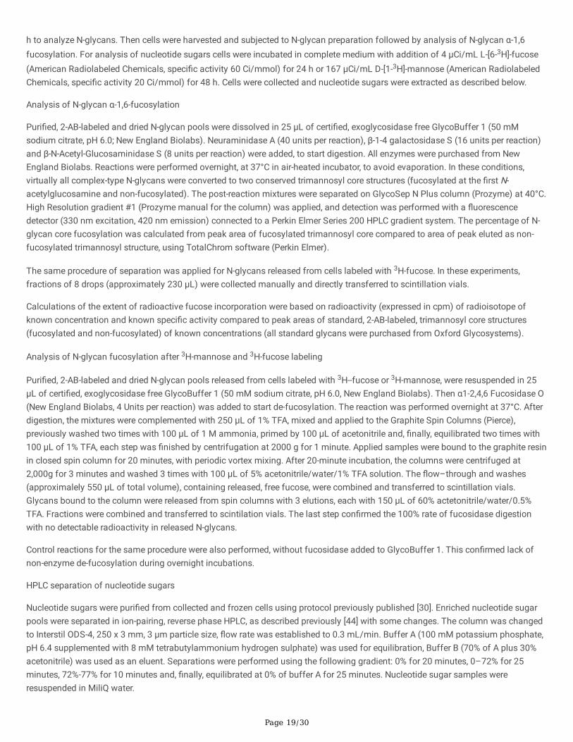
Analysis of N-glycan α-1,6-fucosylation
Puri�ed, 2-AB-labeled and dried N-glycan pools were dissolved in 25 µL of certi�ed, exoglycosidase free GlycoBuffer 1 (50 mMsodium citrate, pH 6.0; New England Biolabs). Neuraminidase A (40 units per reaction), β-1-4 galactosidase S (16 units per reaction)and β-N-Acetyl-Glucosaminidase S (8 units per reaction) were added, to start digestion. All enzymes were purchased from NewEngland Biolabs. Reactions were performed overnight, at 37°C in air-heated incubator, to avoid evaporation. In these conditions,virtually all complex-type N-glycans were converted to two conserved trimannosyl core structures (fucosylated at the �rst N-acetylglucosamine and non-fucosylated). The post-reaction mixtures were separated on GlycoSep N Plus column (Prozyme) at 40°C.High Resolution gradient #1 (Prozyme manual for the column) was applied, and detection was performed with a �uorescencedetector (330 nm excitation, 420 nm emission) connected to a Perkin Elmer Series 200 HPLC gradient system. The percentage of N-glycan core fucosylation was calculated from peak area of fucosylated trimannosyl core compared to area of peak eluted as non-fucosylated trimannosyl structure, using TotalChrom software (Perkin Elmer).
The same procedure of separation was applied for N-glycans released from cells labeled with 3H-fucose. In these experiments,fractions of 8 drops (approximately 230 µL) were collected manually and directly transferred to scintillation vials.
Calculations of the extent of radioactive fucose incorporation were based on radioactivity (expressed in cpm) of radioisotope ofknown concentration and known speci�c activity compared to peak areas of standard, 2-AB-labeled, trimannosyl core structures(fucosylated and non-fucosylated) of known concentrations (all standard glycans were purchased from Oxford Glycosystems).
Analysis of N-glycan fucosylation after 3H-mannose and 3H-fucose labeling
Puri�ed, 2-AB-labeled and dried N-glycan pools released from cells labeled with 3H--fucose or 3H-mannose, were resuspended in 25µL of certi�ed, exoglycosidase free GlycoBuffer 1 (50 mM sodium citrate, pH 6.0, New England Biolabs). Then α1-2,4,6 Fucosidase O(New England Biolabs, 4 Units per reaction) was added to start de-fucosylation. The reaction was performed overnight at 37°C. Afterdigestion, the mixtures were complemented with 250 µL of 1% TFA, mixed and applied to the Graphite Spin Columns (Pierce),previously washed two times with 100 µL of 1 M ammonia, primed by 100 µL of acetonitrile and, �nally, equilibrated two times with100 µL of 1% TFA, each step was �nished by centrifugation at 2000 g for 1 minute. Applied samples were bound to the graphite resinin closed spin column for 20 minutes, with periodic vortex mixing. After 20-minute incubation, the columns were centrifuged at2,000g for 3 minutes and washed 3 times with 100 µL of 5% acetonitrile/water/1% TFA solution. The �ow–through and washes(approximalely 550 µL of total volume), containing released, free fucose, were combined and transferred to scintillation vials.Glycans bound to the column were released from spin columns with 3 elutions, each with 150 µL of 60% actetonitrile/water/0.5%TFA. Fractions were combined and transferred to scintilation vials. The last step con�rmed the 100% rate of fucosidase digestionwith no detectable radioactivity in released N-glycans.
Control reactions for the same procedure were also performed, without fucosidase added to GlycoBuffer 1. This con�rmed lack ofnon-enzyme de-fucosylation during overnight incubations.
HPLC separation of nucleotide sugars
Nucleotide sugars were puri�ed from collected and frozen cells using protocol previously published [30]. Enriched nucleotide sugarpools were separated in ion-pairing, reverse phase HPLC, as described previously [44] with some changes. The column was changedto Interstil ODS-4, 250 x 3 mm, 3 µm particle size, �ow rate was established to 0.3 mL/min. Buffer A (100 mM potassium phosphate,pH 6.4 supplemented with 8 mM tetrabutylammonium hydrogen sulphate) was used for equilibration, Buffer B (70% of A plus 30%acetonitrile) was used as an eluent. Separations were performed using the following gradient: 0% for 20 minutes, 0–72% for 25minutes, 72%-77% for 10 minutes and, �nally, equilibrated at 0% of buffer A for 25 minutes. Nucleotide sugar samples wereresuspended in MiliQ water.

Page 20/30
The sample volumes of 5 µL or less were injected to start separations. The column was connected to Nexera Shimadzu HPLCsystem. Detection was performed at 254 nm with SPD_M30A Diode Array Detector equipped with HS (high-sensitivity) quartz cell.Calculations of nucleotide sugar concentrations were performed with LabSolutions Software (Shimadzu). Standard, high puritynucleotide sugars were purchased from Sigma-Aldrich. The absolute quanti�cation of nucleotide sugars was performed bycomparison of the detected signals to the externally added reference compounds. Next, by referring to the starting number of cellsand to the anticipated volume of a single cell (3 and 3.5 pL per cell for HepG2 and HEK293T, respectively), it was possible to estimatethe intracellular nucleotide sugar concentrations.
The same procedure of ion-pairing reverse-phase separation was applied for nucleotide sugar pools, previously puri�ed from cellslabeled with 3H-fucose and 3H mannose (as described before). In these experiments, fractions of 5 drops (approximately 150 µL)between 26th and 33rd minute of HPLC gradient, were collected manually and directly transferred to scintillation vials. Retentiontimes under separation conditions described above were approximately 27.2 min. and 31.1 min., for GDP-mannose and GDP-fucose,respectively.
Statistical AnalysisStatistical parameters including data plotted (mean ± SD), P values, and statistical tests used are mentioned in Figure Legends.Statistical analyses were performed using Graphpad Prism 6. Data were analyzed by Student’s t-test and Benjamini-Hochbergprocedure or Welch’s correction and by one-way ANOVA with the Tukey post-hoc test.
DeclarationsCompeting interests
All authors declare no competing interests.
Data availability
The datasets generated during and/or analysed during the current study are available from the corresponding author on reasonablerequest.
Author contributions
E.S. performed CRISPR-Cas knockouts, cell culture experiments, including 3H labeling and �uorescence imaging, contributed topreparation of the manuscript, B.S. performed CRISPR-Cas knockouts, cell culture experiments, 3H labeling, �uorescence imaging,contributed to preparation of the manuscript, including all �gures, D.M.-S. wrote the manuscript and strongly contributed to theliterature analysis and data interpretation. M.W. performed qPCR on 96-well plates and took part in manuscript writing and datapresentation, W.W. performed RT-qPCR SYBR Green assays and took part in data interpretation, M.O. conceptuated and supervisedthe research, performed preparation and HPLC separations of N-glycan and metabolite samples, and contribute to preparation of themanuscript. The �nal version of the manuscript was commented on and approved by all authors
Funding
This work is supported by the National Science Centre (Cracow, Poland), 2016/21/B/NZ5/00144 (to M.O.).
References[1] A. Varki, R.D. Cummings, J.D. Esko, P. Stanley, G.W. Hart, M. Aebi, A.G. Darvill, T. Kinoshita, N.H. Packer, J.H. Prestegard, R.L.Schnaar, P.H. Seeberger (Eds.), Essentials of Glycobiology, Cold Spring Harbor Laboratory Press 2015-2017, Cold Spring Harbor (NY),ISBN: 9781621821328.
[2] P.D. Yurchenco, P.H. Atkinson, Equilibration of fucosyl glycoprotein pools in HeLa cells, Biochemistry 16(5) (1977) 944-53,doi:10.1021/bi00624a021.

Page 21/30
[3] P.D. Yurchenco, P.H. Atkinson, Fucosyl-glycoprotein and precursor polls in HeLa cells, Biochemistry 14(14) (1975) 3107-14,doi:10.1021/bi00685a011.
[4] K. Moriwaki, E. Miyoshi, Fucosylation and gastrointestinal cancer, World journal of hepatology 2(4) (2010) 151-61,doi:10.4254/wjh.v2.i4.151.
[5] T.J. Wiese, J.A. Dunlap, M.A. Yorek, L-fucose is accumulated via a speci�c transport system in eukaryotic cells, The Journal ofbiological chemistry 269(36) (1994) 22705-11, doi:10.1016/S0021-9258(17)31703-9.
[6] I. Pastuszak, C. Ketchum, G. Hermanson, E.J. Sjoberg, R. Drake, A.D. Elbein, GDP-L-fucose pyrophosphorylase. Puri�cation, cDNAcloning, and properties of the enzyme, The Journal of biological chemistry 273(46) (1998) 30165-74, doi:10.1074/jbc.273.46.30165.
[7] J.M. Capasso, C.B. Hirschberg, Mechanisms of glycosylation and sulfation in the Golgi apparatus: evidence for nucleotidesugar/nucleoside monophosphate and nucleotide sulfate/nucleoside monophosphate antiports in the Golgi apparatus membrane,Proceedings of the National Academy of Sciences of the United States of America 81(22) (1984) 7051-5,doi:10.1073/pnas.81.22.7051.
[8] N.J. Kuhn, A. White, The role of nucleoside diphosphatase in a uridine nucleotide cycle associated with lactose synthesis in ratmammary-gland Golgi apparatus, The Biochemical journal 168(3) (1977) 423-33, doi:10.1042/bj1680423.
[9] T. Lübke, T. Marquardt, A. Etzioni, E. Hartmann, K. von Figura, C. Körner, Complementation cloning identi�es CDG-IIc, a new type ofcongenital disorders of glycosylation, as a GDP-fucose transporter de�ciency, Nature genetics 28(1) (2001) 73-6,doi:10.1038/ng0501-73.
[10] K. Lühn, M.K. Wild, M. Eckhardt, R. Gerardy-Schahn, D. Vestweber, The gene defective in leukocyte adhesion de�ciency II encodesa putative GDP-fucose transporter, Nature genetics 28(1) (2001) 69-72, doi:10.1038/ng0501-69.
[11] H.O. Ishikawa, T. Ayukawa, M. Nakayama, S. Higashi, S. Kamiyama, S. Nishihara, K. Aoki, N. Ishida, Y. Sanai, K. Matsuno, Twopathways for importing GDP-fucose into the endoplasmic reticulum lumen function redundantly in the O-fucosylation of Notch inDrosophila, The Journal of biological chemistry 285(6) (2010) 4122-4129, doi:10.1074/jbc.M109.016964.
[12] L. Lu, X. Hou, S. Shi, C. Körner, P. Stanley, Slc35c2 promotes Notch1 fucosylation and is required for optimal Notch signaling inmammalian cells, The Journal of biological chemistry 285(46) (2010) 36245-54, doi:10.1074/jbc.M110.126003.
[13] B. Ma, J.L. Simala-Grant, D.E. Taylor, Fucosylation in prokaryotes and eukaryotes, Glycobiology 16(12) (2006) 158r-184r,doi:10.1093/glycob/cwl040.
[14] Y. Gazit, A. Mory, A. Etzioni, M. Frydman, O. Scheuerman, R. Gershoni-Baruch, B.Z. Garty, Leukocyte adhesion de�ciency type II:long-term follow-up and review of the literature, Journal of clinical immunology 30(2) (2010) 308-13, doi:10.1007/s10875-009-9354-0.
[15] A. Etzioni, M. Frydman, S. Pollack, I. Avidor, M.L. Phillips, J.C. Paulson, R. Gershoni-Baruch, Brief report: recurrent severeinfections caused by a novel leukocyte adhesion de�ciency, The New England journal of medicine 327(25) (1992) 1789-92,doi:10.1056/NEJM199212173272505.
[16] A. Dauber, A. Ercan, J. Lee, P. James, P.P. Jacobs, D.J. Ashline, S.R. Wang, T. Miller, J.N. Hirschhorn, P.A. Nigrovic, R. Sackstein,Congenital disorder of fucosylation type 2c (LADII) presenting with short stature and developmental delay with minimal adhesiondefect, Human molecular genetics 23(11) (2014) 2880-7, doi:10.1093/hmg/ddu001.
[17] A. Hüllen, K. Falkenstein, C. Weigel, H. Huidekoper, N. Naumann-Bartsch, J. Spenger, R.G. Feichtinger, J. Schaefers, S. Frenz, D.Kotlarz, T. Momen, R. Khoshnevisan, K.M. Riedhammer, R. Santer, T. Herget, A. Rennings, D.J. Lefeber, J.A. Mayr, C. Thiel, S.B.Wortmann, CONGENITAL DISORDERS OF GLYCOSYLATION WITH DEFECTIVE FUCOSYLATION, Journal of inherited metabolicdisease 44(6) (2021) 1441-1452, doi:10.1002/jimd.12426.

Page 22/30
[18] Y. Helmus, J. Denecke, S. Yakubenia, P. Robinson, K. Lühn, D.L. Watson, P.J. McGrogan, D. Vestweber, T. Marquardt, M.K. Wild,Leukocyte adhesion de�ciency II patients with a dual defect of the GDP-fucose transporter, Blood 107(10) (2006) 3959-66,doi:10.1182/blood-2005-08-3334.
[19] E. van de Vijver, A. Maddalena, Ö. Sanal, S.M. Holland, G. Uzel, M. Madkaikar, M. de Boer, K. van Leeuwen, M.Y. Köker, N.Parvaneh, A. Fischer, S.K. Law, N. Klein, F.I. Tezcan, E. Unal, T. Patiroglu, B.H. Belohradsky, K. Schwartz, R. Somech, T.W. Kuijpers, D.Roos, Hematologically important mutations: leukocyte adhesion de�ciency (�rst update), Blood cells, molecules & diseases 48(1)(2012) 53-61, doi:10.1016/j.bcmd.2011.10.004.
[20] A. Hidalgo, S. Ma, A.J. Peired, L.A. Weiss, C. Cunningham-Rundles, P.S. Frenette, Insights into leukocyte adhesion de�ciency type2 from a novel mutation in the GDP-fucose transporter gene, Blood 101(5) (2003) 1705-12, doi:10.1182/blood-2002-09-2840.
[21] D. Cagdas, M. Yilmaz, N. Kandemir, I. Tezcan, A. Etzioni, Ö. Sanal, A novel mutation in leukocyte adhesion de�ciency typeII/CDGIIc, Journal of clinical immunology 34(8) (2014) 1009-14, doi:10.1007/s10875-014-0091-7.
[22] N. Cooper, Y.T. Li, A. Möller, N. Schulz-Weidner, U.J. Sachs, F. Wagner, H. Hackstein, S. Wienzek-Lischka, M. Grüneberg, M.K. Wild,G. Bein, T. Marquardt, Incidental diagnosis of leukocyte adhesion de�ciency type II following ABO typing, Clinical immunology 221(2020) 108599, doi:10.1016/j.clim.2020.108599.
[23] K.M. Knapp, R. Luu, M. Baerenfaenger, F. Zijlstra, H. Wessels, D. Jenkins, D.J. Lefeber, K. Neas, L.S. Bicknell, Biallelic variants inSLC35C1 as a cause of isolated short stature with intellectual disability, Journal of human genetics 65(9) (2020) 743-750,doi:10.1038/s10038-020-0764-4.
[24] A. Karsan, C.J. Cornejo, R.K. Winn, B.R. Schwartz, W. Way, N. Lannir, R. Gershoni-Baruch, A. Etzioni, H.D. Ochs, J.M. Harlan,Leukocyte Adhesion De�ciency Type II is a generalized defect of de novo GDP-fucose biosynthesis. Endothelial cell fucosylation isnot required for neutrophil rolling on human nonlymphoid endothelium, The Journal of clinical investigation 101(11) (1998) 2438-45,doi: 10.1172/JCI905.
[25] T. Marquardt, K. Lühn, G. Srikrishna, H.H. Freeze, E. Harms, D. Vestweber, Correction of leukocyte adhesion de�ciency type II withoral fucose, Blood 94(12) (1999) 3976-85, doi: 10.1182/blood.V94.12.3976.
[26] L. Sturla, R. Rampal, R.S. Haltiwanger, F. Fruscione, A. Etzioni, M. Tonetti, Differential terminal fucosylation of N-linked glycansversus protein O-fucosylation in leukocyte adhesion de�ciency type II (CDG IIc), The Journal of biological chemistry 278(29) (2003)26727-33, doi: 10.1074/jbc.M304068200.
[27] C.C. Hellbusch, M. Sperandio, D. Frommhold, S. Yakubenia, M.K. Wild, D. Popovici, D. Vestweber, H.J. Gröne, K. von Figura, T.Lübke, C. Körner, Golgi GDP-fucose transporter-de�cient mice mimic congenital disorder of glycosylation IIc/leukocyte adhesionde�ciency II, The Journal of biological chemistry 282(14) (2007) 10762-72, doi:10.1074/jbc.M700314200.
[28] ]M. Olczak, B. Szulc, Modi�ed secreted alkaline phosphatase as an improved reporter protein for N-glycosylation analysis, PLoSONE 16(5) (2021) e0251805, doi:10.1371/journal.pone.0251805.
[29] M. R. Kudelka, A. Antonopoulos, Y. Wang, D. M. Duong, X. Song, N. T. Seyfried, A. Dell, S. M. Haslam, R. D. Cummings, T. Ju,Cellular O-Glycome Reporter/Ampli�cation to explore O-glycans of living cells, Nature methods 13(1) (2016) 81–86,doi:10.1038/nmeth.3675.
[30] J. Räbinä, M. Mäki, E. M. Savilahti, N. Järvinen, L. Penttilä, R. Renkonen, Analysis of nucleotide sugars from cell lysates by ion-pair solid-phase extraction and reversed-phase high-performance liquid chromatography, Glycoconjugate journal 18(10) (2001) 799–805, doi:10.1023/a:1021107602535.
[31] W. van Tol, Discovery of the sugar supply pathways for the O-mannosylation of dystroglycan. On the road for treating musculardystrophy-dystroglycanopathy (doctoral dissertation), Radboud University Nijmegen, the Netherlands (2020),https://repository.ubn.ru.nl/handle/2066/213932.

Page 23/30
[32] K. Moriwaki, K. Noda, T. Nakagawa, M. Asahi, H. Yoshihara, N. Taniguchi, N. Hayashi, E. Miyoshi, A high expression of GDP-fucose transporter in hepatocellular carcinoma is a key factor for increases in fucosylation, Glycobiology 17(12) (2007) 1311-1320,doi:10.1093/glycob/cwm094.
[33] P. Sosicka, B.G. Ng, L.E. Pepi, A. Shajahan, M. Wong, D.A. Scott, K. Matsumoto, Z. Xia, C.B. Lebrilla, R.S. Haltiwanger, P. Azadi, H.H.Freeze, Metabolic heritage mapping: heterogenous pools of cytoplasmic nucleotide sugars are selectively utilized by variousglycosyltransferases, bioRxiv (2021) 2021.11.03.467160, doi:10.1101/2021.11.03.467160
[34] S. H. Park, I. Pastuszak, R. Drake, A. D. Elbein, Puri�cation to apparent homogeneity and properties of pig kidney L-fucosekinase, The Journal of biological chemistry 273(10) (1998) 5685–5691, doi:10.1074/jbc.273.10.5685.
[35] S. W. Coates, T. Jr Gurney, L. W. Sommers, M. Yeh, C. B. Hirschberg, Subcellular localization of sugar nucleotide synthetases, TheJournal of biological chemistry 255(19) (1980) 9225–9229, doi: 10.1016/S0021-9258(19)70550-X.
[36] V. Pareek, Z. Sha, J. He, N. S. Wingreen, S. J. Benkovic, Metabolic channeling: predictions, deductions, and evidence, Molecularcell 81(18) (2021) 3775–3785, doi:10.1016/j.molcel.2021.08.030
[37] F. X. Sullivan, R. Kumar, R. Kriz, M. Stahl, G. Y. Xu, J. Rouse, X. J. Chang, A. Boodhoo, B. Potvin, D. A. Cumming, Molecular cloningof human GDP-mannose 4,6-dehydratase and reconstitution of GDP-fucose biosynthesis in vitro, The Journal of Biological Chemistry273(14) (1998) 8193-8202, doi:10.1074/jbc.273.14.8193.
[38] R. H. Kornfeld, V. Ginsburg, Control of synthesis of guanosine 5'-diphosphate D-mannose and guanosine 5'-diphosphate L-fucosein bacteria, Biochimica et biophysica acta 117(1) (1966) 79–87, doi:10.1016/0304-4165(66)90154-1.
[39] Y. Zhang, Y. Wang, C. Wang, C. Rautengarten, E. Duan, J. Zhu, X. Zhu, J. Lei, C. Peng, Y. Wang, X. Teng, Y. Tian, X. Liu, J. L.Heazlewood, A. Wu, J. Wan, BRITTLE PLANT1 is required for normal cell wall composition and mechanical strength in rice, Journal ofintegrative plant biology 63(5) (2021) 865–877, doi:10.1111/jipb.13050.
[40] D. Maszczak-Seneczko, T. Olczak, L. Wunderlich, M. Olczak, Comparative analysis of involvement of UGT1 and UGT2 splicevariants of UDP-galactose transporter in glycosylation of macromolecules in MDCK and CHO cell lines, Glycoconjugate journal 28(2011) 481–492, doi: 10.1007/s10719-011-9348-z.
[41] B. Bazan, M. Wiktor, D. Maszczak-Seneczko, T. Olczak, B. Kaczmarek, M. Olczak, Lysine at position 329 within a C-terminaldilysine motif is crucial for the ER localization of human SLC35B4, PLoS ONE, 13 (2018) e0207521,doi:10.1371/journal.pone.0207521.
[42] B. Szulc, P. Sosicka, D. Maszczak-Seneczko, E. Skurska, A. Shauchuk, T. Olczak, H. H. Freeze, M. Olczak, Biosynthesis of GlcNAc-rich N- and O-glycans in the Golgi apparatus does not require the nucleotide sugar transporter SLC35A3, The Journal of biologicalchemistry 295 (2020) 16445–16463, doi:10.1074/jbc.RA119.012362.
[43] S.C. Taylor, K. Nadeau, M. Abbasi, C. Lachance, M. Nguyen, J. Fenrich The Ultimate qPCR Experiment: Producing PublicationQuality, Reproducible Data the First Time, Trends in biotechnology 37 (2019) 761-774, doi:10.1016/j.tibtech.2018.12.002.
[44] K. Nakajima, S. Kitazume, T. Angata, R. Fujinawa, K. Ohtsubo, E. Miyoshi, N. Taniguchi, Simultaneous determination of nucleotidesugars with ion-pair reversed-phase HPLC, Glycobiology, 20(7) (2010) 865–871, doi:10.1093/glycob/cwq044.
Figures

Page 24/30
Figure 1
Simpli�ed representation of GDP-fucose metabolism in mammalian cells. GDP-fucose is synthesized in two independent pathways,i.e. de novo (from GDP-mannose) and salvage (from fucose)

Page 25/30
Figure 2
Optimization of the conditions of fucose supplementation. AAL dot blot analysis of the wild-type and SLC35C1 knockout HEK293T(A) and HepG2 (B) cells after 5 days supplementation with (+) or without (-) different fucose concentrations. (C) AAL staining (red) ofHEK293T SLC35C1 knockout supplemented with 5 mM fucose during 48 h. Cell nuclei were counterstained with DAPI. Scale bar 10 =µm. (D-F) Schematic representation of our HPLC-based method for quanti�cation of the α1,6 fucosylation of N-glycans. (G)Quantitative analysis of the α-1,6-fucosylation of N-glycans in HEK293T wild-type and SLC35C1 knockout cells supplemented withdifferent fucose concentrations for indicated time periods
Figure 3
Quanti�cation of the percentage of the core-fucosylated N-glycan structures. HPLC quanti�cation of N-glycans derived fromendogenous HEK293T (A) and HepG2 (B) glycoproteins. N-glycans decorating SEAP glycoprotein over-expressed by HEK293T (C)and HepG2 (D) cell lines. ns, not signi�cant; *, p < 0.05; **, p < 0.01; ***, p < 0.001; ****, p < 0.0001 as determined using one-wayANOVA with the Tukey post-hoc test. Data are represented as mean ± SEM.

Page 26/30
Figure 4
O-glycosylation �ngerprinting of HEK293T (A-F) and HepG2 (G-J) cells. MALDI-TOF mass spectra of permethylated mucin-type Bn-O-glycans secreted to the culture medium were permethylated and analyzed in a positive ion mode. Structural assignments based onbiosynthetic knowledge were prepared using the GlycoWorkBench tool (2.1; EuroCarbDB).

Page 27/30
Figure 5
Quanti�cation of the intracellular GDP-fucose concentration. The intracellular GDP-fucose concentration in HEK293T (A) and HepG2(B) cell lines. ns, not signi�cant; *, p < 0.05; **, p < 0.01; ***, p < 0.001; ****, p < 0.0001 as determined using one-way ANOVA with theTukey post-hoc test. Data are represented as mean ± SEM (C) Time-course analysis (0-24 h) of the GDP-fucose synthesis in the wild-type and SLC35C1 knockout HEK293T cells fed with 5 mM fucose. (D) Analysis of the GDP-fucose degradation in the wild-type andSLC35C1 knockout HEK293T. The cells were �rst cultured for 24 h in the presence of 5 mM fucose and then for another 24 h in afucose-free medium. (E) Disappearance of the fucosylation phenotype visualized by the AAL staining (red). Cell nuclei werecounterstained with DAPI. Scale bar = 10 µm. (F) Dependence of the intracellular GDP-fucose concentration on the concentration ofthe fucose supplemented to the culture medium (HEK293T). (G) Dependence of the percentage of the core fucosylated N-glycans onthe concentration of the fucose supplemented to the culture medium (HEK293T)

Page 28/30
Figure 6
Metabolic labeling of the cellular N-glycans with radioactive fucose. (A) Exemplary representative HPLC chromatograms of thedigested N-glycans isolated from the indicated cell lines. The �uorescence signal from the 2-AB-labeled N-glycans (black solid lines)was overlayed with the radioactivity data (red dots and solid lines). The total radioactivity of the fucosylated N-glycans normalizedfor the amount of the starting material (total protein) from HEK293T (B) and HepG2 (C) cell lines. Percentage of the radioactivefucose incorporated into the fucosylated N-glycans isolated from HEK293T (D) and HepG2 (E) cell lines. To calculate the values thesum radioactivity measured in the HPLC fractions corresponding to the fucosylated species was divided by the total radioactivity ofthe fucose added to the culture medium. Comparison of the relative incorporation of the radioactive fucose derived from the salvagepathway in the wild-type (F) and SLC35C1 knockout (G) HEK293T cells. (H) The total radioactivity of the GDP-fucose normalized fortotal number of HEK293T cells. ns, not signi�cant; *, p < 0.05 as determined using two-tailed unpaired t-test with Welch’s correction.Data are represented as mean ± SEM. ISTD, internal standard (GDP-glucose).

Page 29/30
Figure 7
Quanti�cation of the intracellular GDP-mannose and GDP-fucose concentration in cells fed with mannose. Intracellularconcentrations of GDP-mannose (A) and GDP-fucose (B) in the wild-type and SLC35C1 knockout HEK293T cells cultured for 48 h inthe absence and presence of 5 mM mannose. Comparison of the relative incorporation of the radioactive fucose derived from the denovo pathway in the wild-type (C) and SLC35C1 knockout (D) HEK293T cells. ISTD, internal standard (GDP-glucose). ns, notsigni�cant; *, p < 0.05; **, p < 0.01 as determined using two-tailed unpaired t-test with Welch’s correction. Data are represented asmean ± SEM

Page 30/30
Figure 8
Relative contribution of the de novo and salvage pathways in glycan fucosylation Comparison of the relative incorporation of theradioactive fucose in N-glycans, derived from either the salvage (A) or de novo (B) pathways in the wild-type and SLC35C1 knockoutHEK293T cells. The radioactivity was normalized for the amount of the starting material (total protein). ns, not signi�cant; *, p < 0.05;***, p < 0.001 as determined using two-tailed unpaired t-test with Welch’s correction. Data are represented as mean ± SEM
Supplementary Files
This is a list of supplementary �les associated with this preprint. Click to download.
Supplementarymaterial.pdf