[frontiers in bioscience s4, 335-355, january 1, 2012
TRANSCRIPT

[Frontiers in Bioscience S4, 335-355, January 1, 2012]
335
Curcumin: The multi-targeted therapy for cancer regression Dewan Md Sakib Hossain, Sankar Bhattacharyya, Tanya Das, Gaurisankar Sa Division of Molecular Medicine, Bose Institute, P-1/12 CIT Scheme VII M, Kolkata 700054, India TABLE OF CONTENTS 1. Abstract 2. Introduction 3. Introductory background of curcumin
3.1. Physical and chemical backdrop 3.2. Safety and toxicity 3.3. Biotransformation and bioavailability 3.4. On the way of alternative to main stream medicine
4. Use of curcumin: Multi-targeted therapy for cancer 4.1. Control on carcinogenesis
4.1. 1. Inhibition of tumor initiation by curcumin 4.1. 2. Inhibition of tumor promotion and progression by curcumin
4.2. Regression of tumor 4.2. 1. Induction of tumor cell apoptosis by curcumin 4.2. 2. Inhibition of angiogenesis by curcumin 4.2. 3. Retardation of metastasis by curcumin
4.3. Renovation of depressed immune system by curcumin 4.4. Amelioration of systemic toxicity
4.4. 1. Anti-oxidant properties of curcumin 4.4. 2. Detoxificant properties of curcumin
5. Conclusion 6. Acknowledgment 7. References 1. ABSTRACT
Tumors are multifaceted; in fact, numerous things happen in synchrony to enable tumor promotion and progression. Any type of cancer is associated with the modification of 300–500 normal genes and characterized by the deregulation of cell signaling pathways at multiple steps leading to cancer phenotype. Thus a proper management of tumorigenesis requires the development of multi-targeted therapies. Several adverse effects associated with present day cancer therapies and the thirsts for multi-targeted safe anticancer drug instigate the use of natural polyphenol, curcumin. It appears to involve a blend of anti-carcinogenic, pro-apoptotic, anti-angiogenic, anti-metastatic, immunomodulatory and antioxidant activities. Also the molecular mechanisms implicated for the pleotropic activities of curcumin are diverse and seem to involve a combination of cell signaling pathways at multiple levels of tumorigenesis. Being a potent scavenger of reactive oxygen species, curcumin also ameliorates systemic toxicity in tumor-bearer. Taken together, by placing particular emphasis on molecular basis of tumor promotion and progression this review summarizes the anti-cancer properties of curcumin that may be exploited for successful clinical cancer prevention.
2. INTRODUCTION
The problem with cancer treatment today is that we are trying to fix the car without understanding how the engine works. As a result in spite of an extensive search for safe and efficacious treatments for cancer, it has involved the use of harmful substances, such as poisonous mustargen, chemotherapy and then now targeted therapies (1-3). Also their benefits are often short lived, and various adaptive mechanisms eventually lead to tumor progression. For example, therapies aimed at inhibiting angiogenesis by targeting the vascular endothelial growth factor A, despite positive effects on established localized tumors, inhibition of angiogenesis can result in increased tumor invasion and metastasis (3). In addition to cancer itself being immunosuppressive, so are the chemotherapies often used to treat them grounding another major setback to the modern day cancer therapy (4-7).
Thus if we could understand the precise nature of
the workings of cancer, and all of the different things that the cancer engine needs to drive itself, then we might be able to find ways to stop the engine. Tumors develop from normal cells that have acquired the ability to divide continuously and inappropriately. When a normal cell is

The multi-targeted therapy for cancer
336
Figure 1. Hallmarks of carcinogenesis. The complex process by which normal cells develop into a malignant tumor is known as carcinogenesis. Six essential physiological alterations manifested when a normal cell transforms into malignant one include (i) mitogenic signaling independency and limitless replicative potential (ii) deregulation cell cycle progression (iii) anti-growth signaling insensitivity (iv) evading apoptosis (v) sustaining angiogenesis (v) tissue invasion and metastasis.
induced to divide inharmoniously, it gives rise to a little nest of abnormal cells. Out of that nest of abnormal cells comes another group that is more aggressive and more capable of proliferation, which later on results in tumor formation. The epidemiology and the genetics of cancer have suggested that this conversion of a normal cell into a cancer require multifaceted changes. Like car engines there are so many things that need to happen in synchrony to enable the engine-the tumor- to run. The process of tumorigenesis is initiated as a result of rather rapid and irreversible assault to the cell. Tumorigenesis is the complex process by which normal cells develop into a malignant tumor. In traditional descriptions, it has been divided into 3 stages: initiation, during which normal cells become transformed, promotion where transformed cells become preneoplastic and progression, which is the final step when the preneoplastic cells become neoplastic. While there are many distinct types of cancer, there are believed to be six essential alterations to normal cell physiology, which together define the progression of most human malignancies. They are (i) mitogenic signaling independency and limitless replicative potential, (ii) deregulated cell cycle progression, (iii) anti-growth signaling insensitivity, (iv) evasion of apoptosis, (v) sustaining angiogenesis, (vi) tissue invasion and metastasis (Figure 1). This sequence of events presents many opportunities for intervention, with the aim of preventing, slowing down or reversing the transformation process.
This entire process of tumorigenesis is
characterized by the deregulation of cell signaling pathways at multiple steps. But most current anticancer therapies involve the modulation of a single target. Overall the ineffectiveness, lack of safety, and high cost of today’s mono-targeted therapies have led to a lack of faith in these approaches. As a result, scientists and medical
professionals are increasingly interested in developing multi-targeted therapies. Many plant-based products, however, accomplish multi-targeting naturally and, in addition, are inexpensive and safe compared to synthetic agents. Over the past decade, there has been a significant increase in public and scientific interest in the beneficial effects of chemicals derived from plants, known as phytochemicals, and their role in the maintenance of health and prevention of disease. Polyphenols are amongst the lead chemical substances that fulfill this definition. Polyphenols are derived from many components of the human diet, including peanuts, green and black tea, red wine, olive oil and the spice, turmeric. Many of these natural substances, which were traditionally utilized in ancient medicines for their anti-inflammatory and antioxidant actions, are now being investigated as cardioprotective, antiproliferative, and preventive agents. In particular, traditional agents derived from ancient Indian medicine, such as curcumin from turmeric, chemically known as diferuloylmethane (C21H20O6), has been the subject of hundreds of published papers over the past three decades, studying its antioxidant, anti-toxic, anti-inflammatory, cancer chemopreventive and other potentially chemotherapeutic properties. Because curcumin has been shown to suppress cancer cell proliferation, induce apoptosis, inhibit angiogenesis, suppress the expression of anti-apoptotic proteins while protecting immune system of the tumor bearer - it may have untapped therapeutic value. With regard to the chemoprevention and therapy of many diseases, particularly cancer, this article aims to review the extensive published literature on the use of the natural polyphenol, curcumin, as a single agent and in combinatorial chemoprevention and treatment.
Basic searches of the most commonly
internationally accessed scientific databases using the

The multi-targeted therapy for cancer
337
Figure 2. Curcuma longa plant and chemical structure of curcumin, the active ingradient of rhizome turmeric. (A) Curcuma longa plant and the rhizome turmeric. The tautomerism of curcumin is demonstrated under different physiological conditions with the bis-keto form (bottom) being more predominant than enolate form under acidic and neutral conditions. (B) Chemical structures shown refer to metabolites identified in rodents and humans.
keywords “curcumin” and “turmeric” has demonstrated that over 2600 articles including reviews have been published in English language journals since 1966. Whereas these reviews have generally focused on the potential role of phytochemicals to treat or prevent several diseases, the purpose of this comprehensive review is to offer a broader perspective on the multifaceted potential of curcumin to prevent cancer. In this review, cellular events induced by curcumin that directly influence tumor promotion and progression or indirectly affect the intrinsic defense system of the tumor-bearer are discussed. 3. INTRODUCTORY BACKGROUND OF CURCUMIN 3.1. Physical and chemical backdrop
Curcumin is a component of turmeric; the yellow spice derived from the roots (rhizomes) of the plant Curcuma longa. Curcuma longa is a short-stemmed ginger-like plant, which grows to about 100 cm in height (Figure 2). Curcuma longa grows naturally throughout the Indian subcontinent and in tropical countries, particularly South East Asia. Curcumin (chemical name: (E,E)-1,7-bis (4-hydroxy-3-methoxyphenyl)- 1,6-heptadiene-3, 5 dione) has a molecular weight of 368.38, a melting point of 179-183 °C and chemical formula C21H20O6. Curcumin’s chemical structure includes two methoxyl groups, two phenolic hydroxyl groups and three double conjugated bonds. Curcumin is more soluble in ethanol, dimethylsulfoxide or acetone than it is in water. It is a bis-alpha,beta-unsaturated beta-diketone, which exhibits keto-enol tautomerism (Figure 2) having a predominant keto form in acidic and neutral solutions and stable enol form in alkaline medium (8). The curcuminoids, which constitute approximately 5% of most turmeric preparations, are a mixture of curcumin
(sometimes referred to as Curcumin I), desmethoxycurcumin (Curcumin II) and bisdesmethoxycurcumin (Curcumin III) (9). Recently other curcuminoids like cyclocurcumin or Curcumin IV have been isolated and identified from turmeric (10). Most commercially available preparations of “curcumin” are not pure: approximately 77% diferuloylmethane, 17% demethoxycurcumin, and 6% bisdemethoxycurcumin (11). Under physiological conditions, maximum light absorption by curcumin occurs at 420 nm (12). As a result of light sensitivity demonstrated by several researchers, biological samples containing curcumin should be protected from light. 3.2. Safety and toxicity
Although curcumin and turmeric are natural products used in the diet, the doses administered in clinical trials exceed those normally consumed in diet. This fact underlines the need for systematic safety and toxicity studies. Turmeric is generally recognized as safe by the USA-FDA and curcumin has been granted an acceptable daily intake level of 0.1-3 mg/kg body-weight by the Joint FAO/WHO Expert Committee on Food Additives 1996 (13). Anecdotal reports suggest that dietary consumption of curcumin up to 150 mg/day is not associated with any adverse effects in humans (14). In India, where the average intake of turmeric can be as high as 2.0-2.5 g per day (corresponding to approximately 60-100 mg of curcumin daily), no toxicities or adverse effects have been reported at the population level (15). More recently, no toxicity has been observed in a preclinical study of the administration of 2% dietary curcumin (approximately 1.2 g/kg body-weight) to rats for 14 days (16). In a study performed in India, administration of 1.2 to 2.1 g of oral curcumin to patients with rheumatoid arthritis daily for 2 - 6 weeks did

The multi-targeted therapy for cancer
338
not cause any toxicity (17). In another study of high dose oral curcumin by Cheng and colleagues in Taiwan, administration of up to 8 g daily of curcumin for 3 months to patients with pre-invasive malignant or high-risk pre-malignant conditions had no adverse effects (18). In a phase-I clinical trial of oral curcumin in patients with advanced colorectal cancer in which US National Cancer Institute criteria were used to assess potential toxicity, curcumin was well tolerated at all dose levels up to 3.6 g daily for up to 4 months (19). Although turmeric is often used to treat inflammatory skin conditions in traditional South East Asian medical systems, it should be noted that some reports of allergic dermatitis after contact with curcumin have been published in the scientific literature (20). Allergic reaction to turmeric-related products has also been described in one healthy volunteer enrolled in a Phase-I study for testing the safety of turmeric oil and turmeric extract. 3.3. Biotransformation and bioavailability
Curcumin is biotransformed in the intestinal tract of humans and rodents. The major metabolites of curcumin identified in rat were curcumin glucuronide and curcumin sulfate based on enzymatic hydrolysis studies. Hexahydrocurcumin, hexahydrocurcuminol, and hexahydrocurcumin glucuronide were also present in minor amounts (21). Curcumin-glucuronide, dihydro-curcumin-glucuronide, tetrahydrocurcumin-glucuronide and tetrahydrocurcumin are major metabolites (Figure 2) of curcumin in mice. Data on the pharmacokinetic properties and metabolism of curcumin in humans are very limited. A study of six patients with advanced colorectal cancer dosed with 3.6 g of curcumin daily for up to 3 months yielded 4.3, 5.8, and 3.3 ng/mL mean plasma concentrations of curcumin, curcumin glucuronide, and curcumin sulfate, respectively, 1 h after administration (21). Although Phase-I clinical trials have shown that curcumin is safe even at high doses (12g/day) in humans but exhibit poor bioavailability (22). Despite the considerable promise that curcumin is an efficacious and safe compound for cancer therapy and chemoprevention; it has by no means been embraced by the cancer community as a "panacea for all ills". The single most important reason for this reticence has been the reduced bioavailability. Studies over the past three decades related to absorption, distribution, metabolism and excretion of curcumin have revealed poor absorption and rapid metabolism of curcumin that severely curtails its bioavailability. Since curcumin’s poor systemic bioavailability compromises its potential therapeutic uses, many groups have focused on ways to improve its bioavailability. Use of adjuvant like piperine that interferes with glucuronidation, use of liposomal curcumin, curcumin nanoparticles, use of curcumin phospholipid complex, use of structural analogues of curcumin (e.g., EF-24) are the important approaches to overcome the problems of limited bioavailability (23-27). Also nanoparticle-based drug delivery approaches have the potential for rendering hydrophobic agents like curcumin dispersible in aqueous media, thus circumventing the pitfalls of poor solubility (25). 3.4. On the way from alternative to main stream medicine
A traditional remedy in “Ayurvedic medicine” and ancient Indian healing system that dates back over
5,000 years, turmeric has been used through the ages as an "herbal aspirin" and "herbal cortisone" to relieve discomfort and inflammation associated with an extraordinary spectrum of infectious and autoimmune diseases. Turmeric has been used for thousand of years in Ayurvedic and traditional Chinese medicine. In modern times, curcumin the active constituent of turmeric continues to be used as an alternative medicinal agent in many parts of South East Asia for the treatment of common ailments such as stomachic upset, flatulence, jaundice, arthritis, sprains, wounds and skin infections among many others. Curcumin and turmeric products have been characterized as safe by health authorities such as the Food and Drug Administration in United States of America, Food and Agriculture Organization/World Health Organization. Curcumin has entered scientific clinical trials at the phase-I and phase-II level for its therapeutic efficacy (28). A phase-III study of curcumin along with celecoxib and gemcitabine has recently started at the Tel-Aviv Sourasky Medical Center for patients with metastatic colorectal cancer. 4. USE OF CURCUMIN: MULTI-TARGETED THERAPY OF CANCER
The successful targeted therapy of cancer requires:
Control on carcinogenesis by inhibition of (i) initiation, (i) promotion and (iii) progression of neoplasm. Regression of tumor by (i) induction of apoptosis, (ii) inhibition of angiogenesis and (iii) retarda-tion of metastasis. Renovation of depressed of immune system by (i) protecting and (ii) rejuvenating immune cells. Amelioration of systemic toxicity by enhancement of (i) anti-oxidant and (ii) detoxification systems. Among its many benefits, curcumin possesses all
these above-mentioned potential anticancer therapeutic properties. Here we summarize the key findings related to curcumin’s beneficial effects as a multi-targeted anti cancer medicine. 4.1. Control on carcinogenesis
Carcinogenesis is the complex process by which normal cells develop into a malignant tumor, which includes initiation, promotion and progression. Various stimuli can cause initiation of a cell such as carcinogens, oxidative stress, chronic inflammation, UV radiation and abnormal hormonal stimulation. Curcumin can interfere in the described processes (Figure 3) of carcinogenesis by inhibiting the initiation step or suppressing the promotion and progression stages. 4.1.1. Inhibition of tumor initiation by curcumin
One of the things that set curcumin apart from most other anti-cancer supplements is that this phenolic can actually block carcinogenic chemicals from getting inside cells. Importantly, curcumin can interfere with pesticides

The multi-targeted therapy for cancer
339
Figure 3. Curcumin curtails tumor initiation, promotion and progression. (A) Curcumin inhibits tumor initiation by (i) blocking intracellular entry of chemical carcinogens (ii) facilitating the excretion of carcinogen that gains access into the cytoplasm and (iii) neutralizing ROS production and repairing DNA damage by carcinogen, when left unexcreted. (B) Curcumin inhibits tumor promotion by down regulating the expression of inflammatory cytokines, proliferative and anti-apoptotic proteins.
that mimic estrogen (29). These include DDT and dioxin, two extremely toxic chemicals that contaminate water and
food. Like estrogen, estrogen-mimicking chemicals promote the growth of breast cancer (30). Curcumin blocks

The multi-targeted therapy for cancer
340
the access of estrogen and estrogen mimickers to the cell (31-33). In a study on human breast cancer cells, curcumin reversed growth caused by 17beta-estradiol by 98%. DDT's growth-enhancing effects on breast cancer were blocked about 75% by curcumin. Curcumin's ability to block other chemicals has also been documented. It has been tested against paraquat (weed killer), nitrosamines (in cooked meat and "lunch" meats) and carbon tetrachloride (a solvent in varnish and other products). In all cases, curcumin is able to block the chemical's effect. Curcumin also inhibited tumor initiation by benzo (alpha)pyrene (BalphaP) and 7,12 dimethylbenz (alpha)anthracene (DMBA) (34). Topical application of curcumin strongly inhibited tumor promotion in the skin of DMBA-initiated mice (35-37). Including 0.5% – 2.0% curcumin in the diet decreased BalphaP-induced fore-stomach tumors per mouse by 51% – 53% when it was administered during the initiation period and by 47% – 67% when it was administered during the post-initiation period (38). Including curcumin in the diet decreased the number of N-ethyl-N’-nitro-N-nitrosoguanidine (ENNG)-induced duodenal tumors per mouse (38). Administration of curcumin in the diet decreased the number of azoxymethane (AOM)-induced colon tumors in mice and in rats (38,39). In addition, a deregulated balance between adaptive and innate immunity results in chronic inflammation that paves the way for tumor initiation and progression. Inflammation via the production of reactive oxygen species (ROS) and reactive nitrogen species by activated neutrophils and macrophages, leads to lethal cancer causing mutations (40). Curcumin has been reported to actively inhibit the inflammatory responses while combating cancers in different experimental models (41-47)
Curcumin has the unique ability to fit through a cellular doorway known as the aryl hydrocarbon receptor. This is a feat it shares with estrogen and estrogen-mimicking chemicals. Because it can compete for the same doorway, curcumin has the power to protect against entry of estrogen mimickers (31-33). In case when a carcinogen from dietary and environmental sources gains access to cellular compartment it is subjected to metabolism. The product of the phase-I reaction can be either excreted or activated into toxic metabolite. The toxic metabolite is conjugated to substrates in the diet by phase-II conjugating enzymes such as sulfotransferase and glutathione-S-transferase and then excreted. When unmodified, carcinogens can form a covalent adduct with DNA, resulting in DNA damage. Irreparable damage leads to mutations in critical genes involved in growth, proliferation, and apoptosis, resulting in initiation and subsequent development of cancer. Curcumin exerts anti-cancer effects by increasing the expression of phase-II conjugation enzymes that expel the carcinogens out from the cells (48). Dietary supplementation of curcumin induces phase-II detoxifying enzymes, suggesting that curcumin has chemopreventive efficacy in inhibiting chemical carcinogenesis and other forms of electrophilic toxicity. Curcumin counteracts carcinogen-induced ROS by increasing ornithine decarboxylase, glutathione, antioxidant enzymes, and phase-II metabolizing enzymes (49).
Additionally curcumin has been found to induce HO-1 expression by signaling through (NF-E2)-related factor 2 (Nrf-2) and NFkappaB and thereby has the potential to reduce oxidative stress (50). By modulating cytochrome P450 function, curcumin also reduces the aflatoxin B1-DNA adduct formation, thereby showing its potential to inhibit chemical carcinogenesis (51).
Additionally, curcumin prevents initiation of
tumors either by curtailing the proinflammatory pathways. Curcumin inhibits the induction of nitric oxide synthase in activated macrophages and has been shown to be a potent scavenger of free radicals like nitric oxide (41). In macrophages activated with lipopolysaccharide and the interferon-gamma system, curcumin treatment showed anti-tumorigenic potential by significantly reducing the levels of inducible nitric oxide synthase (iNOS) (41). Curcumin has been found to inhibit cell proliferation and cytokine production by inhibiting NFkappaB target genes involved in the mitogen induction of T-cell proliferation, interleukin-2 production, and nitric oxide generation (52). Radiation-induced inflammation by over-expression of cytokines such as IL-10, IL-6, and IL-18 is accompanied by NFkappaB induction, which is controlled and inhibited by curcumin (53). It also has been shown to down regulate activator protein-1 (AP-1) (53), which regulates the genes for pro-inflammatory mediators. Curcumin inhibits NFkappaB activation by blocking phosphorylation of IkappaB through inactivation of IkappaB kinase complex (54,55). Suppression of AP-1 is due to a direct interaction of curcumin with AP-1 binding to its DNA-binding motif and also due to inhibition of c-Jun and c-fos, components of AP-1 (56,57). Curcumin is also a potent scavenger of a variety of ROS generated at the site of inflammation, including superoxide anion hydroxyl radical, singlet oxygen, nitric oxide and peroxynitrite (42,43). Curcumin has the ability to protect lipids, hemoglobin, and DNA against oxidative degradation (43-46). Curcumin is also a potent inhibitor of ROS-generating enzymes cyclooxygenase and lipoxygenase (47). 4.1.2. Inhibition of tumor promotion and progression by curcumin
The cancer chemopreventive potential of curcumin has also been studied during the stages of tumor promotion and progression. In a series of studies, BalphaP or DMBA were used to induce tumor initiation and 12-O-tetradecanoylphorbol-13-acetate (TPA) used for tumor promotion; all of which were inhibited by curcumin (34-38). Oral curcumin administration has been shown to prevent the development of cancers of the skin, soft palate, stomach, duodenum, colon, liver, lung, and breasts of rodents (39). Topical application of curcumin has been also shown to inhibit the initiation and promotion stages of chemically induced skin cancer (35-37). In particular, the effects of dietary curcumin on colon carcinogenesis have been demonstrated in both chemical and genetic rodent models (58). Inhibition of initiation has been demonstrated in chemical models, incorporating the measurement of DNA adducts formed by BalphaP or by aflatoxin B1, which have been linked with this stage of carcinogenesis (59). Curcumin has been used in the prevention of

The multi-targeted therapy for cancer
341
carcinogenesis in both the model (60,61). In azoxymethane treated mice, which can be used as a model for promotion and progression of colon cancer, oral curcumin produced a significant increase in the apoptotic histological index (62). Curcumin has also shown growth inhibitory effects in vitro in cancer cell lines derived from human prostate, breast, large intestine, bone, bladder and leukaemia (63-71). Curcumin also causes cell cycle arrest in human breast tumor cell line (58,69). Similar type of effects have been observed in breast, kidney, lung, pancreatic, gastric, ovarian, cervical, hepatocellular, lymphoid, myeloid, melanoma, oral epithelial and prostatic cell lines derived from malignant tumors. These findings are compatible with the hypothesis that curcumin inhibits tumorigenesis in the initiation and the promotion/progression stages.
NFkappaB repression, AP-1 inhibition and decreased beta-catenin signaling are the major mechanisms by which curcumin suppresses the promotion and progression of cancer. It has been shown that the activation of NFkappaB, an inducible transcription factor, is critical to the establishment of cancer. Inactive NFkappaB in the cytoplasm is a heterotrimer composed of three subunits p50, p65 RelA and IkappaBalpha. Upon stimulation, IkappaBalpha is phosphorylated by IkappaB kinase complex IKK, followed by ubiquitination-dependent degradation of IkappaBalpha, leading to nuclear translocation, and binding of NFkappaB to a specific DNA sequence. This results in transcription of multiple kappaB-dependent genes including TNF-alpha, IL-6, IL-8 and other chemokines, MHC class II, ICAM-1, inducible nitric oxide synthase (iNOS), Cox-2, as well as, apoptosis suppressing proteins such as Bcl-2 and Bcl-xL which in turn induce cellular transformation, proliferation, differentiation, growth and inflammation (72-75).
Activator protein-1, another important
transcription factor involved in cell proliferation and survival, consists of a homodimer of c-Jun, or a heterodimer of c-Jun/c-Fos family members. Phosphorylation of c-Jun by c-Jun-N-terminal kinases (JNKs; also named stress activated protein kinases, SAPKs) is important for c-Jun transcriptional activity. These kinases (JNK1, JNK2, and JNK3) are members of the mitogen-activated protein kinase (MAPK) family that is involved in cellular responses to mitogen stimulation, environmental stress, proinflammatory cytokines, and apoptotic stimuli. Curcumin also has been shown to inhibit JNK activation. Several studies have shown that curcumin inhibits the activation of NFkappaB and Ap-1, and down-regulates the expression of their target gene products, finally leading to cell cycle arrest and suppression of proliferation (76,77).
Dysregulated beta-catenin is implicated in cancer
progression and poor prognosis (78). beta-catenin in the cytoplasmic pool is phosphorylated by the Axin–adenomatous polyposis coli–glycogen synthase kinase 3beta complex and subjected to degradation by the ubiquitin-proteasome pathway. Non-degraded beta-catenin either enters the nucleus to transactivate the TCF/LEF transcription factor, leading to up-regulation of many genes responsible for cell proliferation. Curcumin has been found
to decrease nuclear beta-catenin and TCF-4 and hence inhibit beta-catenin/TCF signaling in various cancer cell lines. Curcumin induces G2/M phase arrest and apoptosis in colon cancer cells by impairing Wnt signaling and decreasing transactivation of beta-catenin/TCF/LEF, subsequently attenuating tumor progression (78). The antitumor effect of curcumin has been evidenced by its ability to decrease intestinal tumors in an animal model of familial adenomatous polyposis by reducing the expression of the oncoprotein beta-catenin (79). Some human beta-catenin/TCF target genes- including cyclin D, MMP 7, OPN, IL-8, and matrilysin-play a role in tumor promotion and progression (80).
Apart from these curcumin also stimulates the
activity of peroxisome proliferator-activated receptor gamma, which mediates the suppression of gene expression of cyclin D1 and the epidermal growth factor receptor (EGFR) and induces cell differentiation and cell cycle arrest (81). Curcumin also appeared to inhibit the Akt/PI3K pathway, which transmits signals received by the EGFR (82,83). Inhibition of protein kinase C (PKC) function by curcumin has been documented in several independent studies (84). Curcumin inactivates PKC activity in both cytosolic and particulate fractions in vitro by competing with phosphatidylserine. However, the inhibitory effect of curcumin was reduced following pre-incubation with thiol compounds. 4.2. Regression of tumor
A well-established tumor for its propagation defies apoptosis, stimulates angiogenesis and eventually metastasizes to distant sites thereby accounting for multiple organ tumorigenesis. For decades conventional chemo-/radio-therapies have targeted these processes to overwhelm the growing and limitless powers of malignant cells. However due to their toxic side effects, phytochemicals like curcumin that intelligently differentiate between normal and cancer cells has grabbed the attention of researchers worldwide. 4.2.1. Induction of tumor cell apoptosis by curcumin
Apoptosis is an active, energy-dependent process in which the cell participates in its own destruction. It is a genetically controlled and evolutionarily conserved form of cell death, critically important for the normal embryonic development and for the maintenance of tissue homeostasis in the adult organism. Cancer cells are characterized by a deregulated proliferation, and/or an inability to undergo programmed cell death. They evade apoptosis by expressing several anti-apoptotic proteins, down-regulation and mutation of pro-apoptotic genes and alterations in signaling pathways that give them survival advantage and thereby allow them to resist therapy-induced apoptosis. Many researchers including our group have demonstrated the involvement of several pro- and anti-apoptotic molecules in curcumin-induced apoptosis, and ways to sensitize chemoresistant cancer cells to curcumin treatment (85-89). Whereas curcumin may be relatively non-toxic in healthy cells, in vitro studies in cancer cells suggest that curcumin is highly cytotoxic (87). Features of apoptosis such as cell shrinkage, increased membrane permeability,
The multi-targeted therapy for cancer
342
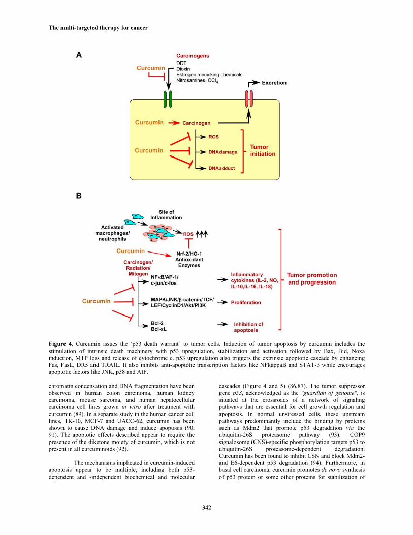
Figure 4. Curcumin issues the ‘p53 death warrant’ to tumor cells. Induction of tumor apoptosis by curcumin includes the stimulation of intrinsic death machinery with p53 upregulation, stabilization and activation followed by Bax, Bid, Noxa induction, MTP loss and release of cytochrome c. p53 upregulation also triggers the extrinsic apoptotic cascade by enhancing Fas, FasL, DR5 and TRAIL. It also inhibits anti-apoptotic transcription factors like NFkappaB and STAT-3 while encourages apoptotic factors like JNK, p38 and AIF.
chromatin condensation and DNA fragmentation have been observed in human colon carcinoma, human kidney carcinoma, mouse sarcoma, and human hepatocellular carcinoma cell lines grown in vitro after treatment with curcumin (89). In a separate study in the human cancer cell lines, TK-10, MCF-7 and UACC-62, curcumin has been shown to cause DNA damage and induce apoptosis (90, 91). The apoptotic effects described appear to require the presence of the diketone moiety of curcumin, which is not present in all curcuminoids (92).
The mechanisms implicated in curcumin-induced apoptosis appear to be multiple, including both p53-dependent and -independent biochemical and molecular
cascades (Figure 4 and 5) (86,87). The tumor suppressor gene p53, acknowledged as the "guardian of genome", is situated at the crossroads of a network of signaling pathways that are essential for cell growth regulation and apoptosis. In normal unstressed cells, these upstream pathways predominantly include the binding by proteins such as Mdm2 that promote p53 degradation via the ubiquitin-26S proteasome pathway (93). COP9 signalosome (CNS)-specific phosphorylation targets p53 to ubiquitin-26S proteasome-dependent degradation. Curcumin has been found to inhibit CSN and block Mdm2- and E6-dependent p53 degradation (94). Furthermore, in basal cell carcinoma, curcumin promotes de novo synthesis of p53 protein or some other proteins for stabilization of

The multi-targeted therapy for cancer
343
Figure 5. Curcumin and the apoptotic network. Apart from p53, curcumin enhances apoptotic death and inhibits cellular proliferation by altering specific key signaling molecules. Such a network organization allows the cell to sense many aspects of the intracellular and extra-cellular milieu, yet ensures that cell death proceeds efficiently once activated. Left: The extrinsic or death receptor pathway is triggered by members of the death receptor super-family such as Fas, TNF etc. Binding of ligand to specific receptors induces trimerization of the receptor, recruitment of specific adaptor proteins (FADD/TRADD) and recruitment and processing of pro-caspase-8 molecules. Caspase-8 can in turn activate Bid, which represents a crosstalk between extrinsic and intrinsic death pathways. Right: Drugs, ionizing radiation, polyphenols etc. can activate the intrinsic death pathway. The intrinsic or mitochondrial pathway is also triggered by DNA damage via p53 activities. The death stimuli result in loss of mitochondrial membrane integrity and release of cytochrome c, Apaf-1 and other pro-apoptotic factors in the cytoplasm. Maintenance or perturbation of mitochondrial membrane potential depends on the ratio between pro-apoptotic (Bax) and anti-apoptotic (Bcl-2), by causing or preventing cytochrome c release. Multiple molecules of cythocrome c, Apaf-1, dATP and procaspase-9 associate to form a supra-molecular complex termed ‘apoptosome’ that activates caspase-9 through autocatalysis. Both the activated caspase-9 and caspase-8 cleave procaspase-3 generating the active caspase-3 that, in turn, activates other executor caspases and cleaves cellular targets. Circular-head-lines indicate that these molecules can be down-regulated by curcumin, where as star-head-lines indicate that these molecules are often up-regulated by curcumin.
p53, and hence enhances its nuclear translocation to transactivate Cip1 and Gadd45 indicating that p53-associated signaling pathway is critically involved in curcumin-mediated apoptotic cell death (95). With time-lapse video-micrography and quantitative imaging approach we have demonstrated that in deregulated cells, curcumin induces p53 dramatically at G2 phase of cell cycle and enhances p53 DNA-binding activity resulting in apoptosis at G2 phase (87). Curcumin also induces p53 expression in colon and breast cancer cells (87, 96-98). Search for downstream of p53 revealed that in mammary epithelial carcinoma and colon adenocarcinoma cells curcumin could increase the expression of the pro-apoptotic protein Bax and decrease the anti-apoptotic protein Bcl-2/Bcl-xL through the phosphorylation at Ser15 and activation of p53. Our results also revealed curcumin induced G2/M arrest and apoptosis of mammary epithelial carcinoma cells via p53-mediated Bax activation (96). All these reports indicate that curcumin can induce cancer cell killing predominantly via p53-mediated pathway.
Increasing reports are indicating that curcumin can block cell cycle progression or even apoptosis in a p53-independent manner as well, especially in the cells that lack functional p53 (99). Curcumin induces apoptosis in p53-null cancer cells (100). It induces melanoma cell apoptosis by activating caspase-8 and caspase-3 via Fas receptor aggregation in a FasL-independent manner, blocks NFkappaB cell survival pathway and suppresses the apoptotic inhibitor XIAP (101). Curcumin inhibits cellular isopeptidases, and cause cell death independently of p53 in isogenic pairs of RKO and HCT 116 cells with differential p53 status (102). It enhances the chemotherapy-induced cytotoxicity in p53-null prostate cancer cell line PC-3, via up-regulation of Cip1 and C/EBPβ expressions and suppression of NFkappaB activation (103). In multiple myloma cells, curcumin induces apoptosis by inhibiting IKK and NFkappaB activity (104) while in colon cancer cells curcumin-induced JNK-dependent apoptosis takes the lead (105). It has also been demonstrated that curcumin-induced apoptosis is mediated through the impairment of

The multi-targeted therapy for cancer
344
ubiquitin-proteasome pathway (106). The constitutive phosphorylation of STAT3 found in certain multiple myeloma cells is abrogated by curcumin treatment, and inhibition of STAT3 by curcumin leads to the induction of apoptosis (107,108). Curcumin-induced rapid ROS generation causes the release of AIF from the mitochondria to the cytosol and nucleus, hence leading to caspase 3-independent apoptosis (109). It also strongly activates AMP-activated protein kinase (AMPK) in a p38-dependent manner in ovarian cancer cells, thus inducing cell death (110). Curcumin has also been shown to induce DNA damage mediated apoptosis by interfering with the activity of topoisomerase II, an enzyme which catalyzes the “unknotting” of DNA during mitosis (90). Other investigators have shown that curcumin delays phosphorylation of histone H2AX (gammaH2AX), a marker of DNA damage, which can result in apoptotic DNA fragmentation (91). 4.2.2. Inhibition of angiogenesis by curcumin
It has been well known for more than half a century that angiogenesis is linked to neoplasia. Angiogenesis, meaning the formation of new vessels, is generally considered to be a crucial step in tumor survival and growth beyond a certain size (about 1-2 mm in diameter). Curcumin inhibits carcinogenesis in different organs and the common link between these actions is its anti-angiogenic effect (111) since curcumin is a direct inhibitor of angiogenesis that down-regulates various pro-angiogenic proteins (112). Earlier it has been reported that curcumin treatment resulted in inhibition of angiogenic differentiation of human umbilical vein endothelial cells on matrigel and endothelial cell infiltration and vessel formation in matrigel plug, indicating the anti-angiogenic activity (113). Subsequently, it has been shown to inhibit bFGF-induced corneal neo-vascularization in mouse cornea (114). This angiostatic efficacy in the cornea has also been observed when curcuminoids are provided to mice in the diet. Recent studies have demonstrated that several other curcumin analogs show inhibitory effect on angiogenesis as seen by chicken chorioallantoic membrane assay, invasion assay, and tube formation assay (115).
Curcumin may inhibit angiogenesis directly and via regulation of these angiogenic growth factors, as well as the genes, angiopoietin-1/-2, HIF-1, HO-1, and the transcriptional factors like NFkappaB (Figure 6) (116-118). It is known that hypoxic stress and transforming growth factor–beta (TGF-beta) activation induces VEGF expression through trascriptional activation of AP-1 and hypoxia-inducible factor-1 (HIF-1) (119). Curcumin is a potent inhibitor of AP-1 activation and recently it has also been found that curcumin is a direct inhibitor of the activity of the HIF-1 trascriptional factor, which induces transcription of many genes involved in angiogenesis in tumors (115,116). Inhibition of angiogenic growth factor production and metalloproteinase generation, both integral to the formation of new vasculature, has also been influenced by curcumin in non-malignant and malignant cells growth (114). Cell adhesion molecules are up-regulated in active angiogenesis and curcumin can block
this effect, adding further dimensions to curcumin’s antiangiogenic effect. 4.2. 3. Retardation of metastasis by curcumin:
Metastasis is a multistage process that requires cancer cells to escape from the primary tumor, survive in the circulation, seed at distant sites and grow. Apart from causing cancer cell death, curcumin has been found to reduce the invasion and subsequent metastasis of cancer cells. Human lung adenocarcinoma cells treated with curcumin show a reduction in cell migration, invasion, and metastatic ability (120). Curcumin also reduced the migration of human cancer cells in Matrigel invasion assay (121,122). It prevents hematogenous breast cancer metastases in immunodeficient mice (123). The TPA-induced invasiveness of breast cancer cells and osteopontin (OPN)-induced non-small cell lung cancer migration has also been retarded by curcumin (124-126).
Local invasion and metastasis during tumor progression require its interaction with the surrounding matrix. Tumor derived factors influence these cellular interactions and subsequent adhesion (127). Similar to the inhibition of angiogenic factors, curcumin has been shown to regulate proteins related to cell-cell adhesion, such as β-catenin, E-cadherin and APC and to inhibit the production of cytokines relevant to tumor growth, e.g. tumour necrosis factor-alpha (TNF-alpha) and interleukin-1 (128). Additionally, curcumin has been shown to reduce the expression of membrane surface molecules such as intracellular adhesion molecule-1, vascular cell adhesion molecule-1 and E-selectin that play a role in cellular adhesion (Figure 6) (129). In human tracheal smooth muscle cells, curcumin treatment has been resulted in significant inhibition of tumor necrosis factor-alpha (TNF-alpha)-induced VCAM-1 expression, which is related to the activation of the MAPK/NFkappaB pathways (130). Curcumin suppresses the expression of matrix metalloproteinases (MMPs), which are important to endothelial cell migration and tube formation, and curtails the invasiveness of cancer cells (Figure 7) (131). Curcumin down regulates expression of MMP-9, that is specifically implicated in the growth and invasiveness of brain tumors, by inhibiting NFkappaB and AP-1 binding to the DNA promoter region (132). Collectively, these results pertaining to direct and indirect inhibition of angiogenesis and attenuation of cell-cell adhesion necessary for malignant behavior render curcumin a promising agent for altering the invasive and metastatic behavior of established malignancy. 4.3. Renovation of depressed immune system by curcumin
Immune dysfunction is well documented during tumor progression and likely contributes to tumor immune evasion. CD8+ cytotoxic T lymphocytes (CTLs) are involved in antigen-specific tumor destruction and CD4+T cells are essential for helping this CD8+ T cell-dependent tumor eradication. Tumors often target and inhibit T-cell function to escape from immune surveillance (4-6,133). Reports of the immunomodulatory power of curcumin in tumor-bearing hosts are inadequate. Some researchers,

The multi-targeted therapy for cancer
345
Figure 6. Curcumin retards tumor angiogenesis and metastasis: Curcumin decreases the expression of angiogenic promoters e.g, HIF-1, TGF-beta, NFkappaB, HO-1 and AP-1 while hinders metastasis by negatively regulating the expression of MMPs, uPA, beta-catenin, ICAM, VCAM and E selectin.
including us, have addressed the immunoprotective potential of curcumin in tumor-bearing hosts (134-138). Curcumin was found to prevent tumor-induced depletion of CD4+/CD8+ T cells, loss of central memory (TCM) and effector memory (TEM) T cells, augmentation of CD4+CD25+FoxP3+, TGF-beta producing and IL-10 producing Treg cells, imbalance of Th1/Tc1-type cytokine-producing T cells, down-regulation of T-cell proliferation (138).
Studies from our laboratory showed that curcumin neutralized tumor-induced oxidative stress,
restored back NFkappaB activity, and inhibited TNF-alpha production, thereby minimizing tumor-induced T-cell apoptosis (136). Further work suggests that curcumin helps in T cell survival both in primary and effecter immune compartments of tumor-bearing hosts by normalizing perturbed of Jak-3/Stat-5 activity via restoration of IL2-receptor gammac chain expression (137). Our ongoing research works also indicate that curcumin reverses T regulatory cell mediated type-2 cytokine bias and dysfunctional dendritic cell mediated T cell apoptosis in tumor microenvironment through inhibiting the tumor-shed immunosuppressive cytokines (unpublished data) (Figure 7).

The multi-targeted therapy for cancer
346
Figure 7. Curcumin rejuvenates tumor-induced imuunosuppression. Tumor derived immunosuppressive factors (PGE2, TGF-beta, gangliosides, TNF-alpha, IL-10 and exosomes) mediate immune dysfunction via induction of (i) IL-10 secreting Treg cells, (ii) tumor-disabled DC, (iii) natural killer cell (NKC) T cell-mediated tumor cytotoxicity and (iv) T cell apoptosis. Curcumin inhibits immune dysfunctions at different stages, thereby ameliorating tumor-induced immune suppression.
Moreover, curcumin treatment of tumor cells lead to the enhancement of ubiquination of tumor exosome proteins, and the reduction of tumor exosome-mediated inhibition of NK cell tumor cytotoxicity (139). From all these observations it is suggested that curcumin may be used alone or can be combined with classical anti-tumor drugs so as to sustain the immune capacity of the host, which can be affected by the disease or the treatment or may be by the both. 4.4. Amelioration of systemic toxicity
Mutilation of antioxidant and detoxification systems during tumor progression has been suggested in much literature. Having strong antioxidant and detoxifier properties curcumin has gripped enormous attention regarding cancer therapy. 4.4.1. Anti-oxidant properties of curcumin
Oxygen radicals are continuously generated within mammalian cells, this being a consequence of the use of oxygen in aerobic respiration. Superoxide is generated within the mitochondria and is sequentially reduced to hydrogen peroxide and hydroxyl radicals. These species damage DNA, producing the mutations that initiate tumors and sustain progression. Consequently, “quenching” of activated oxygen species or preventing the cellular damage they cause to proteins and DNA is an important mechanism to potentially prevent diseases like cancer. Curcumin with its proven anti-inflammatory and antioxidant properties has been shown to have several therapeutic advantages. Its antioxidant activity is comparable to vitamins C and E (140). Similar to other dietary phytochemicals, curcumin may possess pro-oxidant activity or antioxidant effects, dependent on dose and the chemical environment (141).
Curcumin was shown to be a potent scavenger of a variety of reactive oxygen species including superoxide anion radicals, hydroxyl radicals and nitrogen dioxide radicals (142-144). Nitric oxide (NO) is a short-lived, lipophilic molecule generated by enzymes called NO synthases (NOS). Its bioactivity is related to the production of many reactive intermediates. Some of these nitrogen species intermediates can damage DNA directly or interfere with DNA repair via protein damage. It has been shown that curcumin strongly inhibits lipopolysaccharide-induced iNOS gene expression (145). It was also shown to inhibit lipid peroxidation in different animal models (146,147). Curcumin protected oxidative cell injury of kidney cells by inhibiting lipid degradation, lipid peroxidation and cytolysis (148) and also decreased ischemia-induced biochemical changes in heart in a feline model (149). Vascular endothelial cells treated with curcumin prevented oxidant mediated injury by increased heme oxygenase production (150). Curcumin was found to protect rat myocardium against isoprenaline induced myocardial ischemic damage and the protective effect was attributed to its antioxidant properties by inhibiting free radial generation (151,152). It caused a decrease in the degree of degradation of the existing collagen matrix and collagen synthesis, two weeks after the second dose of isoprenaline. These effects were attributed to free radical scavenging properties and inhibition of lysosomal enzyme release by curcumin (153). Treatment with curcumin showed beneficial effects on renal injury by its ability to inhibit the expression of the apoptosis-related genes Fas and Fas-L (154). In clinical studies of colorectal cancer patients, orally administered curcumin have achieved pharmacologically active levels in the colorectum, associated with a decline in oxidative DNA adducts in colorectal tumors (155).

The multi-targeted therapy for cancer
347
Figure 8. Molecular targets of curcumin related to the therapy and prevention of cancer.
4.4.2. Detoxificant properties of curcumin Our bodies have evolved complex systems of
detoxification enzymes. These enzyme systems generally function adequately to minimize the potential of damage from environmental toxicants, drugs, carcinogens or xenobiotics. Enzyme systems involved in detoxification are (i) The Phase-I System: The Phase I detoxification system, composed mainly of the cytochrome P450 supergene family of enzymes, is generally the first enzymatic defense against foreign compounds. Most pharmaceuticals are metabolized through Phase-I biotransformation. In a typical Phase-I reaction, a cytochrome P450 enzyme (CypP450) uses oxygen and, as a cofactor, NADH, to add a reactive group, such as a hydroxyl radical. As a consequence of this step in detoxification, reactive molecules, which may be more toxic than the parent molecule, are produced. If these reactive molecules are not further metabolized by Phase-II conjugation, they may cause damage to proteins, RNA, and DNA within the cell. (ii) The Phase-II System: Phase-II conjugation reactions generally follow Phase-I activation, resulting in a xenobiotic that has been transformed into a water-soluble compound that can be excreted through urine or bile. Several types of conjugation reactions are present in the body, including glucuronidation, sulfation, and glutathione and amino acid conjugation. These reactions require cofactors, which must be replenished through dietary sources. However, several studies have shown evidence of associations between induced Phase-I and/or
decreased Phase-II activities and an increased risk of disease, such as cancer. Also the Pi (pi) isoenzyme of phase-I enzymes is present in very low amounts in normal liver but its expression becomes very high with the onset of carcinogenesis (156). It is now also well recognized that with onset of cancer, there is a concurrent toxic manifestation in the form of oxidative stress within the host. Status of antioxidant enzymes, i.e., superoxide dismutase (SOD), catalase, glutathione-S-transference (GST), peroxidases etc., which protect cells against oxidative damage, is almost invariably altered during carcinogenesis (157). Consequently, accumulated reactive oxygen species interacts with and modifies cellular proteins, lipids and DNA, which results in altered target cell function (158,159). All these finally lead to systemic toxicity in the tumor-bearer resulting in further failure of the treatment. Curcumin feeding in mice has been observed to inhibit P450 enzyme in mammary carcinoma cell line and to induce of detoxifiers, such as epoxide hydrolase and various hepatic GST isoenzymes, thereby indicating its protective role against carcinogens.
Curcumin’s inhibition of cytochrome P450-mediated activation of dimethylben zanthracene resulted in diminished DNA adduct formation (160,161). The ability of curcumin to induce phase-II enzymes appears to be asso-ciated with the presence of the hydroxyl groups at ortho-positions on the aromatic rings and the beta-diketone

The multi-targeted therapy for cancer
348
functionality (162). Though glutathione (GSH) plays a protective role against toxins, carcinogens and reactive oxygen species, it may also be linked with multidrug resistance through its spontaneous reactions with drugs as a co-factor for GST isoenzymes. In contrast to the early stages of carcinogenesis in advanced tumors, GST isoen-zymes (pi, alpha, mu) are aberrantly over-expressed and linked with resistance to chemotherapy (163). In addition to total induction of GST activity, curcumin appears to be capable of inhibiting specific GST isoenzymes (164,165). In the studies of GST isoenzymes, there was a linear association between the level of inhibition by curcumin and the induction of apoptosis. 5. CONCLUSION
The field of phytochemical research has expanded rapidly over the past decade resulting in the preclinical and early clinical development of many promising agents. Among these studying the antioxidant, anti-toxic, anti-inflammatory and cancer chemopreventive properties of curcumin are the subject of hundreds of published papers over the past three decades. Our review attempts to summarize the multifaceted anti-tumor activity of this phytochemical including influence upon key signal transduction pathways (Figure 5) associated with different stages of tumor progression. Due to the failure of conventional cancer therapies in advance stages of cancer and its enormous adverse effects, this phytochemical could be developed as preventive and alternative medicine. There is a hope that in years to come, cancer chemoprevention by this phytochemical in a defined molecular target approach (Figure 8) will play an important role in reducing cancer incidence as well as the number of deaths caused by this disease. 6. ACKNOWLEDGMENT
We thank S. Mohanty for her valuable suggestions during the preparation of this manuscript. This work was supported by research grants from CSIR and DST, Govt. of India. 7. REFERENCES 1. A Tekiner, O Yucel, AK Sargin, O Genc, B Can, T Karayilanoglu, J Karakaya, MA Bayar: The effect of nitrogen mustard on the rat brain and the therapeutic value of proanthocyanidin. Turk Neurosurg 19, 360-366 (2009) 2. R Miller, HL McLeod: Pharmacogenomics of cancer chemotherapy-induced toxicity. J Support Oncol 5, 9-14 (2007) 3. M Pàez-Ribes, E Allen, J Hudock, T Takeda, H Okuyama, F Viñals, M Inoue, G Bergers, D Hanahan, O Casanovas: Antiangiogenic therapy elicits malignant progression of tumors to increased local invasion and distant metastasis. Cancer Cell 15, 220-231 (2009) 4. T Das, G Sa, E Paszkiewicz-Kozik, C Hilston, L Molto, P Rayman, K Biswas, RM Bukowski, JH Finke, CS
Tannenbaum: Renal cell carcinoma tumors induce T cell apoptosis through receptor-dependent and receptor-independent pathways. J. Immunol 180, 4687–4696 (2008) 5. G Sa, T Das, C Moon, CM Hilston, PA Rayman, BI Rini, CS Tannenbaum, JH Finke: GD3, an overexpressed tumor-derived ganglioside, mediates the apoptosis of activated but not resting T cells. Cancer Res 69, 3095–3104 (2009) 6. T Das, G Sa, C Hilston, D Kudo, P Rayman, K Biswas, L Molto, R Bukowski, B Rini, J H. Finke, C Tannenbaum: GM1 and TNFα, overexpressed in renal cell carcinoma, synergize to induce T cell apoptosis. Cancer Res 68, 2014–2023 (2008) 7. L Zitvogel, L Apetoh, F Ghiringhelli, G Kroemer: Immunological aspects of cancer chemotherapy. Nat Rev Immunol 8, 59-73 (2008) 8. GK Jayaprakasha, L Jagan Mohan Rao, KK Sakariah: Chemistry and biological activities of C. longa. Trends in Food Science & Technology 16, 533-548 (2005) 9. World Health Organization. WHO monographs on selected medicinal plants: 1, 115-124 (1999) 10. F Kiuchi, Y Goto, N Sugimoto, N Akao, K Kondo, Y Tsuda: Nematocidal activity of turmeric: synergistic action of curcuminoids. Chem Pharm Bull 41, 1640-1643 (1993) 11. GK Jayaprakasha, L Jagan Mohan Rao, KK Sakariah: Improved HPLC method for the determination of curcumin, demethoxycurcumin, and bisdemethoxycurcumin. J Agric Food Chem 50, 3668-3672 (2002) 12. HH Tonnesen, J Karlsen, GB van Henegouwen: Studies on curcumin and curcuminoids. VIII. Photochemical stability of curcumin. Z Lebensm Unters Forsch 183, 116-122 (1986) 13. National Cancer Institute, DCPC. Clinical development plan: curcumin. J Cell Biochem 26, 72-85 (1996) 14. D Eigner, D Scholz: Ferula asa-foetida and Curcuma longa in traditional medical treatment and diet in Nepal. J Ethnopharmacol 67, 1-6 (1999) 15. N Chainani-Wu: Safety and anti-inflammatory activity of curcumin: a component of tumeric (Curcuma longa) J Altern Complement Med 9, 161-168 (2003) 16. RA Sharma, CR Ireson, RD Verschoyle, KA Hill, ML Williams, C Leuratti, MM Manson, LJ Marnett, WP Steward, A. Gescher: Effects of dietary curcumin on glutathione Stransferase and malondialdehyde-DNA adducts in rat liver and colon mucosa: relationship with drug levels. Clin Cancer Res 7, 1452-1458 (2001) 17. SD Deodhar, R Sethi, RC Srimal: Preliminary study on antirheumatic activity of curcumin (diferuloyl methane) Indian J. Med. Res 71, 632-634 (1980) 18. AL Cheng, CH Hsu and JK Lin: Phase I clinical trial of curcumin, a chemopreventive agent, in patients with

The multi-targeted therapy for cancer
349
high-risk or pre-malignant lesions. Anticancer Res 21, 2895–2900 (2001) 19. RA Sharma, SA Euden, SL Platton: Phase I clinical trial of oral curcumin: biomarkers of systemic activity and compliance. Clin Cancer Res 10, 6847–6854 (2004) 20. S Strimpakos, RA Sharma: Curcumin: preventive and therapeutic properties in laboratory studies and clinical trials. Antioxid Redox Signal 10, 511–545 (2008) 21. C Ireson, S Orr, D. J. Jones: Characterization of metabolites of the chemopreventive agent curcumin in human and rat hepatocytes and in the rat in vivo, and evaluation of their ability to inhibit phorbol ester-induced prostaglandin E2 production. Cancer Res 61, 1058–64 (2001) 22. P Anand, AB Kunnumakkara, RA Newman, BB Aggarwal: Bioavailability of curcumin: problems and promises. Mol Pharm 4, 807-818 (2007) 23. G Shoba, D Joy, T Joseph, M Majeed, R Rajendran, PS Srinivas: Influence of piperine on the pharmacokinetics of curcumin in animals and human volunteers. Planta Med 64, 353-356 (1998) 24. L Li, FS Braiteh, R Kurzrock: Liposome-encapsulated curcumin: in vitro and in vivo effects on proliferation, apoptosis, signaling, and angiogenesis. Cancer 104, 1322-1331 (2005) 25. C Karikar, A Maitra, S Bisht, G Feldmann, S Soni, R Ravi: Polymeric nanoparticle-encapsulated curcumin (“nanocurcumin”): a novel strategy for human cancer therapy. J. Nanobiotechnol 5, 3 (2007) 26. A Liu, H Lou, L Zhao, P Fan: Validated LC/MS/MS assay for curcumin and tetrahydrocurcumin in rat plasma and application to pharmacokinetic study of phospholipid complex of curcumin. J. Pharm. Biomed. Anal 40, 720-727 (2006) 27. H Ohori, H Yamakoshi, M Tomizawa, M Shibuya, Y Kakudo, A Takahashi, S Takahashi, S Kato, T Suzuki, C Ishioka, Y Iwabuchi, H Shibata: Synthesis and biological analysis of new curcumin analogues bearing an enhanced potential for the medicinal treatment of cancer. Mol. Cancer Ther 5, 2563 2571 (2006) 28. SS Han, ST Chung, DA Robertson, D Ranjan, S Bondada: Curcumin causes the growth arrest and apoptosis of B cell lymphoma by downregulation of egr-1, c-myc, bcl-XL, NF-kappa B, and p53. Clin. Immuno.l 93, 152-61 (1999) 29. SP Verma, BR Goldin, PS Lin: The inhibition of the estrogenic effects of pesticides and environmental chemicals by curcumin and isoflavonoids. Environ Health Perspect. 106, 807-812 (1998) 30. JG Brody, KB Moysich, O Humblet, KR Attfield, GP Beehler, RA Rudel: Environmental pollutants and breast cancer: epidemiologic studies. Cancer109, 2667-2711 (2007)
31. S Nishiumi, K Yoshida, H Ashida: Curcumin suppresses the transformation of an aryl hydrocarbon receptor through its phosphorylation. Arch Biochem Biophys. 466, 267-273 (2007) 32. H Choi, YS Chun, SW Kim, MS Kim, JW Park: Curcumin inhibits hypoxia-inducible factor-1 by degrading aryl hydrocarbon receptor nuclear translocator: a mechanism of tumor growth inhibition. Mol Pharmacol. 70, 1664-1671 (2006) 33. SP Verma, E Salamone B Goldin: Curcumin and genistein, plant natural products, show synergistic inhibitory effects on the growth of human breast cancer MCF-7 cells induced by estrogenic pesticides. Biochem Biophys Res Commun 233, 692-696 (1997) 34. N Li, X Chen, J Liao, G Yang, S Wang, Y Josephson, C Han, J Chen, MT Huang, CS Yang. Inhibition of 7,12-dimethylbenz[a]anthracene (DMBA)-induced oral carcinogenesis in hamsters by tea and curcumin. Carcinogenesis 23, 1307-13 (2002) 35. MT Huang, W Ma, YP Lu, RL Chang, C Fischer, PS Manchand, HL Newmark, AH Conney: Effect of curcumin, demethoxycurcumin, bisdemethoxy-curcumin and tetrahydrocurcumin on TPA-induced tumor promotion. Carcinogenesis 16, 2493-2497 (1995) 36. MT Huang, RC Smart, CQ Wong, AH Conney: Inhibitory effect of curcumin, chlorogenic acid, caffeic acid and ferulic acid on tumor promotion in mouse skin by 12-O-tetradecanoylphorbol-13-acetate. Cancer Res 48, 5941-5946 (1988) 37. MT Huang, ZY Wang, CA Georgiadis, JD Laskin, AH Conney: Inhibitory effect of curcumin on tumor initiation by benzo[a] pyrene and 7,12-dimethyl-benz[a]anthracene. Carcinogenesis 13, 947-954 (1992) 38. MT Huang, YR Lou, W Ma, HL Newmark, KR Reuhl, AH Conney: Inhibitory effects of dietary curcumin on forestomach, duodenal and colon carcinogenesis in mice. Cancer Res 54, 5841-5847 (1994) 39. CV Rao, A Riven, B Simi, BS Reddy: Chemoprevention of colon carcinogenesis by dietary curcumin, a naturally occurring plant phenolic compound. Cancer Res 55, 259-266 (1995) 40. JW Pollard: Tumour-educated macrophages promote tumour progression and metastasis. Nat Rev Cancer 4, 71-78 (2004) 41. H Ohsima: Curcumin, an anti-tumor promoter and anti-inflammatory agent, inhibits induction of nitric oxide synthase in activated macrophages. Biochem Biophys Res Commun 206, 533–540 (1995) 42. MNA. Rao: Oxygen radical scavenging activity of curcumin. Int J Pharm 38, 239-240 (1990)

The multi-targeted therapy for cancer
350
43. M Subramanian, MNA Sreejayan Rao, TPA Devasagyam, BB Singh: Diminution of singlet oxygen-induced DNA-damage by curcumin and related antioxidants. Mutat Res 311, 249-255 (1994) 44. RJ Verma: Curcumin ameliorates aflatoxin-induced lipid peroxidation in liver, kidney and testis of mice--an in vitro study. Acta Pol Pharm 64, 413-416 (2007) 45. MK Unnikrishnan, MN Rao: Curcumin inhibits nitrogen dioxide induced oxidation of hemoglobin. Mol Cell Biochem 146, 35-37 (1995) 46. M Iqbal, Y Okazaki, S Okada: Curcumin attenuates oxidative damage in animals treated with a renal carcinogen, ferric nitrilotriacetate (Fe-NTA): implications for cancer prevention. Mol Cell Biochem 324,157-64 (2009) 47. SM Plummer, KA Holloway, MM Manson: Inhibition of cyclooxygenase 2 expression in colon cells by chemopreventive agent curcumin involves inhibition of NF-κB activation via the NIK/IKK signaling complex. Oncogene 18, 6013–6020 (1999) 48. MSD Iqbal, Y Sharma, M Okazaki, S Okada: Dietary supplementation of curcumin enhances antioxidant and phase II metabolizing enzymes in ddY male mice: possible role in protection against chemical carcinogenesis and toxicity. Pharmacol Toxicol 92, 33-38 (2003) 49. Y Okazaki, M Iqbal, S Okada: Suppressive effects of dietary curcumin on the increased activity of renal ornithine decarboxylase in mice treated with a renal carcinogen, ferric nitrilotriacetate. Biochim Biophys Acta 1740, 357-366 (2005) 50.CK Andreadi, LM Howells, PA Atherfold, MM Manson: Involvement of Nrf2, p38, B-Raf, and nuclear factor-{kappa}B, but not phosphatidylinositol 3-kinase, in induction of hemeoxygenase-1 by dietary polyphenols. Mol Pharmacol 69, 1033-1040 (2006) 51. PF Firozi, VS Aboobaker, RK Bhattacharya: Action of curcumin on the cytochrome P450-system catalyzing the activation of aflatoxin B1. Chem Biol Interact 100, 41-51 (1996) 52. VS Yadav, KP Mishra, DP Singh, S Mehrotra, VK Singh: Immunomodulatory effects of curcumin. Immunopharmacol Immunotoxicol 27, 485-497 (2005) 53. A Grandjean-Laquerriere, SC Gangloff, R Le Naour, C Trentesaux, W Hornebeck, M Guenounou: Relative contribution of NF-kappaB and AP-1 in the modulation by curcumin and pyrrolidine dithiocarbamate of the UVB-induced cytokine expression by keratinocytes. Cytokine 18, 168-177 (2002) 54. SS Han, YS Keum, HJ Seo, Y.J. Surh: Curcumin suppresses activation of NF-kappaB and AP-1 induced by
phorbol ester in cultured human promyelocytic leukemia cells. J Biochem Mol Biol 35, 337-342 (2002) 55. C Jobin, CA Bradham, MP Russo, B Juma, AS Narula, DA Brenner, RB Sartor: Curcumin blocks cytokine-mediated NF-kappa B activation and proinflammatory gene expression by inhibiting inhibitory factor I-kappa B kinase activity. Journal of Immunology 163, 3474–3483 (1999) 56. Y Bierhaus, P Zhang, T Quehenberger, M Luther, M Haase, M Muller, N Mackman, R Ziegler, PP Nawroth: The dietary pigment curcumin reduces endothelial tissue factor gene expression by inhibiting binding of AP-1 to the DNA and activation of NF-kappa B. Thrombosis and Haemostasis 77, 772–782 (1997) 57. S Park, DK Lee, CH Yang: Inhibition of fos–jun–DNA complex formation by dihydroguaiaretic acid and in vitro cytotoxic effects on cancer cells. Cancer Letters 127, 23–28 (1998) 58. T Kawamori, R Lubet, VE Steele, GJ Kelloff, RB Kaskey, CV Rao, BS Reddy: Chemopreventive effect of curcumin, a naturally occurring anti-inflammatory agent, during the promotion/progression stages of colon cancer. Cancer Res 59, 597-601 (1999) 59. RA Sharma, PB Farmer: Biological relevance of adduct detection to the chemoprevention of cancer. Clin Cancer Res 10, 4901-12 (2004) 60. S Perkins, RD Verschoyle, KA Hill: Chemopreventive efficacy and pharmacokinetics of curcumin in the Min/+ mouse, a model of familial adenomatous polyposis. Cancer Epidem Biomar Prevent 11, 535–540 (2002) 61. NN Mahmoud, AM Carothers, D Grunberger: Plant phenolics decrease intestinal tumors in an animal model of familial adenomatous polyposis. Carcinogenesis 21, 921–927 (2000) 62. HS Samaha, GJ Kelloff, V Steele: Modulation of apoptosis by sulindac, curcumin, phenylethyl-3-methylcaffeate, and 6-phenylhexyl isothiocyanate: apoptotic index as a biomarker in colon cancer chemoprevention and promotion. Cancer Res 57, 1301–1305 (1997) 63. T Dorai, N Gehani, A Katz: Therapeutic potential of curcumin in human prostate cancer – I. Prostate Cancer Dis 3, 84–93 (2000) 64. AP Chen, J Xu: Activation of PPAR gamma by curcumin inhibits Moser cell growth and mediates suppression of gene expression of cyclin D1 and EGFR. Am J Physiol 288, G447–G456 (2005) 65. ML Kuo, TS Huang, JK Lin: Curcumin, an antioxidant and anti-tumor promoter, induces apoptosis in human leukemia cells. Biochim Biophys Acta 1317, 95–100 (1996)

The multi-targeted therapy for cancer
351
66. K Ozaki, Y Kawata, S Amano: Stimulatory effect of curcumin on osteoclast apoptosis, Biochem Pharmacol 59, 1577–1581 (2000) 67. Chen ZS Zhang, YL Zhang, DY Zhou: Curcumin inhibits cell proliferation by interfering with the cell cycle and inducing apoptosis in colon carcinoma cells. Anticancer Res 19, 3675–3680 (1999) 68. CC Su, JG Lin, TM Li, JG Chung, JS Yang, SW Ip, WC Lin, GW Chen: Curcumin-induced apoptosis of human colon cancer colo 205 cells through the production of ROS, Ca2+ and the activation of caspase-3. Anticancer Res 26, 4379-89 (2006) 69. A Simon, DP Allais, JL Duroux, JP Basly, S Durand-Fontanier, C Delage: Inhibitory effect of curcuminoids on MCF-7 cell proliferation and structure-activity relationships. Cancer Lett 129, 111–116 (1998) 70. MC Jiang, HF Yang Yen, JJY Yen: Curcumin induces apoptosis in immortalized NIH 3T3 and malignant cancer cell lines. Nutr Cancer 26, 111–120 (1996) 71. SC Gautam, YX Yong, KR Pndolia: Nonselective inhibition of proliferation of transformed and nontransformed cells by the anticancer agent curcumin (diferuloylmethane) Biochem Pharmacol 55, 1333–1337 (1998) 72. S Plummer, D Wakelin, M Bouer: Inhibition of growth of colon tumour cells by curcumin correlates with inhibition of the IkB kinase and down regulation of cyclin D1. Br J Cancer 83, 20 (2000) 73. SS Han, ST Chung, DA Robertson: Curcumin causes the growth arrest and apoptosis of B cell lymphoma by downregulation of egr-1, c-myc, bcl-XL, NF-kappa B, and p53. Clin Immunol 93, 152–161 (1999) 74. S Bhaumik, MD Jyothi, A Khar: Differential modulation of nitric oxide production by curcumin in host macrophages and NK cells. FEBS Lett 483, 78–82 (2000) 75. CH Yan, MS Jamaluddin, B Aggarwal: Gene expression profiling identifies activating transcription factor 3 as a novel contributor to the proapoptotic effect of curcumin. Mol Cancer Ther 4, 233–241 (2005) 76. AC Bharti, N Donato, S Singh, BB Aggarwal: Curcumin (diferuloylmethane) down-regulates the constitutive activation of nuclear factor-kappa B and IkappaBalpha kinase in human multiple myeloma cells, leading to suppression of proliferation and induction of apoptosis. Blood 101, 1053-62 (2003) 77. SS Han, YS Keum, HJ Seo, YJ Surh: Curcumin suppresses activation of NF-kappaB and AP-1 induced by
phorbol ester in cultured human promyelocytic leukemia cells. J Biochem Mol Biol 35, 337-42 (2002) 78. M Austinat, R Dunsch, C Wittekind, A Tannapfel, R Gebhardt, F Gaunitz: Correlation between beta-catenin mutations and expression of Wnt-signaling target genes in hepatocellular carcinoma. Mol Cancer 7, 21 (2008) 79. AS Jaiswal, BP Marlow, N Gupta, S Narayan: Beta-catenin-mediated transactivation and cell-cell adhesion pathways are important in curcumin (diferuylmethane)-induced growth arrest and apoptosis in colon cancer cells. Oncogene 21, 8414–8427 (2002) 80. CH Park, ER Hahm, S Park, HK Kim, CH Yang: The inhibitory mechanism of curcumin and its derivative against beta-catenin/Tcf signaling.FEBS Lett 579, 2965–2971 (2005) 81. A Chen, J Xu: Activation of PPAR{gamma} by curcumin inhibits Moser cell growth and mediates suppression of gene expression of cyclin D1 and EGFR. Am J Physiol Gastrointest Liver Physiol 288, G447-G456 (2005) 82. AR Hussain, M Al-Rasheed, PS Manogaran, KA Al-Hussein, LC Platanias, Al KK and S Uddin: Curcumin induces apoptosis via inhibition of PI3'-kinase/AKT pathway in acute T cell leukemias. Apoptosis 11, 245-54 (2006) 83. JH Woo, YH Kim, YJ Choi: Molecular mechanisms of curcumin-induced cytotoxicity: induction of apoptosis through generation of reactive oxygen species, down-regulation of Bcl-XL and IAP, the release of cytochrome c and inhibition of Akt. Carcinogenesis 24, 1199-208 (2003) 84. JY Liu, SJ Lin, JK Lin: Inhibitory effects of curcumin on protein kinase C activity induced by12-O-tetradecanoyl-phorbol-13-acetate in NIH 3T3 cells. Carcinogenesis 14, 857-61 (1993) 85. E Liu, J Wu, W Cao, J Zhang, W Liu, X Jiang, X Zhang: Curcumin induces G2/M cell cycle arrest in a p53-dependent manner and upregulates ING4 expression in human glioma. J Neurooncol 85, 263-270 (2007) 86. JL Watson, A Greenshields, R Hill, A Hilchie, PW Lee, CA Giacomantonio, DW Hoskin: Curcumin-induced apoptosis in ovarian carcinoma cells is p53-independent and involves p38 mitogen-activated protein kinase activation and downregulation of Bcl-2 and survivin expression and Akt signaling. Mol Carcinog 49, 13-24 (2010) 87. T Choudhuri, S Pal, T Das, G Sa: Curcumin selectively induces apoptosis in deregulated cyclin D1-expressed cells at G2 phase of cell cycle in a p53-dependent manner. J Biol Chem 280, 20059-20068 (2005) 88. NM Weir, K Selvendiran, VK Kutala, L Tong, S Vishwanath, M Rajaram, S Tridandapani, S Anant, P Kuppusamy: Curcumin induces G2/M arrest and apoptosis in

The multi-targeted therapy for cancer
352
cisplatin-resistant human ovarian cancer cells by modulating Akt and p38 MAPK. Cancer Biol Ther 6, 178-184 (2007) 89. MC Jiang, HF. Yang-Yen, JJ Yen, JK Lin: Curcumin induces apoptosis in immortalized NIH 3T3 and malignant cancer cell lines. Nutr Cancer 26, 111-20 (1996) 90. C Martin-Cordero, M. Lopez-Lazaro, M Galvez, MJ Ayuso: Curcumin as a DNA topoisomerase II poison. J Enzyme Inhib Med Chem 18, 505-509 (2003) 91. G Mosieniak, M Sliwinska, K Piwocka, E Sikora: Curcumin abolishes apoptosis resistance of calcitriol-differentiated HL-60 cells. FEBS Letters 580, 4653-60 (2006) 92. H Ligeret, S Barthelemy, R Zini, JP Tillement, S Labidalle, D Morin: Effects of curcumin and curcumin derivatives on mitochondrial permeability ransition pore. Free Radic Biol Med 36, 919-29 (2004) 93. AS Coutts, CJ Adams, NB La Thangue: p53 ubiquitination by Mdm2: a never ending tail? DNA Repair (Amst) 8, 483-90 (2009) 94. D Bech-Otschir, R Kraft, X Huang, P Henklein, B Kapelari, C Pollmann, W Dubiel: COP9 signalosome-specific phosphorylation targets p53 to degradation by the ubiquitin system. EMBO J 20,1630-9(2001) 95. SH Jee, SC Shen, CR Tseng, HC Chiu, M.L. Kuo: Curcumin induces a p53-dependent apoptosis in human basal cell carcinoma cells. J Invest Dermatol 111, 656-661 (1998) 96. T Choudhuri, S Pal, ML Agwarwal, T Das, G Sa: Curcumin induces apoptosis in human breast cancer cells through p53-dependent Bax induction. FEBS Lett 512, 334-340 (2002) 97. PJ Moos, K Edes, JE Mullally, FA Fitzpatrick: Curcumin impairs tumor suppressor p53 function in colon cancer cells. Carcinogenesis 25, 1611-1617 (2004) 98. G Song, YB Mao, QF Cai, LM Yao, GL Ouyang, SD Bao: Curcumin induces human HT-29 colon adenocarcinoma cell apoptosis by activating p53 and regulating apoptosis-related protein expression. Braz J Med Biol Res 38, 1791-1798 (2005) 99. MJ Park, EH Kim, IC Park, HC Lee, SH Woo, JY Lee, YJ Hong, CH Rhee, SH Choi, BS Shim, SH Lee, SI Hong: Curcumin inhibits cell cycle progression of immortalized human umbilical vein endothelial (ECV304) cells by up-regulating cyclin-dependent kinase inhibitor, p21WAF1/CIP1, p27KIP1 and p53. Int J Oncol 21, 379-383 (2002) 100. M Shi, Q Cai, L Yao, Y Mao, Y Ming, G Ouyang: Antiproliferation and apoptosis induced by curcumin in human ovarian cancer cells. Cell Biol Int 30, 221-226 (2006)
101. JA Bush, KJ Jr Cheung, G Li: Curcumin induces apoptosis in human melanoma cells through a Fas receptor/caspase-8 pathway independent of p53. Exp Cell Res 271, 305-314 (2001) 102. JE Mullally, FA Fitzpatrick: Pharmacophore model for novel inhibitors of ubiquitin isopeptidases that induce p53-independent cell death. Mol Pharmacol 69, 351-358 (2002) 103. TC Hour, J Chen, CY Huang, JY Guan, SH Lu, YS Pu: Curcumin enhances cytotoxicity of chemotherapeutic agents in prostate cancer cells by inducing p21(WAF1/CIP1) and C/EBPbeta expressions and suppressing NF-kappaB activation. Prostate 51, 211-218 (2002) 104. AC Bharti, N Donato, S Singh, BB Aggarwal: Curcumin (diferuloylmethane) down-regulates the constitutive activation of nuclear factor-kappa B and IkappaBalpha kinase in human multiple myeloma cells, leading to suppression of proliferation and induction of apoptosis. Blood 101, 1053-1062 (2003) 105. GP Collett, FC Campbell: Curcumin induces c-jun N-terminal kinase-dependent apoptosis in HCT116 human colon cancer cells. Carcinogenesis 25, 2183-2189 (2004) 106. NR Jana, P Dikshit, A Goswami, N Nukina: Inhibition of proteasomal function by curcumin induces apoptosis through mitochondrial pathway. J Biol Chem 279, 11680-11685 (2004) 107. LS Cucuzza, M Motta, S Miretti, P Accornero, M Baratta: Curcuminoid-phospholipid complex induces apoptosis in mammary epithelial cells by STAT-3 signaling. Exp Mol Med 40, 647-657 (2008) 108. M Saydmohammed, D Joseph, V Syed: Curcumin suppresses constitutive activation of STAT-3 by up-regulating protein inhibitor of activated STAT-3 (PIAS-3) in ovarian and endometrial cancer cells. J Cell Biochem (2010)[Epub ahead of print]. 109. F Thayyullathil, S Chathoth, A Hago, M Patel, S Galadari: Rapid reactive oxygen species (ROS) generation induced by curcumin leads to caspase-dependent and -independent apoptosis in L929 cells. Free Radic Biol Med 45, 1403-1412 (2008) 110. W Pan, H Yang, C Cao, X Song, B Wallin, R Kivlin, S Lu, G Hu, W Di, Y Wan: AMPK mediates curcumin-induced cell death in CaOV3 ovarian cancer cells. Oncol Rep 20, 1553-1559 (2008) 111. AE Gururaj, M Belakavadi, DA Venkatesh, D Marmé, BP Salimath: Molecular mechanisms of anti-angiogenic effect of curcumin. Biochem Biophys Res Commun 297, 934-942 (2002)

The multi-targeted therapy for cancer
353
112. JL Arbiser, N Klauber, R Rohan, R van Leeuwen, MT Huang, C Fisher, E Flynn, HR Byers: Curcumin is an in vivo inhibitor of angiogenesis. Mol Med 4, 376-383 (1998) 113. D Thaloor, AK Singh, GS Sidhu, PV Prasad, HK Kleinman, RK Maheshwari: Inhibition of angiogenic differentiation of human umbilical vein endothelial cells by curcumin. Cell Growth Differ 9, 305-312 (1998) 114. R Mohan, J Sivak, P Ashton, LA Russo, BQ Pham, N Kasahara, MB Raizman, ME Fini: Curcuminoids inhibit the angiogenic response stimulated by fibroblast growth factor-2, including expression of matrix metalloproteinase gelatinase B. J Biol Chem 275, 10405-10412 (2000) 115. ER Hahm, Y.S. Gho, S Park, C Park, KW Kim, CH Yang: Synthetic curcumin analogs inhibit activator protein-1 transcription and tumor-induced angiogenesis. Biochem Biophys Res Commun 321, 337-344 (2004) 116. MK Bae, SH Kim, JW Jeong, YM Lee, HS Kim, SR Kim, I Yun, SK Bae, KW Kim: Curcumin inhibits hypoxia-induced angiogenesis via down-regulation of HIF-1. Oncol Rep 15, 1557-1562 (2006) 117. W Bao, K Li, S Rong, P Yao, L Hao, C Ying, X Zhang, A Nussler, L Liu: Curcumin alleviates ethanol-induced hepatocytes oxidative damage involving heme oxygenase-1 induction. J Ethnopharmacol 128, 549-553 (2010) 118. YG Lin, AB Kunnumakkara, A Nair, WM Merritt, LY Han, GN Armaiz-Pena, AA Kamat, WA Spannuth, DM Gershenson, SK Lutgendorf, BB Aggarwal, AK Sood: Curcumin inhibits tumor growth and angiogenesis in ovarian carcinoma by targeting the nuclear factor-kappaB pathway. Clin Cancer Res 13, 3423-3430 (2007) 119. DE Sullivan, M Ferris, H Nguyen, E Abboud, AR Brody: TNF-alpha induces TGF-beta1 expression in lung fibroblasts at the transcriptional level via AP-1 activation. J Cell Mol Med 13, 1866-1876 (2009) 120. HW Chen, JY Lee, JY Huang, CC Wang, WJ Chen, SF Su, CW Huang, CC Ho, JJ Chen, MF Tsai, SL Yu, PC Yang: Curcumin inhibits lung cancer cell invasion and metastasis through the tumor suppressor HLJ1. Cancer Res 68, 7428-7438 (2008) 121. R Olszanecki, A Gebska, R Korbut: The role of haem oxygenase-1 in the decrease of endothelial intercellular adhesion molecule-1 expression by curcumin. Basic Clin Pharmacol Toxicol 101, 411-415 (2007) 122. S Yodkeeree, C Ampasavate, B Sung, BB Aggarwal, P. Limtrakul: Demethoxycurcumin suppresses migration and invasion of MDA-MB-231 human breast cancer cell line. Eur J Pharmacol 627, 8-15 (2010) 123. B Bachmeier, AG Nerlich, CM Iancu, M Cilli, E Schleicher, R Vené, R Dell'Eva, M Jochum, A Albini, U Pfeffer: The chemopreventive polyphenol Curcumin
prevents hematogenous breast cancer metastases in immunodeficient mice. Cell Physiol Biochem 19, 137-152 (2007) 124. SE Chuang, ML Kuo, CH Hsu, CR Chen, JK Lin, GM Lai, CY Hsieh, AL Cheng: Curcumin-containing diet inhibits diethylnitrosamine-induced murine hepatocarcinogenesis. Carcinogenesis 21, 331-335 (2000) 125. KW Lee, JH Kim, HJ Lee, YJ Surh: Curcumin inhibits phorbol ester-induced up-regulation of cyclooxygenase-2 and matrix metalloproteinase-9 by blocking ERK1/2 phosphorylation and NF-kappaB transcriptional activity in MCF10A human breast epithelial cells. Antioxid Redox Signal 7, 1612-1620 (2005) 126. S Philip, GC Kundu: Osteopontin induces nuclear factor kappa B-mediated promatrix metalloproteinase-2 activation through I kappa B alpha/IKK signaling pathways, and curcumin (diferulolylmethane) down-regulates these pathways. J Biol Chem 278, 14487–14497(2003) 127. JA Joyce, JW Pollard: Microenvironmental regulation of metastasis. Nat Rev Cancer 9, 239-252 (2009) 128. CH Park, ER Hahm, S Park, HK Kim, CH Yang: The inhibitory mechanism of curcumin and its derivative against beta-catenin/Tcf signaling. FEBS Lett 579, 2965-2971 (2005) 129. C Jae-We, L Kyu-Suk, K. Chang-Wook: Curcumin attenuates the expression of IL-1ß, IL-6, and TNF-α as well as cyclin e in TNF-α-treated hacat cells; NF-κB and MAPKs as potential upstream targets. Int J Mol Med 19, 469-474 (2007) 130. CW Lee, WN Lin, CC Lin: Transcriptional regulation of VCAM-1 expression by tumor necrosis factor-alpha in human tracheal smooth muscle cells: involvement of MAPKs, NF-kappaB, p300, and histone acetylation. J Cell Physio 207, 174-186 (2006) 131. MS Woo, SH Jung, SY Kim: Curcumin suppresses phorbol ester-induced matrix metalloproteinase-9 expression by inhibiting the PKC to MAPK signaling pathways in human astroglioma cells. Biochem Biophys Res Commun 335, 1017-1025 (2005) 132. BB Aggarwal, S Shishodia, Y Takada: Curcumin suppresses the paclitaxel-induced nuclear factor-kappaB pathway in breast cancer cells and inhibits lung metastasis of human breast cancer in nude mice. Clin Cancer Res 11, 7490-7498 (2005) 133. S Chattopadhyay, S Bhattacharyya, B Saha, J Chakraborty , S Mohanty, DMSakib Hossain, S Banerjee, K Das, G Sa, T Das: Tumor-shed PGE(2) impairs IL2Rgammac-signaling to inhibit CD4 T cell survival: regulation by theaflavins. PLoS One 4,e7382. (2009) 134. S Pal, S Bhattacharya, T Choudhuri, GK Datta, T Das, G Sa: Amelioration of immune cell number depletion and potentiation of depressed detoxification system of tumor-

The multi-targeted therapy for cancer
354
bearing mice by curcumin. Cancer Detection Prevention 29, 470-478 (2005) 135. G Sa, T Das: Anti cancer effects of curcumin: cycle of life and death. Cell Div 3, 14 (2008) 136. S Bhattacharyya, D Mandal, GS Sen, S Pal, S Banerjee, L Lahiry, JH Finke, CS Tannenbaum, T Das, G Sa: Tumor-induced oxidative stress perturbs nuclear factor-kappaB activity-augmenting tumor necrosis factor-alpha-mediated T-cell death: protection by curcumin. Cancer Res 67, 362-70 (2007) 137. S Bhattacharyya, D Mandal, B Saha, GS Sen, T Das, G Sa: Curcumin prevents tumor-induced T cell apoptosis through Stat-5a-mediated Bcl-2 induction. J Biol Chem 282, 15954-64 (2007) 138. S Bhattacharyya, MdDS Hossain, S Mohanty, GS Sen, S Chattopadhyay, S Banerjee, J Chakraborty, K Das, D Sarkar, T Das, G Sa: Curcumin reverses T cell-mediated adaptive immune dysfunctions in tumor-bearing hosts. Cell Mol Immunol 2010 [Epub ahead of print]. 139. HG Zhang, H Kim, C Liu, S Yu, J Wang, WE Grizzle, RP Kimberly, S Barnes: Curcumin reverses breast tumor exosomes mediated immune suppression of NK cell tumor cytotoxicity. Biochim Biophys Acta 1773, 1116-23 (2007) 140. S Toda, T Miyase, H Arichi, H Tanizawa, Y Takino: Natural antioxidants. III. Antioxidative components isolated from rhizome of Curcuma longa L. Chemical and Pharmaceutical Bulletin 33, 1725–1728 (1985) 141. A Barik, B Mishra, L. Shen: Evaluation of a new copper(II)-curcumin complex as superoxide dismutase mimic and its free radical reactions. Free Radic Biol Med 39, 811-22 (2005) 142. AC Reddy, BR Lokesh: Studies on the inhibitory effects of curcumin and eugenol on the formation of reactive oxygen species and the oxidation of ferrous iron. Molecular and Cellular Biochemistry 137, 1–8 (1994) 143. MK Unnikrishnan, MN Rao: Curcumin inhibits nitrogen dioxide induced oxidation of hemoglobin. Molecular and Cellular Biochemistry 146, 35–37 (1995) 144. MNS Rao: Nitric oxide scavenging by curcuminoids. Journal of Pharmacy and Pharmacology 49, 105–107 (1997) 145. M Graziewicz, DAWink, F Laval: Nitric oxide inhibits DNA ligase activity: potential mechanisms for NO-mediated DNA damage. Carcinogenesis 17, 2501-5 (1996) 146. AC Reddy, BR Lokesh: Studies on spice principles as antioxidants in the inhibition of lipid peroxidation of rat liver microsomes. Molecular and Cellular Biochemistry 111, 117–124 (1992)
147. MNS. Rao: Curcuminoids as potent inhibitors of lipid peroxidation. Journal of Pharmacy and Pharmacology 46, 1013–1016 (1994) 148. HH Cohly, A Taylor, MF Angel, AK Salahudeen: Effect of turmeric, turmerin and curcumin on H2O2-induced renal epithelial (LLCPK1) cell injury. Free Radical Biology and Medicine 24, 49–54 (1998) 149. M Dikshit, L Rastogi, R Shukla, RC Srimal: Prevention of ischaemia-induced biochemical changes by curcumin and quinidine in the cat heart. Indian Journal of Medical Research 101, 31–35 (1995) 150. R Motterlini, R Foresti, R Bassi, CJ Green: Curcumin, an antioxidant and anti-inflammatory agent, induces heme oxygenase-1 and protects endothelial cells against oxidative stress. Free Radical Biology and Medicine 28, 1303–1312 (2000) 151. C Nirmala, R Puvanakrishnan: Effect of curcumin on certain lysosomal hydrolases in isoproterenol-induced myocardial infarction in rats. Biochemical Pharmacology 51, 47–51 (1996) 152. P Manikandan, M Sumitra, S Aishwarya, BM Manohar, B Lokanadam, R Puvanakrishnan: Curcumin modulates free radical quenching in myocardial ischaemia in rats. International Journal of Biochemistry and Cell Biology 36, 1967–1980 (2004) 153. C Nirmala, S Anand, R Puvanakrishnan: Curcumin treatment modulates collagen metabolism in isoproterenol induced myocardial necrosis in rats. Molecular and Cellular Biochemistry 197, 31–37 (1999) 154. EA Jones, A Shahed, DA Shoskes: Modulation of apoptotic and inflammatory genes by bioflavonoids and angiotensin II inhibition in ureteral obstruction. Urology 56, 346–351 (2000) 155. MM Chan, HI Huang, MR Fenton, D. Fong: In vivo inhibition of nitric oxide synthase gene expression by curcumin, a cancer preventive natural product with anti-inflammatory properties. Biochem Pharmacol 55, 1955-62 (1998) 156. CR Ireson, DJ Jones, S Orr: Metabolism of the cancer chemopreventive agent curcumin in human and rat intestine. Cancer Epidemiol Biomarkers Prev 11, 105-11 (2002) 157. AT Dinkova-Kostova, P Talalay: Relation of structure of curcumin analogs to their potencies as inducers of Phase 2 detoxification enzymes. Carcinogenesis 20, 911-14 (1999) 158. J Nair, S Strand, N Frank, J Knauft, H Wesch, PR Galle, H.Bartsch: Apoptosis and age-dependant induction of nuclear and mitochondrial etheno-DNA adducts in Long-Evans Cinnamon (LEC) rats: enhanced DNA damage

The multi-targeted therapy for cancer
355
by dietary curcumin upon copper accumulation. Carcinogenesis 26, 1307-15 (2005) 159. PK Lala, C Chakraborty: Role of nitric oxide in carcinogenesis and tumour progression. Lancet Oncol 2, 149-56 (2001) 160. G Garcea, DP Berry, DJ Jones: Consumption of the putative chemopreventive agent curcumin by cancer patients: assessment of curcumin levels in the colorectum and their pharmacodynamic consequences. Cancer Epidemiol. Biomarkers Prev 14, 120-125 (2005) 161. HP Ciolino, PJ Daschner, TT Wang, GC Yeh: Effect of curcumin on the aryl hydrocarbon and cytochrome P450 1A1 in MCF-7 human breast carcinoma cells. Biochem Pharmacol 56, 197-206 (1998) 162. AT Dinkova-Kostova, P Talalay: Relation of structure of curcumin analogs to their potencies as inducers of Phase 2 detoxification enzymes. Carcinogenesis 20, 911-14 (1999) 163. A Duvoix, M Schnekenburger, S Delhalle: Expression of glutathione S-transferase P1-1 in leukemic cells is regulated by inducible AP-1 binding. Cancer Lett 216, 207-19 (2004) 164. R Hayeshi, I Mutingwende, W Mavengere, V Masiyanise, S Mukanganyama: The inhibition of human glutathione S-transferases activity by plant polyphenolic compounds ellagic acid and curcumin. Food Chem Toxicol 45, 286-95 (2007) 165. JD Hayes, DJ Pulford: The glutathione S-transferase supergene family: regulation of GST and the contribution of the isoenzymes to cancer chemoprotection and drug resistance. Crit Rev Biochem Mol Biol 30, 445-600 (1995) Abbreviations: AP1: activator protein-1; Apaf-1; apoptosis protease-activating factor-1; bFGF: basic fibroblast growth factor; Bax: Bcl-2-associated X protein; COX-2: cyclooxygenase-2; EGFR: epidermal growth factor receptor; GST: glutathione S-transferase; HIF: hypoxia inducible factor; IκB: inhibitor of κB iNOS: inducible nitric oxide synthase; JNK: c-Jun N-terminal kinase; MAPK: mitogen-activated protein kinase; MMP: matrix metalloproteinase; MDM2: murine double minute 2; NFκB: nuclear factor κB; PI3K: phosphatidylinositol-3-kinase; PKC: protein kinase C; PUMA: p53-upregulated modulator of apoptosis; ROS: reactive oxygen species; STAT: signal transducer and activator of transcription; TGFβ: transforming growth factor beta; VEGF: vascular endothelial growth factor. Key Words: Apoptosis, Angiogenesis, Carcinogenesis, Chemoprevention, Curcumin, Metastasis, Review Send correspondence to: Gaurisankar Sa, Division of Molecular Medicine, Bose Institute, P-1/12 CIT Scheme VII M, Kolkata 700 054, India, Tel: 91-33-2569-3258; Fax: 91-33-2355-3886, E-mail: [email protected] http://www.bioscience.org/current/vol4S.htm