frolich, human anatomy,upprlimb upper limb what is a limb? skeleton joints shoulder/scapula muscles...
TRANSCRIPT

Frolich, Human Anatomy,UpprLimb
UPPER LIMB• What is a limb?• Skeleton• Joints• Shoulder/Scapula
muscles• Brachial plexus—
getting spinal nerves out onto limb
• Muscles—anterior and posterior compartments
• Sensory innervation• Surface anatomy
From Royal Collection da Vinci drawings

Frolich, Human Anatomy,UpprLimb
What is a limb?• Ventral somatic outgrowth of
outer tube– Bones (made of bony tissue,
cartilage, and other tissues)– Joints– Muscles– Nerves (with motor neurons to
muscles, sensory neurons to skin, proprioceptors)
• No viscera--all innervation is somatic (motor or sensory) from ventral ramus of spinal nerve (except autonomics to blood vessels)

Frolich, Human Anatomy,UpprLimb
Upper Limb Skeleton
• Scapula• Humerus• Radius, ulna• Carpals--
proximal, distal• Digits
– Metacarpals– Phalanges

Frolich, Human Anatomy,UpprLimb
JointsJOINT BETWEEN MOVEMENT TYPE

Frolich, Human Anatomy,UpprLimb

Frolich, Human Anatomy,UpprLimb
Muscle origins and insertions• Muscle crosses joint• Origin is usually proximal and
insertion is usually distal• Origin is considered fixed in
analyzing muscle action• Muscle action (concentric) is
given by movement of insertion relative to origin across joint
• Remember:– Sometimes insertion might be
fixed– Muscle may be active
isometrically and concentrically giving different real function during real activity

Frolich, Human Anatomy,UpprLimb
Muscles of Scapula• If INSERTION on
scapula, muscle moves scapula– Trapezius– Rhomboids– Pectoralis Minor– Serratus Ventralis– Levator Scapulae
• If ORIGIN on scapula, muscle moves arm– Teres Major– Latissimus Dorsi (partially
on scapula)

Frolich, Human Anatomy,UpprLimb
Rotator Cuff
• Muscles originate on fossae of scapula
• Help support “open socket” of shoulder joint
• Insert around ball of femur
• Medial and lateral rotation of upper limb
• Typical baseball pitcher injury
• Supraspinatus• Infraspinatus• Teres minor• Subscapularis
Frolich, Human Anatomy,UpprLimb
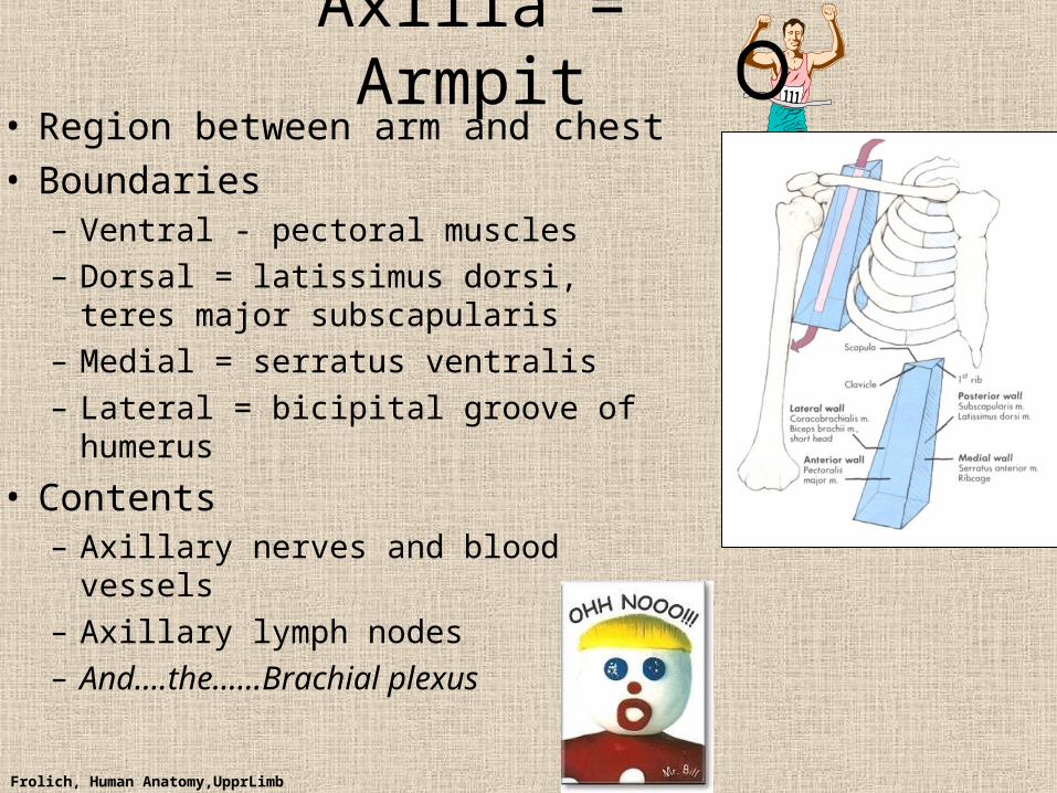
Axilla = Armpit• Region between arm and chest• Boundaries
– Ventral - pectoral muscles– Dorsal = latissimus dorsi, teres major
subscapularis– Medial = serratus ventralis– Lateral = bicipital groove of humerus
• Contents– Axillary nerves and blood vessels– Axillary lymph nodes– And….the……Brachial plexus

Frolich, Human Anatomy,UpprLimb

Frolich, Human Anatomy,UpprLimb
• Posterior Compartment—posterior cord• Anterior compartment—medial, lateral cords• Name of cord is relative to axillary artery
Brachial Plexus
Frolich, Human Anatomy,UpprLimb
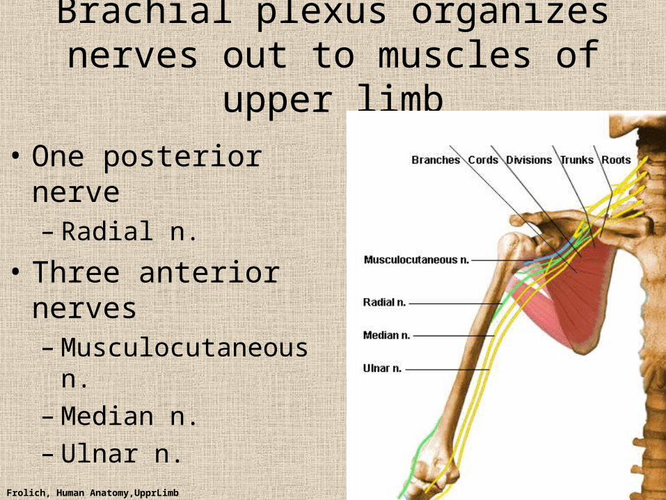
Brachial plexus organizes nerves out to muscles of upper limb
• One posterior nerve– Radial n.
• Three anterior nerves– Musculocutaneous n.– Median n.– Ulnar n.

Frolich, Human Anatomy,UpprLimb
Muscles and nerves by compartment
ANTERIOR POSTERIOR
NERVES M-C, ulnar, median
Radial
MOVEMENT Flexion Extension
MUSCLES Biceps, flexors
Triceps, extensors
TWIST Flexors from medial epicondyle
Extensors from lateral epicondyle

Frolich, Human Anatomy,UpprLimb
POSTERIOR AND ANTERIOR COMPARTMENTS

Frolich, Human Anatomy,UpprLimb
• Biceps—anterior compartment, flexion (M-C n.)• Triceps—posterior compartment, extension (radial n.)

Frolich, Human Anatomy,UpprLimb
Anterior Compartment Forearm--flexors
Flexor Carpi Radialis
Flexor Retinaculum
Medial Epicondyle
Flexor Digitorum Superficialis is deep to other flexors
Flexor Carpi Ulnaris
BrachioradialisPronator Teres
Anterior View
Frolich, Human Anatomy,UpprLimb
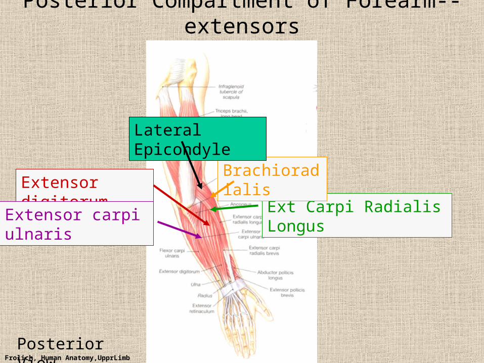
Posterior Compartment of Forearm--extensors
Extensor digitorum
Extensor carpi ulnaris Ext Carpi Radialis Longus
Brachioradialis
Lateral Epicondyle
Posterior View

Frolich, Human Anatomy,UpprLimb
ANTERIOR MUSCLES• M-C nerve
– Biceps– brachialis
• Median nerve– Forearm flexors– Thumb intrinsics (1M$
nerve)
• Ulnar nerve– Flexor carpi ulnaris– Hand intrinsics
POSTERIOR MUSCLES
• Muscles (radial nerve)– Triceps– Anconeus– Brachioradialis– Carpal, digit extensors

Frolich, Human Anatomy,UpprLimb
Sensory from limb (dermatomes/sensory skin segments from spine)
• Dermatomes extend over limbs
• Twisted orientation reflects twisting of limb during development
• Named nerves generally innervate skin over muscles that they innervate

Frolich, Human Anatomy,UpprLimb
Sensory territory of nerves
REMEMBER: Brachial plexus re-directs spinal routes into named nerves covering certain territory
Cutaneous branches of medial cord/ulnar nerve

Frolich, Human Anatomy,UpprLimb
Routes of nerves (in human)
• M-C: between biceps brachii and brachialis• Median: medial/posterior to biceps, branches
into forearm flexors at elbow then to hand through carpal tunnel– Recurrent median (1M$) superficial at wrist to thumb
over thenar emminence) deficit - ape’s hand
• Ulnar: medial in arm, posterior to medial epicondle of humerus (funny bone) down medial forearm medial to carpal tunnel into palm
• Radial: deep posterior arm around lateral epicondyle of humerus to forearm (deep and superficial branches)

Frolich, Human Anatomy,UpprLimb
Ulnar Nerve
Brachial Artery
Median Nerve
Ulnar NerveMedian Nerve
Radial Artery
Musculocutaneous Nerve UlnarArtery
Where’s Radial Nerve?

Frolich, Human Anatomy,UpprLimb
Surface Anatomy of Upper Limb• Biceps + Triceps brachii• Olecranon Process• Medial Epicondyle• Cubital Fossa
– Anterior surface elbow– Contents
• Brachial Artery• Median Nerve
– Boundaries• Medial = Pronator teres• Lateral = Brachioradialis• Superior = Line between epicondyles

Frolich, Human Anatomy,UpprLimb
Surface Anatomy of Upper Limb
• Carpal Tunnel– Carpals concave
anteriorly– Carpal ligament covers it– Contains: long tendons,
Median nerve– Inflammation of tendons =
compression of Median nerve
• Anatomical Snuffbox– Lateral = E.pollicis brevis– Medial = E. pollicis longus– Floor = scaphoid, styloid
of radius– Contains Radial Artery
(pulse)

Frolich, Human Anatomy,UpprLimb
Suggestion: a muscle table organized by
Joint crossed?Nerve innervating?Action?Compartments?All of the above?
MUSCLE ACTION ORIGIN INSERTION INNERVATI ON(cord to nerve)
Biceps Flex, sup. Humerus,glenoid
Radialtuberosity
Medial cord—M-C.

Frolich, Human Anatomy,UpprLimb