fret-based sensor for camkii activity (fresca): a useful tool for … · 2019-06-14 · post...
TRANSCRIPT

1
FRET-based sensor for CaMKII activity (FRESCA): A useful tool for assessing CaMKII activity in response to Ca2+ oscillations in live cells
Goli Ardestani1,2,5 #, Megan C. West3,5 #, Thomas J. Maresca4,5, Rafael A. Fissore1,5, Margaret M. Stratton3,5* From the Department of Veterinary and Animal Sciences1, Veterinary and Animal Sciences Graduate Program2, Department of Biochemistry and Molecular Biology3, Department of Biology4, University of Massachusetts, Amherst, United States5
Running title: CaMKII activity during fertilization
#These authors contributed equally *To whom correspondence should be addressed: Margaret M. Stratton: Department of Biochemistry & Molecular Biology, University of Massachusetts Amherst, Amherst MA 01003; [email protected]; Tel. (413) 545-0631. Keywords: Ca2+-calmodulin-dependent protein kinase II (CaMKII), calcium imaging, biosensor, fertilization, phosphorylation, Camui, reporter, FRESCA (FRET-based Sensor for CaMKII Activity), fertility ______________________________________________________________________________ ABSTRACT Ca2+ oscillations and consequent Ca2+/calmodulin-dependent protein kinase II (CaMKII) activation are required for embryogenesis, as well as neuronal, immunological and cardiac signaling. Fertilization directly results in Ca2+ oscillations, but the resultant pattern of CaMKII activity remains largely unclear. To address this gap, we first employed the one existing biosensor for CaMKII activation. This sensor, Camui, comprises CaMKIIα and therefore solely reports on the activation of this CaMKII variant. Additionally, to detect the activity of all endogenous CaMKII variants simultaneously, we constructed a substrate-based sensor for CaMKII activity, FRESCA (FRET-based Sensor for CaMKII Activity). To examine the differential responses of the Camui and FRESCA sensors, we used several approaches to stimulate Ca2+
release in mouse eggs, including addition of phospholipaseC𝜁 cRNA, which mimics natural fertilization. We found that the Camui response is delayed or terminates earlier than the FRESCA response. FRESCA enables assessment of endogenous CaMKII activity in
real-time by both fertilization and artificial reagents, such as Sr2+, which also leads to CaMKII activation. FRESCA’s broad utility will be important for optimizing artificial CaMKII activation for clinical use to manage infertility. Moreover, FRESCA provides a new view on CaMKII activity, and its application in additional biological systems may reveal new signaling paradigms in eggs, as well as in neurons, cardiomyocytes, immune cells, and other CaMKII-expressing cells. _____________________________________ Calcium is a crucial ubiquitous second messenger in the cell. All electrically coupled cells, such as neurons and cardiomyocytes, and even cells that are not – such as lymphocytes and oocytes/eggs, communicate or are induced to differentiate following intracellular Ca2+ changes caused by Ca2+ release and/or Ca2+ influx through channels whose combined output can result in a single Ca2+ rise or in more complex responses such as oscillations (1-4). Absence of Ca2+ signals lead to severe defects in cell functionality, such as memory deficits in the case of neurons (5), or in the case of eggs, failure of
http://www.jbc.org/cgi/doi/10.1074/jbc.RA119.009235The latest version is at JBC Papers in Press. Published on June 14, 2019 as Manuscript RA119.009235
by guest on April 19, 2020
http://ww
w.jbc.org/
Dow
nloaded from

2
fertilization and initiation of embryo development (6,7).
In oocytes, and other cell types, Ca2+-calmodulin dependent protein kinase II (CaMKII) is responsible for reacting to Ca2+
increases and transducing this signal to downstream molecules. Indeed, it has been shown that neuronal CaMKII has a threshold frequency for activation (8,9). CaMKII has a unique oligomeric structure among the protein kinase family (Fig. 1A). Each subunit of CaMKII is comprised of a kinase domain, a regulatory segment, a variable linker region, and a hub domain (Fig. 1B). The hub domain is responsible for oligomerization, which organizes it into two stacked hexameric (or heptameric) rings to form a dodecameric (or tetradecameric) holoenzyme (8,10,11). In the absence of Ca2+, the regulatory segment binds to and blocks the substrate-binding pocket. Ca2+-calmodulin (Ca2+/CaM) turns CaMKII on by competitively binding the regulatory segment and exposing the substrate-binding pocket (Fig. 1C). Unlike other Ca2+/CaM-sensitive kinases, CaMKII acquires activity that is Ca2+ independent (autonomy) with sustained stimulation by autophosphorylation at Thr 286 (we will use CaMKIIα numbering throughout the manuscript) (Fig. 1C) (12,13). In other words, as long as Thr 286 is phosphorylated, CaMKII will retain activity, even in the absence of Ca2+. It is this property, combined with its oligomeric organization, which provides the sensitivity of CaMKII to specific frequencies of Ca2+ rises.
CaMKII is essential for oocyte activation and initiation of embryo development (Fig. 1D). All vertebrate oocytes are arrested at the time of fertilization at the metaphase (M) stage of meiosis II (MII); henceforth referred to as eggs. This arrest is underpinned by a complex regulation of the activity of the maturation-promoting factor (MPF), which is maintained high during the arrest (14,15). MPF inactivation is required to complete meiosis and initiate the mitotic cycles of early embryogenesis. A prolonged Ca2+ signal – in the form of multiple oscillations – is initiated by the sperm and is responsible for inducing the release of the meiotic arrest at fertilization in all mammals, as preventing their generation
results in failure of MII exit (16). Importantly, Ducibella et. al showed that these Ca2+ oscillations are not redundant, rather, several Ca2+ rises are required for the early signaling events essential for embryogenesis (17). Indeed, it has been hypothesized that these early Ca2+ signals also play a role in long term signaling in the developing embryo (17,18).
It was first shown in Xenopus eggs that the Ca2+ rise induced by the sperm resulted in CaMKII activation, which is required for the initiation of embryogenesis (19). In mammals, this was demonstrated using genetic models where CaMKII was knocked-out and/or downregulated, which resulted in sterile females despite the ability of their eggs to initiate normal Ca2+ oscillations. These studies confirmed the requirement of CaMKII for the initiation of mammalian embryo development (20,21). Despite these findings, the complete profile of CaMKII activity during fertilization in mammals is not known. Using in vitro kinase assays and egg lysates, an elegant series of papers showed that these early Ca2+ rises induced CaMKII activity corresponding with discrete Ca2+ rises during the first hour post fertilization (17,18,22,23). Nevertheless, Ca2+ rises associated with fertilization in mammalian species occur every ~20 min and last longer than 3 hours, and the changes in CaMKII activity associated with all oscillations have not been determined.
In mammalian eggs, the Ca2+ oscillations induced by fertilization that activate CaMKII and promote egg activation occur with characteristic amplitude and frequency (24,25). Interestingly, it has been shown that CaMKII has a threshold frequency for activation (8,9). There are four human CaMKII genes; CaMKIIα and β are predominantly found in the brain, CaMKIIδ is found in the heart and CaMKIIγ is found in multiple organ systems, including the reproductive organs. The kinase and hub domains of all four genes are highly conserved (~95% and 80% identity, respectively), however, the linker connecting the kinase and hub domains is highly variable in length and composition (see Fig. 1B). Details elucidating the importance of the variable linker region remain to be uncovered, but there are several
by guest on April 19, 2020
http://ww
w.jbc.org/
Dow
nloaded from
3
splice variants of each of the four genes, which mostly vary in the linker region. It has been shown that CaMKII activity is tuned by the length of the variable linker (8). Specifically, as the variable linker is lengthened, less Ca2+ is needed for activation (i.e., activation of CaMKII is easier). Mouse eggs express equimolar concentrations of the two versions of CaMKIIγ (γ3 and γJ, Fig.1E) (26). The underlying regulation and contributions of these isoforms in mammalian eggs has not been investigated.
To date, CaMKII activity has only been assessed based on a few Ca2+ rises using in vitro kinase assays and during only the first hour of oscillations, which is considerably shorter than the time scale for normal oscillations in the mouse. Therefore, there is a need to monitor CaMKII activity in live cells and for an extended time, which is what we address here. RESULTS Measuring CaMKII activity in real-time in mouse eggs
Herein, we show CaMKII activity monitored in real time following the induction of Ca2+ responses using several agonists that are capable of initiating embryogenesis. To examine the real-time changes, we expressed the Camui reporter (see Fig. 2A) in mouse eggs. Camui is a Förster resonance energy transfer (FRET)-based biosensor for CaMKII activity, which exploits the conformational change that CaMKII undergoes when it binds to Ca2+/CaM (27) (Fig. 2A). Camui is currently the only biosensor for CaMKIIα activity. Camui expression was robust about ~30 min after cRNA injection, and monitoring was performed 3 hours post injection to attain reasonably stable Camui levels. We first examined the distribution of Camui using confocal microscopy and observed widespread cytoplasmic expression (Fig. 2B). Camui monitoring of ionomycin-induced Ca2+ rises
Given the immediate and large Ca2+ rise caused by the addition of ionomycin, we first tested Camui responses in eggs using this
ionophore. We analyzed the effect of 3 concentrations of ionomycin: 0.5 µM, 2.5 µM and 5 µM. Upon addition of ionomycin to eggs expressing Camui, we observed a decrease in ratiometric FRET (YFP/CFP), indicating CaMKII activation (Fig. 2C-E). As expected with a decrease in FRET, we observed a corresponding increase in CFP fluorescence and decrease in YFP fluorescence (Fig. S3B).
It is clear that CaMKII activity increases (red line) coincident with the increase in Ca2+ (black line) in all conditions. The Ca2+ and Camui responses increased dose-dependently and approximately synchronously, as the large increase in the amount of Ca2+ release caused by increasing ionomycin from 0.5 µM to 2.5 µM, results in a 1.9-fold increase in CaMKII activity (mean amplitude of FRET change) (Fig. 2F). Further increasing ionomycin from 2.5 µM to 5 µM produces nearly no change in total Ca2+ release, although it is very likely that the reporting range of Rhod-2 is saturated at these levels of intracellular Ca2+. The CaMKII activity also appears to remain constant, although this may also represent saturation of the FRET signal (Fig. 2F). Notably, addition of 5 µM ionomycin results in a faster and prolonged duration of activity compared to lower concentrations (Fig. 2G), but it is unclear whether this reflects the extended activation of the enzyme or cellular stress. Camui monitoring of Sr2+ induced oscillations
We next examined the Camui response to Sr2+-induced oscillations (Fig. 3). Addition of 10 mM Sr2+ to the extracellular media in place of external Ca2+ is a common method of parthenogenetic activation in mouse eggs. Also, 10 mM Sr2+ induces highly consistent oscillations in these cells (Fig. 3, black lines); these oscillations initiate all events of egg activation (28-30). The TRPV3 channel has recently been identified as the channel responsible for Sr2+ influx in mouse eggs (30). These oscillations, as reported by Rhod-2 in the following experiments, are most likely to represent a combination of both Ca2+ and Sr2+
by guest on April 19, 2020
http://ww
w.jbc.org/
Dow
nloaded from

4
release, with a progressively greater release of Sr2+ as these measurements were performed in the absence of extracellular Ca2+. Camui reported FRET changes following the initiation of oscillations by Sr2+ (Fig. 3, red lines). Remarkably, the FRET changes were delayed, despite the presence of robust changes in intracellular Ca2+/Sr2+ levels. Roughly 82% of eggs (14/17) did not report significant CaMKII activity until the ~4th Ca2+/Sr2+ rise, where some initial activity is seen at the 3rd rise (Fig. 3B, arrow and inset). In a few eggs (2/10), we observed longer delays, until the 5th rise (Fig. S1).
Another distinctive feature of the Camui response caused by Sr2+ oscillations is that although the initial Camui responses were delayed, once they commenced, they displayed an integrated activation with each subsequent pulse during the first few pulses. We analyzed the mean amplitude for the first three observable FRET changes. From the first to the second FRET change, there was a 1.6-fold increase in CaMKII activity. From the second to the third FRET change, there was a negligible change, and these changes occurred while the amplitude of the Ca2+ peaks progressively decreased and/or remained unchanged (Fig. 3C-E). These data indicate that CaMKII activity, once stimulated by Ca2+/Sr2+ rises, is cooperative in response to the initial rises until saturation is achieved, which is around the 3rd FRET response. This result is consistent with previous data showing that CaMKII activity is highly cooperative in vitro (8,31). As depicted in Figure 3A, a potential explanation for this is phosphorylation at Thr286, which may persist even in the absence of elevated Ca2+. It has been clearly shown that CaMKII with Thr286 phosphorylated has a significantly higher affinity for Ca2+/CaM (32). This would also explain why individual FRET responses outlasts individual Ca2+ rises and do not return to baseline simultaneously (Fig. 3B, Fig. S1, blue horizontal lines). Considerations for endogenous CaMKII in eggs
To date, Camui has been a useful tool to study and understand CaMKII activity in
various cell types and under various conditions. Despite the many insights gained by the widespread use of Camui, it is an over-expressed protein reporter construct and it does not necessarily faithfully report the activation state of endogenous CaMKII in any cell. Camui had not been used to monitor CaMKII activity in mouse eggs until this study. Since the Camui sensor is constructed of a CaMKII variant itself, it will report on this particular variant, in this case is CaMKIIα with a 30-residue linker region, which is not expressed in mammalian eggs (see Fig. 1E). Given that it has been demonstrated that CaMKII activity is tuned by the length of the variable linker (8,33), specifically, as the variable linker is lengthened, less Ca2+ is needed for activation, we hypothesized that Camui may not be reporting faithfully on the endogenous CaMKII in eggs. This assumption is based on the knowledge that mouse eggs express, as mentioned, equimolar concentrations of the two versions of CaMKIIγ (γ3 and γJ), which have 69 and 90 residue variable linkers, respectively (Fig. 1E) (34), considerably longer than the 30-residue linker of CaMKIIα. We suspected this might be the case because of the lack of FRET changes following the initial large changes in Rhod-2 fluorescence induced by Sr2+ oscillations. This possible limitation of the Camui reporter led us to seek additional methods to detect these physiological changes. One option is to re-engineer Camui with the appropriate CaMKII isoform to be studied, however this becomes cumbersome since there are multiple isoforms expressed in a single cell type. To circumvent this problem, we report here the development of a novel biosensor that detects endogenous CaMKII activity in mouse eggs. Development of a novel biosensor for endogenous CaMKII activity
We developed a novel substrate-based sensor for CaMKII activity, FRESCA (FRET based Sensor for CaMKII Activity, Fig. 4A) and monitored CaMKII activity using FRESCA in real-time. We adapted the FRESCA design from an original design for a protein kinase C (PKC) biosensor (CKAR)
by guest on April 19, 2020
http://ww
w.jbc.org/
Dow
nloaded from

5
from Newton and colleagues (35), which has also been adapted to make an Aurora kinase biosensor (36). The premise for this design is to engineer in a conformational change upon phosphorylation of the substrate. Cleverly, this was done by fusing the kinase substrate to a phosphate binding protein (FHA2) (35). FHA2 will bind to this phosphorylated Thr residue and produce a decrease in FRET between the terminal CFP/YFP pair (which in this case is Turquoise and Venus, termed CFP/YFP throughout for simplicity). We cloned in a CaMKII-specific substrate (syntide-2) (37), with a few modifications to provide a better substrate for FHA2 (see Methods section for sequence details). We refer to this peptide as syntide-FRESCA throughout for clarity. Measuring the FRESCA response in HEK293T cells
We first tested FRESCA in HEK293T cells, which express negligible levels of CaMKII. We transfected HEK293T cells with either (i) CaMKII, calmodulin and FRESCA, or (ii) calmodulin and FRESCA. Ionomycin was added to the HEK293T cells to induce Ca2+ release and simultaneously monitored FRET (YFP/CFP). We observed that with CaMKII transfected, the addition of ionomycin causes a reduction in FRET, indicating that CaMKII is active and phosphorylating FRESCA (Fig. S2, red lines). Importantly, we did not observe a FRET change when CaMKII was not co-transfected, demonstrating that FRESCA is selective for the transfected CaMKII and is not being phosphorylated by any intrinsic HEK cell kinases under these conditions (Fig. S2, blue lines). FRESCA monitoring of ionomycin-induced Ca2+ release in mouse eggs
We expressed FRESCA in mouse eggs to measure endogenous CaMKIIγ activity. FRESCA expression in MII eggs resulted in uniform distribution throughout the cytoplasm (Fig. 4B). Upon addition of ionomycin to eggs expressing FRESCA, we observed a FRET decrease indicating CaMKII activity (Fig. 4C-E). The amplitude change is 10-fold less than
what is observed for Camui (0.016 for FRESCA compared to 0.16 for Camui), however, this measurable signal change is sufficient for us to monitor endogenous CaMKIIγ activity in comparison to the CaMKIIα of Camui. It is worth noting that the shape of the FRESCA trace is slightly different from that of Camui for the same stimulus. At the lowest ionomycin concentration (0.5 µM), CaMKII activity appears to perfectly track the Ca2+ rise (Fig. 4C). Conversely, at higher ionomycin concentrations, CaMKII activity is unstable during the duration of the Ca2+ rise, although higher concentrations appear to prolong and increase the FRET response of FRESCA (Fig. 4F). The time to FRET peak was faster with addition of higher ionomycin concentrations (Fig. 4G).
To demonstrate that the changes in FRESCA fluorescence reflect FRET, we plotted the traces of CFP and YFP fluorescence after the addition of ionomycin independently. As expected, the values changed simultaneously but in opposite directions (Fig. S3A). We also expressed YFP cRNA alone and monitored fluorescence changes following stimulation with Sr2+ oscillations. Consistent with the evidence that the changes in FRESCA are the result of FRET, the oscillations did not induce changes in YFP fluorescence corresponding with each Ca2+/Sr2+ elevation (Fig.S4). Specificity of FRESCA in mouse eggs
We sought to show that FRESCA is reporting specifically on CaMKII in eggs as opposed to other CaM kinases or Ca2+ sensitive kinases. A previous study using real-time PCR revealed that transcript levels of CaMKIV were ~1% compared to those of CaMKIIγ the predominant isoform in mouse eggs (38). Additionally, even when overexpressed, CaMKIV was insufficient to lead to egg activation. In this same study, CaMKI levels were undetectable, and CaMKK seems to also be absent in mouse eggs. There has also been a comprehensive mass spectrometry study of total proteins expressed in GV oocytes and MII eggs (39). Similar to the mRNA sequencing, they detected no
by guest on April 19, 2020
http://ww
w.jbc.org/
Dow
nloaded from

6
CaMKI and very low levels of CaMKIV. There is also no evidence for CaMKK or PKA detected in MII eggs in this study, which is indicative of low expression levels in these cells. Finally, it has not been shown that Akt or PKA undergo immediate activation following increases in intracellular Ca2+ rises.
We therefore focused our efforts on CaMKII and PKC, which is also a Ca2+ sensitive kinase in eggs (38,39). To this end, we compared the rates of phosphorylation of FRESCA-syntide and syntide-2 by CaMKII and PKC in vitro (Fig. S5). Consistent with existing data (37), we observed that CaMKII phosphorylates syntide-2 at a 3-fold higher rate compared to PKC at high substrate concentration (0.3 mM). We next tested our new syntide variant, FRESCA-syntide. Importantly, we observe a large increase in specificity with CaMKII phosphorylating FRESCA-syntide at a 44-fold higher rate compared to PKC.
We next performed a series of experiments testing CaMKII and PKC inhibitors, and a PKC activator. CaMKII inhibitors should eliminate the FRET response if CaMKII is the only kinase phosphorylating FRESCA in eggs. We show that the addition AS105, a CaMKII specific ATP competitive inhibitor, significantly reduced FRET, while its inactive analog (AS461) did not affect FRET (Fig. 5A) (40). It is important to note that we also tested a common CaMKII inhibitor, KN93, which significantly reduced the amount of Ca2+ released upon addition of ionomycin (41) (data not shown). In addition to the recent study that showed KN93 binds Ca2+/CaM (42), we chose to not pursue this inhibitor further.
We next tested the PKC inhibitor GO6983 (43), which did not affect the FRESCA signal, indicating that PKC is not phosphorylating FRESCA, which is consistent with our in vitro kinetic data (Fig. 5B, Fig. S5). Eggs do not express conventional PKC isoforms, which is why we chose to use the broad-spectrum PKC inhibitor (GO6983). To ensure that PKC in our system is responding these agents, we measured the PKC response directly using the CKAR biosensor for PKC activity (35). The phorbol ester, PMA, has been shown to
activate PKC in the absence of a Ca2+ stimulus (44), which we also observed (Fig. 5C, E). The CaMKII inhibitor AS105 does not affect the CKAR response to PMA at all (Fig. 5C), but GO6983 completely abolishes the CKAR response (Fig. 5D). Compared side-by-side, PMA strongly and persistently activates CKAR but not FRESCA (Fig. 5E). We note a transient rise in Ca2+ upon PMA addition, during which we also see a corresponding transient FRET response in both CKAR and FRESCA. The FRESCA FRET response immediately returns to baseline, whereas the CKAR FRET change continues to decrease, indicating persistent activation. There is previous evidence showing changes in Ca2+ upon addition of phorbol ester compounds, which is what we attribute this to (45).
Finally, we closely compared the response dynamics of Camui, FRESCA and CKAR in response to ionomycin addition (Fig. S6). It is clear that both Camui and FRESCA show the same response pattern, where the FRET response essentially mirrors the Ca2+ rise. When this is directly compared to CKAR, it is clear that CKAR has a delayed response, and maximal activity is not coincident with maximum Ca2+. This observation is in line with previous studies using CKAR to monitor PKC activity in eggs (46). Taken all together, we show that FRESCA is specifically reporting on endogenous CaMKII activity in the egg. FRESCA monitoring of Sr2+-induced Ca2+ oscillations
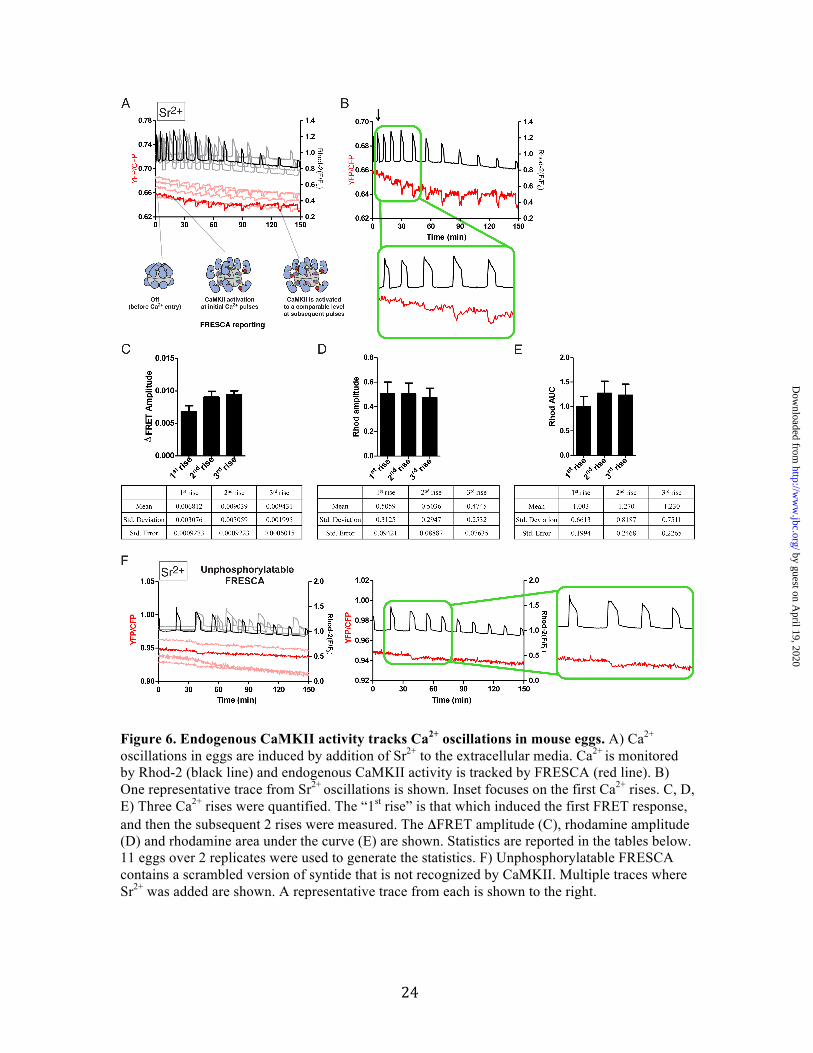
Next, we examined the response of endogenous CaMKII to Sr2+-induced oscillations (Fig. 6). We observed endogenous CaMKIIγ activity (monitored by FRESCA) almost simultaneously with the initiation of oscillations. Indeed, over 70% of eggs (8/11) showed CaMKII activity with the first Ca2+ rise. The other 3 eggs responded at the second Ca2+ rise. Importantly, CaMKII activity is prolonged over time, as FRESCA continues to track each Ca2+ rise for >2 hours (Fig. 6A, B). In contrast, only 18% of the eggs (3/17) expressing Camui showed activity during the first Ca2+ rise.
by guest on April 19, 2020
http://ww
w.jbc.org/
Dow
nloaded from
7
We tested the effect of the PKC inhibitor GO6983 on both FRESCA and CKAR in response to Sr2+-induced oscillations. In this experiment, we initiated oscillations by adding Sr2+ to the media of eggs expressing either FRESCA or CKAR, and then added GO6983 after approximately 90 minutes (Fig. S7). After GO6983 addition, the FRESCA response continues unabated in 100% of the eggs tested whereas the CKAR response is attenuated in 60% of the eggs tested. This indicates that FRESCA is indeed reporting CaMKII activity in response to Sr2+-induced oscillations.
We propose a molecular model to describe this data. Over the course of the first few Ca2+/Sr2+ oscillations, the amplitude of the Rhod-2 signal is constant whereas the FRESCA response and the Rhod-2 AUC increases from the first to the second rise (Fig. 6C-E). Further, once FRESCA signal peaks, it is sustained throughout the remainder of the data collection. This may suggest autophosphorylation of CaMKII at Thr 286, which facilitates activation at subsequent Ca2+ rises by increasing the affinity for Ca2+/CaM (see cartoons in Fig. 6A) (32). Additionally, a prolonged time course of FRESCA response to Sr2+ indicates that FRESCA continues to faithfully track endogenous CaMKII up to 6 hours (Fig. S8).
We generated a scrambled version of syntide and inserted this into the FRESCA construct to generate an unphosphorylatable variant. We expressed this sensor in eggs and added Sr2+ to initiate Ca2+ oscillations. The unphosphorylatable FRESCA did not respond to this stimulus, indicating that the signal change we observe using FRESCA is specific to a phosphorylation event (Fig. 6F). Measuring CaMKII activity under native fertilization conditions
In mammals, fertilization-associated Ca2+ oscillations are induced by the release of sperm’s phospholipase C 𝜁1 (PLC𝜁) into the ooplasm (47). We tested the response of both FRESCA and Camui in response to the expression of PLC𝜁.
Camui monitoring of PLC𝜁-induced Ca2+ oscillations
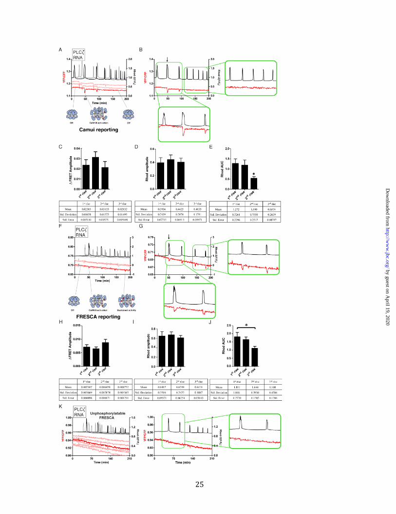
We assessed how Camui would report CaMKII activity induced by native Ca2+ oscillations and compared the response to those induced by Sr2+. To do this, eggs expressing Camui were injected with PLC𝜁 cRNA and Ca2+ and FRET responses were monitored (Fig. 7A, B). We observed that Ca2+ oscillations nearly immediately induced CaMKII activity as monitored by Camui (Fig. 7B, arrow and bottom inset). However, this initial activity abruptly ceased and was not detected in subsequent rises. Only the first and second (and to a less extent, third) Ca2+ rises induced Camui responses despite the presence of robust and frequent Ca2+ oscillations (Fig. 7A). 75% of eggs (9/12) expressing Camui responded to the first or second Ca2+ rise. Additionally, despite the fact that the amplitude of the first Ca2+ rises remains relatively steady, the area under the curve showed a marked decrease after the second Ca2+ rise in these experiments (see Fig. 7E, J). It is worth noting that the abundant expression of Camui may be contributing significantly to the existing CaMKII in the egg, and potentially altering Ca2+/CaM dynamics. These results raised the possibility that Camui is not well suited to detect CaMKII activity initiated by sporadic and low magnitude Ca2+ rises, which are characteristic of mammalian fertilization. Regardless, it remains to be elucidated why Sr2+ induced oscillations are able to protractedly promote robust and persistent Camui responses whereas the Camui response to PLC𝜁-induced oscillations fades rapidly. FRESCA monitoring of PLC𝜁-induced Ca2+ oscillations
We injected PLC𝜁 cRNA into FRESCA expressing eggs, and thereafter began monitoring changes in FRESCA responses (Fig. 7F, G). The initiation of oscillations stimulated the early activity of the endogenous CaMKIIγ, and this activity was detected with each additional rise. 100% of eggs (17/17) expressing FRESCA responded to the first or second Ca2+ rise. Similar to Sr2+ induced oscillations, we observed a relative decrease in
by guest on April 19, 2020
http://ww
w.jbc.org/
Dow
nloaded from

8
the amplitude of the Ca2+ rises over time, yet the FRESCA response was largely maintained (Fig. 7G). These observations are the longest evaluation of CaMKII activity reported following fertilization-like oscillations, as previous studies only reported up to 60 min post-initiation of oscillations (18,23). These results also suggest that all Ca2+ rises induced by fertilization trigger activation of CaMKII (Fig. 7 H-J).
We tested the unphosphorylatable FRESCA variant in eggs injected with PLC𝜁 cRNA. The unphosphorylatable FRESCA did not respond to this stimulus, indicating that the signal change we observe using FRESCA is specific to a phosphorylation event (Fig. 7K).
Finally, we compared the response of CKAR and FRESCA to PLC𝜁 cRNA induced oscillations. Side-by-side comparisons reveal a delayed FRET changes for CKAR compared to FRESCA (Fig. S9). These results are consistent with our observations following the addition of ionomycin (Fig. S6). DISCUSSION
It has been appreciated for decades that both Ca2+ oscillations and CaMKII activation in mouse eggs are crucial to fertilization and initiation of embryo development. Here, we provide an analysis of CaMKII activation in real-time in eggs using FRET-based CaMKII biosensors. Importantly, our new biosensor, FRESCA, allowed us to monitor endogenous CaMKII (CaMKIIγ3 and γJ) activity in real-time as a consequence of different activation stimuli (ionomycin, Sr2+, and PLC𝜁). The FRESCA response was noticeably different from Camui, which reports on the conformational change in the one CaMKII variant expressed in the sensor (in this paper and others, this variant is CaMKIIα). An additional advantage of FRESCA is that it does not alter the intracellular kinase concentrations, as already noted. When different Ca2+ oscillation patterns are induced, we observed subsequent differences in CaMKII activation in both sensors. From our data, it is clear that the (i) agent of Ca2+ oscillations as well as the (ii) specific CaMKII isoform responding, both play a role in CaMKII activation.
The pivotal role of CaMKII activation in causing release of the meiotic arrest and initiation of embryonic development in all species was recently and more specifically evidenced by careful mass spectrometry experiments (48). This study showed that soon after fertilization, and temporally coinciding with the Ca2+ wave, there is a strong increase in protein phosphorylation that far outweighs the biochemical changes caused by protein degradation that accompanies fertilization. Remarkably, the study also found that 25% of the phosphorylated sites matched the minimal phosphorylation motif of CaMKII. It is therefore important to determine how Ca2+ rises turn on CaMKII activity, and what parameter(s) of individual rises within an oscillatory pattern are necessary for periodic and consistent stimulation of its activity. We propose that the magnitude of the initial activation of CaMKII depends on the magnitude of the Ca2+ stimulus and on internal regulation of CaMKII. Knowing the minimal Ca2+ signal that increases the activity of CaMKIIγ is important, as we seek to develop more physiological methods of parthenogenetic activation to treat some cases of infertility.
Our data suggest that the endogenous CaMKIIγ in eggs is potentially more sensitive to Ca2+/CaM than CaMKIIα. This finding is in line with previous data showing that longer linker CaMKII splice variants (CaMKIIγ3 and CaMKIIγJ) are activated at lower concentrations of Ca2+/CaM than shorter linker variants (CaMKIIα) (see Fig. 1E) (8). Additionally, the delayed response seen in the Camui expressing eggs in response to Sr2+ stimulation could also be a result of endogenous CaMKII being activated first (lower EC50 for Ca2+/CaM) thereby competing with Camui for the available activating ligand.
Our study also represents the first characterization of the activation of CaMKII by a common parthenogenetic agonist in the mouse, such as Sr2+. While the FRESCA responses caused by Sr2+ and expression of PLC𝜁 were similar in timing and persistence, the responses caused by these agonists following the expression of Camui were very different. Sr2+ oscillations induced
by guest on April 19, 2020
http://ww
w.jbc.org/
Dow
nloaded from

9
delayed activation of Camui, but once it was activated, each Ca2+/Sr2+ elevation resulted in successive Camui stimulation. Conversely, PLC𝜁-induced oscillations resulted in early activation of Camui followed by a rapid run down, such that after the second or third Ca2+ rise, and despite consistent Ca2+ oscillations, FRET changes in Camui were never again stimulated. These discrepancies likely have to do with the amplitude and/or duration of the Ca2+ rises induced by these two agonists. Learning the parameters of individual Ca2+ rises that are translated into endogenous CaMKII activity will be invaluable as we aim to design better parthenogenetic methods for application in IVF clinics.
Our results clearly show that the FRET changes reported by FRESCA significantly differ from that of CKAR, which reports on PKC activity. In addition to the effects of kinase specific inhibitors and activators, there is a striking difference in the timing of response dynamics to all agonists examined here. This is consistent with previous reports showing CKAR responses in mouse eggs (46). It is not currently understood why CKAR oscillates persistently with Ca2+ release, as the reported predominant PKC isoform present in eggs is not directly activated by Ca2+, and the Ca2+-induced changes in DAG are expected to be minor (49,50).
Addition of up to 10 µM ionomycin is a widely applied practice in IVF labs. Here, we showed that even a lower concentration range (0.5 – 5 µM ionomycin) results in substantially different activity profiles of CaMKII activation. With regard to the Camui experiments, in these FRET measurements we are observing direct activation of Camui (as opposed to endogenous CaMKII). It is clear that the extent of Camui activation is dependent on the amount of Camui present and the magnitude of the stimulus, as there is a significant increase in the amplitude of FRET change from 0.5 to 2.5 µM ionomycin. The FRESCA response (reporting on endogenous protein) at higher concentrations of ionomycin was more complex as it appears not to change significantly, which might reflect saturation of endogenous CaMKII or a negative feedback loop, as demonstrated by the transient nature
of the maximal peak in FRESCA fluorescence change. These results indicate an unexpected effect on CaMKII activity even by increasing the ionomycin by 5-fold (2.5 µM), which will be crucial to elucidate further for clinical application.
More broadly, now that we have demonstrated the utility of FRESCA in mouse eggs, this opens the door to measuring endogenous CaMKII activity in other cell types, such as neurons and cardiomyocytes. Here, we thoroughly addressed the specificity of FRESCA in eggs, but this would need to be done in other cell types as well. CaMKII activation has been heavily studied in vitro (8,31,51), and it is intriguing to also consider the potential effects of subunit exchange in cellular conditions (10,52). It will be necessary to increase the signal to noise ratio of the FRESCA sensor in order to achieve a more robust signal for accurate quantification of kinetics and amplitudes. This should be possible by adjusting the length and/or rigidity of the linker regions in the sensor. Once this is accomplished, we believe that FRESCA will provide new insights into CaMKII activity in cells and allow us to further unravel the complexity of this unique protein kinase. EXPERIMENTAL PROCEDURES Plasmid design
In order to accommodate the requirements for FHA2 binding (53), syntide-2 was modified from PLARTLSVAGLPGKK to PLARALTVAGLPGKK to create syntide-FRESCA. Syntide-FRESCA was generated by annealing GATCCGGCGGCGCCGGCGGCGGCccgctggcgcgcgccctgaccgtggcgggcctgccgggcaaaaaaGGC and GGCCGCCttttttgcccggcaggcccgccacggtcagggcgcgcgccagcggGCCGCCGCCGGCGCCGCCG (IDT), which produced BamHI site at the 5’ end and a NotI site on the 3’ end. This product was phosphorylated (Ambion Pnk), purified (Thermo Fisher) and then ligated using T4 DNA ligase (Invitrogen) into a plasmid dencoding the Aurora kinase FRET sensor (kind gift from Thomas Maresca). The final FRESCA sensor (with syntide-2 in place of
by guest on April 19, 2020
http://ww
w.jbc.org/
Dow
nloaded from

10
the Aurora substrate) was cloned into pCDNA3.1. The unphosphorylatable FRESCA contains a scrambled version of syntide (RKVAAPKGAGLLLPA). We generated the scrambled sequence using an online tool: Mimitopes (http://www.mimotopes.com/). This was cloned using the same technique as was used for cloning syntide-FRESCA.
Enzyme assays
Coupled-kinase assays were performed as previously described (31). Purified peptides (syntide-2 and syntide-FRESCA) were purchased from Lifetein (New Jersey). Purified PKC was purchased from Promega (Cat. V5261). CaMKIIγ was expressed in Rosetta 2(DE3)pLysS competent cells (Millipore) and purified as previously described (52). Fluorescence of NADH (ex. 340 nm/em. 460 nm) was monitored over time for 10 minutes using a Synergy H1 microplate reader (Biotek). For the experiments with syntide-2, the final enzyme concentration (both CaMKII and PKC) was 2.67 nM. For the experiments with syntide-FRESCA, the final enzyme concentration was 10 nM. CaMKIIγ activity was measured with the addition of 1 µM Ca2+/CaM (activated) or an equivalent volume of buffer (control). Total CaMKII activity was corrected by subtracting the background rate without Ca2+/CaM. PKC activity was measured with 1 mM Ca2+ and 140 µM / 3.8 µM phosphatidylserine / diacylglycerol membranes (activated) or an equivalent volume of buffer (the protocol from Dr. Alexandra Newton’s lab website was closely followed, modified from (54)). The lipid mixture was prepared as follows: chloroform solubilized lipids (Avanti) were mixed together at the appropriate ratio, dried under N2, speed-vacuumed for 1.5 hr, and resuspended in 20 mM Hepes pH 7.4 to make a 10X solution. The mixture was vortexed and sonicated in a water bath for 30s to fully resuspend. Rates were calculated as follows: first, the change in fluorescence over the time course was fit with a straight line (y = mx + c) to obtain a slope (m) proportional to the kinetic rate of the reaction. For each reaction, slopes were fit to a sliding window of 5 points (50 seconds) and the maximum observed slope
was used to represent the kinetic rate. Total PKC activity was corrected by subtracting the background rate of all kinase assay components except for PKC to account for the contribution from lipid scattering. This rate was roughly equivalent to the background rate of PKC without addition of Ca2+/lipid, but we only performed one subtraction. Total CaMKII activity was corrected by subtracting the background rate of all assay components except for Ca2+/CaM. HEK 293T Cell culture
All HEK293T cell cultures (kind gift from Dr. Daniel Hebert’s lab) were grown in Dulbecco’s Modified Eagle’s Medium (Sigma) supplemented with 10% fetal bovine serum (Sigma) and maintained at 37°C and 5% carbon dioxide levels. The identity of these cells was authenticated by ATCC using short tandem repeat analysis (CRL-3216 ATC 293T, Lot #63226319). These cells tested negative for mycoplasma. Cells were transfected using Lipofectamine® 2000 Reagent (Invitrogen) and 150 ng of DNA constructs. Collection of mouse eggs
Metaphase II (MII) eggs were collected from the oviducts of 6- to 10-week-old CD-1 female mice 12–14 h after administration of 5 IU of human chorionic gonadotropin (hCG), which was administered 46–48h after the injection of 5 IU of pregnant mare serum gonadotropin (PMSG; Sigma; Saint Louis, MO). Cumulus cells were removed with 0.1% bovine testes hyaluronidase (Sigma). MII eggs were placed in KSOM with amino acids (Millipore Sigma) under mineral oil at 37°C in a humidified atmosphere of 5% CO2 until the time of monitoring. All animal procedures were performed according to research animal protocols approved by the University of Massachusetts Institutional Animal Care and Use Committee. Preparation of cRNAs and Microinjections
The sequences encoding Camui and FRESCA were subcloned into a pcDNA6 vector (pcDNA6/Myc-His B; Invitrogen,
by guest on April 19, 2020
http://ww
w.jbc.org/
Dow
nloaded from
11
Carlsbad, CA) between the XhoI and PmeI restriction sites. Mouse PLC𝜁 (PLCzeta) was a kind gift from Dr. K. Fukami (Tokyo University of Pharmacy and Life Science, Japan) and subcloned into a PCS2+ vector, as previously described by us (Kurokawa et al., 2007). CKAR was ordered from Addgene (Plasmid #14860). Plasmids were linearized with a restriction enzyme downstream of the insert to be transcribed and cDNAs were in vitro transcribed using the T7 or SP6 mMESSAGE mMACHINE Kit (Ambion, Austin, TX) according to the promoter present in the construct. A Poly (A)-tail was added to the mRNAs using a Tailing Kit (Ambion) and poly(A)-tailed RNAs were eluted with RNAase-free water and stored in aliquots at -80 °C. Microinjections were performed as described previously (Lee et al., 2016). cRNAs were centrifuged, and the top 1–2 µl was used to prepare micro drops from which glass micropipettes were loaded by aspiration. cRNA (1 µg/µL) were delivered into eggs by pneumatic pressure (PLI-100 picoinjector, Harvard Apparatus, Cambridge, MA). Each egg received 5–10 pl, which is approximately 1–3% of the total volume of the egg. Injected MII eggs were allowed for translation up to 4h in KSOM. Group of eggs were injected with mouse PLC𝜁 cRNA after 4h of FRET construct injection. FRET and Calcium imaging
To estimate relative changes in the cytoplasmic activity of Camui, FRESCA and/or CKAR, emission ratio imaging of the YFP/CFP was performed using a CFP excitation filter, dichroic beam splitter, CFP and YFP emission filters (Chroma technology, Rockingham, VT; ET436/20X, 89007bs, ET480/40m and ET535/30m). To measure Camui and/or FRESCA activity and [Ca2+]i simultaneously, eggs that had been injected with Camui and/or FRESCA cRNAs were loaded ~ 4 hours post-injection with 1 µM Rhod-2AM supplemented with 0.02% pluronic acid for 20 minutes at RT. Eggs were then immobilized on glass-bottom dishes (MatTek Corp., Ashland, MA) by putting them in protein-free media (which causes the eggs to stick to the glass), and finally placed
on the stage of an inverted microscope. CFP, YFP and Rhod-2 intensities were collected every 20 second by a cooled Photometrics SenSys CCD camera (Roper Scientific, Tucson, AZ). The rotation of excitation and emission filter wheels was controlled using the MAC5000 filter wheel/shutter control box (Ludl) and NIS-elements software (Nikon). Imaging was performed on an inverted epifluorescence microscope (Nikon Eclipse TE 300, Analis Ghent, Belgium) using a 20x objective. For studies where ionomycin was used to induce Ca2+ responses, eggs were transferred into a 360 µl Ca2+-free TL-Hepes drop on a glass bottom dish, after which and following a brief monitoring period to determine baseline [Ca2+]i values, different concentrations of ionomycin were added and Ca2+ responses monitored. For Sr2+ studies, eggs were transferred into a nominally Ca2+- free TL-Hepes, containing 10 mM Sr2+. In cases where [Ca2+]i oscillations were induced by injection of PLC𝜁 cRNA, eggs were placed in TL-Hepes media containing 2 mM Ca2+
within 20 minutes of the injection of PLC𝜁 cRNA which occurred 4 hours post-injection of the FRET constructs (Camui or FRESCA). Pharmacological tests in mouse eggs
Mouse eggs were transferred to Ca2+ free TL-Hepes containing desired concentrations of pharmacological compounds 5 min prior to Ca2+ imaging. FRET (YFP/CFP) was monitored simultaneously with Ca2+ (rhodamine signal). First, we determined how much inhibitor could be added without affecting Ca2+ entry. Concentrations of inhibitors were chosen based on this information as well as what was used in previous studies (see text for references). All media was Ca2+ free. The following concentrations and number of replicates were used: ionomycin/FRESCA (0.5 µM, n=18), ionomycin/CKAR (0.5 µM, n=20), ionomycin/GO6983/FRESCA (0.5 µM/5 µM, n=14), ionomycin/GO6983/CKAR (0.5 µM/5 µM, n=15), PMA/CKAR (1 µM, n=16), PMA/FRESCA (1 µM, n=13), Sr2+/GO6983/FRESCA (10 mM/5 µM, n=9), Sr2+/GO6983/CKAR (10 mM/5 µM, n=7),
by guest on April 19, 2020
http://ww
w.jbc.org/
Dow
nloaded from

12
AS105 (5 µM, n=19), AS461 (5 µM, n=14). AS105 (kind gift from Allosteros Therapeutics, Inc.) has not been used in mouse eggs, so we adjusted the concentration to a level where the Ca2+ release was not affected. Eggs with the first 4 compounds added were stimulated with 0.5 µM ionomycin, while the eggs with AS compounds were stimulated with 2.5 µM ionomycin. Side by side controls were performed under the same conditions (0.5 µM vs. 2.5 µM ionomycin). Confocal imaging
Confocal images were acquired using a Nikon Apo 1.4 NA 60X oil immersion objective on a Nikon A1R confocal TiE microscope stand equipped with a LU-NV laser launch system and DU4 detector system housed in the IALS Nikon Center of Excellence microscopy facility a UMass Amherst. The Galvano scanner was used and the pinhole size was set to 17.88 µm. CFP was excited with the 445 nm laser at 9.2%, 540/30 emission filter, and PMT set to 75. YFP was excited with the 514 nm laser at 5.6%, 585/65 emission filter, and PMT set to 58. Z-stacks images were collected at 1024 x 1024 in 12 bits and the step size was 1.00 µm.
Data processing & statistical analyses
Graphs reporting FRET changes and Ca2+ responses were prepared using the values of the YFP (436x535)/CFP (436x480) ratios on the left axis. No correction was applied here; just the raw ratios are plotted. Rhod-2 values were calculated using the following formula (F)/F0 (actual value at x time/average baseline values for the first 2 minutes of monitoring) and the scale placed on the right axis. For calculation of time to FRET peak, analyses were performed in excel to identify the baseline and highest or lowest value for each peak (Rhod-2 or FRET, respectively). For calculation of area under the curve and all statistics, we used Prism Graphpad. Values from three or more replicates were performed on different batches of eggs with at least 5 eggs per condition per replicate. These are presented as means ± s.e.m and were analyzed by ANOVA. Differences were considered significant at P <0.05. Post-hoc analyses were done using a Tukey multiple comparison test (Prism Graphpad).
Acknowledgements: We thank Changli He for assistance on RNA purification. The microscopy data was gathered in the Light Microscopy Facility and Nikon Center of Excellence at the Institute for Applied Life Sciences, UMass Amherst with support from the Massachusetts Life Sciences Center. We thank Allosteros therapeutics for providing AS105, AS461 and Howard Schulman for helpful discussions. We thank members of the Stratton lab (Roman Sloutsky and Noelle Dziedzic) and Alejandro Heuck for assistance with reagents for the enzyme assays. We also thank Peter Chien, Eric Strieter, Scott Garman for discussions and John Kuriyan for helpful comments on the manuscript. Conflict of interest: The authors declare that they have no conflicts of interest with the contents of this article.
by guest on April 19, 2020
http://ww
w.jbc.org/
Dow
nloaded from

13
REFERENCES 1. Cuthbertson,K.S.,Whittingham,D.G.,andCobbold,P.H.(1981)FreeCa2+
increasesinexponentialphasesduringmouseoocyteactivation.Nature294,754-757
2. Swann,K.,andLai,F.A.(2013)PLCzetaandtheinitiationofCa(2+)oscillationsinfertilizingmammalianeggs.Cellcalcium53,55-62
3. Eisner,D.A.,Caldwell,J.L.,Kistamas,K.,andTrafford,A.W.(2017)CalciumandExcitation-ContractionCouplingintheHeart.Circulationresearch121,181-195
4. Rutecki,P.A.(1992)Neuronalexcitability:voltage-dependentcurrentsandsynaptictransmission.JClinNeurophysiol9,195-211
5. Herring,B.E.,andNicoll,R.A.(2016)Long-TermPotentiation:FromCaMKIItoAMPAReceptorTrafficking.Annualreviewofphysiology78,351-365
6. Escoffier,J.,Lee,H.C.,Yassine,S.,Zouari,R.,Martinez,G.,Karaouzene,T.,Coutton,C.,Kherraf,Z.E.,Halouani,L.,Triki,C.,Nef,S.,Thierry-Mieg,N.,Savinov,S.N.,Fissore,R.,Ray,P.F.,andArnoult,C.(2016)HomozygousmutationofPLCZ1leadstodefectivehumanoocyteactivationandinfertilitythatisnotrescuedbytheWW-bindingproteinPAWP.HumMolGenet25,878-891
7. Yoon,S.Y.,Jellerette,T.,Salicioni,A.M.,Lee,H.C.,Yoo,M.S.,Coward,K.,Parrington,J.,Grow,D.,Cibelli,J.B.,Visconti,P.E.,Mager,J.,andFissore,R.A.(2008)HumanspermdevoidofPLC,zeta1failtoinduceCa(2+)releaseandareunabletoinitiatethefirststepofembryodevelopment.TheJournalofclinicalinvestigation118,3671-3681
8. Chao,L.H.,Stratton,M.M.,Lee,I.H.,Rosenberg,O.S.,Levitz,J.,Mandell,D.J.,Kortemme,T.,Groves,J.T.,Schulman,H.,andKuriyan,J.(2011)AmechanismfortunableautoinhibitioninthestructureofahumanCa2+/calmodulin-dependentkinaseIIholoenzyme.Cell146,732-745
9. DeKoninck,P.,andSchulman,H.(1998)SensitivityofCaMkinaseIItothefrequencyofCa2+oscillations.Science279,227-230
10. Bhattacharyya,M.,Stratton,M.M.,Going,C.C.,McSpadden,E.D.,Huang,Y.,Susa,A.C.,Elleman,A.,Cao,Y.M.,Pappireddi,N.,Burkhardt,P.,Gee,C.L.,Barros,T.,Schulman,H.,Williams,E.R.,andKuriyan,J.(2016)Molecularmechanismofactivation-triggeredsubunitexchangeinCa/calmodulin-dependentproteinkinaseII.eLife5
11. Rosenberg,O.S.,Deindl,S.,Comolli,L.R.,Hoelz,A.,Downing,K.H.,Nairn,A.C.,andKuriyan,J.(2006)OligomerizationstatesoftheassociationdomainandtheholoenyzmeofCa2+/CaMkinaseII.FEBSJ273,682-694
12. Lai,Y.,Nairn,A.C.,andGreengard,P.(1986)AutophosphorylationreversiblyregulatestheCa2+/calmodulin-dependenceofCa2+/calmodulin-dependentproteinkinaseII.ProcNatlAcadSciUSA83,4253-4257
13. Miller,S.G.,andKennedy,M.B.(1986)RegulationofbraintypeIICa2+/calmodulin-dependentproteinkinasebyautophosphorylation:aCa2+-triggeredmolecularswitch.Cell44,861-870
14. Wu,J.Q.,andKornbluth,S.(2008)Acrossthemeioticdivide-CSFactivityinthepost-Emi2/XErp1era.JCellSci121,3509-3514
by guest on April 19, 2020
http://ww
w.jbc.org/
Dow
nloaded from

14
15. Suzuki,M.,Hara,Y.,Takagi,C.,Yamamoto,T.S.,andUeno,N.(2011)MID1andMID2arerequiredforXenopusneuraltubeclosurethroughtheregulationofmicrotubuleorganization(vol137,pg2329,2010).Development138,385-385
16. Miyazaki,S.,Yuzaki,M.,Nakada,K.,Shirakawa,H.,Nakanishi,S.,Nakade,S.,andMikoshiba,K.(1992)BlockofCa2+WaveandCa2+OscillationbyAntibodytotheInositol1,4,5-TrisphosphateReceptorinFertilizedHamsterEggs.Science257,251-255
17. Ducibella,T.,Huneau,D.,Angelichio,E.,Xu,Z.,Schultz,R.M.,Kopf,G.S.,Fissore,R.,Madoux,S.,andOzil,J.P.(2002)Egg-to-embryotransitionisdrivenbydifferentialresponsestoCa2+oscillationnumber.DevelopmentalBiology250,280-291
18. Ozil,J.P.,Markoulaki,S.,Toth,S.,Matson,S.,Banrezes,B.,Knott,J.G.,Schultz,R.M.,Huneau,D.,andDucibella,T.(2005)Eggactivationeventsareregulatedbythedurationofasustained[Ca2+]cytsignalinthemouse.DevBiol282,39-54
19. Lorca,T.,Cruzalegui,F.H.,Fesquet,D.,Cavadore,J.C.,Mery,J.,Means,A.,andDoree,M.(1993)Calmodulin-DependentProteinKinase-IiMediatesInactivationofMpfandCsfUponFertilizationofXenopusEggs.Nature366,270-273
20. Miao,Y.L.,Stein,P.,Jefferson,W.N.,Padilla-Banks,E.,andWilliams,C.J.(2012)Calciuminflux-mediatedsignalingisrequiredforcompletemouseeggactivation.PNatlAcadSciUSA109,4169-4174
21. Chang,H.Y.,Minahan,K.,Merriman,J.A.,andJones,K.T.(2009)Calmodulin-dependentproteinkinasegamma3(CamKIIgamma3)mediatesthecellcycleresumptionofmetaphaseIIeggsinmouse.Development136,4077-4081
22. Ducibella,T.,Schultz,R.M.,andOzil,J.P.(2006)Roleofcalciumsignalsinearlydevelopment.Seminarsincell&developmentalbiology17,324-332
23. Markoulaki,S.,Matson,S.,andDucibella,T.(2004)Fertilizationstimulateslong-lastingoscillationsofCaMKIIactivityinmouseeggs.DevBiol272,15-25
24. Deguchi,R.,Shirakawa,H.,Oda,S.,Mohri,T.,andMiyazaki,S.(2000)SpatiotemporalanalysisofCa(2+)wavesinrelationtothespermentrysiteandanimal-vegetalaxisduringCa(2+)oscillationsinfertilizedmouseeggs.DevBiol218,299-313
25. Fissore,R.A.,Dobrinsky,J.R.,Balise,J.J.,Duby,R.T.,andRobl,J.M.(1992)PatternsofintracellularCa2+concentrationsinfertilizedbovineeggs.BiolReprod47,960-969
26. Hatch,K.R.,andCapco,D.G.(2001)ColocalizationofCaMKIIandMAPkinaseonarchitecturalelementsofthemouseegg:potentiationofMAPkinaseactivitybyCaMKII.MolReprodDev58,69-77
27. Takao,K.,Okamoto,K.,Nakagawa,T.,Neve,R.L.,Nagai,T.,Miyawaki,A.,Hashikawa,T.,Kobayashi,S.,andHayashi,Y.(2005)VisualizationofsynapticCa2+/calmodulin-dependentproteinkinaseIIactivityinlivingneurons.JNeurosci25,3107-3112
by guest on April 19, 2020
http://ww
w.jbc.org/
Dow
nloaded from
15
28. Kline,D.,andKline,J.T.(1992)RepetitiveCalciumTransientsandtheRoleofCalciuminExocytosisandCell-CycleActivationintheMouseEgg.DevelopmentalBiology149,80-89
29. Bosmikich,A.,andWhittingham,D.G.(1995)AnalysisoftheChromosomeComplementofFrozen-ThawedMouseOocytesafterParthenogeneticActivation.MolecularReproductionandDevelopment42,254-260
30. Carvacho,I.,Lee,H.C.,Fissore,R.A.,andClapham,D.E.(2013)TRPV3ChannelsMediateStrontium-InducedMouse-EggActivation.CellReports5,1375-1386
31. Chao,L.H.,Pellicena,P.,Deindl,S.,Barclay,L.A.,Schulman,H.,andKuriyan,J.(2010)IntersubunitcaptureofregulatorysegmentsisacomponentofcooperativeCaMKIIactivation.NatStructMolBiol17,264-272
32. Meyer,T.,Hanson,P.I.,Stryer,L.,andSchulman,H.(1992)Calmodulintrappingbycalcium-calmodulin-dependentproteinkinase.Science256,1199-1202
33. Bayer,K.U.,DeKoninck,P.,andSchulman,H.(2002)Alternativesplicingmodulatesthefrequency-dependentresponseofCaMKIItoCa(2+)oscillations.EMBOJ21,3590-3597
34. Suzuki,T.,Suzuki,E.,Yoshida,N.,Kubo,A.,Li,H.,Okuda,E.,Amanai,M.,andPerry,A.C.(2010)MouseEmi2asadistinctiveregulatoryhubinsecondmeioticmetaphase.Development137,3281-3291
35. Violin,J.D.,Zhang,J.,Tsien,R.Y.,andNewton,A.C.(2003)AgeneticallyencodedfluorescentreporterrevealsoscillatoryphosphorylationbyproteinkinaseC.TheJournalofcellbiology161,899-909
36. Liu,D.,Vader,G.,Vromans,M.J.,Lampson,M.A.,andLens,S.M.(2009)Sensingchromosomebi-orientationbyspatialseparationofauroraBkinasefromkinetochoresubstrates.Science323,1350-1353
37. Hashimoto,Y.,andSoderling,T.R.(1987)Calcium.calmodulin-dependentproteinkinaseIIandcalcium.phospholipid-dependentproteinkinaseactivitiesinrattissuesassayedwithasyntheticpeptide.Archivesofbiochemistryandbiophysics252,418-425
38. Medvedev,S.,Stein,P.,andSchultz,R.M.(2014)Specificityofcalcium/calmodulin-dependentproteinkinasesinmouseeggactivation.CellCycle13,1482-1488
39. Wang,S.F.,Kou,Z.H.,Jing,Z.Y.,Zhang,Y.,Guo,X.Z.,Dong,M.Q.,Wilmut,I.,andGao,S.R.(2010)Proteomeofmouseoocytesatdifferentdevelopmentalstages.PNatlAcadSciUSA107,17639-17644
40. Neef,S.,Steffens,A.,Pellicena,P.,Mustroph,J.,Lebek,S.,Ort,K.R.,Schulman,H.,andMaier,L.S.(2018)Improvementofcardiomyocytefunctionbyanovelpyrimidine-basedCaMKII-inhibitor.Journalofmolecularandcellularcardiology115,73-81
41. Brooks,I.M.,andTavalin,S.J.(2011)Ca2+/calmodulin-dependentproteinkinaseIIinhibitorsdisruptAKAP79-dependentPKCsignalingtoGluA1AMPAreceptors.JBiolChem286,6697-6706
42. Wong,M.H.,Samal,A.B.,Lee,M.,Vlach,J.,Novikov,N.,Niedziela-Majka,A.,Feng,J.Y.,Koltun,D.O.,Brendza,K.M.,Kwon,H.J.,Schultz,B.E.,Sakowicz,R.,
by guest on April 19, 2020
http://ww
w.jbc.org/
Dow
nloaded from
16
Saad,J.S.,andPapalia,G.A.(2019)TheKN-93MoleculeInhibitsCalcium/Calmodulin-DependentProteinKinaseII(CaMKII)ActivitybyBindingtoCa(2+)/CaM.JMolBiol431,1440-1459
43. Gou,X.,Wang,W.,Zou,S.,Qi,Y.,andXu,Y.(2018)ProteinkinaseCepsilonmediatestheinhibitionofangiotensinIIontheslowlyactivatingdelayed-rectifierpotassiumcurrentthroughchannelphosphorylation.Journalofmolecularandcellularcardiology116,165-174
44. Halet,G.(2004)PKCsignalingatfertilizationinmammalianeggs.Biochimicaetbiophysicaacta1742,185-189
45. Cuthbertson,K.S.,andCobbold,P.H.(1985)PhorbolesterandspermactivatemouseoocytesbyinducingsustainedoscillationsincellCa2+.Nature316,541-542
46. Gonzalez-Garcia,J.R.,Machaty,Z.,Lai,F.A.,andSwann,K.(2013)ThedynamicsofPKC-inducedphosphorylationtriggeredbyCa2+oscillationsinmouseeggs.JCellPhysiol228,110-119
47. Saunders,C.M.,Larman,M.G.,Parrington,J.,Cox,L.J.,Royse,J.,Blayney,L.M.,Swann,K.,andLai,F.A.(2002)PLCzeta:asperm-specifictriggerofCa(2+)oscillationsineggsandembryodevelopment.Development129,3533-3544
48. Presler,M.,VanItallie,E.,Klein,A.M.,Kunz,R.,Coughlin,M.L.,Peshkin,L.,Gygi,S.P.,Wuhr,M.,andKirschner,M.W.(2017)ProteomicsofphosphorylationandproteindynamicsduringfertilizationandmeioticexitintheXenopusegg.ProcNatlAcadSciUSA114,E10838-E10847
49. Gangeswaran,R.,andJones,K.T.(1997)UniqueproteinkinaseCprofileinmouseoocytes:lackofcalcium-dependentconventionalisoformssuggestedbyrtPCRandWesternblotting.FEBSletters412,309-312
50. Matsu-Ura,T.,Shirakawa,H.,Suzuki,K.G.N.,Miyamoto,A.,Sugiura,K.,Michikawa,T.,Kusumi,A.,andMikoshiba,K.(2019)Dual-FRETimagingofIP3andCa(2+)revealedCa(2+)-inducedIP3productionmaintainslonglastingCa(2+)oscillationsinfertilizedmouseeggs.SciRep9,4829
51. Rosenberg,O.S.,Deindl,S.,Sung,R.J.,Nairn,A.C.,andKuriyan,J.(2005)StructureoftheautoinhibitedkinasedomainofCaMKIIandSAXSanalysisoftheholoenzyme.Cell123,849-860
52. Stratton,M.,Lee,I.H.,Bhattacharyya,M.,Christensen,S.M.,Chao,L.H.,Schulman,H.,Groves,J.T.,andKuriyan,J.(2013)Activation-triggeredsubunitexchangebetweenCaMKIIholoenzymesfacilitatesthespreadofkinaseactivity.eLife3,e01610
53. Durocher,D.,Taylor,I.A.,Sarbassova,D.,Haire,L.F.,Westcott,S.L.,Jackson,S.P.,Smerdon,S.J.,andYaffe,M.B.(2000)ThemolecularbasisofFHAdomain:phosphopeptidebindingspecificityandimplicationsforphospho-dependentsignalingmechanisms.MolCell6,1169-1182
54. Kikkawa,U.,Takai,Y.,Minakuchi,R.,Inohara,S.,andNishizuka,Y.(1982)Calcium-activated,phospholipid-dependentproteinkinasefromratbrain.Subcellulardistribution,purification,andproperties.JBiolChem257,13341-13348
by guest on April 19, 2020
http://ww
w.jbc.org/
Dow
nloaded from

17
FOOTNOTES Funding was provided by the University of Massachusetts, Amherst and a grant to M.M.S. from the National Institute of General Medical Sciences (5R01GM027616-37) and grants to R.A.F. from the National Institute of Child Health and Human Development (HD051872, HD092499). The abbreviations used are: CaMKII: Ca2+-calmodulin dependent protein kinase II FRESCA: FRET-based Sensor for CaMKII Activity FRET: Förster resonance energy transfer PKC: protein kinase C
Figure 1. CaMKII: an essential enzyme. A) CaMKII is an oligomeric complex turned on by Ca2+/CaM binding, which facilitates autophosphorylation at Thr 286. B) Each CaMKII subunit is comprised of a kinase domain, regulatory segment which houses the Ca2+/CaM binding domain, a variable linker region, and a hub domain. Herein, we use the numbering for CaMKIIα. C) The regulatory segment of CaMKII maintains its off state in the absence of calcium by blocking its substrate-binding pocket. Ca2+/CaM competes with the regulatory segment, thereby activating the kinase and allowing for Thr286 phosphorylation, which yields autonomous activity. Even when the calcium stimulus diminishes, CaMKII stays on as long as Thr 286 is phosphorylated. D) In mammalian eggs, addition of PLC𝜁 or Sr2+ leads to stimulation of calcium oscillations and CaMKII activation. CaMKII is expected to be “off” in the absence of Ca2+ and turn “on” after Ca2+ levels rise. E) Linear sequences of the two CaMKII isoforms reported in eggs (CaMKII γ3 and γJ) and Camui (CaMKIIα). Color codes are the same as in panel B, the CaM footprint has been removed for clarity.
by guest on April 19, 2020
http://ww
w.jbc.org/
Dow
nloaded from

18
Figure 2. Simultaneously monitoring Ca2+ influx and CaMKII activity using Camui. A) Camui is an existing biosensor for CaMKII activity, which exploits the conformational change of CaMKII binding to Ca2+/CaM to report on activity using FRET. B) Camui expression in mouse MII eggs shows a widespread cytoplasmic distribution. Z-stacks images were collected at 1024 x 1024 in 12 bits and the step size was 1.00 µm. Scale bar is equivalent to 50 µm. C-E) Changes in Ca2+ are monitored using Rhod-2 (black) and CaMKII activity is monitored using Camui (red). Multiple traces are shown after 0.5 µM (C), 2.5 µM (D), or 5 µM (E) ionomycin is added. One
by guest on April 19, 2020
http://ww
w.jbc.org/
Dow
nloaded from
19
representative trace is shown to the right of each plot. F, G) Quantification of Camui response to ionomycin addition. F) ΔFRET amplitude indicates the overall change in FRET during the duration of the Ca2+ signal. G) Time to FRET peak indicates how long it takes Camui to reach maximum ΔFRET signal after addition of ionomycin and increase in Ca2+. Statistics are reported in the adjacent tables, differences were considered significant at P <0.05 (*) using One-way ANOVA. Post-hoc analyses were done using a Tukey multiple comparison test (Prism Graphpad). Number of eggs/replicates for each condition is as follows, 0.5 µM ionomycin: 16/4, 2.5 µM ionomycin: 21/5, 5 µM ionomycin: 10/3.
by guest on April 19, 2020
http://ww
w.jbc.org/
Dow
nloaded from
20
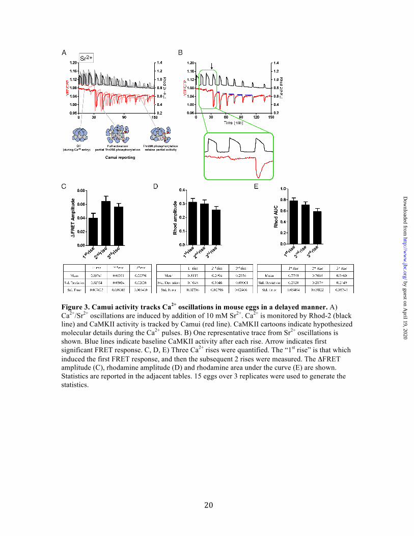
Figure 3. Camui activity tracks Ca2+ oscillations in mouse eggs in a delayed manner. A) Ca2+/Sr2+ oscillations are induced by addition of 10 mM Sr2+. Ca2+ is monitored by Rhod-2 (black line) and CaMKII activity is tracked by Camui (red line). CaMKII cartoons indicate hypothesized molecular details during the Ca2+ pulses. B) One representative trace from Sr2+ oscillations is shown. Blue lines indicate baseline CaMKII activity after each rise. Arrow indicates first significant FRET response. C, D, E) Three Ca2+ rises were quantified. The “1st rise” is that which induced the first FRET response, and then the subsequent 2 rises were measured. The ΔFRET amplitude (C), rhodamine amplitude (D) and rhodamine area under the curve (E) are shown. Statistics are reported in the adjacent tables. 15 eggs over 3 replicates were used to generate the statistics.
by guest on April 19, 2020
http://ww
w.jbc.org/
Dow
nloaded from

21
Figure 4. Monitoring endogenous CaMKII activity using FRESCA. A) A cartoon of a substrate-based CaMKII biosensor is shown: FRET Sensor for CaMKII Activity (FRESCA). Active CaMKII phosphorylates its substrate (syntide), which then acts as a substrate for FHA2 (phosphate binding domain). This induces a conformational change in the sensor as a
by guest on April 19, 2020
http://ww
w.jbc.org/
Dow
nloaded from
22
consequence of CaMKII activity. B) Confocal image of FRESCA expression in a mouse MII egg. Z-stacks images were collected at 1024 x 1024 in 12 bits and the step size was 1.00 µm. Scale bar is equivalent to 50 µm. C, D, E) Changes in Ca2+ are monitored using Rhod-2 (black) and CaMKII activity is monitored using FRESCA (red). Multiple traces are shown after 0.5 µM (C), 2.5 µM (D), or 5 µM (E) ionomycin is added. One representative trace is shown to the right of each plot. F, G) Quantification of FRESCA response to ionomycin addition. ΔFRET amplitude (F) indicates the overall change in FRET during the duration of the Ca2+ signal. Time to FRET peak (G) indicates how long it takes FRESCA to reach maximum ΔFRET signal after addition of ionomycin and increase in Ca2+. Statistics are reported in the adjacent tables, differences were considered significant at P <0.05 (*) using One-way ANOVA. Post-hoc analyses were done using a Tukey multiple comparison test (Prism Graphpad). Number of eggs/replicates for each condition is as follows, 0.5 µM ionomycin: 13/4, 2.5 µM ionomycin: 21/5, 5 µM ionomycin: 8/3.
by guest on April 19, 2020
http://ww
w.jbc.org/
Dow
nloaded from

23
Figure 5. FRESCA specificity in mouse eggs. Various compounds were added to mouse eggs expressing FRESCA and CKAR. In all, Ca2+ was monitored using Rhod-2 (hashed lines) and FRET was monitored by YFP/CFP ratio (solid lines). Colors correspond to the labeled bar graph. The bar graphs below show the quantification of the plots in A and B, where the values were corrected by subtracting the fluorescence from non-injected controls. Differences were considered significant at P <0.05 (*) using one-way ANOVA comparing the 2.5 µM ionomycin data and 0.5 µM ionomycin data in separate analyses. Post-hoc analyses were done using a Tukey multiple comparison test (Prism Graphpad). A) CaMKII inhibitor: AS105 (5 µM) and inactive analog of this inhibitor: AS461 (5 µM) were added to mouse eggs and stimulated with 2.5 µM ionomycin. B) PKC inhibitor: GO6983 (5 µM) was added to mouse eggs expressing either FRESCA or CKAR and stimulated with 0.5 µM ionomycin. These were directly compared to FRESCA and CKAR alone and a non-injected control with 0.5 µM ionomycin. C) A PKC specific activator: PMA (1 µM) was added to mouse eggs expressing CKAR, with or without the addition of the CaMKII inhibitor AS105 or GO6983 (D). E) PMA is added to eggs expressing FRESCA or CKAR. FRET values were normalized to 1.0 for comparison. Number of eggs used in each condition is reported in the methods section.
by guest on April 19, 2020
http://ww
w.jbc.org/
Dow
nloaded from
24
Figure 6. Endogenous CaMKII activity tracks Ca2+ oscillations in mouse eggs. A) Ca2+ oscillations in eggs are induced by addition of Sr2+ to the extracellular media. Ca2+ is monitored by Rhod-2 (black line) and endogenous CaMKII activity is tracked by FRESCA (red line). B) One representative trace from Sr2+ oscillations is shown. Inset focuses on the first Ca2+ rises. C, D, E) Three Ca2+ rises were quantified. The “1st rise” is that which induced the first FRET response, and then the subsequent 2 rises were measured. The ΔFRET amplitude (C), rhodamine amplitude (D) and rhodamine area under the curve (E) are shown. Statistics are reported in the tables below. 11 eggs over 2 replicates were used to generate the statistics. F) Unphosphorylatable FRESCA contains a scrambled version of syntide that is not recognized by CaMKII. Multiple traces where Sr2+ was added are shown. A representative trace from each is shown to the right.
by guest on April 19, 2020
http://ww
w.jbc.org/
Dow
nloaded from

26
Figure 7. FRESCA, but not Camui, continues to report CaMKII activation by Ca2+ oscillations induced by PLC𝜁 Ca2+ oscillations are induced by injection of PLC𝜁 cRNA. CaMKII activity is monitored using FRESCA or Camui (FRET, red lines) and Ca2+ is monitored using Rhod-2 (black lines). A) An overlay of 3 representative eggs using Camui as the reporter of CaMKIIα activity. Cartoon depictions of hypothesized states of CaMKII are shown below. Red circles indicate Thr286 phosphorylation. B) One representative trace from PLC𝜁-induced oscillations and Camui reporting is shown. Insets highlight the first and last pulses. C-E) Quantification of Rhod-2 and FRET signals for Camui during PLC𝜁 induced Ca2+ rises. Three Ca2+ rises were quantified. The “1st rise” is that which induced the first FRET response, and then the subsequent 2 rises were measured. The ΔFRET amplitude (C), rhodamine amplitude (D) and rhodamine area under the curve (E) are shown. Statistics are reported in the tables below. Differences were considered significant at P <0.05 (*) using One-way ANOVA. 12 eggs over 3 replicates were used to generate the statistics. Post-hoc analyses were done using a Tukey multiple comparison test (Prism Graphpad). F) An overlay of 4 representative eggs using FRESCA as the reporter of endogenous CaMKII activity activity. G) One representative trace from PLC𝜁-induced oscillations and FRESCA reporting is shown. Insets highlight the first and last pulses. H-J) Quantification of Rhod-2 and FRET signals for FRESCA during PLC𝜁 induced Ca2+ rises, as in C-E. The ΔFRET amplitude (H), rhodamine amplitude (I) and rhodamine area under the curve (J) are shown. Statistics are reported in the tables below. Differences were considered significant at P <0.05 (*) using One-way ANOVA. Post-hoc analyses were done using a Tukey multiple comparison test (Prism Graphpad). 17 eggs over 3 replicates were used to generate the statistics. K) Unphosphorylatable FRESCA contains a scrambled version of syntide that is not recognized by CaMKII. Multiple traces where PLC𝜁 cRNA was injected are shown. A representative trace from each is shown to the right.
by guest on April 19, 2020
http://ww
w.jbc.org/
Dow
nloaded from

StrattonGoli Ardestani, Megan C. West, Thomas J. Maresca, Rafael A. Fissore and Margaret M.
oscillations in live cells2+CaMKII activity in response to CaFRET-based sensor for CaMKII activity (FRESCA): A useful tool for assessing
published online June 14, 2019J. Biol. Chem.
10.1074/jbc.RA119.009235Access the most updated version of this article at doi:
Alerts:
When a correction for this article is posted•
When this article is cited•
to choose from all of JBC's e-mail alertsClick here
by guest on April 19, 2020
http://ww
w.jbc.org/
Dow
nloaded from