freehand c1 lateral mass screw fixation technique: our experience
TRANSCRIPT

Available online at www.sciencedirect.com
Surgical Neurology 72 (2009) 676–681www.surgicalneurology-online.com
Technique
Freehand C1 lateral mass screw fixation technique: our experienceSerkan Simsek, MD, PhD, Kazim Yigitkanli, MD⁎, Hakan Seckin, MD, PhD, Çetin Akyol, MD,
Deniz Belen, MD, Murad Bavbek, MDNeurosurgery Department, Ministry of Health, Diskapi Educational and Research Hospital, Ankara 06110, Turkey
Received 20 November 2008; accepted 11 June 2009
Abstract Background: Although C1 lateral mass fixation technique is frequently performed in upper cervical
Abbreviations: CTartery; MRI, magnetic
⁎ Corresponding aE-mail address: d
0090-3019/$ – see frodoi:10.1016/j.surneu.2
instabilities, it requires the guidance of fluoroscopic imaging. The fluoroscopy guidance is time-consuming and has the risks of accumulative radiation. Biplane fluoroscopy is also difficult in uppercervical pathologic conditions because of the use of cranial fixations. This study aimed todemonstrate that unicortical C1 lateral mass screws could be placed safely and rapidly withoutfluoroscopy guidance.Methods: Between 2002 and 2008, 32 C1 lateral mass screws were inserted in 17 consecutive patientswith various pathologic conditions involving either atlantoaxial or occipitocervical instability.Results: C1 screw lengths ranged from 18 to 32 mm. The atlantoaxial fixation was performed in 13patients, and C1 lateral mass screws were added to the occipitocervical construct in 3 patients, to theposterior cervical construct in 2 patients, and to the cervicothoracic construct in 1 patient. In 2patients, because C1 lateral mass screws could not be inserted unilaterally, C1 pedicle screw analogswere inserted. There were no screw malpositions or neurovascular complications related to screwinsertion. Operation time and intraoperative bleeding of the isolated atlantoaxial fixations wereretrospectively evaluated. The mean follow-up was 32.3 months (range, 7-59 months). No screwloosening or construct failure was observed within this period. Postoperatively, 4 patientscomplained of hypoesthesia, whereas one patient had superficial wound infection.Conclusion: C1 lateral mass screws may be used safely and rapidly in upper cervical instabilitieswithout intraoperative fluoroscopy guidance and the use of the spinal navigation systems.Preoperative planning and determining the ideal screw insertion point, the ideal trajections, and thelengths of the screws are the most important points.© 2009 Elsevier Inc. All rights reserved.
Keywords: C1 lateral mass; C1 pedicle screw; Atlas; Freehand fixation; Lateral mass screw; Occipitocervical fixation
1. Introduction
The optimal treatment of atlantoaxial instability isreduction and stabilization of the C1-2 joint. Achievementof long-term stabilization has been one of the majorproblems in the treatment of these types of pathologicconditions. Hence, atlantal lateral mass screw fixation
, computerized tomography; ICA, internal carotidresonance imaging; VA, vertebral artery.uthor. Tel.: +90 505 589 48 20; fax: +90 312 517 31 [email protected] (K. Yigitkanli).
nt matter © 2009 Elsevier Inc. All rights reserved.009.06.015
technique is being widely used for various atlantoaxialproblems such as traumatic, degenerative, and tumorousdisorders [2,7,10,11,13,17,18,23]. The method was clini-cally introduced by Goel and Laheri [10] in 1994, and laterpopularized by Harms and Melcher [13], who adapted thistechnique to polyaxial screw and rod systems. Although thetechnique is frequently performed in upper cervical instabil-ities, it requires the guidance of fluoroscopic imaging.Fluoroscopy guidance is time-consuming and has the risks ofaccumulative radiation. Biplane fluoroscopy is also difficultin upper cervical pathologic conditions because of the use ofthe cranial fixations.

677S. Simsek et al. / Surgical Neurology 72 (2009) 676–681
Here, we report a series of 17 patients with differentunderlying causes, who underwent insertion of 32 C1 lateralmass screws. In all the patients, preoperative CT and MRIwere performed to determine the ideal screw lengths andtrajectories of the C1 lateral mass screws. The purpose of thisstudy was to show the safety and the accuracy of freehandscrew insertion of unicortical C1 lateral mass screws.
2. Clinical material and methods
C1 lateral mass screw insertion technique was applied on17 (4 female, 13 male) consecutive patients (mean age, 40years; age range, 6-74 years) between 2002 and 2008. Themean follow-up period was 32.3 months (range, 7-59months). The indications for the surgery were odontoidfractures, os odontoideum, basilar invagination, tumor,infection, and kyphosis reconstruction. In 13 patients,atlantoaxial fixation was performed alone, whereas it wasperformed in 3 patients as a part of occipitocervical fixation,in 2 patients as a part of posterior cervical construct, and in 1patient as a part of posterior cervicothoracic fixation. In 4 ofthe odontoid fracture patients, temporary fixation wasplanned; thus, atlantoaxial fixation without fusion wasperformed, and the atlantoaxial joints of these patientswere not dissected or decorticated. The other 13 patientsunderwent dissection and distraction of the C1-2 joint withplacement of allograft spacers.
Because it is highly important to evaluate the morpho-metry of the atlas, all the patients were routinely screenedwith plain radiographs, CT, and MRI preoperatively. The CTand MRI scans of the atlas lateral mass were carefullyexamined for the ideal screw insertion positions and lengths.
3. Surgical technique
The patients were placed in the prone position with ahead holder under cervical traction, and the arms weretucked on both sides. The shoulders were retracted caudallyby using adhesive tapes. The head was elevated up 15° toease venous return. A midline incision was made betweenthe occipital protuberance and C3 spinous process. Theincision was extended caudally considering the lowestcervical vertebral level that would be involved duringfixation. To expose the lateral margins of the cervical facetjoints, bilateral subperiosteal dissection of the cervicalmusculature was performed.
The C1 lamina was dissected bilaterally to expose thecourse of the vertebral artery at the retroarticular foramenover the C1 lamina. The vertebral artery was not dissected orretracted in any of the patients. Inferior to the lamina, the C1inferior articular process and C2 superior articular processwere dissected through their lateral margins, by cutting offthe C2 root bilaterally. At this stage, there was huge venousbleeding from the perivertebral venous plexus. It wascontrolled by bipolar coagulation and by using hemostatic
agents. Laterally, lateral border of the inferior articularprocess (posterior surface of the lateral mass), and medially,the medial border of the lateral mass were identified. Afterreduction of the fractures, the articular facets of the C1-2joints were dissected bilaterally except in 4 patients withodontoid fractures, in whom we performed only fixationwithout fusion. The joint capsule of C1-2 was widelyremoved by using a microdrill, and the large pieces of boneallografts were packed into the joints.
On the coronal plane, midline of the lateral mass at theintersection of the posterior arch and the C1 lateral masswas used as the ideal entrance point. To create additionalspace and to facilitate C1 lateral mass screw insertion, theinferior surface of the posterior arch was drilled. The entrypoint was marked with a 2 mm high-speed burr. Then,drilling was performed with a 3-mm drill bit, while thedissector was placed at the medial border of the lateralmass. Approximately 15° medial and 15° cephalic trajec-tions, determined on the preoperative CT scans, wereselected. C1 posterior arch was taken as a reference pointfor the C1 screw trajectory on the cephalic plane. C1-2 jointdissection is also helpful at this stage for the orientation ofthe C1 lateral mass anatomy and the cephalic and medialtrajection of the screw.
The ideal lengths of the unicortical screws, taking thereference point as 2 mm behind the anterior cortex of theanterior edge of the lateral mass, were calculated preopera-tively. After confirming the integrity of the hole by using aprobe, sequential probing and drilling of the hole wereperformed gradually and cautiously so that the drill bit didnot penetrate beyond the anterior cortex of the C1 vertebra.In the next step, the hole was initially tapped with a 3.0-mmtip. The bleeding from the hole was controlled with rapidinsertion of polyaxial screws of 3.5-mm diameter (Vertex,Medtronic Sofamor Danek, Memphis, Tenn) (Fig. 1).Because of the overlying posterior arch of the C1, the longerscrews were used to achieve the adaptation of the rod (18- to32-mm polyaxial screws).
In 2 patients, because of the huge venous bleeding, C1pedicle analog screws of 24 mm in length were insertedunilaterally. During the operation, soft tissue attaching theposterior arch was dissected, and the course of the vertebralartery at the retroarticular foramen over the C1 lamina wasexposed. On the coronal plane, midline of the lateral massand the C1 lateral mass was used as the ideal entrance point.High-speed drill were used to remove the dorsal cortex of theposterior arch at the entry point. The inferior border of theposterior arch was exposed as a landmark for screwplacement. The entry point was marked with a 2 mm high-speed burr. Then, drilling was performed with a 3-mm drillbit, while the dissector was placed between the vertebralartery and the cranial edge of the arch. The direction of thescrew placement was perpendicular to the coronal plane andabout 5° cephalad to the transverse plane, which weredetermined on the preoperative CT scans. After confirmingthe integrity of the hole by using a probe, sequential probing

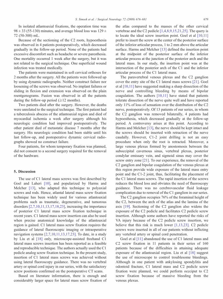
Fig. 1. Imaging studies of a 38-year-old man after trauma. A: Lateral x-ray of the craniovertebral region showing displaced type II odontoid fracture andatlantoaxial dislocation. B: Intraoperative photograph demonstrating final position of C1-2 polyaxial screw/rod construct without fusion material. C:Postoperative axial CT scan of the C1 showing the screws inside the lateral mass. D: Postoperative lateral radiograph of the patient.
678 S. Simsek et al. / Surgical Neurology 72 (2009) 676–681
and drilling of the hole were performed gradually andcautiously so that the drill bit did not penetrate the lateralmass medially and laterally. In the next step, the hole wasinitially tapped with a 3.0-mm tip. The bleeding from thehole was controlled with rapid insertion of polyaxial screwsof 3.5-mm diameter (Vertex, Medtronic Sofamor Danek,Memphis, Tenn). The lengths of the 2 screws inserted in atlasvia posterior arch (C1 pedicle analog screws) were 24 mmand 28 mm.
Thirty-four C2 pedicle screws were inserted into the axis,without any neurovascular injury. Their lengths ranged from16 mm to 26 mm.
The instrumentation was finalized with fixation of therods on the heads of the screws after distraction ormanipulation of the C1 and C2 vertebrae individually(Fig. 1). Intraoperative C-arm fluoroscopy was used toverify the screw positions and alignment of the atlantoaxialor occipitocervical deformities.
The patients were scanned by CT immediately afterthe surgery and then followed up in the postoperative 1st,
3rd, 6th, 12th, and 24th months. Fusion was assessedusing clinical criteria and plain radiographs during thefollow-up period.
4. Results
Thirty-two atlas lateral mass screws and 2 C1 pedicleanalog screws were inserted in 17 patients. No vertebralartery injury or cerebrospinal fluid leakage occurred duringthe screw insertion. Although C1 lateral mass and C2 pediclescrew fixation was planned for one patient with ankylosingspondylitis and odontoid fracture, only occciput to C2 screwfixation could be performed because of the massive bleedingfrom the venous plexus. Satisfactory screw positions werenoted on postoperative CT scans. No revision surgery wasperformed in our series. In 2 patients, bilateral C1 lateralmass screws were overpenetrating the C1 anterior cortex forapproximately 2 mm. However, these patients had noneurovascular injury.
679S. Simsek et al. / Surgical Neurology 72 (2009) 676–681
In isolated atlantoaxial fixations, the operation time was98 ± 33 (55-130) minutes, and average blood loss was 129 ±72 (50-300) mL.
Because of the sectioning of the C2 roots, hypoesthesiawas observed in 4 patients postoperatively, which decreasedgradually in the follow-up period. None of the patients hadexcessive discomfort such as headache or severe paresthesia.One mortality occurred 1 week after the surgery, but it wasnot related to the surgical technique. One superficial woundinfection was treated medically.
The patients were maintained in soft cervical orthoses for2 months after the surgery. All the patients were followed upby using dynamic radiographs. Neither construct failure norloosening of the screws was observed. No implant failures orsliding in flexion and extension was observed on the plainroentgenograms. Fusion was observed in all the patientsduring the follow-up period (≥12 months).
Two patients died after the surgery. However, the deathswere unrelated to the surgical technique. The first patient hada tuberculosis abscess of the atlantoaxial region and died ofmyocardial ischemia a week after surgery although hisneurologic condition had improved postoperatively. Theother patient died of metastatic disease 7 months after thesurgery. His neurologic condition had been stable until hislast follow-up, and postoperative dynamic cervical radio-graphs showed no construct failure.
Four patients, for whom temporary fixation was planned,did not consent to a second surgery required for the removalof the hardware.
5. Discussion
The use of C1 lateral mass screws was first described byGoel and Laheri [10], and popularized by Harms andMelcher [13], who adapted this technique to polyaxialscrews and rods. Hence, atlantal lateral mass screw fixationtechnique has been widely used for various atlantoaxialproblems such as traumatic, degenerative, and tumorousdisorders [2,7,10,11,13,17,18,23], increasing the importanceof posterior C1 lateral mass screw fixation technique inrecent years. C1 lateral mass screw insertion can also be usedwhen precise anatomical knowledge of the atlantoaxialregion is gained. C1 lateral mass screw fixation requires theguidance of lateral fluoroscopic imaging or intraoperativenavigation systems [2,7,10,11,13,17,23]. To date, in a studyby Liu et al [18] only, microscope-assisted freehand C1lateral mass screws insertion has been reported as a feasibleand reproducible technique. The authors actually used the C1pedicle analog screw fixation technique. In the present study,insertion of C1 lateral mass screws was achieved withoutusing lateral fluoroscopy guidance. There was no vertebralartery or spinal cord injury in our series, with the satisfactoryscrew positions confirmed on the postoperative CT scans.
Based on literature information, there is enough andconsiderably larger space for lateral mass screw fixation of
the atlas compared to the masses of the other cervicalvertebrae and the C2 pedicle [1,4,8,9,15,21,25]. The query isto locate the ideal screw insertion point. Goel et al [10,11]prefer to insert the screw at the center of the posterior surfaceof the inferior articular process, 1 to 2 mm above the articularsurface. Harms and Melcher [13] defined the insertion pointat the midpoint of the posterior surface of the inferiorarticular process at the junction of the posterior arch and thelateral mass. In our study, the insertion point was at theintersection of the posterior arch of the C1 and the inferiorarticular process of the C1 lateral mass.
The paravertebral venous plexus and the C2 ganglioncover the entry site of the C1 lateral mass screws [21]. Goelet al [10,11] have suggested making a sharp dissection of thenerve and controlling bleeding by means of bipolarcoagulation. The authors [10,11] have stated that patientstolerate dissection of the nerve quite well and have reportedonly 11% of loss of sensation over the distribution of the C2nerve, postoperatively. Of 13 patients of our series, in whomthe C2 ganglion was removed bilaterally, 4 patients hadhypoesthesia, which decreased gradually at the follow-upperiod. A controversy arises at this point. According toHarms and Melcher [13], the nerve should be kept intact andthe screws should be inserted with retraction of the nervecaudally. However, C1-2 joint drilling is not an easyprocedure when only the root is retracted. Moreover, alarge venous plexus formed by anostomosis between thesuboccipital cavernous sinus, vertebral plexus, posteriorcondylar emissary vein, and sigmoid sinus may cover thescrew entry zone [21]. To our experience, the removal of theC2 ganglion and bipolar coagulation of the venous plexus atthis region provide wide exposure of the lateral mass entrypoint and the C1-2 joint, thus, facilitating the placement ofthe C1 lateral mass screws. The rapid insertion of the screwsreduces the blood loss and obviates the need of fluoroscopyguidance. There was no cerebrovascular fluid leakagecomplication due to removal of the C1 ganglion in our series.
The C2 ganglion occupies 76% of the foraminal height ofthe C2, between the arch of the atlas and the lamina of theaxis [19]. Sectioning of the C2 ganglion also widens theexposure of the C2 pedicle and facilitates C2 pedicle screwinsertion. Although some authors have reported the risks ofVA injury because of the C2 pedicle screw insertion, webelieve that this risk is much lower [1,13,23]. C2 pediclescrews were inserted in all of our patients without inflictingany vertebral artery or spinal cord penetration.
Goel et al [11] abandoned the use of C1 lateral mass andC2 screw fixation in 11 patients in their series of 160patients because of the difficulties in attaining adequateexposure of the atlantoaxial region. Liu et al [18] suggestthe use of microscope to control troublesome bleedings.Although in one patient with ankylosing spondylitis andodontoid fracture, C1 lateral mass and C2 pedicle screwfixation were planned, we could perform occciput to C2screw fixation because of massive bleeding from thevenous plexus.

680 S. Simsek et al. / Surgical Neurology 72 (2009) 676–681
Atlas is capable for screw fixation via its posterior archand lateral mass without neurovascular injury [17,24]. Thewidth of the posterior arch at the location of the vertebralartery groove is relatively broad enough to hold a 3.5 mmdiameter screw [24]. The individual variations of theposterior arch should be made clear by the surgeon beforethe operation. The dimension of the screw should bedetermined according to the thickness of the posterior archat the vertebral artery groove, which guarantees the successof the screw placement [24]. For example, posteriorponticulus of the C1 posterior arch occurs in 9.4% of thepopulation [22]. The posterior ponticulus may give a falseidea of a thicker posterior C1 arch, and if it is taken as theinsertion point of the C1 pedicle screws, the risk of thevertebral artery injury is very high [22]. In 2 patients, C1pedicle analog screws were used because of the massivebleeding from the venous plexus, unilaterally. Thistechnique can be an effective alternative in atlantoaxialfixation especially to prevent troublesome venous bleedingand to decrease the blood loss during the procedure.Considering these potential intraoperative complications,preoperative planning for the fixation of the C1 pedicle oroccipitocervical fixation is important to manage anyanomalies of the region preoperatively.
Various trajectory and length of the suggested C1 lateralmass screws have been defined in earlier anatomical studies[15,21,25]. Goel et al [10,11] suggested 15° of medialangulation with the 15° of cephalic angulation as ideal.Harms and Melcher [13] defined the screw trajectory as theanterior arch of C1 under fluoroscopy control. In the studyby Rocha et al [21], the maximum angle of medializationfrom the midline was calculated as 16.7° ± 1.3° (range,14.6°-20.7°). Hong and colleagues [15] reported the screwangulation as 14.7° relative to the axial plane. In the study byWang and Samudrala [25], maximum screw medializationfor the large bulk of the C1 lateral mass ranged between 25°and 45° (mean, 33°), with the screw length of 15.1 to 29 mm.Screw length also varies depending on the starting point aswell as the vertical and cephalic angulations of the screw.Accordingly, it may be difficult to insert a C1 lateral masswithout the risk of overpenetration. Considering thesevariations, awareness of potential complications due tooverpenetrated screws in different trajections is of utmostimportance. We inserted screws in 15° of medial andcephalic angulations. The C1 posterior arch was taken as areference point for the C1 screw trajectory on the cephalicplane. The dissector at the medial edge of the entry zone wasanother reference point for the medial trajection of thescrews. In 13 of our patients, C1-2 facet dissection anddecortication increased our orientation to the C1 lateral massanatomy and the trajections of the screws.
According to Harms and Melcher [13], the C1 lateralmass should be screwed bicortically. In the anatomicalstudy by Doherty and Heggeness [8], the thickest and mostdense cortical bone was found in the anterior cortex of theanterior ring, suggesting that the strongest screws would be
placed bicortically. Goel et al [10,11] and Harms andMelcher [13] had 100% of fusion in their series. Noconstruct failures were seen in the series of Liu et al [18], inwhich the researchers used unicortical screws as in ourstudy. In the study by Eck et al [12], the strength of thebicortical screws was statistically higher than that of theunicortical screws. Nevertheless, the authors recommendedthe use of unicortical screws because of potentialcatastrophic complications. The increased fusion rate inseries of atlantoxial fixations may also be related to thedrilling, decortication, and bone placement into the C1-2joint. In 4 of our patients, we did not use bone grafts andperformed only internal fixation after the reduction of thedeformities. In 13 patients of our series, demineralized bonematrix combined with allograft spacers were used toincrease the fusion rate after reduction of the deformities.
Analysis of biomechanical results showed that theatlantoaxial construct is effective for the fixation of theatlantoaxial junction. Although C1-2 construct is wellagainst flexion, lateral bending, and axial rotation, it doesnot resists well in extension [16]. Adding an interspinouscable/graft to the C1-2 construct significantly lowers therange of motion during extension [16]. So the combinationof atlantoaxial construct with cable/graft when possible maybe used to reduce the range of motion and provides a scaffoldfor new bone growth. The increased fusion rate in our serieswithout using an interspinous cable/graft may be related tograft placement into the C1-2 joint.
Internal carotid artery may be located within 1 mm of theideal exit point of the C1 lateral mass screw [5,6]. ICAinjuries due to overpenetrated transarticular screws havebeen reported in the literature [3,5]. Therefore, the depth ofthe screw inside the lateral mass is an important issue.Currier et al [5,6] suggested that medial angulation of thescrew in the lateral mass of the C1 might increase the safetyof the internal carotid artery injuries. Hong et al [14] reporteda case of hypoglossal nerve palsy due to the C1 lateral massscrew fixation. The authors suggested a more mediallyangled trajection of the screw to avoid the hypoglossal nervepalsy and use of unicortical screws [14]. Thus, we paid greatattention to medial angulations while the dissector remainedat the medial edge of the lateral mass. We calculated the ideallengths of the lateral mass screws preoperatively for thetrajectories selected. Because of the possible risks ofoverpenetrated C1 lateral mass screws, we inserted uni-cortical screws. Intraoperatively, sequential probing anddrilling of the hole was cautiously performed to feel the“crunching” sensation, as was stated earlier by Liu et al [18].Feeling the dense anterior cortex of the atlas is of utmostimportance. Despite all the precautions, in 2 of our patientswith bilateral C1 screws, the screws had overpenetrated by 2mm without causing any neurovascular injuries, as seen onthe postoperative axial CT of the C1. Hypoglossal nerve orICA injury was not observed in our series.
Atlas lateral mass screws may provide additional fixationforces for the occipitocervical constructs, increasing the

681S. Simsek et al. / Surgical Neurology 72 (2009) 676–681
resistance to construct failure [20,26]. In 3 patients of ourseries, we were able to add the C1 lateral mass screws to theoccipitocervical construct. C1 lateral mass screws allow thesurgeon to individually manipulate the C1 with respect to C2and the occiput [17].
6. Conclusions
We suggest that unicortical C1 lateral mass screws may besafely and effectively used in atlantoaxial and occipitocervi-cal fusion procedures without the use of intraoperativenavigation or fluoroscopy guidance. These techniques aretime-consuming and thus, increase the intraoperative bleed-ing from the venous plexus. In addition, fluoroscopy exposesboth the patient and the surgical team to harmful radiation.Our study also confirms that unicortical C1 lateral massscrews are sufficiently resistant to construct failure. Sequen-tial probing and drilling of the hole should be performedgradually and cautiously so that the drill bit does notpenetrate beyond the anterior cortex of the C1 vertebra.Anatomical knowledge of the region and preoperativeplanning using imaging studies and determination of theproper surgical technique are essential. Learning curve isalso required to perform this technique without fluoroscopy,and we suggest using fluoroscopy, for the surgeonsparticularly early on in their experience. The surgeon shouldalso be ready preoperatively and note the anomalies of theregion, to determine the use of any other fixation techniquesuch as C1 pedicle screw fixation.
References
[1] AbouMA, Solanki G, Casey AT, Crockard HA. Variation of the groovein the axis vertebra for the vertebral artery: implications forinstrumentation. J Bone Joint Surg Br 1997;79:820-3.
[2] Aryan HE, Newman CB, Nottmeier EW, Acosta Jr FL, Wang VY,Ames CP. Stabilization of the atlantoaxial complex via C-1 lateral massand C-2 pedicle screw fixation in a multicenter clinical experience in102 patients: modification of the Harms and Goel techniques.J Neurosurg Spine 2008;8:222-9.
[3] Bogaerde MV, Viaene P, Thijs V. Iatrogenic perforation of the internalcarotid artery by a transarticular screw: an unusual case of repetitiveischemic stroke. Clin Neurol Neurosurg 2007;109:466-9.
[4] Christensen DM, Eastlack RK, Lynch JJ, Yaszemski MJ, Currier BL.C1 anatomy and dimensions relative to lateral mass screw placement.Spine 2007;83:844-8.
[5] Currier BL, Todd LT, Maus TP, Fisher DR, Yaszemski MJ. Anatomicrelationship of the internal carotid artery to the C1: a case report ofcervical reconstruction for chordoma and pilot study to assess the riskof screw fixation of the atlas. Spine 2003;28:E461-7.
[6] Currier BL, Maus TP, Eck JC, Larson DR, Yaszemski MJ. Relationshipof the internal carotid artery to the anterior aspect of the C1.
Implications for C1-C2 transarticular and C1 lateral mass fixation.Spine 2008;33:635-9.
[7] Deen HG, Birch BD, Wharen RE, Reimer R. Lateral mass screw-rodfixation of the cervical spine: a prospective clinical series with one yearfollow-up. The Spine Journal 2003;3:489-95.
[8] Doherty BJ, Heggeness MH. The quantitative anatomy of the atlas.Spine 1994;15:2497-500.
[9] Dong Y, Xia Hong M, Jianyi L, Yuan Lin M. Quantitative anatomy ofthe lateral mass of the atlas. Spine 2003;28:860-3.
[10] Goel A, Laheri V. Plate and screw fixation for atlanto-axialsubluxation. Acta Neurochir (Wien) 1994;129:47-53.
[11] Goel A, Deasi KI, Muzumdar DP. Atlantoaxial fixation using plate andscrew method: a report of 160 treated patients. Neurosurgery 2002;51:1351-7.
[12] Eck JC, Walker MP, Currier BL, Chen Q, Yaszemski MJ, An KN.Biomechanical comparison of unicortical versus bicortical C1 lateralmass screw fixation. J Spinal Disord Tech 2007;20:505-8.
[13] Harms J, Melcher RP. Posterior C1-2 fusion with polyaxial screw androd fixation. Spine 2001;26:2467-71.
[14] Hong JT, Lee SW, Son BC, Sung JH, Kim IS, Park CK. Hypoglossalnerve palsy after posterior screw placement on the C-1 lateral mass.Case report. J Neurosurg Spine 2006;5:83-5.
[15] Hong X, Dong Y, Yunbing C, Quingshui Y, Shizheng Z, Jingfa L.Posterior screw placement on the lateral mass of atlas. An AnatomicStudy. Spine 2004;29:500-3.
[16] Hott JS, Lynch JJ, Chamberlain RH, Sonntag VK, Crawford NR.Biomechanical comparison of C1-2 posterior fixation techniques.J Neurosurg Spine 2005;2:175-81.
[17] Jea A, Taylor MD, Dirks PB, Kulkarni AV, Rutka JT, Drake JM.Incorporation of C-1 lateral mass screws in occipitocervical andatlantoaxial fusions for children 8 years of age or younger. J Neurosurg2007;107:178-83.
[18] Liu G, Buchowski JM, Shen H, Yeom JS, Riew KD. The feasibility ofmicroscope-assisted “free-hand” C1 lateral mass screw insertionwithout fluoroscopy. Spine 2008;33:1042-9.
[19] Lu J, Ebraheim NA. Anatomical considerations of C2 nerve rootganglion. Spine 1998;23:649-52.
[20] Oda J, Abumi K, Sell LC, Haggerty CJ, CunninghamBW,McAfee PC.Biomechanical evaluation of five different occipito-atlanto-axialfixation techniques. Spine 1999;24:2377-82.
[21] Rocha R, Safavi-Abbasi S, Reis C, Theodore N, Bambakidis N, deOliveira E, Sonntag VK, Crawford NR. Working area, safety zones,and angles of approach for posterior C-1 lateral mass screw placement:a quantitative anatomical and morphometric evaluation. J NeurosurgSpine 2007;6:247-54.
[22] Simsek S, Yigitkanli K, Comert A, Acar HI, Seckin H, Er U, Belen D,Tekdemir I, Elhan A. Posterior osseous bridging of C1. J Clin Neurosci2008;15:686-8.
[23] Stulik J, Vyskocil T, Sebesta P, Kryl J. Atlantoaxial fixation using thepolyaxial screw-rod system. Eur Spine J 2007;16:479-84.
[24] Tan M, Wang H, Wang Y, Zhang G, Yi P, Li Z, Wei H, Yang F.Morphometric evaluation of screw fixation in atlas via posterior archand lateral mass. Spine 2003;28:888-95.
[25] Wang MY, Samudrala S. Cadaveric morphometric analysis for atlantallateral mass screw placement. Neurosurgery 2004;54:1436-40.
[26] Wolfla CE, Salerno SA, Yoganandan N, Pintar FA. Comparison ofcontemporary occipitocervical instrumentation techniques with andwithout C1 lateral mass screws. Neurosurgery 2007;61(3 Suppl):87-93.