fractures and physical therapy management
TRANSCRIPT
FRACTURES AND PHYSICAL THERAPY MANAGEMENT
HELEN BUCHANAN, SPT
PHYT 734 Fall 2019
Adapted from previous presentation by Jonathan Valbuena, PT

OBJECTIVES
1. Understand an overview of bone tissue, risk factors for fracture, and general healing times.
2. Be able to describe various fractures by using specific terminology based on MOI, classification, location, fracture pattern, and position/alignment.
3. Appreciate signs/symptoms of possible fractures and the Ottawa Rules for Knee and Ankle suspected fractures.
4. Be able to describe the Salter-Harris Classification system for epiphyseal plate injuries.
5. Be able to apply principles of fracture management for periods of immobilization and post-immobilization in clinical practice.

BRIEF OVERVIEW OF BONE1
¡ Composition¡ Fiber = Type I collagen
¡ Ground Substance = GAGs, hyaluronic acid, calcium salts
¡ Cells = Osteoblasts and Osteoclasts
¡ Types of Bone¡ Cortical
¡ CancellousImage from OpenStaxA

HOW DO BONES BREAK?
¡ Trauma
¡ Fatigue or Stress
¡ Pathology
Image from Getty ImagesB

BONE AND STRESS-STRAIN
Figures from TurnerC

RISK FACTORS FOR FRACTURE2
¡ Sudden impact¡ Ex: trauma, accidents, assault
¡ Osteoporosis¡ Women > Men
¡ History of falls¡ Especially with increased age, low BMI, and low levels of PA
¡ Repetitive stress¡ Repeated microtrauma
¡ Pathology¡ Abnormal fragile bone from neoplastic, poor health, or disease
condition

ASSOCIATED FRACTURE DAMAGE
¡ Edema¡ Ecchymosis¡ Hemorrhage¡ Ruptured tendons/ligaments¡ Severed nerves¡ Damaged blood vessels¡ Injury to organs and muscle/soft tissues¡ Joint dislocation

BUT HOW DID A BONE BREAK?
Image from Barefoot MedsD

MECHANISM OF INJURY1
¡ Speed of Loading¡ ↑ Speed of loading = ↑ ultimate strength and
stiffness of bone
¡ ↑ Energy release at failure
¡ = More potential for soft tissue and other associated damage

MECHANISM OF INJURY1
¡ Frequency of Loading¡ ↑ Frequency associated with ↓ ultimate strength
of tissue
¡ = Cumulative microtrauma leads to failure

DEFINING AND DESCRIBING FRACTURES
¡ Classification
¡ Location
¡ Fracture Patterns/Types
¡ Position/Alignment

IMAGING ABC’s
¡ A = alignment
¡ B = bones
¡ C = cartilage space
¡ s = soft tissue
Image from RadiopaediaE

CLASSIFICATION
¡ Open ¡ Closed
Image from Lex MedicusF

LOCATION
¡ Specific bone
¡ Location within a bone
¡ Proximal
¡ Distal

FRACTURE PATTERNS/ TYPES
¡ Transverse
¡ Linear
¡ Oblique
¡ Spiral
¡ Comminuted
¡ Avulsion
¡ Greenstick
¡ Torus/Buckle
¡ Stress
¡ Pathologic

***
Figure from Dream LifeG

POSITION AND ALIGNMENT
¡ Non-displaced
¡ Superior/Inferior displacement
¡ Anterior/Posterior displacement
¡ Medial/Lateral displacement
¡ Distracted
¡ Rotation

POSITION AND ALIGNMENT
Image from Kisner et al2

HOW TO PUT IT ALL TOGETHER
¡ Convention description:
¡ Distal segment position in relation to proximal segment
¡ Angulation named by direction of angle apex
¡ Describe by classification, location, fracture line, and position/alignment

NAME THAT FRACTURE!Images from RadiopaediaH
AP view Lateral view

NAME THAT FRACTURE!Images from RadiopaediaI
AP view Lateral view

NAME THAT FRACTURE!Images from Jon HackeJ
PA view
Lateral view

NAME THAT FRACTURE!Images from RadiopaediaK
AP view Lateral view

WHO NEEDS A BREAK…?

CAUSES AND TYPES OF FRACTURES2
Force Effect on Bone Type of Fracture
Bending Long bone bends causing failure on convex side of bend
Transverse or oblique fracture; Greenstick fracture in children
Twisting (torsional) Spiral tension failure in long bone
Spiral fracture
Traction Tension failure from pull of ligament or muscle
Avulsion fracture
Crushing (compression)
Usually in cancellous bone Compression fracture, torus (buckle) fracture in children
Repetitive Microtrauma Small crack in bone Fatigue or stress fracture
Normal force on abnormal bone
E.g. osteoporosis, bony tumor, or other diseased bone
Pathological fracture
Table adapted from Kisner et al2

SIGNS/SYMPTOMS OF POSSIBLE FRACTURE2
¡ Swelling, deformity, or abnormal movement¡ May or may not be visibly obvious!
¡ Sharp, localized tenderness at site¡ Localized pain aggravated by movement and/or
weight-bearing¡ Muscle guarding with PROM¡ Decreased functional use of segment¡ History of MOI
¡ Fall, direct blow, twisting injury, or other trauma

OTTAWA ANKLE RULE
¡ A decision aid for excluding fractures of the ankle and midfoot
¡ Accurate predictor3
¡ 36% reduction in unnecessary radiographs

OTTAWA ANKLE RULE
¡ Applies to ages 2 to 55¡ Tender over posterior 6cm OR tip of the medial or
lateral malleolus¡ Base of 5th metatarsal¡ Navicular¡ Inability to bear weight (4 steps) BOTH immediately
AND in the ED¡ Limping is OK
Adapted From: Paul Mintken PT, DPT, OCS
Denise Stelzner PT, MBAUniversity of Colorado PT


OTTAWA KNEE RULE
¡ To determine whether a knee injury needs an X-ray
¡ Accurate predictor?
¡ 26% reduction in unnecessary radiographs4

OTTAWA KNEE RULE5
¡ Radiographs indicated if any of the 5 are present:¡ Age 55 or older
¡ Isolated tenderness over patella
¡ Tenderness over fibular head
¡ Unable to flex knee >90°
¡ Unable to bear weight immediately or in the ED for 4 steps

EPIPHYSEAL PLATE = GROWTH PLATE
¡ Area of growth in long bone
¡ Between epiphysis and metaphysis of long bones
Image from LLRSL
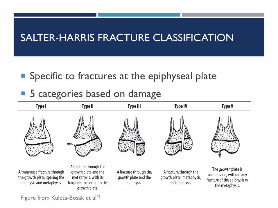
SALTER-HARRIS FRACTURE CLASSIFICATION
¡ Specific to fractures at the epiphyseal plate
¡ 5 categories based on damage
Figure from Kuleta-Bosak et alM

SALTER-HARRIS TYPE I
¡ Shearing injury1
¡ Epiphysis is completely separated from metaphysis
¡ Rarely manipulated, requires immobilization
¡ Good prognosis
Figure from Kuleta-Bosak et alM

SALTER-HARRIS TYPE II
¡ Shearing and bending injury1
¡ Epiphysis and growth plate partially separated from metaphysis, fracture through part of metaphysis
¡ Most common
¡ Manipulated and immobilized
¡ Good prognosis
Figure from Kuleta-Bosak et alM

SALTER-HARRIS TYPE III
¡ Fracture through epiphysis, separates part of epiphysis and growth plate from metaphysis
¡ Rare
¡ Typically requires surgical reduction
Figure from Kuleta-Bosak et alM

SALTER-HARRIS TYPE IV
¡ Fracture through epiphysis and into metaphysis crossing growth plate
¡ Surgery required
¡ Potential for premature closing of growth plate if not perfectly aligned
Figure from Kuleta-Bosak et alM

SALTER-HARRIS TYPE V
¡ Epiphysis crushed into metaphysis, crushing the growth plate
¡ Poor prognosis, stunted growth
¡ Difficult to diagnose, needs to be treated with NWB immediately1
Figure from Kuleta-Bosak et alM

BONE GENERAL HEALING TIME2
¡ Varies with age, location and type of fracture, displacement, surgical involvement, soft tissue damage, and vascularity around the repair
¡ Children = 4-6 weeks
¡ Adolescents = 6-8 weeks
¡ Adults = 10-18 weeks
¡ Smoking and NSAIDs negatively affect bone healing!

STAGES OF FRACTURE HEALING6
¡ Inflammatory¡ Begins immediately after bone injury with formation
of fibrin clot or hematoma¡ Repair
¡ Begins a few days after bone injury¡ “soft callus” formation for 6 weeks¡ “hard callus” formation for 12 weeks
¡ Remodeling¡ Can continue for months or years after bone injury

POTENTIAL COMPLICATIONS2
¡ Swelling
¡ Fat embolism
¡ Skin ulceration, vascular compromise
¡ Nerve injury
¡ Infection
¡ Delayed union or Malunion
¡ Refracture
¡ Problems with fixation devices

PERIOD OF IMMOBILIZATION2
¡ Bone requires immobilization for healing
¡ But connective tissue weakens, articular cartilage degrades, muscle atrophies, at risk for contracture, and slowed circulation
¡ Early “non-destructive” motion is ideal but usually not feasible unless an internal fixation device is present to stabilize

IMMOBILIZATION: PT MANAGEMENT2
¡ Be on alert for complications
¡ General exercises for uninvolved portions of the body to minimize soft tissue degradation¡ Especially important for bed rest with traction!
¡ If LE involved, educate in alternative modes of mobility within WBing limitations¡ Crutches, walker, WC – clinical judgment is key

EXTERNAL FIXATION
Images from Schepers T, RammeltN

IMMOBILIZATION: PT SUMMARY2
Plan of Care Intervention1. Educate the patient 1. Teach functional adaptations and
safe ambulation, bed mobility
2. Decrease effects of inflammation during acute period
2. Ice, elevation
3. Decrease effects of immobilization
3. Intermittent muscle setting, active ROM to joints above and below immobilized region
4. It patient is confined to bed, maintain ROM and strength in major muscle groups
4. ROM activities to all areas not immobilized, resistive exercises to major muscle groups not immobilized especially in preparation for future ambulation

POST-IMMOBILIZATION2
¡ Following immobilization, many impairments likely:
¡ Decreased ROM, joint play, muscle flexibility
¡ Muscle atrophy and poor endurance
¡ Painful movement early on
¡ Inelastic scar tissue?

POST-IMMOBILIZATION: PT MANAGEMENT2
¡ Consult physician for clinical vs. radiological healing expectations
¡ Match interventions to impairments¡ Joint mobilization
¡ Muscle stretching, strengthening and endurance¡ PNF: proximal hold-relax and agonist-contraction
¡ Functional activities
¡ Scar tissue mobilization for soft tissue injuries

POST-IMMOBILIZATION: PT SUMMARY2
¡ Plan of Care1. Educate the patient
2. Provide protection until radiologically healed
3. Initiate active exercises
4. Increase joint and soft tissue mobility
5. Increase strength and muscle endurance
6. Improve cardiorespiratory fitness

POST-IMMOBILIZATION: PT SUMMARY2
¡ Interventions1. Inform patient of:
a. Limitations
b. Home exercises/Reinforce protection
2. PWB in LE injuries, non-stressful activities in UE injuries
3. Active ROM, gentle multi-angle isometrics
4. Joint play and muscle stretching
5. Resistive and repetitive exercises as ROM increases
6. Safe aerobic exercises that do not stress fracture site

ANY QUESTIONS?
Image from SNLO

REFERENCES
Original Presentation: Valbuena, J. Lower Extremity Fractures/TJR and Physical Therapy Management. October 22, 2018; Chapel Hill, NC.
1. Gross, M. Bone – Lectures 1 & II. Lecture presented as VoiceThread; created on July 11 and 13, 2008; Chapel Hill, NC.
2. Kisner C, Thorp JN, Holtgrefe K.. Joint, Connective Tissue, and Bone Disorders and Their Management: Fractures and Posttraumatic Immobilization. In: Therapeutic Exercise Foundations and Techniques. 7th ed. Philadelphia, PA: F.A. Davis Company; 2018:350-354.
3. Stiell IG, Greenberg GH, McKnight RD, et al. A study to develop clinical decision rules for the use of radiography in acute ankle injuries. Ann Emerg Med 1992 Apr;21(4):384-90. doi 10.1016/s0196-0644(05)82656-3.
4. Stiell IG, Wells GA, Hoag RH, Sivilotti ML, et al. Implementation of the Ottawa Knee Rule for the use of radiography in acute knee injuries. JAMA 1997 Dec 17;278(23):2075-9. doi 10.1001/jama.1997.03550230051036.
5. Bachman LM, Kolb E, Koller MT, et al. Accuracy of Ottawa ankle rules to exclude fractures of the ankle and mid-foot: systematic review. Ann IntMed.2004;140:1221-124 doi 10.1136/bmj.326.7368.417.
6. Stages of healing. Available at: https://elentra.healthsci.queensu.ca/assets/modules/msk_bonemorph_agg/66d64b9728004605842f/66d64b9728004605842f.htm. Accessed October 23, 2019.

IMAGES AND FIGURES
A. https://openstax.org/books/anatomy-and-physiology/pages/6-3-bone-structure
B. https://www.wsj.com/articles/SB10001424127887323296504578396832875546130
C. Turner CH. Bone strength: current concepts. Ann N Y Acad Sci. 2006;1068:429-446. doi:10.1196/annals.1346.039
D. https://barefootmeds.wordpress.com/2012/10/01/studying-skeleton/
E. https://radiopaedia.org/cases/radial-neck-fracture-and-sail-sign?lang=us
F. ttp://pathologies.lexmedicus.com.au/pathologies/femur-shaft-fracture
G. Dream Life for Nurses. Available at: https://www.pinterest.com/pin/812266482762856540/?nic=1.
H. https://radiopaedia.org/cases/tibia-fracture-importance-of-2-views?lang=us
I. https://radiopaedia.org/cases/oblique-tibial-fracture?lang=us
J. Guide Radiology Forearm Answers from Jon Hacke
K. https://radiopaedia.org/cases/humeral-fracture-spiral-wedge-midshaft?lang=us
L. Limb Lengthening and Reconstruction Society. Available at: https://llrs.org/information-for-patients/specific-conditions/growth-plate-arrest/
M. Kuleta-Bosak E, Bożek P, Kluczewska E, Tomaszewski R, Machnik-Broncel J. Salter-Harris type II fracture of the femoral bone in a 14-year-old boy – case report. undefined. 2010
N. Schepers T, Rammelt S. Complex foot injury: early and definite management. Foot Ankle Clin 2017;22(1):193-213. doi:10.1016/j.fcl.2016.09.014.
O. SNL Haunted Elevator. Available at: https://www.youtube.com/watch?v=rS00xWnqwvI