formation, maturation, and disorders of white matter
TRANSCRIPT

Formation, Maturation, and Disorders of White Matter
A. James Barkovich, 1 Gilles Lyon, and Philippe Evrard
From the University of California, San Francisco (AJB), and the University of Louvain Medical School, Brussels, Belgium (GL and PE)
The cerebral white matter is composed primarily of myelinated and unmyelinated axons, which transmit chemically mediated electrical signals; oligodendrocytes, which are the myelin forming cells; astrocytes; and, in infants, a few neurons in the subcortical region . The axonal fibers almost invariably travel in fiber bundles that can be broken down into three essential types: projection fibers that carry afferent and efferent impulses between the cortex and distant loci; association fibers that interconnect cortical regions of the same hemisphere; and commissural fibers that interconnect corresponding cortical regions of the two hemispheres (1). The purpose of this manuscript is to elucidate the mechanisms by which cerebral fiber tracts form and to discuss the conditions that result from aberrations in their formation, particularly those that have neuroradiologic implications.
Axonal Formation and Early Axonal Guidance
The white matter of the cerebral hemispheres is first visible as the intermediate zone, an area located between the subventricular germinal matrix and the developing cortical plate, as early as the second half of the first trimester. In its earliest stages, the white matter consists primarily of radial glial fibers and the neurons migrating upon them (2-5). Although there is evidence that limited axonogenesis begins during neuronal migration, the majority of axonogenesis does not com-
1 Address reprint requests to Dr Barkovich, Neuroradiology Section,
Box 0628, UCSF, Room L358, 505 Parnassus Ave., San Francisco, CA
94943-0628.
Index terms: Brain, growtH and development; white matter, abnormalities
and anomalies; Pediatric neuroradiology
AJNR 13:447-461, Mar/ Apr 1992 0195-6108/92/1302-0447
© American Society of Neuroradiology
447
mence until the neurons have arrived at their final destinations in the cerebral cortex (3). The most important structure in the proper generation and guidance of axons through the developing brain is the "growth cone ," the motile sensory apparatus situated at the tip of the advancing axon (6). Interaction with specific molecules on cell membranes causes polymerization of actin filaments in the growth cone tip, causing growth of the cone in the specific direction of the interaction (7, 8). Lack of interaction with specific molecular targets causes eventual retraction of the cone, with breakdown of the actin polymers (6). In some instances, the growth cone may receive inhibitory signals, causing active avoidance of specific targets (9). Thus, the growth cone provides a mechanism for axonal elongation and guidance in response to specific cues in its immediate environs.
The external or internal cues that stimulate initial axonal extension from the neural cell body have not yet been fully elucidated. It is known that initial axon extension begins adjacent to a focus of microtubule polymerization within the cell body and that the microtubules grow into the developing axon, presumably causing its elongation (1 0). Therefore, the structural organization of the cell at the time of neuronal differentiation may establish the initial site and direction of axonal extension (11). However, environmental cues, both from the surfaces of adjacent cells and from the extracellular matrix, seem to play major roles in axonal growth and guidance thereafter.
Axons from early differentiating neurons may respond to different environmental cues than those that develop later. The earliest neurons lie on a bed of astrocytes, astrocytic precursors, and neuroepithelial cells. Axons formed in these early stages initially travel through mesenchymal and epithelial regions that are devoid of other axons. Therefore, they respond to molecular markers on

448
and around the astrocytic precursors, neuroepithelial cells, and mesenchymal cells, which act as stimuli for axon outgrowth (12) (Figs. 1 and 2). The two best characterized cell surface molecules are neural cell adhesion molecule (N-CAM) and N-cadherin. Both are glycoproteins that are abundant on the cell membranes of developing vertebrate neural ectoderm and on axons of differentiated neurons (13-15). Both bind to glycoproteins of the same type on other cell membranes. Antibodies to these two glycoproteins inhibit axonogenesis on cellular substrates in vivo (13, 15). It thus appears that these two molecules faciMate initial axonal growth; however, the ubiquitous presence of N-CAM and N-cadherin in most tissues of the developing central nervous system makes it likely that these molecules aid primarily in the initial outgrowth of axons but not in specific axonal guidance.
Laminin, an extracellular matrix glycoprotein, promotes axon extension in vivo (16) and may
ECM ---+----l---+--+---
N-CAM
Neural epithelium
ECM ---+---t--t----+--
N-cadherin ca++
Neural epithelium
Fig. 1. Initial axon outgrowth. In early stages after axon formation, growth cones (G) and axonal bodies respond to cell surface markers on and around astrocytic precursors, neuroepithelial cells, and mesenchymal cells. The two best characterized cell surface molecules are N-CAM (A) and N-cadherin (B). ECM, extracellular matrix.
AJNR: 13, March/ April 1992
ECM---+--~--~--~-
Laminin t t t t lntegrin
Neural epithelium Fig. 2. Laminin is an extracellular matrix glycoprotein that
promotes axonal extension by interacting with axonal surface glycoproteins called integrins. The integrins can change their configurations and, therefore, may interact with different cell surface and extracellular matrix molecules as development proceeds. G, growth cone; ECM, extracellular matrix.
contribute to axonal guidance, as well. Laminin is believed to promote axonal extension by interacting with axonal surface glycoproteins called integrins (Fig. 2). lntegrins are composed of a variable combination of alpha and beta subunits; it is postulated that specific binding of axons to various molecules in the extracellular matrix and on cell surfaces results from the ability of neurons to produce specific combinations of integrin subunits and, later, to modify these combinations. Such axonal surface marker versatility may lead to specific guidance of the axon through the developing brain ( 11, 17).
Subsequent Axonal Elongation and Guidance
Although the initial guidance of the vertebrate axons is modulated primarily by axonal adhesion via the homophilic mechanisms of N-CAM and N-cadherin and the heterophilic mechanism of laminin with integrins, other mechanisms begin to contribute as the axon elongates. The path of the axon can be influenced by nonpermissive substrates on cell surfaces and in the extracellular matrix, and by cell surface molecules that frankly inhibit advancement of growth cones, a characteristic known as contact inhibition (9) (Fig. 3). For example, two surface proteins have been identified on oligodendrocytes that inhibit axonal growth in cell culture (18). Molecules that inhibit axonal growth are also present on some axonal surfaces and may contribute to the selective grouping of some axons in bundles (fasciculation) (1). The fact that some developing axons travel
AJNR: 13, March/ April 1992
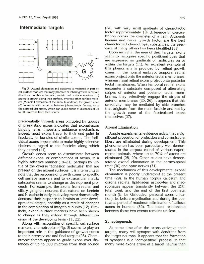
Intermediate Targets
RtmE I -c-
I -c-
Fig. 3. Axonal elongation and guidance is mediated in part by cell surface markers that may promote or inhibit growth in certain directions. In this schematic, certain cell surface markers (m) promote growth along their surface, whereas other surface markers (R) inhibit extension of the axon. In addition , the growth cone (G) interacts with certain substrates (chemotropic factors, c) in the extracellular space, which can guide axons at distances of up to 300 microns from their source.
preferentially through areas occupied by groups of preexisting axons indicates that axonal-axon binding is an important guidance mechanism. Indeed, most axons travel to their end point in fascicles, ie, bundles of similar axons. The individual axons appear able to make highly selective choices in regard to the fascicles along which they extend (11).
Growth cones seem to discriminate between different axons, or combinations of axons, in a highly selective manner (19-21), perhaps by virtue of the diverse "adhesion molecules" that are present on the axonal surfaces. It is interesting to note that the response of growth cones to specific cell surface markers and to extracellular matrix substrates seems to change as development proceeds. For example, the axons from retinal and ciliary ganglion neurons that extend on laminin and N-cadherin early in development dramatically decrease their response to laminin at later developmental stages, possibly as a result of changes in the combination of integrin subunits ( 12). Similarly, axonal surface markers have been shown to change as they extend through different regions of the developing brain (11, 22).
Along with recognition of specific cell surface markers, chemotropism (Fig. 3) seems to play an important role in the guidance of growth cones to their intermediate and final targets (23). Chemotropic factors appear to guide axons over distances of up to 300 microns from their source
449
(24), with very small gradients of chemotactic factor (approximately 1% difference in concentration across the diameter of a cell) . Although laminin and nerve growth factor are the best characterized chemotropic substances, the presence of many others has been identified (11 ).
Upon arrival in the area of their targets , axons seem to recognize specific positional cues that are expressed as gradients of molecules on or within the targets (11). An excellent example of this phenomena is provided by retinal growth cones. In the normal embryo, temporal retinal axons project onto the anterior tecta! membranes, whereas nasal retinal axons project onto posterior tecta! membranes. When temporal retinal axons encounter a substrate composed of alternating stripes of anterior and posterior tecta! membranes, they selectively engage the stripes of anterior membranes (25, 26). It appears that this selectivity may be mediated by side branches that originate from the main fascicle and not by the growth cone of the fasciculated axons themselves (27).
Axonal Elimination
Ample experimental evidence exists that a significant proportion of projection and commissural fibers are eliminated during development. This phenomenon has been particularly well demonstrated in the corpora callosi of various experimental animals, where up to 70% of axons are eliminated (28, 29). Other studies have demonstrated axonal elimination in the cortico-spinal tract (30) and optic nerves (31 ).
The mechanism of this developmental axonal elimination is poorly understood at the present time (29). In the human corpus callosum and corona radiata, lipid-laden astrocytes and macrophages appear transiently between the 25th fetal week and the end of the first postnatal month (E. Le Galloudec, personal communication), ie, before myelination and during the postulated period of maximum elimination of callosal axons in humans (32). The exact relationship between these two events remains unclear.
Synaptogenesis
At some time after the axons arrive at their targets, many will synapse with dendrites from the target neurons. It appears that the formation of synapses is a "competitive" process, in that many more axons arrive at a target neuron than

450
actually form synapses (33, 34). The factors involved in the specificity of synapse formation are not completely understood, but it is postulated that a complex interaction of the cell body with all of its axonal and dendritic connections is involved in the selection process (33, 35).
An excessive number of synapses are formed during brain development. The excess is eliminated by a combination of processes known collectively as "retrogressive events": cell death, axonal pruning, and synaptic elimination (2, 36). It appears that the number of synapses in the brain increases until maximum synaptic density is achieved at approximately 6-12 months after birth. The level then decreases at a slow level until puberty, when a rapid decrease to the adult level is achieved (2, 37).
Myelination
The final process in the development of the cerebral white matter is myelination. In the central nervous system, myelin is produced by oligodendrocytes in the form of flat processes extending outward from the oligodendrocyte cell body. These flat processes are then wrapped around nearby axons in a spiral fashion . A single oligodendrocyte provides and maintains segments of myelin for as many as 50 axons (38). An important concept, however, is that the contribution to each myelin sheath from a single oligodendrocyte is only a segment of the myelin sheath of that axon; unmyelinated portions, known as nodes of Ranvier, are situated between the myelinated segments. In general, thicker axons have longer myelin segments and thicker myelin sheaths (39). As a result, transmission of nerve impulses is faster in larger myelinated axons ( 40). The presence of myelin increases the resistance and lowers the capacitance of the underlying axonal membrane (40, 41). As a result, the action potential jumps from node to node through the myelin sheath, considerably increasing the conduction velocity.
The chemical composition of myelin is different from that of most membranes. The lipids (cholesterol, glycolipids, and phospholipids) that make up a very large part (70%) of the membrane (42, 43) impart a high stability and compactness to the myelin membrane. Any change in the chemical composition of the myelin will result in a less stable structure that is more susceptible to being broken down by the normal metabolic processes of the brain.
AJNR: 13, March/ April 1992
The factors initiating myelin production have not been fully elucidated. However, it is known that myelination is retarded by preventing the conduction of neural impulses through axons (44). Furthermore, oligodendrocytes in cell culture produce myelin much more efficiently when extracts of neural cells are added to the culture (45, 46). Therefore, it appears that both neural impulses and some as yet unknown cellular communication between neurons and oligodendrocytes (ie, surface markers, chemotactic factors, or a combination thereof) stimulate the process of myelination.
Myelination of the brain occurs in an orderly, predetermined sequence. As a rule, myelination occurs in a caudal to rostral gradient. The spinal nerve roots and spinal cord begin to myelinate during the second trimester in utero. Toward the end of the second trimester and beginning of the third trimester, myelination begins in the brain stem and, by birth, considerable meylin is present in the brain stem, superior and inferior cerebellar peduncles, posterior limb of the internal capsule, and the corona radiata in the region of the rolandic fissure (47, 48). In the central nervous system, myelination of fiber systems mediating sensory input to the thalami and cerebral cortex precedes that correlating the sensory input into movement. Therefore, in the brain stem, the median longitudinal fasciculus, lateral and medial lemnisci, and inferior and superior cerebellar peduncles, which transmit vestibular, acoustic, tactile, and proprioceptive sense, are myelinated at birth, whereas the middle cerebellar peduncles, which integrate cerebral activities into the cerebellum, acquire myelin later and more slowly. Similarly, in the cerebrum, the geniculate and calcarine (optic), postcentral (somatesthetic), and precentral (propriokinesthetic) regions acquire myelin early, whereas the posterior parietal, frontal, and temporal areas, which integrate the sensory experience, acquire myelin later (47, 48).
MR of Myelination
Brain maturation occurs at different rates and at different times on T1-weighted images than on T2-weighted images of the brain (Fig. 4). The exact reasons for these differences have not been entirely worked out. However, it is known that the T 1 shortening correlates temporally with the increase in cholesterol and glycolipids known to accompany the formation of myelin from oligodendrocytes. Furthermore, the T2 shortening cor-

AJNR: 13, March/April1992 451
8 c D
E F G H Fig. 4. MR of myelination. A-D, Normal myelination in a 2 month old. A, Axial spin-echo (500/15) image at the level of the internal capsules shows mild hyperintensity in the posterior limbs of the internal
capsules (arrows) and in the surrounding thalami and putamina as the only areas of this brain section in which the process of myelination can be detected.
B, Axial spin-echo (500/15) image at the level of the centrum ovale shows minimal hyperintensity along the corticospinal tracts (arrows) .
C, Axial spin-echo (3000/120) image at the same level as A shows a small amount of hypointensity in the posterior aspects of the posterior limbs of the internal capsules (open arrows), in the ventrolateral thalami (closed arrows), and in the posterior putamina (small arrows).
D, Axial spin-echo (3000/120) image at the same level as B shows no hypointensity in the white matter at this age. E and F, Normal myelination in a 6-month-old. E, Axial spin-echo (500/15) image at the same level as A and C. Note that T1 shortening is now present in the anterior and posterior
limbs of the internal capsules (open arrows), the genu and splenium of the corpus callosum (closed arrows), and the optic radiations (curved arrows). The subcortical white matter is now isointense to the cortical gray matter frontally and mimics pachygyria.
F, Axial spin-echo (500/15) image at the same level as Band D shows significantly more T1 shortening in the center of the centra semiovale than in B. Some subcortical white matter is showing T1 shortening in the perirolandic regions (arrows).
G and H, Normal myelination in an 18-month-old. G, Axial spin-echo (2800/80) image. Compare with C. The changes of myelination are nearly complete. The genu and splenium of
the corpus callosum and the internal capsules are now hypointense, indicating myelination. Most of the subcortical white matter is myelinated by this age, as well.
H, Axial spin-echo (2800/80) image. Compare with D. Myelination is nearly complete. Note that the subcortical frontal white matter (arrows) is not yet hypointense; the frontal and temporal lobes myelinate quite late.
relates temporally with the tightening of the spiral of myelin around the axon, i.e., the maturation of the myelin sheath (49). Both cholesterol and glycolipids are hydrophilic; that is both sets of compounds hydrogen bond strongly with water
molecules. Therefore, it is likely that the initial T1 shortening results from an increase in the amount of bound water in the brain (and a consequent decrease in the amount of free water) resulting from hydrogen bonding of free water to

452
the accumulating building blocks of myelin. The changes on the long TR/TE images probably reflect changes in water distribution resulting from the tightening of the spiral of myelin around the axon.
A number of different approaches have been used in assessing myelination of the newborn brain by magnetic resonance (MR) imaging. Some authors (50, 51) have been largely descriptive. Others (49, 52) have tried to quantitate myelination and create milestones of normal myelination by which delayed myelination can be identified. Other authors have described stages of myelination (53) or have analyzed the images in terms of , patterns and then attempted to use pattern recognition to assess the degree of myelination and any delay in myelination (54, 55). We assess myelination through the use of established normal milestones. That is, certain portions of the brain are normally myelinated by certain specific times. If myelination has not occurred by that specific time, we consider myelination in that patient to be delayed. The accompanying table (Table 1) lists a set of milestones for normal brain maturation (myelination) as assessed by MR imaging at 1.5 T.
Disorders of White Matter Development
Disorders of white matter development comprise a number of genetically determined or acquired diseases affecting the growth and maturation of axons, the formation and maintenance of myelin , and all injuries occurring during maturation. Disorders of myelination are considered separately in this manuscript and will be discussed in a subsequent section.
One difficulty in the identification of developmental white matter disorders is the inability to distinguish specific fiber tracts, other than the cerebral commissures, by routine imaging and gross pathologic methods. Therefore, the present discussion concentrates on those disorders that
TABLE 1: Milestones for normal myelination on MR at 1.5 T
Structure Short TR/TE Images Long TR/ TE Images
Cerebellar white matter
Ca llosal splenium
Callosal genu
Anterior limb of internal
capsule
Frontal white matter
(deep)
Adult pattern
3 mo 3- 5 mo
4 mo 6 mo
6 mo 8mo
2-3 mo 11 mo
3-6 mo 11-1 4 mo
8 mo 18 mo
AJNR: 13, March/ April 1992
result from either lack of production of white matter or secondary destruction of white matter with consequent atrophy of the centrum ovale.
Prenatal and Perinatal Destructive White Matter Lesions
Although many causes of periventricular white matter damage have been documented, including genetic disorders (56), radiation (57), hydrocephalus (58), and inborn errors of metabolism (59, 60), the most common cause of cerebral white matter injury in infants is periventricular leukomalacia . The term periventricular leukomalacia refers to a state in which the periventricular white matter is destroyed and then resorbed during the pre- or perinatal period in premature infants (61, 62). It has been established that, in almost all cases, periventricular leukomalacia is a result of ischemia or hemorrhage (61-63). The cause of the ischemia is controversial (65-66) and may be the result of either anatomical or physiologic factors . Periventricular leukomalacia is discussed in detail in another manuscript in this volume and will not be detailed here.
Late prenatal or perinatal hemorrhage secondary to fete-maternal platelet isoimmunization or to a deficiency of coagulation factors can cause severe central white matter damage. Periventricular leukomalacia, asphyxia at term, and such late prenatal/perinatal hemorrhage can give rise to progressively expanding cysts in the white matter (Figs. 5-7).
In theory, brain injury occurring during the second trimester of pregnancy, prior to the time of astrocyte generation, results in liquefaction necrosis without any glial response (67, 68). Thus, peri ventricular leukomalacia in a premature infant born before the beginning of the third trimester will show minimal to absent T2 prolongation in the periventricular white matter on MR scans (63).
Hereditary Metabolic and Toxic Disorders of Myelination
Hereditary disorders of myelination will not be discussed in depth, as many of them result from disorders of lysosomes, mitochondria, or peroxisomes, which are subjects of another manuscript in this issue. Only the basic principles concerning dysmyelination , hypomyelination, and demyelination will be addressed.
Disorders of myelination can theoretically result from abnormalities of or injury to oligoden-

AJNR: 13, March/ April 1992
6 Fig. 5. White matter damage secondary to hypoxic ischemic
injury. There is cavitation (arrows) of the white matter subjacent to the injured cortex in this asphyxiated infant.
Fig. 6. Diffuse multicystic necrosis of the white matter (and, to a lesser extent, gray matter) resulting from asphyxia. (Reprinted with permission from Lyon, G. Les encephalopathies congenitale nonevolutives. Louvain Medical 1970;89:351-353).
drocytes, from abnormal synthesis and deposition of myelin, from impaired maintenance of the myelin, or from a combination thereof. For example, in Krabbe disease (globoid cell leukodystrophy) a buildup of the toxic substance psychosine occurs within the oligodendrocyte, resulting in cell death and subsequent loss of myelin (69, 70). Some congenital metabolic disorders interfere with myelin synthesis due to a shortage of myelin precursors. Pelizaeus-Merzbacher disease probably belongs in this group (71 ). Certain congenital metabolic disorders cause a disturbance in myelin maintenance. In metachromatic leukodystrophy, for example, the accumulation of sulfatides within the lysosomes of oligodendrocytes probably triggers demyelination (72). Other metabolic disorders lead to the production of
453
abnormal myelin as a result of the incorporation of abnormal molecular components into the myelin sheath; the presence of these faulty metabolic components causes the myelin to be weak and to break down more easily (55, 73-75). Toxic disorders may cause demyelinatiqn in a similar manner, as lipophilic toxins accumulate in myelin and disrupt its stability (76).
Hereditary diseases may affect the white matter by other mechanisms. Widespread, occasionally cystic, necrosis of the white matter occurs in Leigh's disease and other mitochondrial disorders. In cerebral white matter hypoplasia, a rare familial condition that will be discussed briefly in the section on anomalies of the corpus callosum, the cerebral white matter, corpus callosum, and pyramidal tracts are diffusely hypogenetic. Demyelination and white matter atrophy may also be the result of a diffuse loss of cortical neurons (Wallerian degeneration) or malnutrition, which can lead to disturbed myelin synthesis (77).
Normal and Abnormal Development of the Cerebral Commissures
After closure of the neural tube at the end of the fourth gestational week, the rostral end of the neural tube is called the primitive lamina terminalis. This midline structure, the site of closure of the anterior neuropore, extends from the developing optic chiasm to the velum transversum (78). At a gestational age of 6 to 8 weeks, when the fetus has attained a 15 to 30-mm crown rump length, a rapid increase in thickness occurs in the
Fig. 7. Cystic white matter injury secondary to hemorrhage. Axial spin-echo (600/20) image shows a large homogeneous region (arrows) , isointense with cerebrospinal fluid, in the left temporo-occipital region. (Reprinted with permission from Barkovich (101) .)

454
dorsal end of the primitive lamina terminalis. This densely cellular region was labeled the "lamina reuniens" by His (cited in Ref. 79). The lamina reuniens will eventually develop into a commissural plate, through which the axons of the three cerebral commissures, the anterior commissure, the hippocampal commissure, and the corpus callosum, will migrate (79, 80) (Fig. 8). The corpus callosal fibers connect neocortical structures, whereas the hippocampal commissural (psalterium) fibers connect the archecortical structures in the hippocampus, subiculum, and parahippocampus (5, 81). Classically, the anterior commissure has been described as a paleocortical commissure, connecting the basal regions of the telencephalon, the entorhinal and the pyriform cortices (5). Recent work in rhesus monkeys (81) indicates that the anterior commissure connects neocortical structures and that a fourth cerebral commissure, the basal telencephalic commissure, lies at the anterior margin of the anterior commissure and interconnects paleocortical regions.
Axons from the developing cortex navigate through the immature brain by mechanisms outlined earlier in this manuscript. A puzzling aspect of their migration has been the mechanism of
Fornix fibers A .
Corpus callosum
(.-- An terior ~ comm1ssure /
Fig. 8. Formation of the cerebral commissures. The cerebral commissures form by growth of axons from the cortex of the cerebral hemispheres across the midline through a central commissural plate.
A, The fibers of the anterior commissure cross first, with the pioneer fibers first seen in the midline at about 10 weeks gestational age. The first fibers of the hippocampal commissure cross the midline during the 11th gestational week, whereas the callosal fibers are first detected during the 12th week.
8-D, The corpus callosum grows mainly anterior to posterior, with the posterior genu forming first, followed by the body, splenium, anterior genu, and rostrum. As the brain grows, the hippocampal commissure undergoes a relative posterior displacement. (Reprinted with permission from Barkovich (85).)
AJNR: 13, March/ April 1992
crossing of the interhemispheric fissure. Rakic and Yakovlev (79) and Sidman and Rakic (5) postulated fusion of the medial hemispheric walls adjacent to a commissural plate or "massa commissuralis," formed by mesenchymal cells from the meninx primitiva and cells of an undetermined type migrating from the medial hemispheric wall. In a study of mouse embryos, Silver et at (82) described the migration of primitive glial cells through the fused medial walls of the hemispheres immediately rostral to the primitive lamina terminalis to form a bridge-like structure, spanning the interhemispheric fissure. They noted these interhemispheric bridges were situated in the regions of the initial crossing of the anterior commissure, hippocampal commissure, and corpus callosum and termed these bridges "glial slings." In rat embryos, Altman and Bayer (83) noted early glial cells migrating across the interhemispheric fissure in the region of the future corpus callosum. Therefore, it appears that pioneer cerebral commissural fibers cross the midline with the help of early glial cells, and are guided either by cell surface markers or by chemotactic substances expressed into the extracellular space. Subsequent commissural fibers presumably travel in fascicles, following molecular markers on the surfaces of the pioneer axons. The mystery as to how the early glial cells are guided to the interhemispheric fissure remains to be solved.
The earliest commissural fibers to cross the midline are those of the anterior commissure. These fibers can be seen growing medially from the anteroventral portion of the insula and through the primordial basal ganglia as early as the sixth gestational week (5). They cross in the rostral end of the massa commissuralis during the tenth gestational week (40-mm crown rump length) (5, 79). The hippocampal commissure is the next to form. Starting in the 11th gestational week, it crosses through the commissural plate at a site dorsal to the anterior commissure (5, 79). Near the beginning of the 12th gestational week (50- to 60-mm crown rump length), pioneer callosal fibers begin to enter the massa commissuralis. By 12 to 13 weeks gestational age, a definite corpus callosum is formed in the portion of the massa commissuralis that will become the posterior portion of the callosal genu (79). Growth continues over the next 5 to 7 weeks in an anterior to posterior direction, with formation of the callosal body and splenium (Figs. 8 and 9). The corpus callosum also grows anteriorly from

AJNR: 13, March/ April 1992 455
A 8
c D E
Fig. 9. Development of the corpus callosum. Midline sagittal MR images of fetal brains. A and B, Thirteen-week fetus. The corpus begins to form just anterior to the foramina of Monro. At this age, only the posterior genu
and anterior body (arrows) are formed. C, Fifteen to sixteen-week fetus. The midportion of the genu, the posterior body , and part of the splenium (arrows) have formed. D and£, Eighteen-week fetus. Most of the splenium (open arrows) , the anterior genu, and part of the rostrum (closed arrow) have
formed.
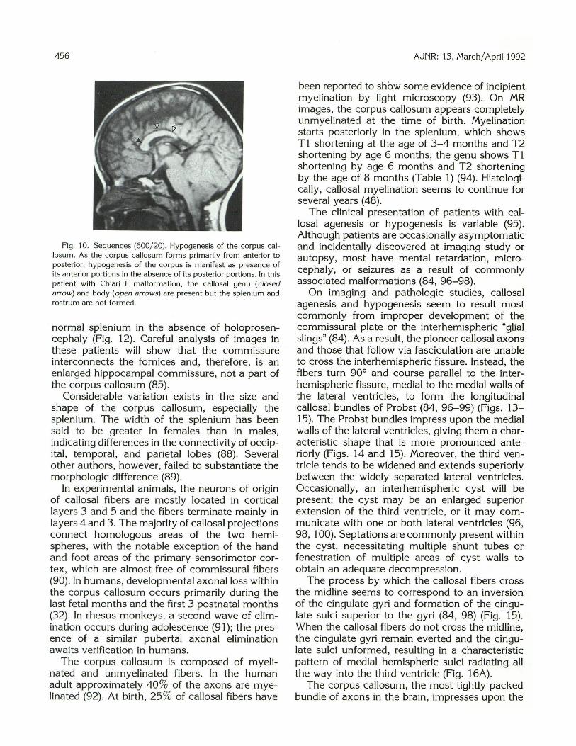
the location of the crossing of the initial pioneer fibers to form the rostral portion of the genu and the callosal rostrum (79). Because the posterior growth occurs more rapidly than the anterior growth (reflecting the later growth of the frontal lobes), the rostrum is the last part of the corpus callosum to form, after the genu, body, and splenium (79, 84). As a result of this characteristic sequence of growth, hypogenesis of the corpus callosum, in which callosal formation is incomplete, manifests as presence of the early formed portions (genu, genu and body, or genu, body and splenium) with absence of the portions
formed later (posterior body, splenium and rostrum, splenium and rostrum, or rostrum alone) (84) (Fig. 10). The sole exceptions to this rule are patients with holoprosencephaly, in which the posterior corpus callosum appears to form in the absence of the genu, rostrum, and, usually, anterior body (85) (Fig. 11 ). Although this interhemispheric commissure of holoprosencephaly has been called a "pseudosplenium" and a "pseudocallosum" (86), it appears to consist of nearly normal transverse neocortical callosal fibers (85, 87). Occasionally, patients with callosal agenesis will have a large commissure in the region of the
456
Fig. 10. Sequences (600/ 20). Hypogenesis of the corpus callosum. As the corpus callosum forms primarily from anterior to posterior, hypogenesis of the corpus is manifest as presence of its anterior portions in the absence of its posterior portions. In this patient with Chiari II malformation, the callosal genu (closed arrow) and body (open arrows) are present but the splenium and rostrum are not formed.
normal splenium in the absence of holoprosencephaly (Fig. 12). Careful analysis of images in these patients will show that the commissure interconnects the fornices and, therefore, is an enlarged hippocampal commissure, not a part of the corpus callosum (85).
Considerable variation exists in the size and shape of the corpus callosum, especially the splenium. The width of the splenium has been said to be greater in females than in males, indicating differences in the connectivity of occipital, temporal, and parietal lobes (88). Several other authors, however, failed to substantiate the morphologic difference (89).
In experimental animals, the neurons of origin of callosal fibers are mostly located in cortical layers 3 and 5 and the fibers terminate mainly in layers 4 and 3. The majority of callosal projections connect homologous areas of the two hemispheres, with the notable exception of the hand and foot areas of the primary sensorimotor cortex , which are almost free of commissural fibers (90) . In humans, developmental axonal loss within the corpus callosum occurs primarily during the last fetal months and the first 3 postnatal months (32). In rhesus monkeys, a second wave of elimination occurs during adolescence (91 ); the presence of a similar pubertal axonal elimination awaits verification in humans.
The corpus callosum is composed of myelinated and unmyelinated fibers. In the human adult approximately 40% of the axons are myelinated (92). At birth , 25 % of callosal fibers have
AJNR: 13, March/ Apri11992
been reported to show some evidence of incipient myelination by light microscopy (93). On MR images, the corpus callosum appears completely unmyelinated at the time of birth. Myelination starts posteriorly in the splenium, which shows T1 shortening at the age of 3-4 months and T2 shortening by age 6 months; the genu shows T1 shortening by age 6 months and T2 shortening by the age of 8 months (Table 1) (94). Histologically, callosal myelination seems to continue for several years (48).
The clinical presentation of patients with callosal agenesis or hypogenesis is variable (95). Although patients are occasionally asymptomatic and incidentally discovered at imaging study or autopsy , most have mental retardation , microcephaly, or seizures as a result of commonly associated malformations (84, 96-98).
On imaging and pathologic studies, callosal agenesis and hypogenesis seem to result most commonly from improper development of the commissural plate or the interhemispheric "glial slings" (84). As a result, the pioneer callosal axons and those that follow via fasciculation are unable to cross the interhemispheric fissure. Instead, the fibers turn 90° and course parallel to the interhemispheric fissure , medial to the medial walls of the lateral ventricles, to form the longitudinal callosal bundles of Probst (84, 96-99) (Figs. 13-15). The Probst bundles impress upon the medial walls of the lateral ventricles, giving them a characteristic shape that is more pronounced anteriorly (Figs. 14 and 15). Moreover, the third ventricle tends to be widened and extends superiorly between the widely separated lateral ventricles. Occasionally, an interhemispheric cyst will be present; the cyst may be an enlarged superior extension of the third ventricle, or it may communicate with one or both lateral ventricles (96, 98, 1 00). Septations are commonly present within the cyst, necessitating multiple shunt tubes or fenestration of multiple areas of cyst walls to obtain an adequate decompression.
The process by which the callosal fibers cross the midline seems to correspond to an inversion of the cingulate gyri and formation of the cingulate sulci superior to the gyri (84, 98) (Fig. 15). When the callosal fibers do not cross the midline, the cingulate gyri remain everted and the cingulate sulci unformed, resulting in a characteristic pattern of medial hemispheric sulci radiating all the way into the third ventricle (Fig. 16A).
The corpus callosum, the most tightly packed bundle of axons in the brain, impresses upon the

AJNR : 13, March/ April 1992
B
A B
lateral ventricles to give them their characteristic shape. Absence of the corpus results in characteristic deformities of the lateral ventricles ' (80, 97, 98). As a result of the absence of the genu, the frontal horns are straighter and more widely separated than normal (Fig. 168) and the foramina of Monro are elongated. Anteriorly, the firm, deep gray matter nuclei help the frontal horns to retain their normal size. Posteriorly, absence of the callosal splenium results in a variable, occasionally dramatic, enlargement of the trigones and occipital horns of the lateral ventricles (colpocephaly) (Fig. 168) (80, 96, 97, 101). Marked enlargment of the posterior ventricular system most likely results from a lack of organization of the parietal and occipital white matter in the absence of the splenium and forceps major (97, 98). The bodies of the lateral ventricles are affected when the callosal body is absent, resulting in strai(:Jht, parallel lateral ventricles (97). The temporal horns are usually enlarged, q§ yvel!,
457
Fig. 11. Holoprosencephaly. Presence of a splenium in the absence of the callosal genu and body.
A, Sagittal spin-echo (600/ 20) image shows presence of the callosal splenium (arrows) in the absence of the rostrum, genu, and body.
B, A xial spin-echo (2800/ 8) image shows absence of the anterior interhemispheric fi ssure and hypoplastic frontal horns, confirming holoprosencephaly.
Fig. 12. Callosal hypogenesis with large hippocampal commissure.
A, Sagittal spin-echo (600/20) image shows an apparent atypical callosal dysgenesis with a genu (open arrow) and splenium (closed arrow).
B, Coronal spin-echo (600/20) image shows that the "splenium" is actually a hippocampal commissure (arro ws), connecting the fornices, not neocortical structures.
possibly as the result of a hypogenesis , or other malformation, of the hippocampal formation ( 1 02). In patients with callosal agenesis, the hippocampal formation is small and vertically positioned, and the temporal horn is enlarged primarily in its inferolateral aspect (Fig. 158), possibly as a result of incomplete formation of the fibers of the tapetum (103). In contradistinction , patients with hydrocephalus will have a horizontally-oriented hippocampus of normal size, and the temporal horns will show symmetrical enlargement ( 1 03).
As the axons of the anterior commissure and the hippocampal commissure presumably cross the interhemispheric fissure by a process similar to those of the corpus callosum, it is not surprising that the smaller anterior and hippocampal commissures are frequently abnormal in callosal agenesis. The anterior commissure is usually present and occasionally hypertrophied (11 %) or hypoplastic (18%) (96, 104), whereas the hippo-

458
Fig. 13. Probst bundles. In callosal agenesis, . the axons that normally cross in the corpus callosum (dotted lines) form bundles that course parallel to the interhemispheric fissure (solid lines), medial to the bodies of the lateral ventricles. (Reprinted with permission from Barkovich (1 01.)
. Septum ·: pellucidum
·: ...
Fig. 14. Schematic of coronal section through acallosal brain . The bodies of the lateral ventricles have a crescentic shape as a result of being impressed upon by the medially lying Probst bundles. The third ventricle extends upward between the lateral ventricles, sometimes entering the interhemispheric fissure. The fornices are laterally displaced.
campal commissure, although usually absent, is sometimes present and very large (85, 96, 104). Hypogenesis of the corpus callosum with an associated large hippocampal commissure (Fig. 12) can be separated from normal callosal formation with secondary destruction or degeneration of the midportion of the callosal body by inspection of
AJNR: 13, March/ April 1992
the cingulate gyrus. A normally inverted cingulate gyrus with normal cingulate sulcus will be present in those patients with secondary callosal destruction, whereas those patients with callosal hypogenesis will have cingulate gyrus inversion and a normal cingulate sulcus only as far posteriorly as the corpus callosum has formed. This concept can be extended to all cases in which questions arise as to primary callosal hypogenesis versus secondary callosal destruction. In addition, inspection of the cerebral hemispheres will usually show hemispheric brain injury in those cases in which the corpus is secondarily destroyed.
Agenesis of the corpus callosum with longitudinal Probst bundles must be distinguished from extreme congenital hypoplasia or atrophy of the corpus seen in congenital white matter hypoplasia, a progressive familial disease that presents in childhood with severe encephalopathy. In this disorder, a minute contingent of well-myelinated fibers crosses the midline and Probst bundles are
8 Fig. 15. Callosal agenesis. Presence of Probst bundles, enlarge
ment of the temporal horns, and lack of inversion of the cingulate gyri.
A, Normal patient; coronal SE (600/20). The cingulate gyri are normally inverted, pointing slightly upward (arrows). Normal appearance of frontal horns.
8 , Acallosal patient; coronal SE (600/20). Cingulate gyri remain everted, pointing slightly downward (open arrows). Probst bundles (large arrows) compress the medial aspects of the lateral ventricles, giving them a crescentic appearance. Note that the temporal horns are enlarged, primarily inferiorly and laterally (closed arrows).

AJNR: 13, March/ April 1992
A
8 Fig. 16. Callosal agenesis with colpocephaly. A, Sagittal spin-echo (600/15) image shows absence of the
corpus callosum with medial hemispheric sulci (arrows) radiating all the way into the third ventricle as a result of the lack of inversion of the cingulate gyri.
8 , Axial spin-echo (3000/120) image shows marked enlargement of the trigones of the lateral ventricles (open arrows) with normal sized fronta l horns. The fronta l horns (closed arrows) are straighter and more widely separated than normal. This infant, who presented with seizures, had a normal head size (40th percentile).
absent. The cerebral white matter is hypoplastic, and the pyramidal tracts are absent in the medulla. No evidence of demyelination, dysmyelination, or degeneration of cortical neurons is seen on pathologic studies. Oligodendrocytes are nor-
459
mal (1 05-1 07). The ongm of congenital white matter hypoplasia has been postulated to be a primary defect of axonal development consisting of a pathologic extension of the normal phenomenon of axonal elimination (1 07). On computed tomography, the ventricular deformity is similar to that of classical callosal agenesis. Although different varieties of congenital callosal anomalies need further study and clarification, recognition on MR imaging of primary hypogenesis of the corpus callosum and of the cerebral white matter is important, because this condition apparently tends to recur in families, especially when associated with absence of the medullary pyramids (1 07).
Poorly described defects of the corpus callosum, usually with absence of the medullary pyramids, have been described in several familial metabolic disorders, including mitochondrial diseases ( 1 08).
Acknowledgment
We gratefully acknowledge the help of Dr Pierre Gressens.
References
1. Carpenter M . Sutin J . Human neuroanatomy. 8th ed. Baltimore:
Williams & Wilkins. 1983:36- 38
2. Caviness V Jr. Normal development of the cerebral neocortex. In:
Evrard P, Minkowski A . eds. Developmental neurobiology. New
York: Raven , 1989:1- 10
3. Marin-Padilla M. Early ontogenesis of the human cerebral cortex.
In: Peter A , Jones E. eds. Cerebral cortex. Vol. 7, Development and
maturation of the cerebral cortex. New York: Plenum, 1988:1-34
4. Rakic P. Neuronal migration and contact guidance in the primate
telencephalon. Postgrad Med J 1978;54:25-40
5. Sidman RL. Rakic P. Development of the human central nervous
system. In: Haymaker W, Adams RD. eds. Histology and histopa
thology of the nervous system. Springfield, II : Thomas, 1982:73
6. Smith S. Neuronal cytomechanics: the actin-based motility of
growth cones. Science 1988;242: 708-715
7. Norris C, Kalil K. Morphology and cellular interactions of growth
cones in the developing corpus ca llosum. J Camp Neural 1990;
293:268-281 8. Lockerbie R. Biochemical pharmacology of isolated neuronal growth
cones: implications for synaptogenesis. Brain Res Rev 1990;15:
145-165
9. Patterson P. On the importance of being inhibited, or say ing no to
growth cones. Neuron 1988; 1 :263-267
10. Spiegelman B, Lopata M, Kirschner M . Aggregation of microtubule
in itiation sites preceding neurite outgrowth in mouse neuroblastoma
cells. Cell 1979;16:253- 263
11. Dodd J , Jessell TM. Axon guidance and patterning of neuronal
projections in vertebrates. Science 1988;242:692-699
12. Tomasell i K . Neugebauer K. Bixby J, Lilien J , Reichardt L. N
cadherin and integrins: two receptor systems that mediate neuronal
process outgrowth on astrocy te surfaces. Neuron 1988; I :33-43

460
13. Takeichi M. The cadherins: cell -cell adhesion molecules controll ing
animal morphogenesis. Development 1988; 102:639-655
14. Linnemann D, Bock E. Cell adhesion molecules in neural develop
ment. Dev 1'/eurosci 1989; 11 :149-173
15. Hatta K, Takagi S, Fujisawa H, Takeichi M. Spatial and temporal
expression pattern of N-cadherin cell adhesion molecules correlated
with morphogenetic processes of chicken embryos. Dev Bioi 1987;
120:2 15-227
16. Hall D, Neugebauer K, Reichardt L. Embryon ic neural retinal cell
response to extracellular matrix proteins: developmental changes
and effects of the cell substratum attachment antibody (CSA T). J
Cell Bioi 1987; 104:623-634
17. Ruoslahti E, Pierschbacher M. New perspectives in cell adhesion:
RGD and integrins. Science 1987;218:491-497
18. Caroni P, Schwab M . Two membrane protein fractions from rat
central myelin with inhibitory properties for neurite growth and
fibroblast spreading. J Cell Bioi 1988; 106:1281-1288
19. Hammarback J , Letourneau P. Neurite extension across regions of
low cell-substratum adhesiv ity: implications for the guidepost hy
pothhesis of axonal pathfinding. Dev Bioi 1986; 11 7:655-662
20. Hammarback J, McCarthy J, Palm S, Furcht L, Letourneau P.
Growth cone guidance by substrate-bound Jaminin pathways is
correlated with neuron-_to-pathway adhesivity. Dev Bioi 1988; 126:
29-39
21. Kuwada J . Cell recogn ition by neurona l growth cones in a simple
vertebrate embryo. Science 1986;233: 7 40-7 46
22. Dodd J , Morton S, Karagogeos D, Yamamoto M, Jessell T . Spatial
regulation of axonal glycoprotein expression on subsets of embry
onic spinal neurons. Neuron 1988; 1:105-11 6
23. Heffner C, Lumsden A , O'Leary D. Target control of collateral
extension and directional axon growth in the mammalian brain.
Science 1990;247 :217- 220
24. Eichele G, Thaller C. Characterization of concentration gradients of
a morphogenetically active retinoid in the chick limb bud. J Cell
Bioi 1987;105:1917-1923
25. Walter J , Henke-Fahle S, Bonhoeffer F. Avoidance of posterior
tecta! membranes by temporal retinal axons. Development 1987;
101 :909-913
26. Walter J , Kern-Veits B, Huf J , Stolze B, Bonhoeffer F. Recognition
of position~specific properties of tecta! cell membranes by retinal
axons in vitro. Development 1987;101:695-696
27. Hogan D, Berman N. Growth cone morphology, axon trajectory, and
branching patterns in the neonatal rat corpus callosum. Dev Brain
Res 1990;53:283-287
28. Innocenti G. Growth and reshaping of axons in the establishment of
visual callosal connections. Science 1981 ;212:824- 826
29. Berbel P, Innocenti G. The development of the corpus callosum in
cats: a light- and electron-microscopic study. J Comp 1'/euro/ 1988;
176:132-156
30. O'Leary D, Stanfield B. A transient pyramidal tract projection from
the visual cortex in the hamster and its removal by selective
collateral elimination. Dev Brain Res 1986;27:87-99
31. Williams R, Bastiani B, Lia B, Chalupa L. Growth cones, dying axons,
and developmental fluctuations in the fiber population of the eat's
optic nerve. J Camp 1'/euro/ 1986; 146:32-69
32. Clarke S, Kreftsik R, van der Leos H, Innocenti G. Forms and
measures of adult and developing human corpus ca llosum: is there
sexual dimorphism? J Camp 1'/euro/ 1989;280:213-230
33. Nelson P, Fields R, Yu C, Neale E. Mechanisms involved in activity
dependent synapse formation in mammalian central nervous system
cell cultures. J 1'/eurobio/ 1990;21: 138-156
34. Rakic P, Riley K . Overproduction and elimination of retinal axons in
the fetal rhesus monkey. Science 1983;219:1441-1444
35. Lichtman J, Balice-Gordon R. Understanding synaptic competition
in theory and practice. J 1'/eurobio/ 1990;21 :99-106
AJNR: 13, March/ April 1992
36. Cowan W, Fawcett J , O'Leary D, Stanfield B. Regressive events in
neurogenesis. Science 1984;225: 1258-1265
37. Zecev ic N, Bourgeois J-P, Rakic P. Changes in synaptic density in
motor cortex of rhesus monkey during fetal and postnata l life. Dev
Brain Res 1989;50: 11-32
38. Baloga L. Oligodendrocytes, key cells in myelination and target in
demyelinating diseases. J f'leurosci Res 1985; 14:1-20
39. Friede R, Bischhausen R. How are sheath dimensions affected by
axon caliber and internode length? Brain Res 1982;235:335-350
40. Ritchie J . Physiological basis of conduction in myelinated nerve
fibers. In: Morell P, ed. Myelin. 2nd ed . New York: Plenum, 1984:
117-145
41. Ludin H. Function of myelin in the normal nerve fiber. 1'/europedia
trics 1984;15(suppl):21-23
42. Norton W, Cammer W. Isolation and characterization of myelin. In:
Morell P, ed. Myelin. 2nd ed. New York: Plenum, 1984:147-195
43. Braun P. Molecular organization of myelin. In: Morell P, Myelin. 2nd
ed. New York : Plenum, 1984:97-116
44. O'Brien J . Lipids and myelination. in: Himwich W, ed. Developmental
neurobiology. Springfield, IL: Thomas, 1970:262-286
45. Pettman B, Delaunoy J , Courageot J , Devilliers G, Sensenbrenner
M . Rat brain cells in culture: effects of brain extracts on the
development of oligodendroglial-like cells. Dev Bioi 1980; 75:
278-287
46. Bhat S, Barbarese E, Pfeiffer S. Requirement for nonoligodendrocyte
cell signals for enhanced myelinogenic gene expression in long-term
cultures of purified rat oligodendrocytes. Proc /'/at/ Acad Sci USA
1981 ;78:1283-1287
47. Brody B, Kinney H, Kloman A, Gilles F. Sequence of central nervous
system myelination in human infancy. I. An autopsy study of
myelination. J 1'/europatho/ Exp 1'/euro/ 1987;46:283-301
48. Yakovlev P, Lecours A. The myelogenetic cycles of regional matu
ration of the brain. In: Minkowski A, ed. Regional development of
the brain in early life. Oxford, England: Blackwell , 1967:3-70
49. Barkovich AJ, Kjos BO, Jackson JD.E. , Norman D. Normal matu
ration of the neonatal and infant brain: MR imaging at 1.5 T .
Radiology 1988; 166:173-1 80
50. Holland BA, Haas DK, Norman D, Brant-Zawadzki M , Newton TH.
MRI of normal brain maturation. AJ/'JR 1986;7:201-208
51. Johnson M, Pennock J , Bydder G, eta!. Clinical NMR imaging of
the brain in children: normal and neurologic disease. AJR 1983;
141:1005-1018
52. McArdle CB, Richardson CJ, Nicholas DA, Mirfakhraee M, Hayden
CK, Amparo EG. Developmental features of the neonatal brain: MR
imaging. I. Gray-white matter differentiation and myelination. Ra
diology 1987; 162:223-229
53. Dietrich R, Bradley W , Zaragoza E, et a!. MR evaluation of early
myelination patterns in normal and developmentally delayed infants.
AJ/'JR 1988;9:69-76
54. Martin E, Kilkinis R, Zuerrer M , et a!. Developmental stages of
human brain: an MR study. J Comput Assist Tomogr 1988;12:
917-922
55, Valk J , van der Knaap M. Myelination and retarded myelination. In:
Valk J , van der Knaap M , eds. Magnetic resonance of myelin,
myelination, and myelin disorders. Heidelberg , Germany: Springer
Verlag, 1989:26-65
56. Williams D, Elster A, Cox T. Cranial MR imaging in rhizomelic
chondrodysplasia punctata. AJ/'JR 1991;12:363-365
57. Valk P, Dillon W. Radiation injury of the brain. AJ/'JR 1991 ;12:
45-62
58. Weller R, Williams B. Cerebral biopsy and assessment of brain
damage in hydrocephalus. A rch Dis Child 1975;50:763-768
59. Shaw D, Maravilla K, Weinberger E, Garretson J , Trahms C, Scott
C. MR imaging of phenylketonuria. AJ/'JR 1991;12:403-406

AJNR: 13, March/ April 1992
60. Brismar J, Aqeel A , Brismar G, Coates R, Gascon G, Ozand P. Maple
syrup urine disease: findings on CT and MR scans of the brain in I 0
infants. AJNR 1990; II: 1219-1228
61. Flodmark 0, Lupton B, Li D, et al. MR imaging of periventricu lar
leukomalacia in childhood. AJNR 1989; I 0: 111 - 11 8
62. Volpe J: Neurology of the newborn. Philadelphia: Saunders, 1987
63. Barkovich AJ, Truwit CL. MR of perinatal asphyxia: correlation of
gestational age with pattern of damage. AJNR 1990; II
64. Kuban K, Gilles F. Human telencephalic angiogenesis. Ann Neural
1985; 17:539-548
65. Mayer P, Kier E. The controversy of the periventricular white matter
circulation: a review of the anatomic literature. AJNR 1991;12:
223-228
66. Rodriguez J , Claus D, Verellen G, Lyon G. Periventricular leukoma
lacia with disappearance of peri ventricu lar cysts at ultrasound scan
ning: ultrasonic and neuropathological correlations. Dev Med Child
Neurol 1990;32:359-367
67. Gilles FH, Leviton A, Dooling EC. The developing human brain.
Boston: John Wright-PSG, 1983
68. Raybaud C. Destructive lesions of the brain Neuroradiology 1983;
25:265- 291
69. Suzuki K. Biochemical pathogenesis of genetic leukodystrophies:
compari son of metachromatic leukodystrophy and globoid cell leu
kodystrophy (Krabbe's disease). Neuropediatrics 1984; 15(suppl):
32- 36
70. Morrell P, Wiesmann U. A correlative synopsis of the leukodystro
phies. Neuropediatrics 1984; 15(supp1):62-65
7 1. van der Knaap M, Valk J. The reflection of histology in MR imaging
of Pelizaeus-Merzbacher disease. AJNR 1989; I 0:99-103
72. Kolodny E. Metachromatic leukodystrophy and multiple sulfatase
deficiency. In: Scriver C, Beaudet A , Sly W, Valle D, eds. The
metabolic basis of inherited diseases. New York: McGraw Hill, 1989:
1721-1750
73. Swaiman K, Menkes J , DeVivo D, Prensky A. Metabolic disorders
of the central nervous system. In: Swaiman K, Wright F, eds. The
practice of pediatric neurology. 2nd ed. St. Louis: Mosby, 1982:
472-600
74. Norton W, Cammer W. Chemical pathology of diseases involving
myelin. In: Morell P, ed. Myelin. 2nd ed. New York: Plenum, 1984:
369-403
75. van der Knaap M . Myelination and myelin disorders: a magnetic
resonance study in infants, children and young adul ts. Thesis. Free
University of Amsterdam and University of Utrecht, Netherlands,
1991
76. Wiggins R. Myelination: a critical stage in development. Neurotoxi
cology 1986;7:103-120
77. Wiggins R. Myelin development and nutritional insufficiency. Brain
Res Rev 1982;4:151-175
78. Muller F, O'Rahilly R. The development of the human brain and the
closure of the rostral neuropore at stage 11. Anal Embryo/ 1986;
175:205-222
79. Rakic P, Yakovlev Pl. Development of the corpus callosum and
cavum septae in man. J Camp Neuro/1968;132:45-72
80. Loeser J , Alvord E. Agenesis of the corpus callosum . Brain 1968;
91:553-570
81 . Lamantia A, Rakic P. Cytologica l and quantitative characteristics of
four cerebral commissures in the rhesus monkey. J Camp Neural
1990;291 :520-537
82. Silver J, Lorenz S, Wahlsten D, Coughlin J. Axonal guidance during
development of the great cerebral commissures: descriptive and
experimental studies, in vivo, on the role of preformed glial path
ways. J Camp Neuro/ 1982;210:10-29
83. Altman J , Bayer S. Horizontal compartmentation in the germinal
matrices and intermediate zone of the embryonic rat cerebra l cortex.
Exp Neuro/1 990; 107:36-47
461
84. Barkovich AJ, Norman D. Anomalies of the corpus callosum: cor
relation with further anomalies of the brain. AJNR 1988;9:493-501
85. Barkovich AJ . Apparent atypica l callosal dysgenesis: analysis of MR
findings in six cases and their relationship to holoprosencephaly .
AJNR 1990; II :333- 340
86. Probst FP. The prosencephalies. Berlin: Springer-Verlag, 1979:46
87. Fleming G, Norman R. Arrhinencephaly with incomplete separation
of the cerebral hemispheres. J M ent Sci 1942;88:341 -355
88. Holloway R, Lacoste M. Sexual dimorphism in the human corpus
callosum: an ex tension and repl ication study. Human Neurobiol
1986;5:87-91
89. Weis S, Weber G, Wenger E, Kimbacher M. The Controversy about
a sexual dimorphism of the human corpus callosum. lnt J Neurosci
1989;47: 169-173
90. Ramaekers G, Njiokiktjien C. The child 's corpus callosum. Amster
dam: Suyi , 1991 :382
91. Lamantia A, Rakic P. Axon overproduction and elimination in the
corpus callosum of the developing rhesus monkey. J Neurosci 1990;
10:2156-2175
92. Tomasch J . Size, distribution and number of fibers in the human
corpus ca llosum. Anal Rec 1954;119:119-135
93. Gilles F, Shankle W, EC D. Myelinated tracts: growth patterns. In:
Gilles F, Leviton A , Dooling E, eds. The developing human brain:
growth and epidemiologic neuropathology. Boston : John Wright
PSG, 1983:117-183
94. Barkovich A, Kjos B. Normal postnatal development of the corpus
ca llosum as demonstrated by MR imaging. AJNR 1988;9:487-491
95. Warkany J, Lemire R, Cohen MJ. Mental retardation and congenital
malformations of the central nervous system. Chicago: Yearbook,
1981 :224-243
96. Probst FP. Agenesis of the corpus callosum. Acta Radio/ 1973;
331 (suppl): 1-150
97. Kendall BE. Dysgenesis of the corpus callosum. Neuroradiology
1983;25:239-256
98. Friede RL. Develop;T,ental neuropathology. 2nd ed . Berlin: Springer
Verlag, 1989
99. Probst M. Uber den Bau des vollstandigen balkenlosen Grosshirns,
sowie uber Mikrogyrie und Heterotopie der grauen Substanz. Arch
Psychiatr Nervenkr 1901 ;34:709-786
100. Barth P, Uylings H, Stam F. ln terhemisphera l neuroepithelial (glio
ependymal) cysts , associated with agenesis of the corpus callosum
and neocortica l maldevelopment: a case study. Child 's Brain 1984;
11:3 12-319
101. Barkovich AJ. Pediatric neuroimaging. New York: Raven, 1990
102. Atlas S, Zimmerman R, Bilaniuk L, et al. Corpus callosum and limbic
system: neuroanatomic MR eva luation of developmental anomalies.
Radiology 1986; 160:355-362
103. Baker L, Barkovich A . The large temporal horn : MR analysis in
developmental brain anomalies versus hydrocephalus. AJNR 1992;
13:115-122
104. Loeser J , Alvord E. Clinicopathological correlations in agenesis of
the corpus ca llosum. Neurology 1968; 18:745-756
105. Chattha A , Richardson E. Cerebral white matter hypoplasia. Arch
Neuro/ 1977;34:137-140
106. Guazzi G, Stoppoloni G, Ventruto V, DiIorio G. lmmaturite neuronale
corticale avec agenesie des grandes commissures interhemisphe
riques et hypoplasie des voies optico-pyramides chez trois enfants
issus d'une meme famille. A cta Neural (Napoli) 1974;39:659-674
107. Lyon G, Arita F, Gallodec.E L, Vallee L, Misson J , Ferriere G. A
disorder of axonal development, necrotizing myopathy, cardiomy
opathy, and cataracts: a new famil ial disease. Ann Neuro/1990;27:
193-199
I 08. Kolodny E. Agenesis of the corpus callosum: a marker for inherited
metabol ic disease? (editorial). Neurology 1989;39