force fields for md simulations der waals interaction classical molecular dynamics bond definitions,...
TRANSCRIPT
Force Fields for MD simulations
• Topology/parameter files• Where do the numbers an MD code uses
come from?• How to make topology files for ligands,
cofactors, special amino acids, …• How to obtain/develop missing
parameters.

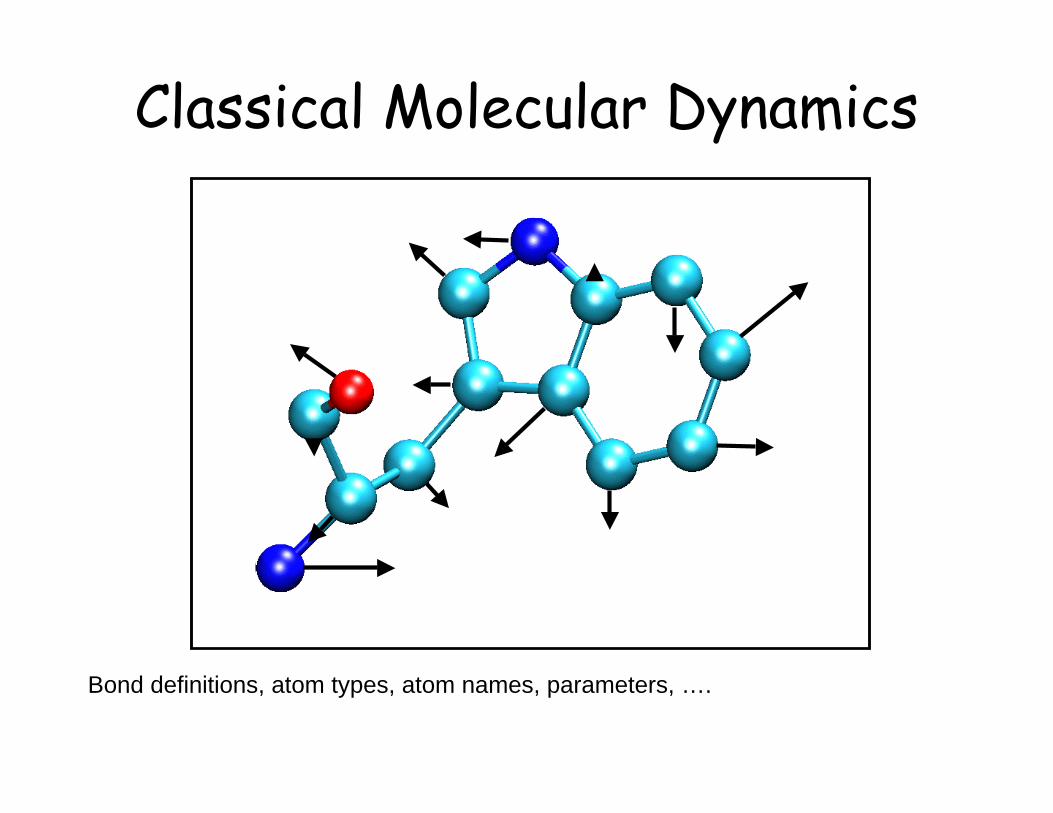
Classical Molecular Dynamics
ij
ji
r
qqrU
04
1)(
!"=
Coulomb interaction
!!
"
#
$$
%
&
''
(
)
**
+
,-
''
(
)
**
+
,=
6
min,
12
min,2)(
ij
ij
ij
ij
ijr
R
r
RrU .
van der Waals interaction
Classical Molecular Dynamics
Bond definitions, atom types, atom names, parameters, ….

Energy Terms Described inthe CHARMm Force Field
Bond Angle
Dihedral Improper
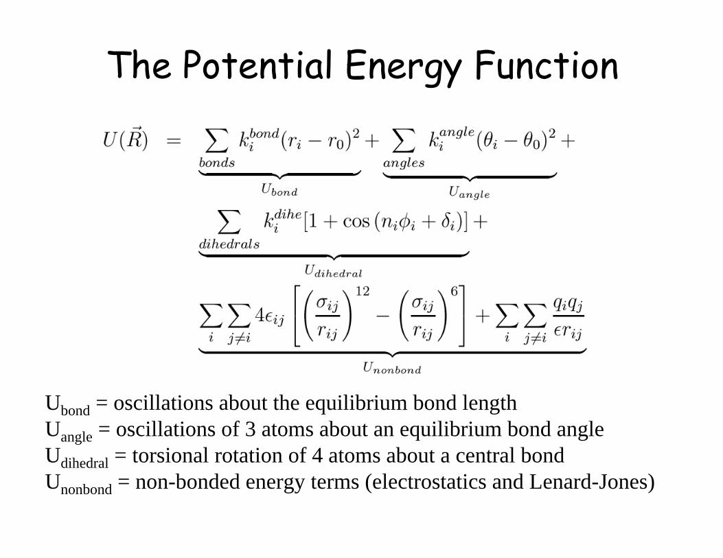
The Potential Energy Function
Ubond = oscillations about the equilibrium bond length
Uangle = oscillations of 3 atoms about an equilibrium bond angle
Udihedral = torsional rotation of 4 atoms about a central bond
Unonbond = non-bonded energy terms (electrostatics and Lenard-Jones)
!
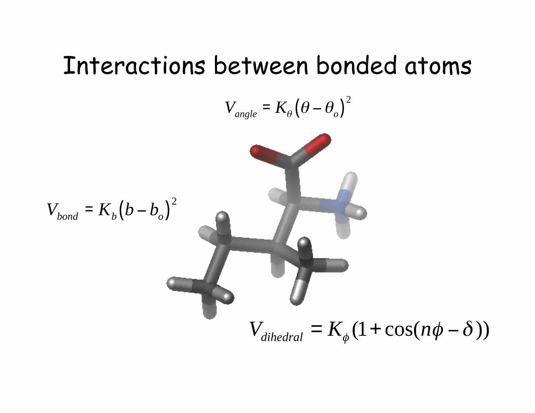
Vbond
= Kb
b " bo( )
2
!
Vangle
= K" " #"o( )2
))cos(1( !"" #+= nKVdihedral
Interactions between bonded atoms

Bond Energy versus Bond length
0
100
200
300
400
0.5 1 1.5 2 2.5
Bond length, Å
Po
ten
tial
En
erg
y,
kcal/
mo
l
Single Bond
Double Bond
Triple Bond
Chemical type Kbond bo
C-C 100 kcal/mole/Å2 1.5 Å
C=C 200 kcal/mole/Å2 1.3 Å
C=C 400 kcal/mole/Å2 1.2 Å
( )2obbond bbKV !=
Bond angles and improper terms have similar quadratic forms, but withsofter spring constants. The force constants can be obtained fromvibrational analysis of the molecule (experimentally or theoretically).

Dihedral energy versus dihedral angle
0
5
10
15
20
0 60 120 180 240 300 360
Dihedral Angle, degrees
Po
ten
tial
En
erg
y,
kcal/
mo
l
K=10, n=1
K=5, n=2
K=2.5, N=3
))cos(1( !"" #+= nKVdihedral
δ = 0˚
Dihedral Potential

qi: partial atomic charge
D: dielectric constant
ε: Lennard-Jones (LJ, vdW) well-depth
Rmin
: LJ radius (Rmin
/2 in CHARMM)
Combining rules (CHARMM, Amber)
Rmin i,j
= Rmin i
+ Rmin j
εi,j
= SQRT(εi * ε
j )
!!
"
#
$$
%
&
''
(
)
**
+
,-
''
(
)
**
+
,+.
6
min,
12
min,2
4 ij
ij
ij
ij
nonbonded
ij
ij
ji
r
R
r
R
Dr
qq/
0
Nonbonded Parameters

Electrostatic Energy versus Distance
-100
-80
-60
-40
-20
0
20
40
60
80
100
0 1 2 3 4 5 6 7 8
Distance, Å
Inte
racti
on
en
erg
y,
kcal/
mo
l
q1=1, q2=1
q1=-1, q2=1
From MacKerell
Note that the effect is long range.
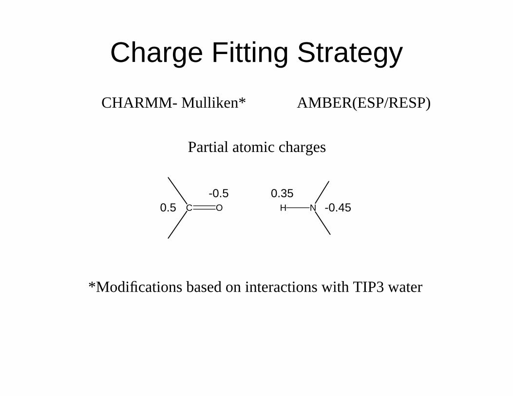
CHARMM- Mulliken* AMBER(ESP/RESP)
Partial atomic charges
C O H N0.5
-0.5 0.35
-0.45
*Modifications based on interactions with TIP3 water
Charge Fitting Strategy

CHARMM Potential Function
geometry
parameters
PDB file
PSF file
Parameter file
Topology

File Format/Structure
• The structure of a pdb file• The structure of a psf file• The topology file• The parameter file• Connection to potential energy terms
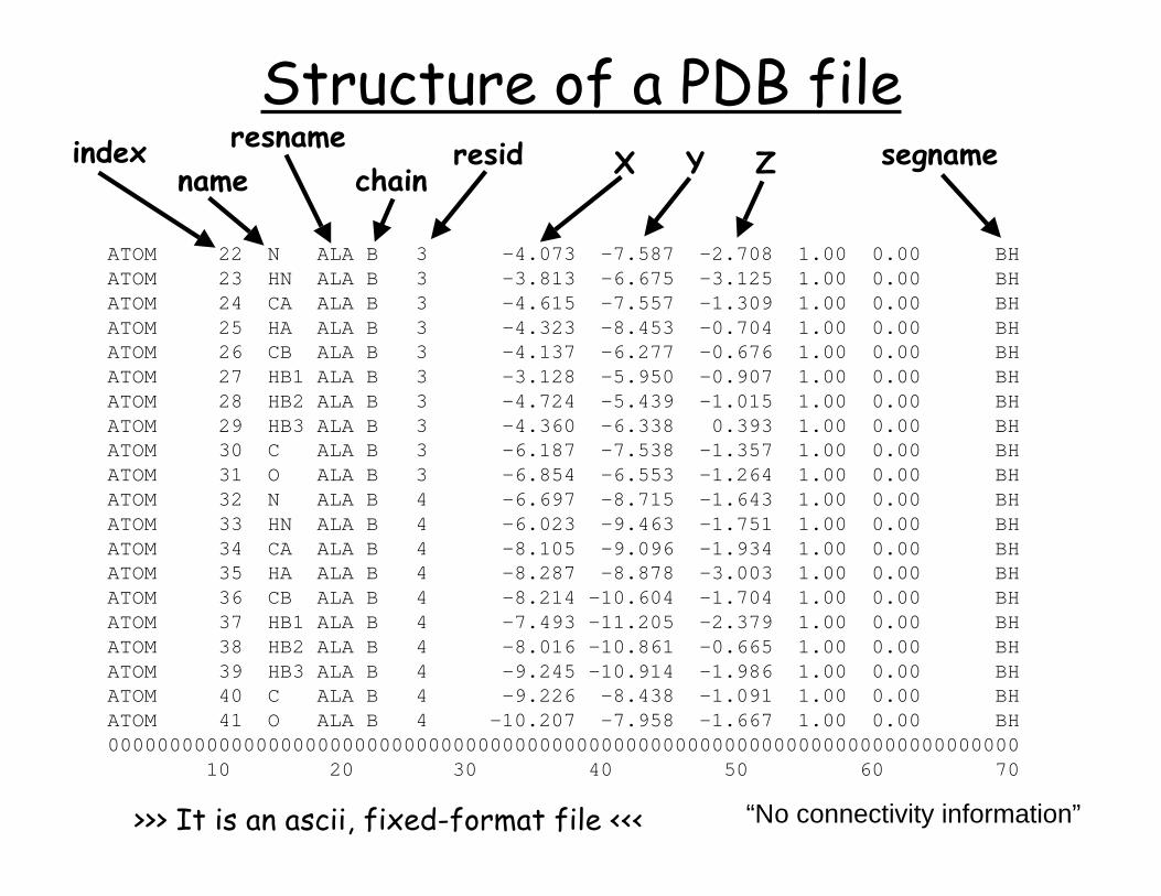
ATOM 22 N ALA B 3 -4.073 -7.587 -2.708 1.00 0.00 BH ATOM 23 HN ALA B 3 -3.813 -6.675 -3.125 1.00 0.00 BH ATOM 24 CA ALA B 3 -4.615 -7.557 -1.309 1.00 0.00 BH ATOM 25 HA ALA B 3 -4.323 -8.453 -0.704 1.00 0.00 BH ATOM 26 CB ALA B 3 -4.137 -6.277 -0.676 1.00 0.00 BH ATOM 27 HB1 ALA B 3 -3.128 -5.950 -0.907 1.00 0.00 BH ATOM 28 HB2 ALA B 3 -4.724 -5.439 -1.015 1.00 0.00 BH ATOM 29 HB3 ALA B 3 -4.360 -6.338 0.393 1.00 0.00 BH ATOM 30 C ALA B 3 -6.187 -7.538 -1.357 1.00 0.00 BH ATOM 31 O ALA B 3 -6.854 -6.553 -1.264 1.00 0.00 BH ATOM 32 N ALA B 4 -6.697 -8.715 -1.643 1.00 0.00 BH ATOM 33 HN ALA B 4 -6.023 -9.463 -1.751 1.00 0.00 BH ATOM 34 CA ALA B 4 -8.105 -9.096 -1.934 1.00 0.00 BH ATOM 35 HA ALA B 4 -8.287 -8.878 -3.003 1.00 0.00 BH ATOM 36 CB ALA B 4 -8.214 -10.604 -1.704 1.00 0.00 BH ATOM 37 HB1 ALA B 4 -7.493 -11.205 -2.379 1.00 0.00 BH ATOM 38 HB2 ALA B 4 -8.016 -10.861 -0.665 1.00 0.00 BH ATOM 39 HB3 ALA B 4 -9.245 -10.914 -1.986 1.00 0.00 BH ATOM 40 C ALA B 4 -9.226 -8.438 -1.091 1.00 0.00 BH ATOM 41 O ALA B 4 -10.207 -7.958 -1.667 1.00 0.00 BH 00000000000000000000000000000000000000000000000000000000000000000000000000 10 20 30 40 50 60 70
indexname
resnamechain
resid X Y Z segname
>>> It is an ascii, fixed-format file <<<
Structure of a PDB file
“No connectivity information”
Checking file structures
• PDB file
• Topology file
• PSF file
• Parameter file

Parameter Optimization StrategiesCheck if it has been parameterized by somebody else
Literature
Minimal optimizationBy analogy (i.e. direct transfer of known parameters)Quick, starting point
Maximal optimizationTime-consumingRequires appropriate experimental and target data
Choice based on goal of the calculationsMinimal database screening NMR/X-ray structure determinationMaximal free energy calculations, mechanistic studies, subtle environmental effects

• Identify previously parameterized compounds
• Access topology information – assign atom types,
connectivity, and charges – annotate changes
CHARMM topology (parameter files)
top_all22_model.inp (par_all22_prot.inp)
top_all22_prot.inp (par_all22_prot.inp)
top_all22_sugar.inp (par_all22_sugar.inp)
top_all27_lipid.rtf (par_all27_lipid.prm)
top_all27_na.rtf (par_all27_na.prm)
top_all27_na_lipid.rtf (par_all27_na_lipid.prm)
top_all27_prot_lipid.rtf (par_all27_prot_lipid.prm)
top_all27_prot_na.rtf (par_all27_prot_na.prm)
toph19.inp (param19.inp)
NA and lipid force fields have new LJ
parameters for the alkanes,
representing increased optimization of
the protein alkane parameters. Tests
have shown that these are compatible
(e.g. in protein-nucleic acid
simulations). For new systems is
suggested that the new LJ parameters
be used. Note that only the LJ
parameters were changed; the internal
parameters are identical
Getting Started
www.pharmacy.umaryland.edu/faculty/amackere/force_fields.htm
NH
N
NHO
OH
NH
N
NHO
OH
A B C
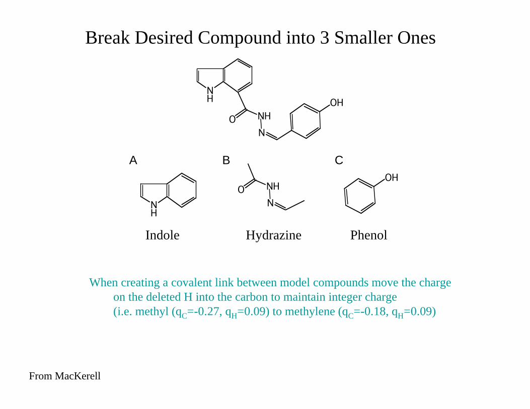
When creating a covalent link between model compounds move the charge
on the deleted H into the carbon to maintain integer charge
(i.e. methyl (qC=-0.27, qH=0.09) to methylene (qC=-0.18, qH=0.09)
Break Desired Compound into 3 Smaller Ones
Indole Hydrazine Phenol
From MacKerell
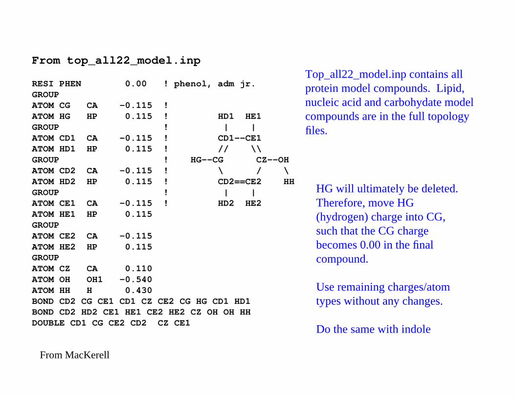
From top_all22_model.inp
RESI PHEN 0.00 ! phenol, adm jr.GROUPATOM CG CA -0.115 !ATOM HG HP 0.115 ! HD1 HE1GROUP ! | |ATOM CD1 CA -0.115 ! CD1--CE1ATOM HD1 HP 0.115 ! // \\GROUP ! HG--CG CZ--OHATOM CD2 CA -0.115 ! \ / \ATOM HD2 HP 0.115 ! CD2==CE2 HHGROUP ! | |ATOM CE1 CA -0.115 ! HD2 HE2ATOM HE1 HP 0.115GROUPATOM CE2 CA -0.115ATOM HE2 HP 0.115GROUPATOM CZ CA 0.110ATOM OH OH1 -0.540ATOM HH H 0.430BOND CD2 CG CE1 CD1 CZ CE2 CG HG CD1 HD1BOND CD2 HD2 CE1 HE1 CE2 HE2 CZ OH OH HHDOUBLE CD1 CG CE2 CD2 CZ CE1
HG will ultimately be deleted.
Therefore, move HG
(hydrogen) charge into CG,
such that the CG charge
becomes 0.00 in the final
compound.
Use remaining charges/atom
types without any changes.
Do the same with indole
Top_all22_model.inp contains all
protein model compounds. Lipid,
nucleic acid and carbohydate model
compounds are in the full topology
files.
From MacKerell
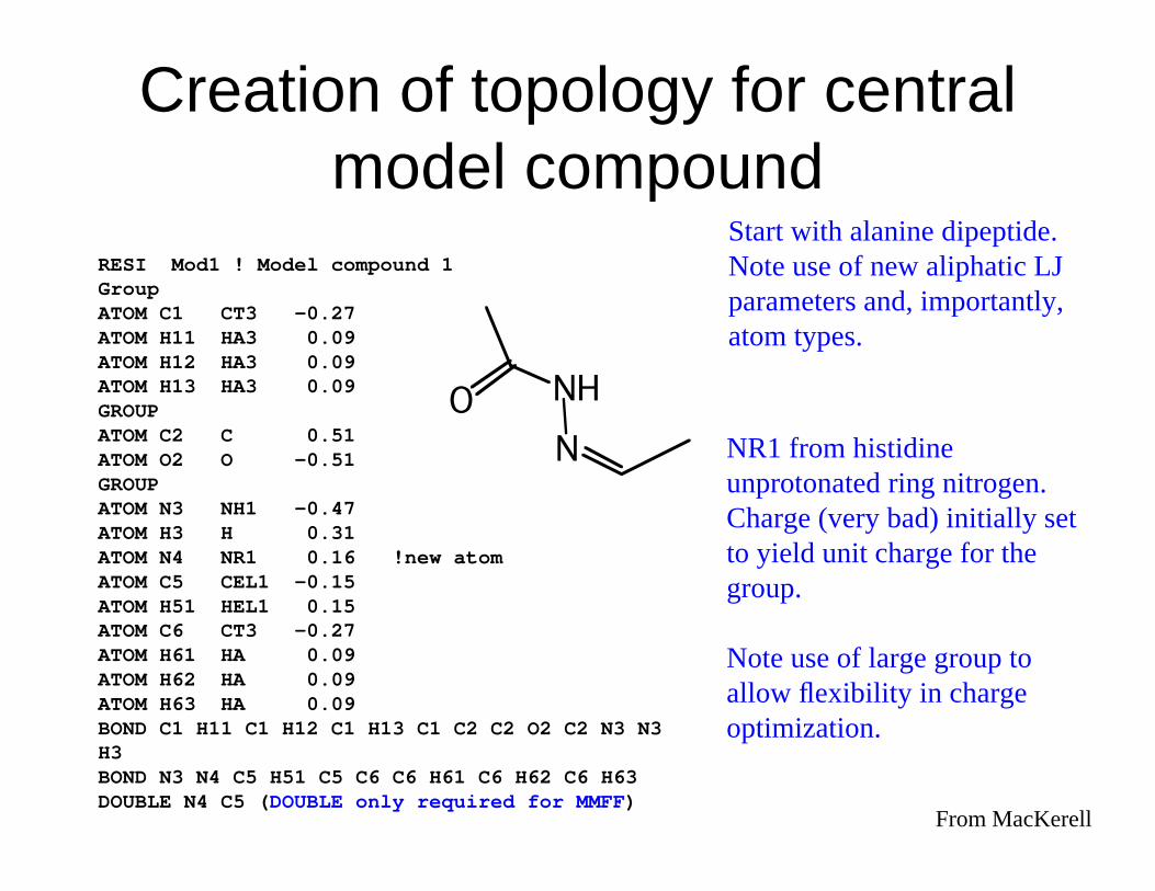
RESI Mod1 ! Model compound 1GroupATOM C1 CT3 -0.27ATOM H11 HA3 0.09ATOM H12 HA3 0.09ATOM H13 HA3 0.09GROUPATOM C2 C 0.51ATOM O2 O -0.51GROUPATOM N3 NH1 -0.47ATOM H3 H 0.31ATOM N4 NR1 0.16 !new atomATOM C5 CEL1 -0.15ATOM H51 HEL1 0.15ATOM C6 CT3 -0.27ATOM H61 HA 0.09ATOM H62 HA 0.09ATOM H63 HA 0.09BOND C1 H11 C1 H12 C1 H13 C1 C2 C2 O2 C2 N3 N3H3BOND N3 N4 C5 H51 C5 C6 C6 H61 C6 H62 C6 H63DOUBLE N4 C5 (DOUBLE only required for MMFF)
Start with alanine dipeptide.
Note use of new aliphatic LJ
parameters and, importantly,
atom types.
NR1 from histidine
unprotonated ring nitrogen.
Charge (very bad) initially set
to yield unit charge for the
group.
Note use of large group to
allow flexibility in charge
optimization.
N
NHO
From MacKerell
Creation of topology for centralmodel compound

1. RESP: HF/6-31G overestimates dipole
moments (AMBER)
2. Interaction based optimization (CHARMM)
Partial Atomic Charge Determination
Method Dependent Choices
For a particular force field do NOT change the QM
level of theory. This is necessary to maintain
consistency with the remainder of the force field.
From MacKerell

Starting charges??
Mulliken population analysis
Analogy comparison
Final charges (methyl, vary qC to maintain integer charge, q
H = 0.09)
interactions with water (HF/6-31G*, monohydrates!)
N
NO
H
CH3
H
CH3
From MacKerell

Comparison of analogy and optimized charges
N
NHO
Name Type Analogy Optimized
C1 CT3 -0.27 -0.27
H11 HA3 0.09 0.09
H12 HA3 0.09 0.09
H13 HA3 0.09 0.09
C2 C 0.51 0.58
O2 O -0.51 -0.50
N3 NH1 -0.47 -0.32
H3 H 0.31 0.33
N4 NR1 0.16 -0.31
C5 CEL1 -0.15 -0.25
H51 HEL1 0.15 0.29
C6 CT3 -0.27 -0.09
H61 HA 0.09 0.09
H62 HA 0.09 0.09
H63 HA 0.09 0.09

NH
N
NHO
OH
N
NHO
Dihedral optimization based on QM potential
energy surfaces (HF/6-31G* or MP2/6-31G*).
NH
N
NHO
OHNH
NH2O
HN
OH
From MacKerell

Parameterization of unsaturated lipids• All C=C bonds are cis, what does rotation about neighboring
single bonds look like?
Courtesy of Scott Feller, Wabash College

DHA conformations from MD• rotational barriers are
extremely small• many conformers are
accessible w/ shortlifetimes
Courtesy of Scott Feller, Wabash College
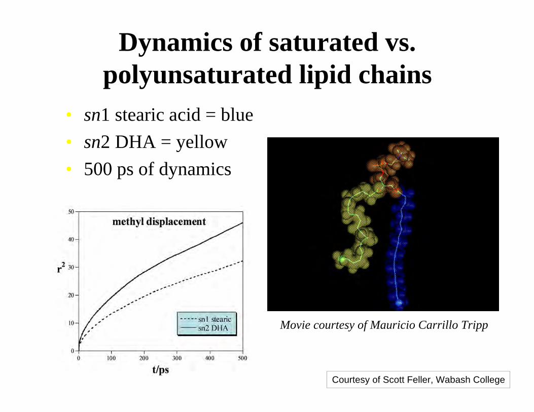
Dynamics of saturated vs.polyunsaturated lipid chains
• sn1 stearic acid = blue
• sn2 DHA = yellow
• 500 ps of dynamics
Movie courtesy of Mauricio Carrillo Tripp
Courtesy of Scott Feller, Wabash College
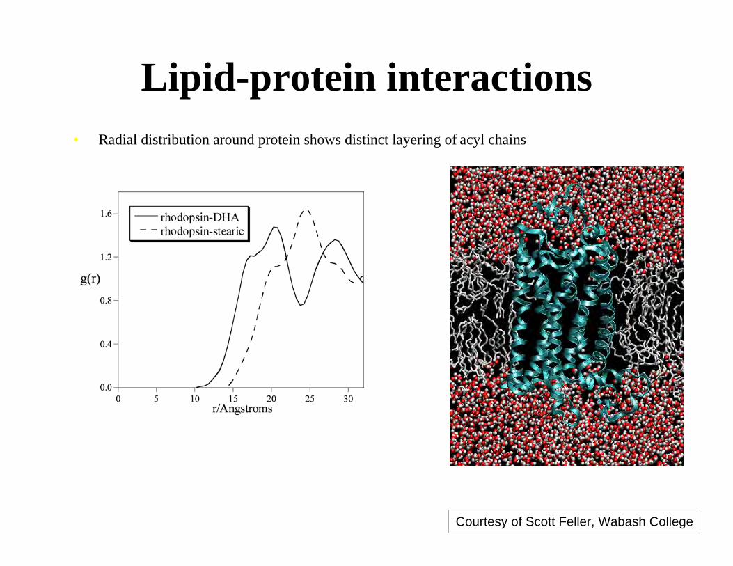
Lipid-protein interactions• Radial distribution around protein shows distinct layering of acyl chains
• DHA penetrates deeper into the protein surface
Courtesy of Scott Feller, Wabash College

Lipid-protein interactions• Decomposition of non-bonded interaction shows rhodopsin is strongly
attracted to unsaturated chain
• All hydrophobic residues are stabilized by DHA
4.2-2.4-10.3TRP
3.8-3.0-11.4ALA
1.8-10.4-18.6TYR
2.4-9.7-22.8MET
1.8-13.0-23.1LEU
2.5-9.6-24.0VAL
3.0-10.1-30.0ILE
2.0-22.6-44.9PHE
ratioUstearicUDHAresname
Courtesy of Scott Feller, Wabash College

Origin of protein:DHA attraction
• Flexibility of the DHA chain allows solvation
of the rough protein surface to occur with little
intra-molecular energy costCourtesy of Scott Feller, Wabash College
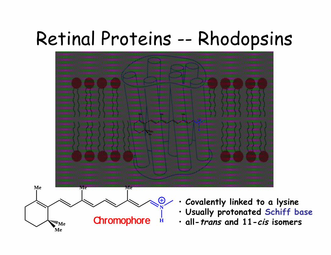
Retinal Proteins -- Rhodopsins
N
Me Me Me
MeMe
H
N
Me Me Me
MeMe
H
• Covalently linked to a lysine• Usually protonated Schiff base• all-trans and 11-cis isomersChromophore
N
Me Me Me
MeMe
H
⊕⊕⊕⊕⊕⊕⊕
N
Me Me Me
MeMe
H
7 9 11 13 15
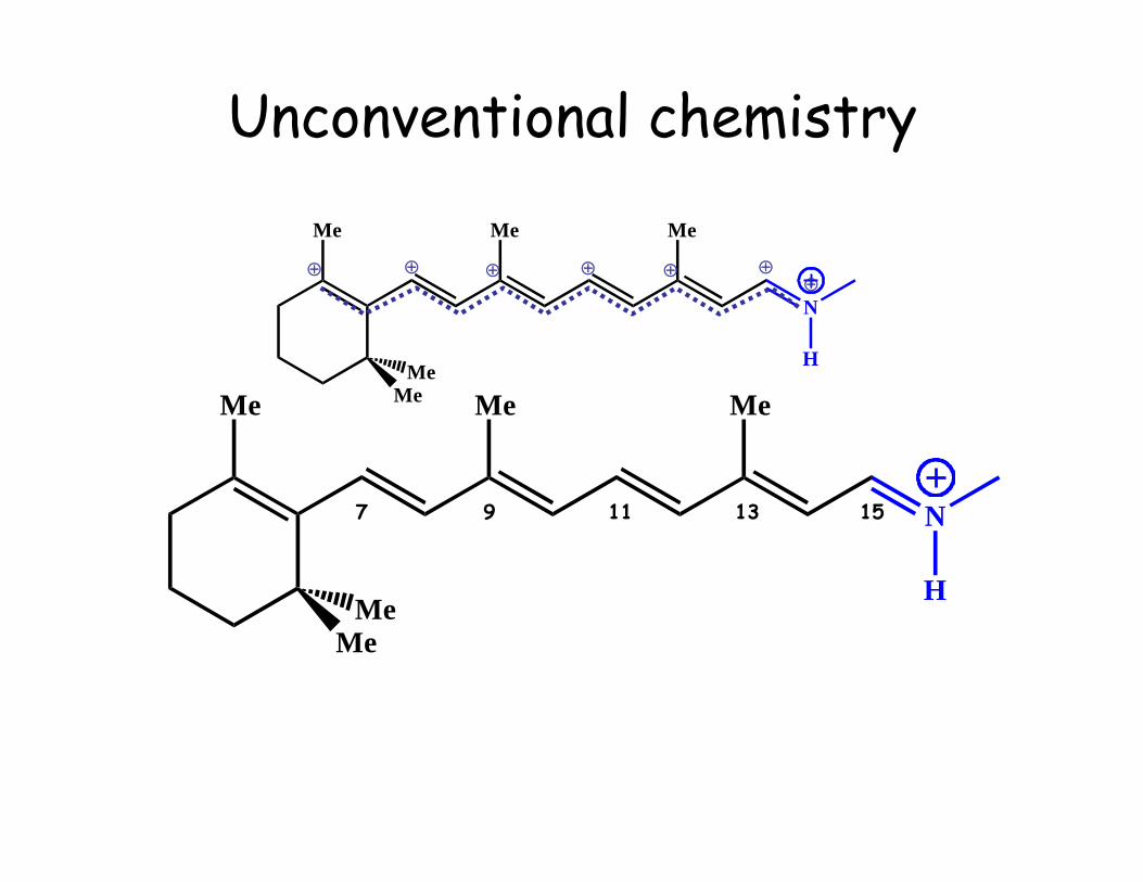
Unconventional chemistry

N1
C2C3
C4C5
C6C7
C8C9
C10C11
C12
C6
C1
C2
C3
C4
C5C7C8
C9
C10
C11C12
C13C14
C15N16
Lys216
H
B1B2
B3B4
B5
B6
+
Isomerization Barriers in retinal
DFT/6-31G**
S0
S1
KBR
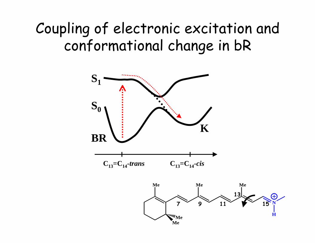
C13=C14-trans C13=C14-cis
Coupling of electronic excitation andconformational change in bR
N
Me Me Me
MeMe
H
7 9 1113
15

Inducing isomerization
500 nm~50 kcal/mole

Classical RetinalIsomerization in Rhodopsin
Twist Propagation
N
O
H N
H
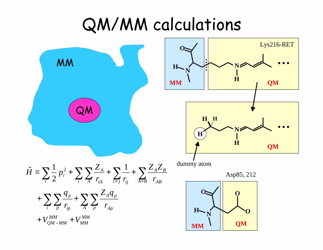
…MM QM
N
H
…QM
H
H H
dummy atom
MM
MM
MM
MMQM
A p Ap
pA
i p ip
p
ji BA AB
BA
iji A iA
A
i
i
VV
r
qZ
r
q
r
ZZ
rr
ZpH
++
++
+++=
!
> >
""""
" """"1
2
1ˆ 2
N
O
H
MM
O
O
QM
Lys216-RET
Asp85, 212
QM/MM calculations
QM
MM

Ab Initio QM/MM Excited State MD Simulation
QM
Quantum mechanical (QM)treatment of the chromophore,and force field (MM) treatment
of the embedding protein

QM/MM calculation of ATP hydrolysis

Coarse grain modeling of lipids
9 particles!
150 particles