for peer review only - connecting repositories · 2017-11-13 · for peer review only...
TRANSCRIPT

For Peer Review O
nly
Detection of human pathogenic Fusarium species in hospital
and communal sink biofilms by using a highly specific monoclonal antibody
Journal: Environmental Microbiology and Environmental Microbiology Reports
Manuscript ID EMI-2015-1627.R1
Manuscript Type: EMI - Research article
Journal: Environmental Microbiology
Date Submitted by the Author: 07-Jan-2016
Complete List of Authors: Al-Maqtoofi, Marwan; University of Exeter, Biosciences Thornton, Christopher; University of Exeter, Biosciences
Keywords: fungi, microbes and surfaces, new tools/technological developments, biofilm biology
Wiley-Blackwell and Society for Applied Microbiology
For Peer Review O
nlyFusarium-specific monoclonal antibody
1
Detection of human pathogenic Fusarium species in hospital and 1
communal sink biofilms by using a highly specific monoclonal 2
antibody 3
4
Marwan Al-Maqtoofi1,2 and Christopher R. Thornton
1* 5
1 Biosciences, Geoffrey Pope Building, University of Exeter, Stocker Road, Exeter, EX4 4QD, UK. 6
2 University of Basrah, College of Science, Biology Department, Basrah, Iraq. 7
*Corresponding author: Tel: +44 (0)1392 725172. Fax: +44 (0)1392 263434. E-mail address: 8
10
Summary 11
The fungus Fusarium is well known as a plant pathogen, but has recently emerged over recent years 12
as an opportunistic pathogen of humans. Habitats providing direct human exposure to infectious 13
propagules are largely unknown, but there is growing evidence that plumbing systems are sources 14
of human pathogenic strains in the Fusarium solani species complex (FSSC) and Fusarium 15
oxysporum species complexes (FOSC), the most common groups infecting humans. Here, we use a 16
newly developed Fusarium-specific monoclonal antibody (mAb ED7) to track FSSC and FOSC 17
strains in sink drain biofilms by detecting its target antigen, an extracellular 200kDa heat-stable 18
carbohydrate, in saline swabs. The diagnostic antigen was detectable in 52% of swab samples 19
collected from sinks across a University campus and a tertiary care hospital. The mAb was 100% 20
accurate in detecting FSSC, FOSC and F. dimerum species complex (FDSC) strains that were 21
present, as mixed fungal communities, in 83% of sink drain biofilms. Specificity of the ELISA was 22
Page 1 of 87
Wiley-Blackwell and Society for Applied Microbiology
For Peer Review O
nlyFusarium-specific monoclonal antibody
2
confirmed by sequencing of the internally transcribed spacer 1 (ITS1)-5.8S-ITS2 rRNA-encoding 1
regions of culturable yeasts and moulds that were recovered using mycological culture, while 2
translation elongation factor (TEF)-1α analysis of Fusarium isolates included FSSC 1-a, FOSC 33 3
and FDSC ET-gr, the most common clinical pathotypes in each group. 4
5
Originality-Significance Statement 6
• Development of a monoclonal antibody (mAb ED7) specific to Fusarium, a fungal genus 7
containing human and plant pathogens 8
• Fusarium diagnostic antigen detected in swabs of sink drain biofilms, with 100% accuracy 9
of mAb-based ELISA confirmed by ITS sequencing of mixed fungal communities 10
comprising human pathogenic yeasts and moulds 11
• First report describing the use of a highly specifc mAb to track human pathogenic fusaria, 12
demonstrating widespread occurrence of pathogen in communal and hospital sinks with 13
potential for nosocomial and community acquired infections 14
15
16
17
18
19
20
21
22
23
Page 2 of 87
Wiley-Blackwell and Society for Applied Microbiology
For Peer Review O
nlyFusarium-specific monoclonal antibody
3
Introduction 1
Species in the fungal genus Fusarium are ubiquitous environmental moulds, and pathogens of both 2
plants and animals (Zhang et al., 2006; Thornton and Wills, 2015). In immunocompromised 3
humans, such as patients with haematological malignancies and hematopoietic stem cell and solid 4
organ transplant recipients, Fusarium species are significant emerging pathogens, causing a 5
frequently fatal disseminated disease known as fusariosis with an associated mortality rate of 50-6
75% (Girmenia et al., 2000; Musa et al., 2000; Boutati and Anaissie, 1997; Dignani and Anaissie, 7
2004; Jensen et al., 2004; Nucci and Anaissie, 2007). In some tertiary cancer centres, Fusarium has 8
emerged as the second most common mould pathogen after Aspergillus (Walsh and Groll, 1999; 9
Muhammed et al., 2011). Regardless of human immune status, Fusarium species can cause 10
localised nail infections (onychomycosis)(Arrese et al., 1996), bone and joint infections (Koehler et 11
al., 2014), infections of burn wounds (Latenser, 2003), skin infections (Nucci and Anaissie, 2002; 12
Gurusidappa and Mamatha, 2011), and are the most frequent cause of mycotic eye infections known 13
as fungal keratitis (Jurkunas et al., 2009), leading to progressive corneal destruction and 14
endophthalmitis, with loss of vision or even loss of the affected eye (Dursun et al., 2003; Edelstein 15
et al., 2012). 16
A recent multistate outbreak of fungal keratitis in the USA and in Singapore and Hong Kong 17
was associated with contact lens solution contaminated with multiple strains of Fusarium and which 18
led to visual loss in many patients and the need for corneal transplantation (Chang et al., 2006). 19
While such outbreaks are rare, disseminated Fusarium infections and keratomycoses have increased 20
in frequency over the past decade (Koehler et al., 2014) and an increasing body of evidence 21
suggests that the main environmental sources of human pathogenic Fusarium species are 22
contaminated water systems (Doggett, 2000; Anaissie et al., 2002; Anaissie et al., 2011; Mehl and 23
Page 3 of 87
Wiley-Blackwell and Society for Applied Microbiology
For Peer Review O
nlyFusarium-specific monoclonal antibody
4
Epstein, 2008). A number of studies have recovered pathogenic Fusarium species from plumbing 1
fixtures and it is hypothesised that microbial biofilms on fixtures may serve as important reservoirs 2
of infectious Fusarium propagules in hospitals and homes (Mehl and Epstein, 2008; Short et al., 3
2011). 4
Identification of environmental reservoirs of human pathogenic mouldmolds including 5
Fusarium has typically relied on nucleic acid-based technologies following recovery of fungi using 6
selective media (Anaissie et al., 2002; Mehl and Epstein, 2008; Short et al., 2011; Anaissie et al., 7
2011; Rougeron et al., 2014). Recently, highly specific monoclonal antibodies (mAb) have been 8
used to identify pathogenic species or species complexes in environmental samples containing 9
mixed populations of yeasts and mouldmolds (Thornton, 2009; Davies and Thornton, 2014; 10
Thornton et al., 2015). While mAb-based approaches similarly rely on culture for recovery of fungi 11
from environmental samples, detection of diagnostic antigens in crude culture extracts using genus- 12
or species-specific mAbs offers an attractive alternative approach to pathogen detection, particularly 13
when combined with unsophisticated diagnostic modalities such as lateral-flow technology 14
(Thornton, 2008; Thornton, 2012). 15
In this study, we set out to determine whether a newly developed Fusarium-specific mAb 16
(ED7) could be used to track the fungus by detecting a water-soluble diagnostic antigen in swabs of 17
communal and hospital sinks. By using the mAb in an enzyme-linked immunsorbent assay 18
(ELISA), we show that it can differentiate Fusarium species from other unrelated yeast and 19
mouldmold pathogens of humans present in mixed fungal communities encountered in sink 20
biofilms. The ELISA represents a simple method for specific detection of Fusarium species in 21
environmental reservoirs and for identifying plumbing systems contaminated with the fungus. 22
Page 4 of 87
Wiley-Blackwell and Society for Applied Microbiology
For Peer Review O
nlyFusarium-specific monoclonal antibody
5
Results 1
Production of hybridoma cell lines, isotyping of mAb and specificity 2
A single fusion was performed and 389 hybridoma cell lines were screened for specificity against a 3
range of clinically relevant yeasts and molds (Table S1)mAb production. The aim was to identify 4
cell lines secreting mAbs specific to Fusarium that could be used to track the fungus in 5
environmental samples containing mixed species of human pathogenic fungi. To this end, aA single 6
cell line, ED7, produced was identified that produced mAbs belonging to the immunoglobulin class 7
M (IgM), which was genus-specific, reacting in ELISA tests with antigens from Fusarium species 8
and with the F. solani teleomorph Haematonectria haematococca only (Figs. 1A and 1B). It did not 9
cross-react with antigens from a wide range of unrelated mouldmold and yeast species (Fig. 1A). 10
11
Western blotting of the ED7 antigen and epitope antigen characterization 12
Gel electrophoresis and western blotting studies showed that mAb ED7 binds to a major 13
extracellular antigen with molecular weight of ~200 kDa which is secreted extracellularly by both 14
F. solani and F. oxysporum (Fig. 2A). Fusarium solani antigens were subjected to enzymatic (Fig. 15
2B and Fig. 2C), heat (Fig. 2D) and chemical (Fig. 2E) modifications in order to characterise the 16
epitope bound by ED7. Reductions in mAb binding following treatment with pronase shows that its 17
epitope consists of protein, while reductions with trypsin indicate a protein epitope containing 18
positively charged lysine and arginine side chains. The lack of reduction in ED7 binding following 19
digestion of immobilized antigen with trypsin (Fig. 2B) and pronase (Fig. 2C) shows that it does not 20
bind to a protein epitope. Reductions in mAb binding following heat treatment shows that an 21
epitope is heat labile. There was no significant reduction in ED7 binding over 70 min of heating, 22
showing that its epitope is heat stable (Fig. 2D). Reductions in mAb binding following chemical 23
Formatted: Font: Italic
Formatted: Font: Italic
Formatted: Font: Italic
Page 5 of 87
Wiley-Blackwell and Society for Applied Microbiology
For Peer Review O
nlyFusarium-specific monoclonal antibody
6
digestion of an antigen with periodate shows that its epitope is carbohydrate and contains vicinal 1
hydroxyl groups. The pronounced reductions in ED7 binding following periodate oxidation shows 2
that its epitope consists of carbohydrate residues (Fig. 2E). Taken together, Binding of mAb ED7 to 3
its target antigen was unaffected by pronase (Fig. 2B) or trypsin (Fig. 2C) digestion or by heating 4
(Fig. 2D). tThese results, combined with significant reductions in antibody binding following 5
periodate oxidation (Fig. 2E), indicate that mAb ED7 binds to an extracellular antigen and that its 6
epitope is a heat stable carbohydrate moeityepitope containing with vicinal hydroxyl groups. 7
8
Immunofluorescence and immunogold electron microscopy 9
Immuno-localisation studies using IF showed that the ED7 antigen was present on the surface of 10
spores and hyphae (Figs. 2F-I and 2G), while IEM showed that the antigen was present in the spore 11
and hyphal cell wall and in an extracellular fibrillar matrix surrounding both (Figs. 2JH-LJ). In the 12
TEM image shown in Fig. 2J, 56% of gold particles were distributed in the fibrillar matrix 13
surrounding the cell, while 40% and 4% of gold particles were distributed in the cell wall and 14
cytoplasm respectively. This shows that the ED7 antigen is predominantly extracellular or located 15
within the cell wall. 16
17
18
19
Immunodetection of Fusarium species in sink swabs and identification of fungi by analysis of the 20
ITS regions of the rRNA-encoding gene unit and by Translation Elongation Factor-1α PCR 21
Monoclonal antibody ED7 was highly specific for the three human pathogenic species of Fusarium, 22
F. solani, F. oxysporum and F. dimerum, which were culturable from 75% of the sink swabs (Table 23
Formatted: Level 1, Widow/Orphan control,Adjust space between Latin and Asian text,Adjust space between Asian text and numbers
Page 6 of 87
Wiley-Blackwell and Society for Applied Microbiology
For Peer Review O
nlyFusarium-specific monoclonal antibody
7
1 and Table S2)2 and summarised in Table 3). ELISA tests of the saline sink swabs showed that 1
52% contained detectable levels of Fusarium antigen (Table 1 and Table S23), with ELISA 2
absorbance values in the range ≥0.100 (the threshold value for antigen detection) and up to 1.500. 3
In four hospital samples (samples S47, S48 and S49 from ophthalmology and sample S64 from 4
oncology) Fusarium strains could not be recovered for identification by ITS sequencing despite 5
detection of the diagnostic antigen in swab samples with absorbance values of 0.264, 0.530, 0.187 6
and 0.193 respectively (Table 12). This was likely due to the Fusarium isolates being outgrown in 7
the mixed culture plates by faster growing or more abundant unrelated fungi. Importantly, mAb 8
ED7 was shown not to cross-react with unrelated fungi (axenic culture absorbance values of ≤0.100 9
in all cases) including the human pathogenic yeast or yeast-like fungi Candida, Exophiala, 10
Meyerozyma, Rhodotorula, Trichosporon, the human pathogenic hyaline or dematiaceous molds 11
Aspergillus, Phialophora, Phoma, Trichoderma, and the human pathogenic mucormycete Mucor 12
(Table 12). The remaining 93% of samples positive for Fusarium antigen, either at the swab stage 13
or following periods of biological amplification in mixed or axenic cultures (Table S2), yielded 14
strains of the three Fusarium species. There was 100% concordance between Fusarium genus 15
identification by ELISA and species identification by ITS sequencing (Table 13). The species of F. 16
solani and F. oxypsporum recovered from sink swabs were subsequently shown by TEF-1α PCR 17
(Supporting Data Set 1) to belong to F. solani species complex (FSSC) 1-a, 1-c, 2-a, 2-v, 5-d, 5-k, 18
9-a, 15-a, 20-d and F. oxysporum (FOSC) species complexes 16, 33, 99, 111, 126, 134, 183 (Table 19
S3Table 4 and Appendix 1). All of the recovered F. dimerum isolates belonged to the F. dimerum 20
species complex (FDSC) ET-gr (Table S3). ITS analysis of axenic cultures (Table 1 3) showed that 21
a number of sink samples (e.g. S2, S6, S17, S19, S21, S24, S30, S38) contained mixtures of 22
Fusarium species, while ITS and TEF-PCR analysis (Table 1 and Table S3Tables 3, 4 and 23
Page 7 of 87
Wiley-Blackwell and Society for Applied Microbiology
For Peer Review O
nlyFusarium-specific monoclonal antibody
8
Appendix 1) showed that others contained mixtures of species complexes of the same species (e.g. 1
S8, S9, S25, S28). Monoclonal antibody ED7 was able to detect all of the Fusarium species 2
complexes recovered in this study. 3
In addition to drain swabs, water samples were collected from the taps of sinks in the 4
hospital haematology and oncology units and from the main water tanks feeding the ophthalmology 5
unit. The ED7 diagnostic antigen could not be detected in any of the water samples directly and, 6
while all of the samples yielded fungi, only two of the tap samples (oncology W57 and W60) 7
contained Fusarium strains that belonged to FDSC ET-gr. and which were detectable by ELISA at 8
the mixed culture stage (Table 12). The sink biofilms corresponding to these water samples were 9
also positive at the swab ELISA stage (Table 1). 10
11
12
13
14
15
16
17
18
19
20
21
22
23
Page 8 of 87
Wiley-Blackwell and Society for Applied Microbiology
For Peer Review O
nlyFusarium-specific monoclonal antibody
9
1
Discussion 2
The genus Fusarium comprises ubiquitous environmental mouldmolds capable of infecting plants 3
and humans (Zhang et al., 2006). Unlike agriculture, where the most economically damaging 4
pathogens are considered to be F. graminearum and F. oxysporum (Dean et al., 2012), the species 5
most commonly cited as human pathogens belong to the Fusarium solani species complex (FSSC, 6
responsible for 50% of reported infections in humans), followed by strains in the Fusarium 7
oxysporum species complex (FOSC)(Torres and Kontoyiannis, 2011). The Fusarium dimerum 8
species complex (FDSC) is less frequently reported as causing human disease, but it is similarly 9
capable of causing disseminated infections in immunocompromised patients (Bigley et al., 2004; 10
Schroers et al., 2009). 11
While the natural habitats of plant pathogenic Fusarium strains are well characterised as soil 12
and decaying plant material, habitats providing direct human exposure to infectious propagules are 13
largely unexplored. The increasing frequency of opportunistic fungal infections in humans means 14
that improved surveillance methods are needed to identify environmental reservoirs of pathogens to 15
limit the exposure of vulnerable individuals to potentially infective propagules. For Fusarium, there 16
is a growing body of evidence to suggest that domestic and municipal water systems are potential 17
reservoirs of human pathogenic strains in the FSSC, FOSC and FDSC groups (Short et al., 2011). 18
Accurate techniques that can be used to identify the fungus in environmental samples 19
containing mixed populations of fungi are currently lacking and, while nucleic acid-based 20
technologies have been developed for the differentiation of Fusarium from other human pathogenic 21
species and to identify Fusarium species complexes, such techniques have typically been used in 22
retrospective analysis of axenic cultures collected during human and environmental population 23
Page 9 of 87
Wiley-Blackwell and Society for Applied Microbiology
For Peer Review O
nlyFusarium-specific monoclonal antibody
10
studies (Bouchara et al., 2009; Steinmann et al., 2011; Lackner et al., 2012). Furthermore, these 1
studies have often employed Fusarium-selective media that eliminate other fungi present in 2
polymicrobial communities (Short et al., 2011). While monoclonal antibodies (mAbs) and antibody 3
fragments have been developed for detecting and differentiating Fusarium species in vitro or in 4
planta (Wong et al., 1988; Arie et al., 1991, 1995; Danks et al., 1996; Hayashi et al., 1998; Hu et 5
al., 2012, 2013), no attempts have been madeused to use mAbs to track human pathogenic strains in 6
environmental samples. Jensen et al. (2011) recently reported the development of Fusarium-7
specific mAbs for immunohistochemical diagnosis of fusariosis. The IgM mAbs, which recognise 8
51 and 63 kDa antigens, reacted strongly with fungal elements in both experimentally infected 9
animals and biopsy samples from patients with fusariosis sepsis and dissemination to the skin. 10
In this prospective study, we set out to determine whether human pathogenic species of 11
Fusarium could be identified in sink drains directly by using crude antigen extracts of biofilms and 12
detection using a genus-specific immunoglobulin M (IgM) mAb, ED7, that binds to an n 13
extracellular ~200kDa carbohydrate antigen present on the surface of spores and hyphae. While the 14
function of the antigen is currently unknowUsing mAb-based ELISAn , we we were able, in 15
Enzyme-Linked Immunosorbent Assay (ELISA) tests, able to detect its presence the diagnostic 16
antigen in 52% of swab samples and, following biological amplification of biofilms on a non-17
selective mycological medium, were able to identify additional biofilm samples containing 18
pathogenic strains of Fusarium. This is the first time, to our knowledge, that a mAb-based detection 19
method has been used to track Fusarium in environmental samples. The mAb was able to 20
differentiate Fusarium from a wide spectrum of unrelated fungi, including the human pathogens 21
Aspergillus (Thornton and Wills, 2015), Candida, Geotrichum, Rhodotorula and Trichosporon 22
(Davies and Thornton, 2014; Miceli et al., 2011), Cyphellophora and Phialophora (Feng et al., 23
Page 10 of 87
Wiley-Blackwell and Society for Applied Microbiology

For Peer Review O
nlyFusarium-specific monoclonal antibody
11
2014), Exophiala (Zeng et al., 2007), Trichoderma (Sandoval-Denis et al., 2014), Engyodontium 1
(Macêdo et al., 2007; Thamke et al., 2015) and Mucor (Petrikkos et al., 2012), several of which 2
have been reported previously to inhabit biofilms in water distribution systems (Dogget, 2000). The 3
100% accuracy of the ED7 ELISA, confirmed by using ITS sequencing and TEF PCR analysis of 4
recovered isolates, demonstrates its robustness in ddetecting potentially infectious Fusarium species 5
in polymicrobial communities. Importantly, mAb ED7 reacted with all of the species complex 6
strains isolated including the most common clinical pathotypes of Fusarium, FSSC 1-a, FOSC 33 7
and FDSC ET-gr (Schroers et al., 2009; Short et al., 2011). 8
While the ED7 ELISA was able to identify Fusarium to the level of genus only, the 9
simplicity of the mAb-based approach to detection, even when combined with a standard 10
mycological isolation procedure, means that a recognised environmental niche of this group of 11
pathogenic fungi can be monitored readily. The widespread occurrence of human pathogenic 12
Fusarium species in sinks of a tertiary care hospital and sinks of a heavily populated university 13
campus, show that indoor plumbing-associated biofilms and water sources are an unseen source of 14
Fusarium infectious propagules for nosocomial and community-acquired infections of vulnerable 15
individuals, an observation consistent with previous studies (Annaisie et al., 2011; Short et al., 16
2011). While no cases of fusariosis were reported during the course of this study, tThe close 17
proximity of the patients to hospital sinks colonised with both pathogenic fusaria and with other 18
opportunistic fungal pathogens is a serious concern given the known vulnerability of 19
immunocompromised individuals to invasive fungal infections. 20
21
22
23
Page 11 of 87
Wiley-Blackwell and Society for Applied Microbiology
For Peer Review O
nlyFusarium-specific monoclonal antibody
12
1
Acknowledgements 2
M. Al-Maqtoofi was funded by a Ministry of Higher Education and Scientific Research (MOHESR) 3
of Iraq studentship (No. S634), to whom we are grateful. The authors would also like to thank the 4
RD&E hospital for allowing us to sample patient sinks. 5
6
Conflicts of Interest 7
We declare that none of the authors involved in writing this paper have any conflicts of interest with 8
respect to the content of this article. 9
10
11
12
13
14
15
16
17
18
19
20
21
22
23
24
25
26
27
28
29
30
31
32
33
34
35
36
Page 12 of 87
Wiley-Blackwell and Society for Applied Microbiology
For Peer Review O
nlyFusarium-specific monoclonal antibody
13
1
2
3
4
5
6
7
8
9
10
11
12
Experimental procedures 13
14
Ethics statement 15
All animal work described in this study was conducted under a UK Home Office Project License, 16
and was reviewed by the institution’s Animal Welfare Ethical Review Board (AWERB) for 17
approval. The work was carried out in accordance with The Animals (Scientific Procedures) Act 18
1986 Directive 2010/63/EU, and followed all the Codes of Practice which reinforce this law, 19
including all elements of housing, care, and euthanasia of the animals. Permission for sink sampling 20
at the Royal Devon and Exeter Hospital was granted by the Director of Infection Prevention and 21
Control. 22
23
Fungal culture 24
Fungi (Table S1) were routinely cultured on Potato Dextrose Agar (PDA: 70139; Sigma)), 25
Sabouraud Dextrose Agar (SDA: Sabouraud Dextrose Broth (SDB: S3306; Sigma) containing 2% 26
(w/v) agar),A), Malt Yeast extract Agar (MYA: Y3127; Sigma), or Oatmeal Agar (OA: O3506; 27
Sigma), sterilized by autoclaving at 121oC for 15 min. Cultures were grown at 26oC under a 16 h 28
fluorescent light regime. 29
Page 13 of 87
Wiley-Blackwell and Society for Applied Microbiology
For Peer Review O
nlyFusarium-specific monoclonal antibody
14
1
Development of mAb, preparation of immunogen, and immunisation regime 2
BALBalb/c mice were immunized with soluble antigens prepared from lyophilized mycelium of a 3
human pathogenic strain of Fusarium solani species complex 1-a (CBS strain 224.34). Conidia 4
were suspended in water after 10-day old PDA slant cultures were flooded with 5 ml dH2O and 5
gently agitated with an inoculation loop. Conidial suspensions were then filtered through Miracloth 6
to remove mycelium and transferred to 1.5 ml micro-centrifuge tubes. The conidia were washed 7
three times with dH2O by repeated vortexing and centrifugation at 14,462 g for 5 min and finally 8
suspended in dH2O to give a concentration of 106 conidia ml
-1 solution. Flasks containing 100 ml of 9
sterilized Potato Dextrose Broth (potato dextrose broth (PDB: P6685; Sigma) ) were inoculated 10
with 200 µl of the conidial suspension and incubated with shaking (75 rpm) for 48 h at 26oC. 11
Hyphal biomass was collected on Miracloth, snap frozen in liquid N2, and lyophilized. Culture 12
filtrates were retained for gel electrophoresis and western blotting studies and stored at -20oC until 13
required. One mg of lyophilized biomass was suspended in 1 ml phosphate buffered saline (PBS: 14
0.8% NaCl; 0.02% KCl; 0.115% Na2HPO4; 0.02% KH2PO4; pH7.2) and the resultant suspension 15
centrifuged for 5 min at 14,462 g. The supernatant, containing solubilized antigens, was used as the 16
immunogen and as a source of antigens for hybridoma screening assays. For immunization, 6-wk-17
old BALB/c female white mice were given four intraperitoneal injections (300 µl per injection) of 18
antigen extract containing 2.3 mg protein ml-1 PBS at 2-wk intervals and a single booster injection 19
five days before fusion. 20
21
Production and screening of hybridomas and determination of antibody specificity 22
Formatted: Superscript
Page 14 of 87
Wiley-Blackwell and Society for Applied Microbiology
For Peer Review O
nlyFusarium-specific monoclonal antibody
15
Hybridoma cells were produced by the method described elsewhere (Thornton, 2001) and the 1
supernatants were screened by Eenzyme-Llinked Iimmunosorbent Aassay (ELISA) against antigens 2
immobilized to the wells of Maxisorp microtitre plates (442404; Nunc)(50 µl per well). For 3
antibody specificity tests, fungi were grown on replicate agar slopes and surface washings 4
containing water-soluble antigens prepared as described in Thornton (2001). Protein concentrations, 5
determined spectrophotometrically at 280 nm (Nanodrop, Agilent Technologies Limited, Berkshire, 6
UK), were adjusted to 60 µg ml-1
buffer. Fifty µl volumes were then used to coat the wells of 7
microtitre plates. After incubating overnight at 4oC, wells were washed four times with PBST (PBS 8
containing Tween-20, 0.05% (v/v)), and once each with PBS and dH2O and then air-dried at 23oC 9
in a laminar flow hood. The plates were stored in sealed plastic bags at 4oC in preparation for 10
screening of hybridoma supernatants by ELISA as described below. 11
12
Plate-Trapped-Antigen-Enzyme-Linked Immunosorbent Assay 13
Wells containing immobilized antigens were incubated successively with hybridoma tissue culture 14
supernatant (TCS) for 1 h, followed with goat anti-mouse polyvalent (immunoglobulin classes IgG, 15
IgA, and IgM) peroxidase conjugate (A-0412; Sigma Chemical Company, Poole, United Kingdom) 16
diluted 1 in 1000 in PBST for a further hour. Bound antibody was visualized by incubating wells 17
with tetramethyl benzidine (TMB: T-2885; Sigma) substrate solution (Thornton, 2001) for 30 min. 18
The reactions were stopped by the addition of 3 M H2SO4 and . aAbsorbance values were 19
determined at 450 nm with an MRX automated microplate reader (Dynex Technologies, 20
Billingshurst, UK). Wells were given four 5-min rinses with PBST between incubations and a final 21
rinse with PBS before addition of the substrate solution. . Working volumes were 50 µl per well, 22
and control wells were incubated with tissue culture medium (TCM) containing 10% (v/v) fetal 23
Page 15 of 87
Wiley-Blackwell and Society for Applied Microbiology
For Peer Review O
nlyFusarium-specific monoclonal antibody
16
bovine serum. All incubation steps were performed at 23oC in sealed plastic bags. The threshold for 1
detection of the antigen in ELISA was determined from control means (2 x TCM absorbance 2
values)(Sutula et al., 1986). These values were consistently in the range 0.050-0.100. Consequently, 3
absorbance values >0.100 were considered as positive for the detection of antigen. 4
5
Determination of Ig subclass and cloning procedure 6
The Ig class of mAbs was determined by using antigen-mediated ELISA. Wells of microtitre plates 7
coated with F. solani CBS224.34 water-soluble antigens from surface washings were incubated 8
successively with hybridoma supernatant ED7 TCS for 1 h, followed with goat anti-mouse IgG1, 9
IgG2a, IgG2b, IgG3, IgM, or IgA-specific antiserum (ISO-2; Sigma) diluted 1 in 3000 in PBST for 30 10
min and rabbit anti-goat peroxidase conjugate diluted 1 in 1000 (A-5420; Sigma) for a further 30 11
min. Bound antibody was visualized with TMB substrate as described above. Hybridoma cells lines 12
were sub-cloned three times by limiting dilution, and cell lines were grown in bulk in a non-13
selective medium preserved by slowly freezing in fetal bovine serum/dimethyl sulfoxide (92:8 14
[v/v]), and stored in liquid nitrogen. 15
16
Gel electrophoresis and Western blotting 17
For sodium-dodecyl-sulphate-polyacrylamide gel electrophoresis (SDS-PAGE), culture filtrates 18
from 2-d-old PDB shake cultures of F. solani CBS224.34 and F. oxysporum f.sp. lycopersici 19
CBS167.30, prepared as described, were diluted in Laemmli buffer (Laemmli, 1970) and were 20
denatured by heating at 95oC for 10 min. Antigens were separated in 4-20% (w/v) polyacrylamide 21
gradient gels (161-1159; Bio-Rad) for 1.5 h at 23oC (165V) under denaturing conditions, and pre-22
stained broad range markers (161-0318; Bio-Rad) were used for molecular weight determinations. 23
Page 16 of 87
Wiley-Blackwell and Society for Applied Microbiology
For Peer Review O
nlyFusarium-specific monoclonal antibody
17
For wWesterns, separated antigens were transferred electrophoretically to a PVDF membrane (162-1
0175; Bio-Rad). The membranes were blocked for 16 h at 4oC with PBS containing 1% (w/v) 2
bovine serum albumin (BSA) and incubated with hybridoma supernatantED7 TCS diluted 1 in 2 3
with PBS containing 0.5% (w/v) BSA (PBSA) for 2 h at 23oC. After washing three times with PBS, 4
membranes were incubated for 1 h with goat anti-mouse IgM (µ-chain specific) alkaline 5
phosphatase conjugate (A-9688; Sigma), diluted 1 in 15,000 in PBSA. After the membranes were 6
washed twice with PBS and once with PBST, the bound antibodies were visualized by incubation in 7
BCIP/NBT substrate solution. Reactions were stopped by immersion in dH2O and air-dried between 8
sheets of Whatman filter paper. 9
10
Characterization of antigen by enzymatic and chemical modifications and by heating 11
Water-soluble antigens from surface washings of slopes of F. solani CBS224.34 were prepared as 12
described. Heat stability studies were conducted by placing tubes of solubilised antigen solubilised 13
antigen from three replicate cultures of F. solani CBS224.34 in a boiling water bath. At 10 min 14
intervals, samples were removed, centrifuged at 14,462 g 14,500 rpm for 5 min, and antigens 15
immobilised to the wells of microtitre plates for assay by ELISA as described. For periodate 16
oxidation, microtitre wells containing immobilised antigens from surface washings of the 17
fungusantigens were incubated with 50 µl of sodium meta-periodate solution (20 mM NaIO4 in 50 18
mM sodium acetate buffer (pH4.5)) or acetate buffer only (control) at 4°C in sealed plastic bags. 19
Plates were given four 3-min PBS washes before processing by ELISA as described. For protease 20
digestions, microtitre wells containing immobilised antigen were incubated with 50 µl of pronase 21
(protease XIV; 9 mg ml−1 in PBS) or trypsin (1 mg ml−1 in Milli-Q H2O) solution or Milli-Q H2O or 22
PBS only controls respectively for 4 h at 37°C or 4°C. Plates were given four 3-min rinses with 23
Page 17 of 87
Wiley-Blackwell and Society for Applied Microbiology
For Peer Review O
nlyFusarium-specific monoclonal antibody
18
PBS and then assayed by ELISA with hybridoma supernatantED7 TCS as described. 1
2
Immunofluorescence and immunogold electron microscopy 3
For immunfluorescence (IF), sterilised slides were coated with a washed spore suspensions of F. 4
solani CBS224.34 containing 1% (w/v) glucose solution and incubated at 26°C for 16 h to allow 5
spore germination and formation of germ tubes. After air-drying, the slides cells were fixed to the 6
slides as described in Thornton (2001) and incubated with hybridoma supernatantED7 TCS or TCM 7
only (negative control) for 1 h, followed by three 5 min PBS washes. Slides were then incubated 8
with goat anti-mouse polyvalent fluorescein isothiocyanate (FITC) conjugate (diluted 1 in 40 in 9
PBS)(F1010; Sigma) for 30 min. Slides were given three 5 min washes with PBS and mounted in 10
PBS-glycerol mounting medium (F4680; Sigma) before overlaying with coverslips. All incubation 11
steps were performed at 23°C in a humid environment to prevent evaporation and slides were stored 12
in the dark, at 4°C, prior to examination using an epifluorescence microscope (Olympus IX81) 13
fitted with 495 nm (excitation) and 518 nm (emission) filters for FITC. For immunogold electron 14
microscopy (IEM) , the method spores were embedded in LR White resin and immunostained by 15
using hybridoma supernatant or TCM control and anti-mouse polyvalent 20nm gold conjugate 16
according to the technique described in Thornton & Talbot (2001) was used. Spores and hyphae of 17
F. solani were prepared by incubating washed conidia in 1% (w/v) glucose solution at 26°C for 16 18
h to allow spore germination and formation of germ tubes. Cells were embedded in LR White resin 19
(Agar Scientific Ltd.) and ultra thin sections prepared for immunolabeling. Sections immobilized to 20
nickel grids were blocked by immersion in PBST containing 1% (w/v) BSA (PBST-BSA) which 21
had been sterile filtered through a 0.2 µm filter. The grids were washed three times (3 min each) in 22
sterile filtered PBST and then incubated in ED7 TCS or TCM only (negative control) for 1 h. After 23
Formatted: Font: Italic
Formatted: Font: Symbol
Page 18 of 87
Wiley-Blackwell and Society for Applied Microbiology

For Peer Review O
nlyFusarium-specific monoclonal antibody
19
four washes (3 min each) with sterile filtered PBST, the grids were incubated for a further hour in 1
PBST-BSA containing a 1:20 dilution of goat anti-mouse 20 nm gold conjugate (EM.GAF20; BBI 2
Solutions). The grids were washed four times (3 min each) in sterile filtered PBST and then placed 3
on Whatman filter paper to dry. Dried grids were then incubated for 20 min in 2% (w/v) uranyl 4
acetate solution followed by 2% (w/v) lead citrate solution for 4 min. Working volumes were 100 µl 5
and incubation and washing steps were carried out at at 23oC. Immunostained samples were 6
examined using a Jeol JEM 1400 transmission electron microscope fitted with a Gatan ES 100W 7
CCD camera. 8
9
Statistical analysis 10
Unless otherwise stated, numerical data were analysed using the statistical programme Minitab 11
(Minitab 16, Minitab®, Coventry, UK). Analysis of variance (ANOVA) was used to compare 12
means of more than two data sets and Post-hoc Tukey-Kramer analysis was then performed to 13
distinguish which sets were significantly different from one another. 14
15
Sampling from drains 16
A total of 65 sinks were swabbed, comprising 32 sinks across the ICU, ITU, haematology, oncology 17
and ophthalmology units of the Royal Devon and Exeter tertiary care hospital (Exeter, Devon, UK) 18
and 33 restroom sinks located around the University of Exeter campus (Exeter, Devon, UK). In 19
addition, cold-water samples were collected from taps connected to the sinks in the haematology 20
and oncology unit, and from the two main water tanks feeding the ophthalmology unit. To isolate 21
fungi from sink biofilms, sterile cotton buds (Boots, UK) wetted with PBS were used to scour the 22
inner surfaces of sink drainpipes for approximately 10 s. Swabs with visible detritus were immersed 23
Formatted: Font: Symbol
Page 19 of 87
Wiley-Blackwell and Society for Applied Microbiology
For Peer Review O
nlyFusarium-specific monoclonal antibody
20
in 1.5-ml micro-centrifuge tubes containing 1 ml PBS to dislodge biofilm debris, and the sealed 1
tubes transferred to the laboratory for processing by ELISA and mycological culture. 2
3
Immunodetection of Fusarium species in sink swabs and identification of fungi by analysis of the 4
ITS regions of the rRNA-encoding gene unit and Translation Elongation Factor-1α PCR 5
Biofilm debris was pelleted by centrifugation at 14,462 g for 5 min and 50 µl samples of 6
supernatant transferred to the wells of microtitre plates for assay by ELISA (Table 12; Swab-7
ELISAand Table S2; Swab-ELISA) as described. The biofilm pellet was re-suspended in 1 ml 8
dH2O, 200 µl samples spread on the surface of PDA containing the 1 µg ml-1
of the broad-spectrum 9
antibiotic rifampicin, and the plates incubated for 2 d at 26oC under a 16 h fluorescent light regime. 10
Fungi in these mixed culture plates were separated on the basis of gross morphological 11
characteristics and axenic slope cultures generated following sub-culture on PDA. Crude antigen 12
extracts were prepared as surface washings from mixed cultures and from axenic cultures and 13
assayed by ELISA (Table 1 and Table S22; Mixed culture-ELISA and Axenic culture-ELISA, 14
respectively) as described. 15
Fungal DNA was extracted from axenic culture material by using the CTAB method (Chow 16
& Kafer, 1993) and fungi were identified by sequencing of the ITS1-5.8S-ITS2 region of the rRNA-17
encoding gene unit (White et al., 1990) according to procedures described elsewhere (Thornton et 18
al., 2002), using the primers ITS1ext (5’-GTAACAAGGTTTCCGTAGGTG-3’) and ITS4ext (5’-19
TTCTTTTCCTCCGCTTATTGATATGC-3’). Species identity was predicted based on >95% 20
sequence identity (E-value = 0.0)(Altschul et al. 1997) of the ITS1-5.8S-ITS2 region of recovered 21
species to species recorded in GenBank. Fusarium species were further identified to species 22
complex level by using the forward primer ef-1 (5’-ATGGGTAAGGA(A/G)GACAAGAC-3’) and 23
Page 20 of 87
Wiley-Blackwell and Society for Applied Microbiology

For Peer Review O
nlyFusarium-specific monoclonal antibody
21
reverse primer ef-2 (5’-GGA(G/A)GTACCAGT(G/C)ATCATGTT-3’), which amplify an ~700 bp 1
region of Translation Elongation Factor 1-alpha (TEF-1α), the single-locus identification tool in 2
Fusarium (Geiser et al., 2004). PCR reactions were carried out in a total volume of 25 µl consisting 3
of 1 µl DNA at a concentration of 30 - 75 ng µl-1, 12.5 µl of GoTaq® Green Master Mix DNA 4
polymerase (Promega, MF7112), 9.5 µl of nuclease free water (Promega) and 1 µl of each primer at 5
20 pmol. The following cycling parameters were used: an initial denaturation step at 95 °C for 8 6
min; 35 cycles of 15 sec at 95 °C (denaturation); 20 s at 54 °C (annealing), 1 min at 72 °C 7
(extension) followed by a final 5 min extension step at 72 °C. Phylogenetic sub-groups of Fusarium 8
species were determined by interrogation of the FUSARIUM-ID v. 1.0 database 9
(http://isolate.fusariumdb.org)(O’Donnell et al., 2010), with the newly acquired TEF-1α sequences 10
(Supporting Data Set 1Appendix 1). 11
12
Nucleotide sequence accession numbers 13
Newly determined ITS sequences were submitted to GenBank and the ITS accession numbers 14
KT876496 to KT876723 were obtained. Species designations of recovered fungi are shown in Table 15
12. 16
17
18
19
20
21
22
23
Page 21 of 87
Wiley-Blackwell and Society for Applied Microbiology
For Peer Review O
nlyFusarium-specific monoclonal antibody
22
1
2
3
4
5
6
7
References 8
Altschul, S.F., Madden, T.L., Schaffer, A.A., Zhang, Z., Miller, W., and Lipman, D.J. (1997) 9
Gapped BLAST and PSI-BLAST: a new generation of protein database programs. Nucleic 10
Acids Res 25: 3389-3402. 11
Anaissie, E.J., Kuchar, R.T., Rex, J.H., Francesconi, A., Kasai, M., Muller, F.M., et al. (2011) 12
Fusariosis associated with pathogenic Fusarium species colonization of a hospital water 13
system: a new paradigm for the epidemiology of opportunistic mouldmold pathogens. Clin 14
Infect Dis 33: 1871-1878. 15
Anaissie, E.J., Penzak, S.R., and Dignani, M.C. (2002) The hospital water supply as a source of 16
nosocomial infections: a plea for action. Arch Intern Med 162: 1483-1492. 17
Arie, T., Hayashi, Y., Nagatani, A., Furuya, M., and Yamaguchi, I. (1991) Production and partial 18
characterization of monoclonal antibodies against Fusarium oxysporum 860926a. Ann 19
Phytopathol Soc Jpn 57: 696-701. 20
Arie, T., Hayashi, Y., Yoneyama K, Nagatani, A., Furuya, M., and Yamaguchi, I. (1995) Detection 21
of Fusarium spp. in plants with monoclonal antibody. Ann Phytopathol Soc Jpn 61: 311-317. 22
Page 22 of 87
Wiley-Blackwell and Society for Applied Microbiology
For Peer Review O
nlyFusarium-specific monoclonal antibody
23
Arrese, J.E., Piérard-Franchimont, C., and Piérard, G.E. (1996) Fatal hyalohyphomycosis following 1
Fusarium onychomycosis in an immunocompromised patient. Am J Dermatopathol 18: 196-2
198. 3
Bigley, V.H., Duarte, R.F., Gosling, R.D., Kibbler, C.C., Seaton, S., and Potter, M. (2004) 4
Fusarium dimerum infection in a stem cell transplant recipient treated successfully with 5
voriconazole. Bone Marrow Transplant 34: 815-817. 6
Bouchara, J-P., Hsieh, H.Y., Croquefer, S., Barton, R., Marchais, V., Pihet, M., et al. (2009) 7
Development of an oligonucleotide array for direct detection of fungi in sputum samples from 8
patients with cystic fibrosis. J Clin Microbiol 47: 142-152. 9
Boutati, E.I., and Anaissie, E.J. (1997) Fusarium, a significant emerging pathogen in patients with 10
haematological malignancies: ten years experience at a cancer centre and implications for 11
management. Blood 90: 999-1008. 12
Chang, D.C., Grant, G.B., O’Donnell, K., Wannemuehler, K.A., Noble-Wang, J., Rao, C.Y., et al. 13
(2006) Multistate outbreak of Fusarium keratitis associated with use of a contact lens solution. 14
JAMA 296: 953-963. 15
Chow, T.Y.K., and Käfer, E. (1993) A rapid method for isolation of total nucleic acids from 16
Aspergillus nidulans. Fungal Genet Newsl 40: 25-27. 17
Danks, J.N., Rizvi, R.H., Barker, I., Turner, J.A., Rahman, S., and Northway, B.J. (1996) Specific 18
monoclonal antibodies to Fusarium species and Michrodochium nivale. Food Agric Immunol 8: 19
249-68. 20
Davies, G., and Thornton, C.R. (2014) Differentiation of the emerging human pathogens 21
Trichosporon asahii and Trichosporon asteroides from other pathogenic yeasts and 22
mouldmolds by using species-specific monoclonal antibodies. PLoS ONE 9: e84789. 23
Formatted: Font: Times, 12 pt
Formatted: Font: Times, 12 pt, Italic
Formatted: Font: Times, 12 pt
Formatted: Font: Times, 12 pt, Italic
Formatted: Font: Times, 12 pt
Page 23 of 87
Wiley-Blackwell and Society for Applied Microbiology
For Peer Review O
nlyFusarium-specific monoclonal antibody
24
Dignani, M.C., and Anaissie, E. (2004) Human fusariosis. Clin Microbiol Infect 10: 67-75. 1
Doggett, M.S. (2000) Characterisation of fungal biofilms within a municipal water distribution 2
system. Appl Env Microbiol 66: 1249-1251. 3
Dursun, D., Fernandez, V., and Miller, D. (2003) Advanced Fusarium keratitis progressing to 4
endophthalmitis. Cornea 22: 300-303. 5
Edelstein, S.L., Akduman, L., Durham, B.H., Fothergill, A.W., and Hsu, H.Y. (2012) Resistant 6
Fusarium keratitis progressing to endophthalmitis. Ophthalmology Contact Lens 38: 331-335. 7
Feng, P., Qiaoyun, L., Najafzadeh, M.J., Gerrits van den Ende, A.H.G., Sun, J., Li, R., et al. (2014) 8
Cyphellophora and its relatives in Phialophora: biodiversity and possible role in human 9
disease. Fungal Diversity 65: 17-45. 10
Geiser, D.M., del Mar Jiménez-Gasco, M., Kang, S., Makalowska, I., Veeraraghavan, N., Ward, 11
T.D., et al. (2004) FUSARIUM-ID v.1.0: A DNA sequence database for identifying Fusarium. 12
Eur J Pl Pathol 110: 473-479. 13
Girmenia, C., Pagano, L., Corvatta, L., Mele, L., Del, A., Favero, P., et al. (2000) The 14
epidemiology of fusarioses in patients with haematological diseases. Br J Haematol 111: 272-15
276. 16
Gurusidappa, S.B., and Mamatha, H.S. (2011) Fusarial skin lesion in immunocompromised. Ind J 17
Cancer 48: 116-117. 18
Hayashi, Y., Arie, T., Yoneyama, K., and Yamaguchi, I. (1998) Characterisation of the antigenic 19
determinant on Fusarium oxysporum recognized by a genus-specific monoclonal antibody. J 20
Gen Appl Microbiol 44: 43-47. 21
Page 24 of 87
Wiley-Blackwell and Society for Applied Microbiology
For Peer Review O
nlyFusarium-specific monoclonal antibody
25
Hu, Z-Q., Liu, J-L., Li, H-P., Xing, S., Xue, S., Zhang, J-B., et al. (2012) Generation of a highly 1
reactive chicken-derived single-chain variable fragment against Fusarium verticillioides by 2
phage display. Int J Mol Sci 13: 7038-7056. 3
Hu, Z-Q., Li, H-P., Zhang, J-B., Huang, T., Liu, J-L., Xue, S., et al. (2013) A phage-displayed 4
chicken single-chain antibody fused to alkaline phosphatase detects Fusarium pathogens and 5
their presence in cereal grains. Anal Chim Acta 764: 84-92. 6
Jensen, T.G., Gahrn-Hansen, B., Arendrup, M., and Bruun, B. (2004) Fusarium fungaemia in 7
immunocompromised patients. Clin Microbiol Infect 10: 499-501. 8
Jensen, H.M.E., Aalbæk, B., Jungersen, G., Hartvig, T., Moser, C., Rozell, B.L., et al. (2011) 9
Immunohistochemical diagnosis of fusariosis with monoclonal antibodies. Mycoses 54: s54-10
s55. 11
Jurkunas, U., Behlau, I., and Colby, K. (2009) Fungal keratitis: changing pathogens and risk factors. 12
Cornea 28: 638-643. 13
Koehler, P., Tacke, D., and Cornely, O.A. (2014) Bone and joint infections by Mucorales, 14
Scedosporium, Fusarium and even rarer fungi. Crit Rev Microbiol. doi: 15
10.3109/1040841X.2014.910749. 16
Lackner, M., Najafzadeh, M.J., Sun, J., Lu, Q., and de Hoog, G.S. (2012) Rapid identification of 17
Pseudallescheria and Scedosporium strains by using rolling circle amplification. Appl Environ 18
Microbiol 78: 126-133. 19
Latenser, B.A. (2003) Fusarium infections in burn patients: a case report and review of the 20
literature. J Burn Care Rehabil 24: 285-288. 21
Macêdo, D.P.C., Neves, R.P., de Souza-Motta, C.M., and Magalhães, O.M.C. (2007) Engyodontium 22
album fungaemia: the first reported case. Braz J Microbiol 38: 110-112. 23
Formatted: Font: Times, 12 pt
Formatted: Font: Times, 12 pt
Formatted: Font: Times, 12 pt, Italic
Formatted: Font: Times, 12 pt
Formatted: Font: Times, 12 pt, Italic
Formatted: Font: Times, 12 pt
Formatted: Font: Times, 12 pt, Italic
Formatted: Font: Times, 12 pt
Page 25 of 87
Wiley-Blackwell and Society for Applied Microbiology
For Peer Review O
nlyFusarium-specific monoclonal antibody
26
Mehl, H.L., and Epstein, L. (2008) Sewage and community shower drains are environmental 1
reservoirs of Fusarium species complex group 1, a human and plant pathogen. Env Microbiol 2
10: 219-227. 3
Miceli, M.H., Díaz, J.A., and Lee, S.A. (2011) Emerging opportunistic yeast infections. Lancet 4
Infect Dis 11: 142-151. 5
Muhammed, M., Carneiro, H., Coleman, J., and Mylonakis, E. (2011) The challenge of managing 6
fusariosis. Virulence 2: 91-96. 7
Musa, H.O., Al Eisa, A., Halim, M., Sahovic, E., Gyger, M., Chaudhri, N., et al. (2000) The 8
spectrum of fusarium infection in immunocompromised patients with haematological 9
malignancies and in non-immunocompromised patients; A single institution experience over 10 10
years. Br J Haematol 108: 544-548. 11
Nucci, M., and Anaissie, E. (2002) Cutaneous infection by Fusarium species in healthy and 12
immunocompromised hosts: implications for diagnosis and management. Clin Infect Dis 35: 13
909-920. 14
Nucci, N., and Anaissie, E. (2007) Fusarium infections in immuncompromised patients. Clin 15
Microbiol Rev 20: 695-704. 16
O’Donnell, K., Sutton, D.A., Rinaldi, M.G., Sarver, B.A.J., Arunmozhi Balajee, S., Schroers, H-J., 17
et al. (2010) Internet-accessible DNA sequence database for identifying Fusaria from human 18
and animal infections. J Clin Microbiol 48: 3708-3718. 19
Petrikkos, G., Skiada, A., Lortholary, O., Roilides, E., Walsh, T.J., and Kontoyiannis, D.P. (2012) 20
Epidemiology and clinical manifestations of mucormycosis. Clin Infect Dis 54: S23-S24. 21
Page 26 of 87
Wiley-Blackwell and Society for Applied Microbiology
For Peer Review O
nlyFusarium-specific monoclonal antibody
27
Rougeron, A., Schuliar, G., Leto, J., Sitterle, E., Landry, D., Bougnoux, M.E., et al. (2014) Human-1
impacted areas of France are environmental reservoirs of the Pseudallescheria 2
boydii/Scedosporium apiospermum species complex. Env Microbiol 17: 1039-1048. 3
Sandoval-Denis, M., Sutton, D.A., Cano-Lira, J.F., Gene, J., Fothergill, A.W., Wiederhold, N.P., et 4
al. (2014) Phylogeny of the clinically relevant species of the emerging fungus Trichoderma and 5
their antifungal susceptibilities. J Clin Microbiol 52: 2112-2125. 6
Schroers, H.-J., O’Donnell, K., Lamprecht, S.C., Kammophthalmologyr, P.L., Johnson, S., Sutton, 7
D.A., et al. (2009) Taxonomy and phylogeny of the Fusarium dimerum species group. 8
Mycologia 101: 44-70. 9
Short, D.P.G., O’Donnell, K., Zhang, N., Juba, J.H., and Geiser, D.M. (2011) Widespread 10
occurrence of diverse human pathogenic types of the fungus Fusarium detected in plumbing 11
drains. J Clin Microbiol 49: 4264-4272. 12
Steinmann, J., Schmidt, D., Buer, J., and Rath, P-M. (2011) Discrimination of Scedosporium 13
prolificans against Pseudallescheria boydii and Scedosporium apiospermum by semiautomated 14
repetitive sequence-based PCR. Med Mycol 49: 475-483. 15
Sutula, C.L., Gillett, J.M., Morrisey, S.M., and Ramsdell, D.C. (1986) Interpreting ELISA data and 16
establishing the positive-negative threshold. Plant Dis 70: 722-26. 17
Thamke, D.C., Mediratta, D.K., Dhabarde, A., and Shukla, A.K. (2015) Mycotic keratitis due to 18
Engyodontium album: first case report from India. Indian J Med Microbiol 33: 303-304. 19
Thornton, C.R. (2001) Immunological methods for fungi. In Molecular and Cellular Biology of 20
Filamentous Fungi, A Practical Approach. Talbot, N.J. (ed.). Oxford, UK: University Press, 21
pp. 227-257. 22
Formatted: Font: Times, 12 pt
Formatted: Font: Times, 12 pt, Italic
Formatted: Font: Times, 12 pt
Formatted: Font: Times, 12 pt, Italic
Formatted: Font: Times, 12 pt
Formatted: Font: Times, 12 pt, Italic
Formatted: Font: Times, 12 pt
Formatted: Font: Times, 12 pt, Italic
Formatted: Font: Times, 12 pt
Formatted: Font: Times, 12 pt, Italic
Formatted: Font: Times, 12 pt
Page 27 of 87
Wiley-Blackwell and Society for Applied Microbiology
For Peer Review O
nlyFusarium-specific monoclonal antibody
28
Thornton, C.R. (2008) Development of an immunochromatographic lateral flow device for rapid 1
serodiagnosis of invasive aspergillosis. Clin Vacc Immunol 15: 1095-1105. 2
Thornton, C.R. (2009) Tracking the emerging human pathogen Pseudallescheria boydii by using 3
highly specific monoclonal antibodies. Clin Vacc Immunol 16: 756-64. 4
Thornton, C.R. (2012) Serological techniques for diagnosis. In Fungal Plant Pathogens, Principles 5
and Protocols. Lane, C., Beales, P., Hughes, K. (eds.). Wallingford, UK: CABI, pp. 159-177. 6
Thornton, C.R., and Wills, O.E. (2015) Immunodetection of fungal and oomycete pathogens: 7
Established and emerging threats to human health, animal welfare, and global food security. 8
Crit Rev Microbiol. 41: 27-51. 9
Thornton, C.R., Ryder, L.S., Le Cocq, K., and Soanes, D.M. (2015) Identifying the emerging 10
human pathogen Scedosporium prolificans by using a species-specific monoclonal antibody 11
that binds to the melanin biosynthetic enzyme tetrahydroxynaphthalene reductase. Env 12
Microbiol 17: 1023-1048. 13
Torres, H.J., and Kontoyiannis, D. (2011) Hyalohyphomycosis (hyaline mouldmolds). In Essentials 14
of Clinical Mycology, Kauffman, C.A., Pappas, P.G., Sobel, J.D., and Dismukes, W.E. (eds.). 15
New York, USA: Springer Science+Business Media, pp. 281-304. 16
Walsh, T.J., and Groll, A.H. (1999) Emerging fungal pathogens: evolving challenges to 17
immunocompromised patients for the twenty-first century. Transpl Infect Dis 1:247-261. 18
Wong, W.C., White, M., and Wright, I.G. (1988) Production of monoclonal antibodies to Fusarium 19
oxysporum f.sp. cubense race 4. Lett Appl Microbiol 6: 39-42. 20
Zeng, J.S., Sutton, D.A., Fothergill, A.W., Rinaldi, M.G., Harrak, M.J., and de Hoog, G.S. (2007) 21
Spectrum of clinically relevant Exophiala species in the Unites States. J Clin Microbiol 45: 22
3713-3720. 23
Page 28 of 87
Wiley-Blackwell and Society for Applied Microbiology

For Peer Review O
nlyFusarium-specific monoclonal antibody
29
Zhang, N., O’Donnell, K., Sutton, D.A., Nalim, F.A., Summerbell, R.C., Padhye, A.A., et al. (2006) 1
Members of the Fusarium solani species complex that cause infections in both humans and 2
plants are common in the environment. J Clin Microbiol 44: 2186-2190. 3
4
5
6
7
8
9
10
11
12
13
14
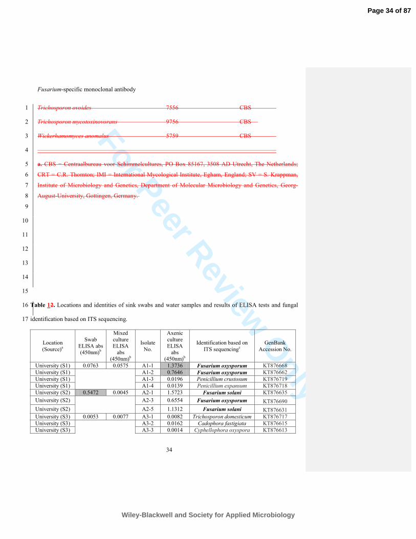
Table 1. Details of fungi used in mAb ED7 specificity tests. 15
16
Organism Isolate no. Sourcea 17
18
Aspergillus cervinus 537.65 CBS 19
Aspergillus ficuum 555.65 CBS 20
Aspergillus flavus 91856iii IMI 21
Aspergillus fumigatus AF293 SK 22
Aspergillus nidulans A4 FGSC 23
Aspergillus niger 102.40 CBS 24
Page 29 of 87
Wiley-Blackwell and Society for Applied Microbiology
For Peer Review O
nlyFusarium-specific monoclonal antibody
30
Aspergillus oryzae 672.92 CBS 1
Aspergillus restrictus 116.50 CBS 2
Aspergillus terreus var. terreus 601.65 CBS 3
Botrytis cinerea R2 CRT 4
Byssochlamys nivea 153.59 CBS 5
Candida glabrata 4692 CBS 6
Candida krusei 5590 CBS 7
Candida parapsilosis 8836 CBS 8
Candida tropicalis 1920 CBS 9
Cryptococcus neoformans (Serotype D) 5728 CBS 10
Cunninghamella elegans 151.80 CBS 11
Filobasidiella bacillispora 10865 CBS 12
Filobasidiella neoformans 10490 CBS 13
14
Table 1. continued 15
16
Organism Isolate no. Sourcea 17
18
Filobasidiella neoformans 10496 CBS 19
Fusarium acutatum 402.97 CBS 20
Fusarium anthophilum 222.76 CBS 21
Fusarium avenaceum 386.62 CBS 22
Fusarium cerealis 134.80 CBS 23
Fusarium chlamydosporium var. chlamydosporium 491.77 CBS 24
Page 30 of 87
Wiley-Blackwell and Society for Applied Microbiology
For Peer Review O
nlyFusarium-specific monoclonal antibody
31
Fusarium culmorum 256.51 CBS 1
Fusarium dimerum var. dimerum 108944 CBS 2
Fusarium incarnatum 678.77 CBS 3
Fusarium nygamai 140.95 CBS 4
Fusarium oxysporum f.sp. cucurbitacearum 254.52 CBS 5
Fusarium oxysporum f.sp. lycopersici 167.30 CBS 6
Fusarium oxysporum f.sp. marmaris 420.80 CBS 7
Fusarium oxysporum f.sp. radicis-lycopersici 872.95 CBS 8
Fusarium oxysporum f.sp. vasinfectum 409.90 CBS 9
Fusarium proliferatum var. proliferatum 181.30 CBS 10
Fusarium sacchari 183.32 CBS 11
Fusarium solani 224.34 CBS 12
Fusarium solani
109696 CBS 13
14
Table 1. continued 15
16
Organism Isolate no. Source 17
18
Fusarium solani 188.34 CBS 19
Fusarium solani 115659
CBS 20
Fusarium solani 117608 CBS 21
Fusarium solani 119223
CBS 22
Fusarium solani var. petroliphilum 102256 CBS 23
Fusarium verticillioides 102699 CBS 24
Page 31 of 87
Wiley-Blackwell and Society for Applied Microbiology
For Peer Review O
nlyFusarium-specific monoclonal antibody
32
Geotrichum candidum 115.23 CBS 1
Haematonectria haematococca 114067 CBS 2
Haematonectria haematococca 119603 CBS 3
Haematonectria haematococca 130692 CBS 4
Lichtheimia corymbifera T14A (FJ713070) CRT 5
Magnusiomyces capitatus 207.83 CBS 6
Mucor circinellioides f.sp. circinellioides E2A (FJ713065) CRT 7
Neosartorya fischeri var. fischeri 687.71 CBS 8
Paecilomyces variotii 339.51 CBS 9
Penicillium cyclopium 123.14 CBS 10
Penicillium islandicum 338.48 CBS 11
Penicillium spinulosum 346.61 CBS 12
Pichia norvegensis 6564 CBS 13
14
Table 1. continued 15
16
Organism Isolate no. Source 17
18
Pseudallescheria boydii 835.96 CBS 19
Pythium insidiosum 673.85 CBS 20
Rhizomucor miehei MG4(2) (FJ713069) CRT 21
Rhizopus stolonifer 389.95 CBS 22
Rhodosporidium toruloides 6016 CBS 23
Rhodotorula mucilaginosa 326 CBS 24
Page 32 of 87
Wiley-Blackwell and Society for Applied Microbiology
For Peer Review O
nlyFusarium-specific monoclonal antibody
33
Scedosporium apiospermum 117407 CBS 1
Scedosporium aurantiacum 121926 CBS 2
Scedosporium aurantiacum 118934 CBS 3
Scedosporium prolificans 102176 CBS 4
Sporidiobolus salmonicolor 6781 CBS 5
Trichoderma hamatum GD12 (AY247559) CRT 6
Trichosporon asahii var. asahii 8973 CBS 7
Trichosporon asahii var. asahii 5286 CBS 8
Trichosporon asahii var. asahii 7632 CBS 9
Trichosporon asahii var. asahii 5599 CBS 10
Trichosporon asteroides 6183 CBS 11
Trichosporon asteroides 7623 CBS 12
Trichosporon asteroides 2481 CBS 13
14
Table 1. continued 15
16
Organism Isolate no. Source 17
18
Trichosporon asteroides 7624 CBS 19
Trichosporon cutaneum 2466 CBS 20
Trichosporon inkin 7630 CBS 21
Trichosporon inkin 7655 CBS 22
Trichosporon loubieri 7065 CBS 23
Page 33 of 87
Wiley-Blackwell and Society for Applied Microbiology
For Peer Review O
nlyFusarium-specific monoclonal antibody
34
Trichosporon ovoides 7556 CBS 1
Trichosporon mycotoxinovorans 9756 CBS 2
Wickerhamomyces anomalus 5759 CBS 3
4
a. CBS = Centraalbureau voor Schimmelcultures, PO Box 85167, 3508 AD Utrecht, The Netherlands; 5
CRT = C.R. Thornton; IMI = International Mycological Institute, Egham, England; SV = S. Krappman, 6
Institute of Microbiology and Genetics, Department of Molecular Microbiology and Genetics, Georg-7
August-University, Gottingen, Germany. 8
9
10
11
12
13
14
15
Table 12. Locations and identities of sink swabs and water samples and results of ELISA tests and fungal 16
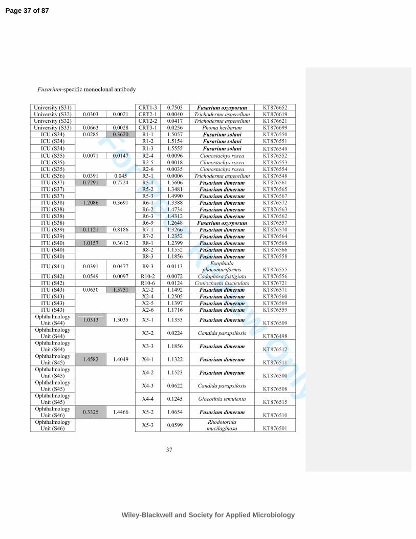
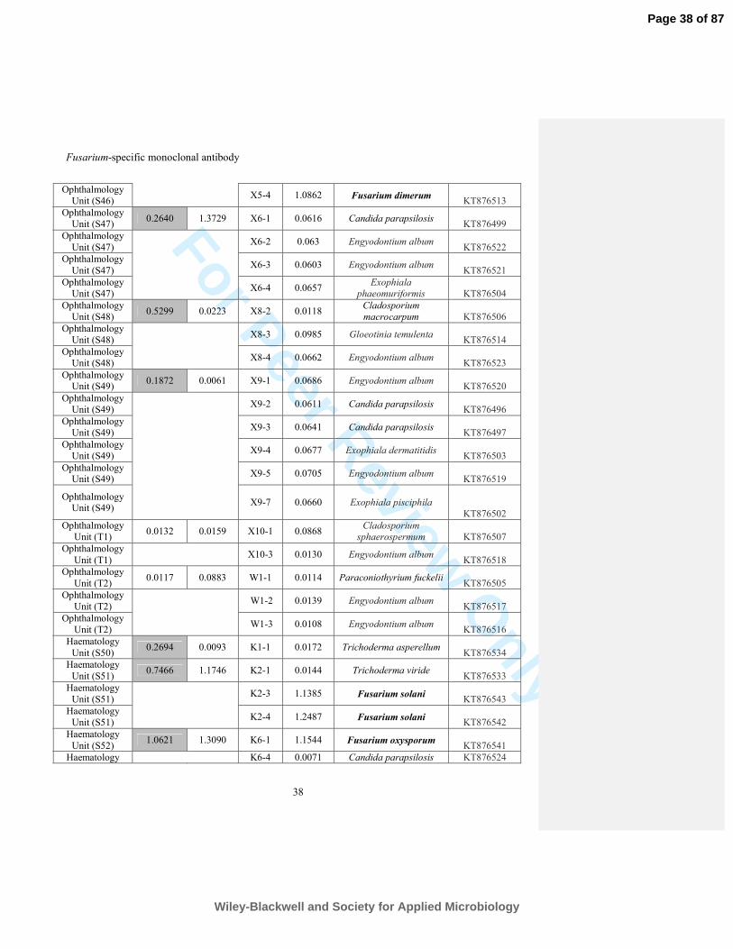
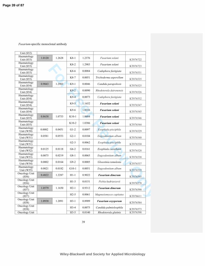
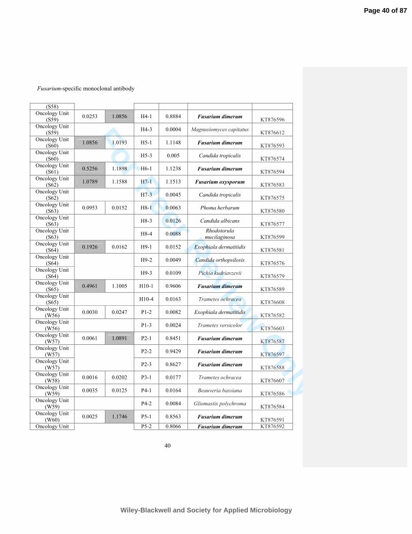
identification based on ITS sequencing. 17
Location
(Source)a
Swab
ELISA abs
(450nm)b
Mixed
culture
ELISA
abs
(450nm)b
Isolate
No.
Axenic
culture
ELISA
abs
(450nm)b
Identification based on
ITS sequencingc
GenBank
Accession No.
University (S1) 0.0763 0.0575 A1-1 1.3736 Fusarium oxysporum KT876668
University (S1) A1-2 0.7646 Fusarium oxysporum KT876662
University (S1) A1-3 0.0196 Penicillium crustosum KT876719
University (S1) A1-4 0.0139 Penicillium expansum KT876718
University (S2) 0.5472 0.0045 A2-1 1.5723 Fusarium solani KT876635
University (S2) A2-3 0.6554 Fusarium oxysporum KT876690
University (S2) A2-5 1.1312 Fusarium solani KT876631
University (S3) 0.0053 0.0077 A3-1 0.0082 Trichosporon domesticum KT876717
University (S3) A3-2 0.0162 Cadophora fastigiata KT876615
University (S3) A3-3 0.0014 Cyphellophora oxyspora KT876613
Page 34 of 87
Wiley-Blackwell and Society for Applied Microbiology
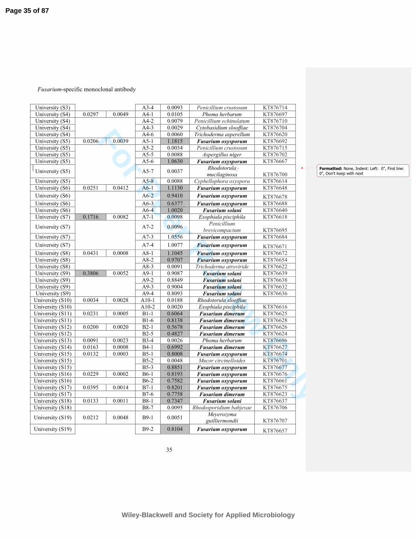
For Peer Review O
nlyFusarium-specific monoclonal antibody
35
University (S3) A3-4 0.0093 Penicillium crustosum KT876714
University (S4) 0.0297 0.0049 A4-1 0.0105 Phoma herbarum KT876697
University (S4) A4-2 0.0079 Penicillium echinulatum KT876710
University (S4) A4-3 0.0029 Cytobasidium slooffiae KT876704
University (S4) A4-6 0.0060 Trichoderma asperellum KT876620
University (S5) 0.0206 0.0039 A5-1 1.1815 Fusarium oxysporum KT876692
University (S5) A5-2 0.0034 Penicillium crustosum KT876715
University (S5) A5-5 0.0088 Aspergillus niger KT876702
University (S5) A5-6 1.0630 Fusarium oxysporum KT876667
University (S5) A5-7 0.0037 Rhodotorula
mucilaginosa KT876700
University (S5) A5-8 0.0088 Cyphellophora oxyspora KT876614
University (S6) 0.0251 0.0412 A6-1 1.1130 Fusarium oxysporum KT876648
University (S6) A6-2 0.9410 Fusarium oxysporum KT876678
University (S6) A6-3 0.6377 Fusarium oxysporum KT876688
University (S6) A6-4 1.0020 Fusarium solani KT876640
University (S7) 0.1716 0.0082 A7-1 0.0098 Exophiala pisciphila KT876618
University (S7) A7-2 0.0096 Penicillium
brevicompactum KT876695
University (S7) A7-3 1.0556 Fusarium oxysporum KT876684
University (S7) A7-4 1.0077 Fusarium oxysporum KT876671
University (S8) 0.0431 0.0008 A8-1 1.1045 Fusarium oxysporum KT876672
University (S8) A8-2 0.9707 Fusarium oxysporum KT876654
University (S8) A8-3 0.0091 Trichoderma atroviride KT876622
University (S9) 0.3806 0.0052 A9-1 0.9087 Fusarium solani KT876639
University (S9) A9-2 0.8849 Fusarium solani KT876638
University (S9) A9-3 0.9004 Fusarium solani KT876632
University (S9) A9-4 0.8093 Fusarium solani KT876636
University (S10) 0.0034 0.0028 A10-1 0.0188 Rhodotorula slooffiae
University (S10) A10-2 0.0020 Exophiala pisciphila KT876616
University (S11) 0.0231 0.0005 B1-1 0.6064 Fusarium dimerum KT876625
University (S11) B1-6 0.8138 Fusarium dimerum KT876628
University (S12) 0.0200 0.0020 B2-1 0.5678 Fusarium dimerum KT876626
University (S12) B2-5 0.4827 Fusarium dimerum KT876624
University (S13) 0.0091 0.0023 B3-4 0.0026 Phoma herbarum KT876696
University (S14) 0.0163 0.0008 B4-1 0.6992 Fusarium dimerum KT876627
University (S15) 0.0132 0.0003 B5-1 0.8008 Fusarium oxysporum KT876674
University (S15) B5-2 0.0048 Mucor circinelloides KT876701
University (S15) B5-3 0.8851 Fusarium oxysporum KT876677
University (S16) 0.0229 0.0002 B6-1 0.8193 Fusarium oxysporum KT876676
University (S16) B6-2 0.7582 Fusarium oxysporum KT876661
University (S17) 0.0395 0.0014 B7-1 0.8201 Fusarium oxysporum KT876675
University (S17) B7-6 0.7758 Fusarium dimerum KT876623
University (S18) 0.0133 0.0011 B8-1 0.7347 Fusarium solani KT876637
University (S18) B8-7 0.0095 Rhodosporidium babjevae KT876706
University (S19) 0.0212 0.0048 B9-1 0.0051 Meyerozyma
guilliermondii KT876707
University (S19) B9-2 0.8104 Fusarium oxysporum KT876657
Formatted: None, Indent: Left: 0", First line: 0", Don't keep with next
Page 35 of 87
Wiley-Blackwell and Society for Applied Microbiology
For Peer Review O
nlyFusarium-specific monoclonal antibody
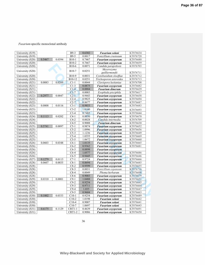
36
University (S19) B9-3 0.6302 Fusarium solani KT876634
University (S19) B9-5 0.0017 Penicillium crustosum KT876720
University (S20) 0.5467 0.0394 B10-1 0.7467 Fusarium oxysporum KT876680
University (S20) B10-2 0.7667 Fusarium oxysporum KT876659
University (S20) B10-6 0.0071 Candida parapsilosis KT876703
University (S20) B10-7 0.0251 Meyerozyma
guilliermondii KT876711
University (S20) B10-9 0.0031 Cystobasidium slooffiae KT876712
University (S20) B10-12 0.0251 Trichosporon asteroides KT876713
University (S21) 0.0083 0.0269 C1-1 0.0044 Clavispora lusitaniae KT876708
University (S21) C1-3 0.8875 Fusarium oxysporum KT876682
University (S21) C1-4 0.8884 Fusarium dimerum KT876629
University (S21) C1-7 0.0003 Exophiala pisciphila KT876617
University (S22) 0.2977 0.0047 C2-1 0.9443 Fusarium oxysporum KT876658
University (S22) C2-4 0.9825 Fusarium oxysporum KT876694
University (S22) C2-7 0.8677 Fusarium oxysporum KT876687
University (S23) 0.0808 0.0116 C3-1 0.9853 Fusarium oxysporum KT876683
University (S23) C3-2 1.0160 Fusarium oxysporum KT876693
University (S23) C3-4 0.7485 Fusarium oxysporum KT876644
University (S24) 0.1113 0.0202 C4-1 0.8930 Fusarium oxysporum KT876670
University (S24) C4-2 0.0028 Candida intermedia KT876709
University (S24) C4-5 0.9008 Fusarium dimerum KT876630
University (S25) 0.5741 0.0097 C5-1 0.9874 Fusarium oxysporum KT876652
University (S25) C5-2 1.0996 Fusarium oxysporum KT876656
University (S25) C5-3 1.1236 Fusarium oxysporum KT876649
University (S25) C5-4 1.0355 Fusarium oxysporum KT876669
University (S25) C5-5 1.0672 Fusarium oxysporum KT876660
University (S26) 0.0603 0.0348 C6-1 1.0135 Fusarium oxysporum KT876665
University (S26) C6-2 0.9362 Fusarium oxysporum KT876681
University (S26) C6-3 0.8898 Fusarium oxysporum
University (S26) C6-4 1.0577 Fusarium oxysporum KT876686
University (S26) C6-5 0.9445 Fusarium oxysporum KT876646
University (S27) 0.1279 0.0113 C7-1 0.9724 Fusarium oxysporum KT876666
University (S28) 0.0607 0.0035 C8-1 0.8694 Fusarium oxysporum KT876685
University (S28) C8-2 0.9599 Fusarium oxysporum KT876655
University (S28) C8-3 0.0031 Penicillium expansum KT876716
University (S28) C8-4 0.0049 Phoma herbarum KT876698
University (S28) C8-6 0.9003 Fusarium oxysporum KT876679
University (S29) 0.0310 0.0001 C9-1 1.0488 Fusarium oxysporum KT876651
University (S29) C9-2 0.9254 Fusarium oxysporum KT876645
University (S29) C9-3 0.9711 Fusarium oxysporum KT876664
University (S29) C9-4 1.035 Fusarium oxysporum KT876647
University (S29) C9-5 0.9604 Fusarium oxysporum KT876663
University (S30) 0.1002 0.0333 C10-1 0.9254 Fusarium oxysporum KT876689
University (S30) C10-2 1.0198 Fusarium solani KT876642
University (S30) C10-4 0.9007 Fusarium solani KT876641
University (S30) C10-7 1.0697 Fusarium solani KT876643
University (S31) 0.6175 0.1129 CRT1-1 0.4639 Fusarium oxysporum KT876691
University (S31) CRT1-2 0.9086 Fusarium oxysporum KT876650
Page 36 of 87
Wiley-Blackwell and Society for Applied Microbiology
For Peer Review O
nlyFusarium-specific monoclonal antibody
37
University (S31) CRT1-3 0.7503 Fusarium oxysporum KT876652
University (S32) 0.0303 0.0021 CRT2-1 0.0040 Trichoderma asperellum KT876619
University (S32) CRT2-2 0.0417 Trichoderma asperellum KT876621
University (S33) 0.0663 0.0028 CRT3-1 0.0256 Phoma herbarum KT876699
ICU (S34) 0.0285 0.3620 R1-1 1.5057 Fusarium solani KT876550
ICU (S34) R1-2 1.5154 Fusarium solani KT876551
ICU (S34) R1-3 1.5555 Fusarium solani KT876549
ICU (S35) 0.0071 0.0147 R2-4 0.0096 Clonostachys rosea KT876552
ICU (S35) R2-5 0.0018 Clonostachys rosea KT876553
ICU (S35) R2-6 0.0035 Clonostachys rosea KT876554
ICU (S36) 0.0391 0.045 R3-1 0.0006 Trichoderma asperellum KT876548
ITU (S37) 0.7291 0.7724 R5-1 1.5606 Fusarium dimerum KT876561
ITU (S37) R5-2 1.3481 Fusarium dimerum KT876565
ITU (S37) R5-3 1.4990 Fusarium dimerum KT876567
ITU (S38) 1.2086 0.3691 R6-1 1.3388 Fusarium dimerum KT876572
ITU (S38) R6-2 1.4734 Fusarium dimerum KT876563
ITU (S38) R6-3 1.4312 Fusarium dimerum KT876562
ITU (S38) R6-9 1.2648 Fusarium oxysporum KT876557
ITU (S39) 0.1121 0.8186 R7-1 1.3266 Fusarium dimerum KT876570
ITU (S39) R7-2 1.2352 Fusarium dimerum KT876564
ITU (S40) 1.0157 0.3612 R8-1 1.2399 Fusarium dimerum KT876568
ITU (S40) R8-2 1.1552 Fusarium dimerum KT876566
ITU (S40) R8-3 1.1856 Fusarium dimerum KT876558
ITU (S41) 0.0391 0.0477 R9-3 0.0113 Exophiala
phaeomuriformis KT876555
ITU (S42) 0.0549 0.0097 R10-2 0.0072 Cadophora fastigiata KT876556
ITU (S42) R10-6 0.0124 Coniochaeta fasciculata KT876721
ITU (S43) 0.0630 1.5751 X2-2 1.1492 Fusarium dimerum KT876571
ITU (S43) X2-4 1.2505 Fusarium dimerum KT876560
ITU (S43) X2-5 1.1397 Fusarium dimerum KT876569
ITU (S43) X2-6 1.1716 Fusarium dimerum KT876559
Ophthalmology Unit (S44)
1.0313 1.5035 X3-1 1.1353 Fusarium dimerum KT876509
Ophthalmology
Unit (S44) X3-2 0.0224 Candida parapsilosis
KT876498
Ophthalmology
Unit (S44) X3-3 1.1856 Fusarium dimerum
KT876512
Ophthalmology
Unit (S45) 1.4582 1.4049 X4-1 1.1322 Fusarium dimerum
KT876511
Ophthalmology Unit (S45)
X4-2 1.1523 Fusarium dimerum KT876500
Ophthalmology
Unit (S45) X4-3 0.0622 Candida parapsilosis
KT876508
Ophthalmology
Unit (S45) X4-4 0.1245 Gloeotinia temulenta
KT876515
Ophthalmology
Unit (S46) 0.3325 1.4466 X5-2 1.0654 Fusarium dimerum
KT876510
Ophthalmology Unit (S46)
X5-3 0.0599 Rhodotorula mucilaginosa KT876501
Page 37 of 87
Wiley-Blackwell and Society for Applied Microbiology
For Peer Review O
nlyFusarium-specific monoclonal antibody
38
Ophthalmology
Unit (S46) X5-4 1.0862 Fusarium dimerum
KT876513
Ophthalmology
Unit (S47) 0.2640 1.3729 X6-1 0.0616 Candida parapsilosis
KT876499
Ophthalmology
Unit (S47) X6-2 0.063 Engyodontium album
KT876522
Ophthalmology
Unit (S47) X6-3 0.0603 Engyodontium album
KT876521
Ophthalmology
Unit (S47) X6-4 0.0657
Exophiala
phaeomuriformis KT876504
Ophthalmology
Unit (S48) 0.5299 0.0223 X8-2 0.0118
Cladosporium
macrocarpum KT876506
Ophthalmology
Unit (S48) X8-3 0.0985 Gloeotinia temulenta
KT876514
Ophthalmology
Unit (S48) X8-4 0.0662 Engyodontium album
KT876523
Ophthalmology Unit (S49)
0.1872 0.0061 X9-1 0.0686 Engyodontium album KT876520
Ophthalmology
Unit (S49) X9-2 0.0611 Candida parapsilosis
KT876496
Ophthalmology
Unit (S49) X9-3 0.0641 Candida parapsilosis
KT876497
Ophthalmology
Unit (S49) X9-4 0.0677 Exophiala dermatitidis
KT876503
Ophthalmology Unit (S49)
X9-5 0.0705 Engyodontium album KT876519
Ophthalmology Unit (S49)
X9-7 0.0660 Exophiala pisciphila
KT876502
Ophthalmology
Unit (T1) 0.0132 0.0159 X10⁻¹ 0.0868
Cladosporium
sphaerospermum KT876507
Ophthalmology
Unit (T1) X10-3 0.0130 Engyodontium album
KT876518
Ophthalmology
Unit (T2) 0.0117 0.0883 W1-1 0.0114 Paraconiothyrium fuckelii
KT876505
Ophthalmology
Unit (T2) W1-2 0.0139 Engyodontium album
KT876517
Ophthalmology
Unit (T2) W1-3 0.0108 Engyodontium album
KT876516
Haematology
Unit (S50) 0.2694 0.0093 K1-1 0.0172 Trichoderma asperellum
KT876534
Haematology
Unit (S51) 0.7466 1.1746 K2-1 0.0144 Trichoderma viride
KT876533
Haematology
Unit (S51) K2-3 1.1385 Fusarium solani
KT876543
Haematology
Unit (S51) K2-4 1.2487 Fusarium solani
KT876542
Haematology
Unit (S52) 1.0621 1.3090 K6-1 1.1544 Fusarium oxysporum
KT876541
Haematology K6-4 0.0071 Candida parapsilosis KT876524
Page 38 of 87
Wiley-Blackwell and Society for Applied Microbiology
For Peer Review O
nlyFusarium-specific monoclonal antibody
39
Unit (S52)
Haematology
Unit (S53) 1.0120 1.2628 K8-1 1.2376 Fusarium solani
KT876722
Haematology
Unit (S53) K8-2 1.2983 Fusarium solani
KT876723
Haematology Unit (S53)
K8-6 0.0084 Cadophora fastigiata KT876531
Haematology
Unit (S53) K8-7 0.0051 Trichoderma asperellum
KT876535
Haematology
Unit (S54) 0.9843 1.2903 K9-1 0.0046 Candida parapsilosis
KT876525
Haematology
Unit (S54) K9-2 0.0090 Rhodotorula dairenensis
KT876526
Haematology Unit (S54)
K9-4 0.0073 Cadophora fastigiata KT876532
Haematology
Unit (S54) K9-5 1.1652 Fusarium solani
KT876547
Haematology
Unit (S54) K9-6 1.0316 Fusarium solani
KT876545
Haematology
Unit (S55) 0.8638 1.0753 K10-1 1.0684 Fusarium solani
KT876546
Haematology Unit (S55)
K10-2 1.0386 Fusarium solani KT876544
Haematology
Unit (W50) 0.0082 0.0451 G1-2 0.0097 Exophiala pisciphila
KT876529
Haematology
Unit (W51) 0.0581 0.0553 G2-1 0.0104 Engyodontium album
KT876540
Haematology
Unit (W51) G2-3 0.0062 Exophiala pisciphila
KT876530
Haematology
Unit (W52) 0.0125 0.0118 G6-2 0.0161 Exophiala castellanii
KT876528
Haematology
Unit (W53) 0.0075 0.0219 G8-1 0.0065 Engyodontium album
KT876538
Haematology
Unit (W54) 0.0002 0.0164 G9-2 0.0005 Gloeotinia temulenta
KT876537
Haematology
Unit (W55) 0.0021 0.0182 G10-1 0.0051 Engyodontium album
KT876539
Oncology Unit
(S56) 0.4822 1.2287 H1-1 0.9022 Fusarium dimerum
KT876595
Oncology Unit
(S56) H1-3 0.0151 Pichia kudriavzevii
KT876578
Oncology Unit
(S57) 1.0579 1.1650 H2-1 0.9312 Fusarium dimerum
KT876590
Oncology Unit
(S57) H2-5 0.0061 Magnusiomyces capitatus
KT876611
Oncology Unit
(S58) 1.0938 1.2091 H3-1 0.8909 Fusarium oxysporum
KT876584
Oncology Unit
(S58) H3-4 0.0075 Candida palmioleophila
KT876573
Oncology Unit H3-5 0.0140 Rhodotorula glutinis KT876598
Page 39 of 87
Wiley-Blackwell and Society for Applied Microbiology
For Peer Review O
nlyFusarium-specific monoclonal antibody
40
(S58)
Oncology Unit
(S59) 0.0253 1.0856 H4-1 0.8884 Fusarium dimerum
KT876596
Oncology Unit
(S59) H4-3 0.0004 Magnusiomyces capitatus
KT876612
Oncology Unit (S60)
1.0856 1.0193 H5-1 1.1148 Fusarium dimerum KT876593
Oncology Unit
(S60) H5-3 0.005 Candida tropicalis
KT876574
Oncology Unit
(S61) 0.5256 1.1898 H6-1 1.1238 Fusarium dimerum
KT876594
Oncology Unit
(S62) 1.0789 1.1588 H7-1 1.1513 Fusarium oxysporum
KT876583
Oncology Unit (S62)
H7-3 0.0045 Candida tropicalis KT876575
Oncology Unit
(S63) 0.0953 0.0152 H8-1 0.0063 Phoma herbarum
KT876580
Oncology Unit
(S63) H8-3 0.0126 Candida albicans
KT876577
Oncology Unit
(S63) H8-4 0.0088
Rhodotorula
mucilaginosa KT876599
Oncology Unit (S64)
0.1926 0.0162 H9-1 0.0152 Exophiala dermatitidis KT876581
Oncology Unit
(S64) H9-2 0.0049 Candida orthopsilosis
KT876576
Oncology Unit
(S64) H9-3 0.0109 Pichia kudriavzevii
KT876579
Oncology Unit
(S65) 0.4961 1.1005 H10-1 0.9606 Fusarium dimerum
KT876589
Oncology Unit
(S65) H10-4 0.0163 Trametes ochracea
KT876608
Oncology Unit
(W56) 0.0030 0.0247 P1-2 0.0082 Exophiala dermatitidis
KT876582
Oncology Unit
(W56) P1-3 0.0024 Trametes versicolor
KT876603
Oncology Unit
(W57) 0.0061 1.0891 P2-1 0.8451 Fusarium dimerum
KT876587
Oncology Unit
(W57) P2-2 0.9429 Fusarium dimerum
KT876597
Oncology Unit
(W57) P2-3 0.8627 Fusarium dimerum
KT876588
Oncology Unit
(W58) 0.0016 0.0202 P3-1 0.0177 Trametes ochracea
KT876607
Oncology Unit
(W59) 0.0035 0.0125 P4-1 0.0164 Beauveria bassiana
KT876586
Oncology Unit
(W59) P4-2 0.0084 Gliomastix polychroma
KT876584
Oncology Unit
(W60) 0.0025 1.1746 P5-1 0.8563 Fusarium dimerum
KT876591
Oncology Unit P5-2 0.8066 Fusarium dimerum KT876592
Page 40 of 87
Wiley-Blackwell and Society for Applied Microbiology
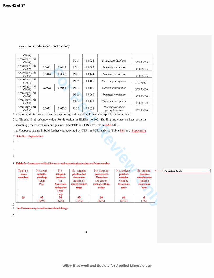
For Peer Review O
nlyFusarium-specific monoclonal antibody
41
a. S, sink; W, tap water from corresponding sink number; T, water sample from main tank. 1
b. Threshold absorbance value for detection in ELISA ≥0.100. Shading indicates earliest point in 2
sampling process at which antigen was detectable in ELISA tests with mAb ED7. 3
c. Fusarium strains in bold further characterized by TEF-1α PCR analysis (Table S34 and Supporting 4
Data Set 1Appendix 1). 5
6
7
8
Table 3. Summary of ELISA tests and mycological culture of sink swabs. 9
Total no.
sinks
swabbed
No. swab
samples
yielding
fungi
(%)a
No.
samples
positive
for
Fusarium
antigen at
swab
stage
No. samples
positive for
Fusarium
antigen by
mixed culture
stage
No. samples
positive for
Fusarium
antigen by
axenic culture
stage
No. antigen-
positive
samples
yielding
Fusarium
spp.
No. antigen-
positive
samples not
yielding
Fusarium
spp.
65
65
(100%)
34
(52%)
37
(57%)
54
(83%)
50
(93%)
4
(7%)
10 a. Fusarium spp. and/or unrelated fungi. 11
12
(W60)
Oncology Unit
(W60) P5-3 0.0024 Piptoporus betulinus
KT876609
Oncology Unit
(W62) 0.0011 0.0417 P7-1 0.0097 Trametes versicolor
KT876605
Oncology Unit (W63)
0.0044 0.0060 P8-1 0.0144 Trametes versicolor KT876606
Oncology Unit
(W63) P8-2 0.0106 Stereum gausapatum
KT876601
Oncology Unit
(W64) 0.0022 0.0163 P9-1 0.0101 Stereum gausapatum
KT876600
Oncology Unit
(W64) P9-2 0.0068 Trametes versicolor
KT876604
Oncology Unit (W64)
P9-3 0.0140 Stereum gausapatum KT876602
Oncology Unit (W65)
0.0051 0.0280 P10-1 0.0032 Phaeophlebiopsis peniophoroides KT876610
Formatted Table
Page 41 of 87
Wiley-Blackwell and Society for Applied Microbiology
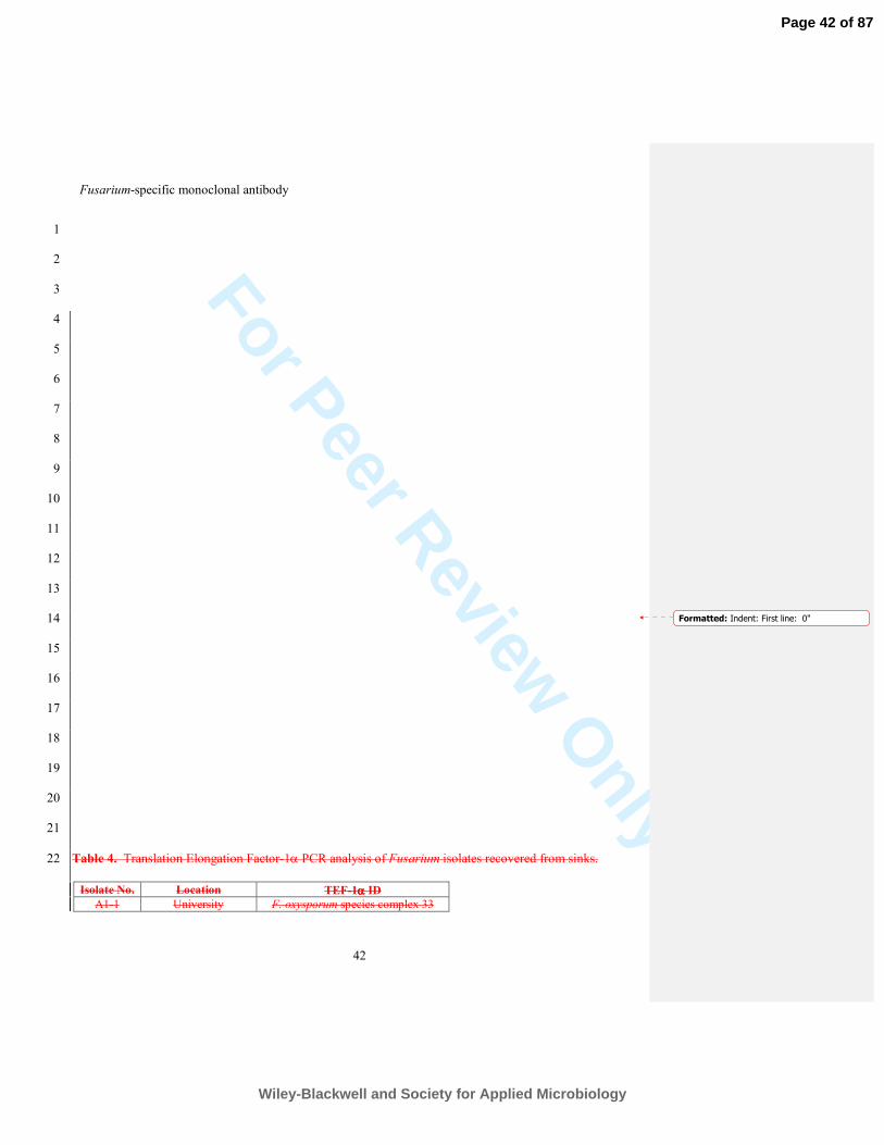
For Peer Review O
nlyFusarium-specific monoclonal antibody
42
1
2
3
4
5
6
7
8
9
10
11
12
13
14
15
16
17
18
19
20
21
Table 4. Translation Elongation Factor-1α PCR analysis of Fusarium isolates recovered from sinks. 22
Isolate No. Location TEF-1αααα ID
A1-1 University F. oxysporum species complex 33
Formatted: Indent: First line: 0"
Page 42 of 87
Wiley-Blackwell and Society for Applied Microbiology
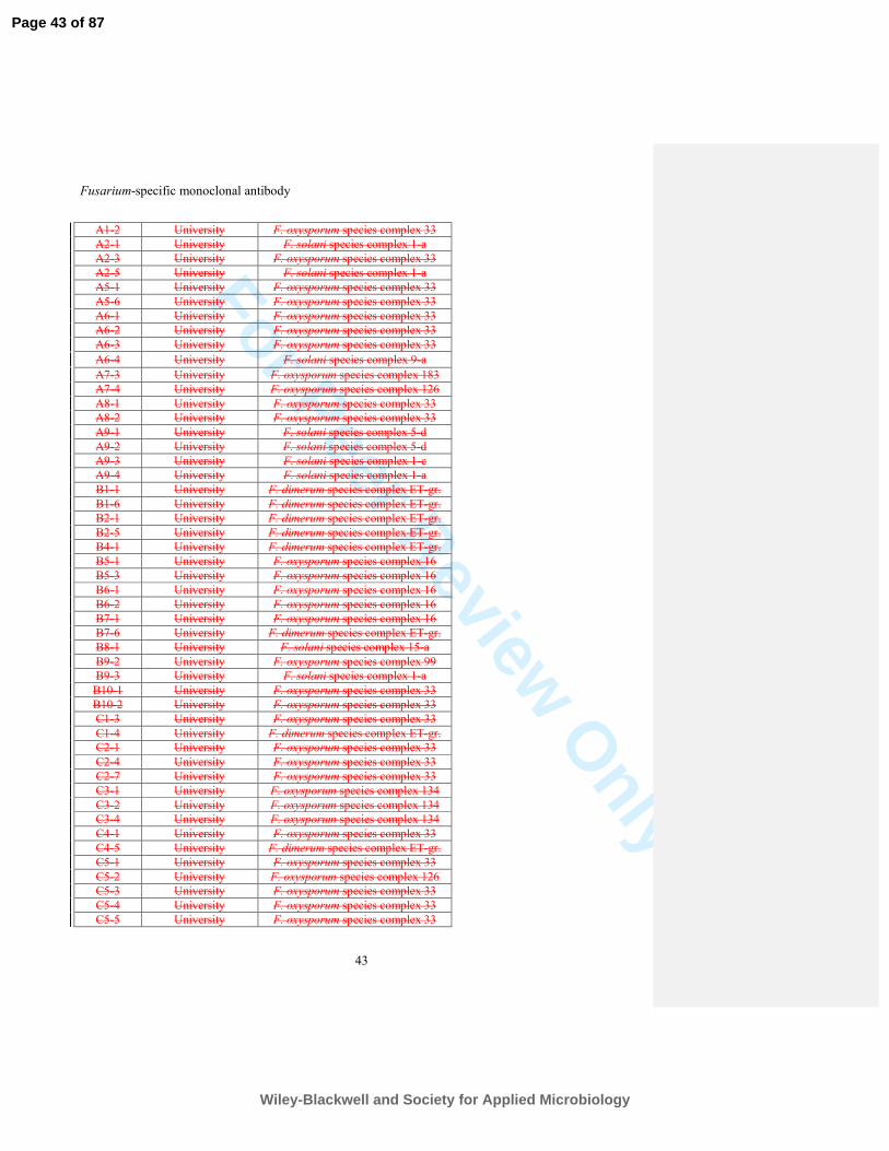
For Peer Review O
nlyFusarium-specific monoclonal antibody
43
A1-2 University F. oxysporum species complex 33
A2-1 University F. solani species complex 1-a
A2-3 University F. oxysporum species complex 33
A2-5 University F. solani species complex 1-a
A5-1 University F. oxysporum species complex 33
A5-6 University F. oxysporum species complex 33
A6-1 University F. oxysporum species complex 33
A6-2 University F. oxysporum species complex 33
A6-3 University F. oxysporum species complex 33
A6-4 University F. solani species complex 9-a
A7-3 University F. oxysporum species complex 183
A7-4 University F. oxysporum species complex 126
A8-1 University F. oxysporum species complex 33
A8-2 University F. oxysporum species complex 33
A9-1 University F. solani species complex 5-d
A9-2 University F. solani species complex 5-d
A9-3 University F. solani species complex 1-c
A9-4 University F. solani species complex 1-a
B1-1 University F. dimerum species complex ET-gr.
B1-6 University F. dimerum species complex ET-gr.
B2-1 University F. dimerum species complex ET-gr.
B2-5 University F. dimerum species complex ET-gr.
B4-1 University F. dimerum species complex ET-gr.
B5-1 University F. oxysporum species complex 16
B5-3 University F. oxysporum species complex 16
B6-1 University F. oxysporum species complex 16
B6-2 University F. oxysporum species complex 16
B7-1 University F. oxysporum species complex 16
B7-6 University F. dimerum species complex ET-gr.
B8-1 University F. solani species complex 15-a
B9-2 University F. oxysporum species complex 99
B9-3 University F. solani species complex 1-a
B10-1 University F. oxysporum species complex 33
B10-2 University F. oxysporum species complex 33
C1-3 University F. oxysporum species complex 33
C1-4 University F. dimerum species complex ET-gr.
C2-1 University F. oxysporum species complex 33
C2-4 University F. oxysporum species complex 33
C2-7 University F. oxysporum species complex 33
C3-1 University F. oxysporum species complex 134
C3-2 University F. oxysporum species complex 134
C3-4 University F. oxysporum species complex 134
C4-1 University F. oxysporum species complex 33
C4-5 University F. dimerum species complex ET-gr.
C5-1 University F. oxysporum species complex 33
C5-2 University F. oxysporum species complex 126
C5-3 University F. oxysporum species complex 33
C5-4 University F. oxysporum species complex 33
C5-5 University F. oxysporum species complex 33
Page 43 of 87
Wiley-Blackwell and Society for Applied Microbiology
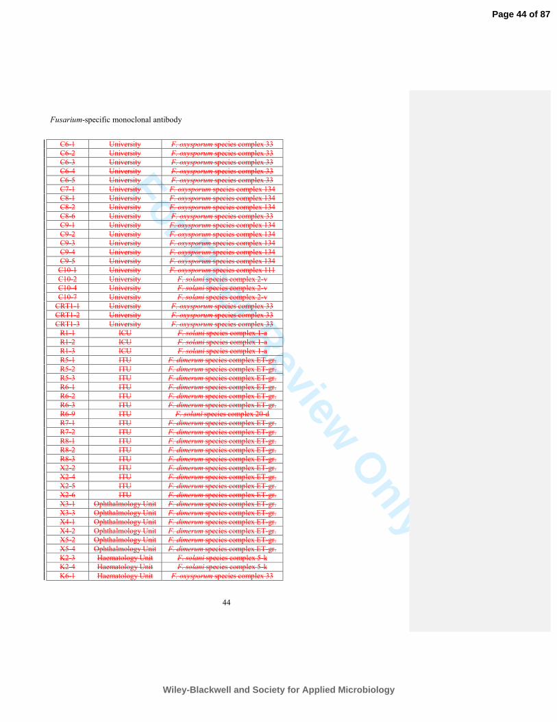
For Peer Review O
nlyFusarium-specific monoclonal antibody
44
C6-1 University F. oxysporum species complex 33
C6-2 University F. oxysporum species complex 33
C6-3 University F. oxysporum species complex 33
C6-4 University F. oxysporum species complex 33
C6-5 University F. oxysporum species complex 33
C7-1 University F. oxysporum species complex 134
C8-1 University F. oxysporum species complex 134
C8-2 University F. oxysporum species complex 134
C8-6 University F. oxysporum species complex 33
C9-1 University F. oxysporum species complex 134
C9-2 University F. oxysporum species complex 134
C9-3 University F. oxysporum species complex 134
C9-4 University F. oxysporum species complex 134
C9-5 University F. oxysporum species complex 134
C10-1 University F. oxysporum species complex 111
C10-2 University F. solani species complex 2-v
C10-4 University F. solani species complex 2-v
C10-7 University F. solani species complex 2-v
CRT1-1 University F. oxysporum species complex 33
CRT1-2 University F. oxysporum species complex 33
CRT1-3 University F. oxysporum species complex 33
R1-1 ICU F. solani species complex 1-a
R1-2 ICU F. solani species complex 1-a
R1-3 ICU F. solani species complex 1-a
R5-1 ITU F. dimerum species complex ET-gr.
R5-2 ITU F. dimerum species complex ET-gr.
R5-3 ITU F. dimerum species complex ET-gr.
R6-1 ITU F. dimerum species complex ET-gr.
R6-2 ITU F. dimerum species complex ET-gr.
R6-3 ITU F. dimerum species complex ET-gr.
R6-9 ITU F. solani species complex 20-d
R7-1 ITU F. dimerum species complex ET-gr.
R7-2 ITU F. dimerum species complex ET-gr.
R8-1 ITU F. dimerum species complex ET-gr.
R8-2 ITU F. dimerum species complex ET-gr.
R8-3 ITU F. dimerum species complex ET-gr.
X2-2 ITU F. dimerum species complex ET-gr.
X2-4 ITU F. dimerum species complex ET-gr.
X2-5 ITU F. dimerum species complex ET-gr.
X2-6 ITU F. dimerum species complex ET-gr.
X3-1 Ophthalmology Unit F. dimerum species complex ET-gr.
X3-3 Ophthalmology Unit F. dimerum species complex ET-gr.
X4-1 Ophthalmology Unit F. dimerum species complex ET-gr.
X4-2 Ophthalmology Unit F. dimerum species complex ET-gr.
X5-2 Ophthalmology Unit F. dimerum species complex ET-gr.
X5-4 Ophthalmology Unit F. dimerum species complex ET-gr.
K2-3 Haematology Unit F. solani species complex 5-k
K2-4 Haematology Unit F. solani species complex 5-k
K6-1 Haematology Unit F. oxysporum species complex 33
Page 44 of 87
Wiley-Blackwell and Society for Applied Microbiology
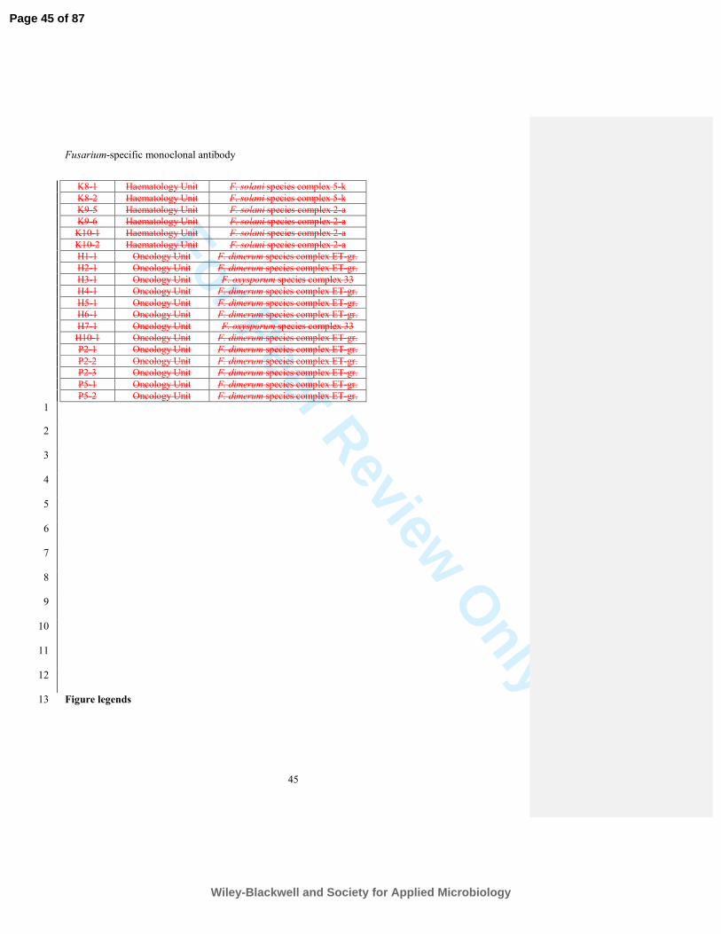
For Peer Review O
nlyFusarium-specific monoclonal antibody
45
K8-1 Haematology Unit F. solani species complex 5-k
K8-2 Haematology Unit F. solani species complex 5-k
K9-5 Haematology Unit F. solani species complex 2-a
K9-6 Haematology Unit F. solani species complex 2-a
K10-1 Haematology Unit F. solani species complex 2-a
K10-2 Haematology Unit F. solani species complex 2-a
H1-1 Oncology Unit F. dimerum species complex ET-gr.
H2-1 Oncology Unit F. dimerum species complex ET-gr.
H3-1 Oncology Unit F. oxysporum species complex 33
H4-1 Oncology Unit F. dimerum species complex ET-gr.
H5-1 Oncology Unit F. dimerum species complex ET-gr.
H6-1 Oncology Unit F. dimerum species complex ET-gr.
H7-1 Oncology Unit F. oxysporum species complex 33
H10-1 Oncology Unit F. dimerum species complex ET-gr.
P2-1 Oncology Unit F. dimerum species complex ET-gr.
P2-2 Oncology Unit F. dimerum species complex ET-gr.
P2-3 Oncology Unit F. dimerum species complex ET-gr.
P5-1 Oncology Unit F. dimerum species complex ET-gr.
P5-2 Oncology Unit F. dimerum species complex ET-gr.
1
2
3
4
5
6
7
8
9
10
11
12
Figure legends 13
Page 45 of 87
Wiley-Blackwell and Society for Applied Microbiology
For Peer Review O
nlyFusarium-specific monoclonal antibody
46
Figure 1. Specificity of ED7 determined by Enzyme-Linked Immunosorbent Assay tests of 1
surface washings containing water-soluble antigens from Fusarium species and related and 2
unrelated yeasts and molds. (A) ELISA aELISA absorbance values at 450 nm for from specificity 3
screening tests using mAb ED7 and antigens from F. solani and unrelated yeasts and mouldmolds 4
(A), and (B) for antigens from the F. solani teleomorph Haematonectria. haematococca and related 5
Fusarium species (B). Wells were coated with 60 µg protein ml-1 buffer. Bars are the means of three 6
biological replicates ± standard errors and . tThe threshold absorbance value for detection of antigen 7
in ELISA is ≥0.100 (indicated by lines on graphs). Numbers in parentheses after species names 8
denote strain numbers with further details of strains provided in Table S1. 9
10
Figure 2. Characterisation Characterisation of the ED7 antigen and its epitope and spatial 11
distribution of the antigen in spores and hyphae bound by mAb ED7. (A) Western immunoblot 12
with mAb ED7 using culture fluid from 2-d-old PDB cultures of F. solani CBS224.34 (lane 1) and 13
F. oxysporum f.sp. lycopersici CBS167.30 (lane 2). Wells were loaded with 1.6 µg of protein. Mr 14
denotes molecular weight in kDa. Note the major extracellular antigen with molecular weight of 15
~200 kDa. (B) Absorbance values from ELISA tests with mAb ED7 using immobilized 16
antigensurface washings containing water-soluble antigens immobilized to the wells of microtitre 17
plates and treated with trypsin or PBS only (control) at 4oC and 37
oC. Bars are the means of three 18
biological replicates ± standard errors and bars with the same letter are not significantly different at 19
p<0.001 (ANOVA and Tukey-Kramer test). (C) Absorbance values from ELISA tests with mAb 20
ED7 using surface washings containing water-soluble antigens immobilized to the wells of 21
microtitre plates and immobilized antigen treated with pronase or Milli-Q H2O only (control) at 4oC 22
and 37oC. Bars are the means of three biological replicates ± standard errors and bars with the same 23
Formatted: Font: Bold
Formatted: Font: Italic
Formatted: Font: Not Bold
Formatted: Font: Not Bold
Formatted: Font: Bold
Formatted: Font: Bold
Formatted: Subscript
Page 46 of 87
Wiley-Blackwell and Society for Applied Microbiology
For Peer Review O
nlyFusarium-specific monoclonal antibody
47
letter are not significantly different at p<0.001 (ANOVA and Tukey-Kramer test). (D) Absorbance 1
values from ELISA tests with mAb ED7 following Stabilityheating of the water-soluble ED7 2
antigen following heating of surface washings antigen at 100oC over a 70 min period. Treated 3
antigen was subsequently immobilized to the wells of microtitre plates and assayed by ELISA. Bars 4
are the means of three biological replicates ± standard errors and bars with the same letter are not 5
significantly different at p<0.001 (ANOVA and Tukey-Kramer test). (E) Absorbance values from 6
ELISA tests with ED7 using surface washings containing water-soluble antigens immobilized to the 7
wells of microtitre plates and Absorbance values from ELISA tests with mAb ED7 and immobilized 8
antigen treated with periodate (open circles) or with acetate only control (closed circles) at 4oC over 9
a 20 h period. Each point is the mean of three biological replicates ± standard errors. (F to and IG) 10
Photomicrographs of F. solani CBS224.34 immunostained with mAb ED7 or TCM control and goat 11
anti-mouse polyvalent Ig fluorescein isothiocyanate (FITC) conjugate. (F) Brightfield image of 12
germinated conidium with hypha probed with ED7 followed by fluorochrome conjugate (scale bar 13
= 6 µm); (G) Same field of view as panel F but examined under epifluorescence. Note intense 14
staining of the cell wall of microconidium and hypha;. Scale bar = 6 µm. (H) Brightfield image of 15
germinated conidium with hypha probed with TCM (negative control) followed by FITC conjugate 16
(scale bar = 3 µm); (I) Same field of view as panel H but examined under epifluorescence. Note 17
lack of staining, demonstrating specific binding of ED7 to surface antigen. (H-J-L) Immunogold 18
labeling of sections of conidia and hyphae of F. solani CBS224.34. (JH) Transverse section of 19
conidium incubated with mAb ED7 and anti-mouse immunoglobulin 20 nm gold particles, showing 20
antigen in the cell wall and in an extracellular fimbrial matrix surrounding the spore (scale bar = 21
250 nm); (KI) Longitudinal section of hypha incubated with mAb ED7 and anti-mouse 22
immunoglobulin 20 nm gold particles, showing antigen in the cell wall and in an extracellular 23
Formatted: Not Highlight
Formatted: Not Highlight
Page 47 of 87
Wiley-Blackwell and Society for Applied Microbiology

For Peer Review O
nlyFusarium-specific monoclonal antibody
48
matrix surrounding the cell (scale bar = 100 nm); (LJ) Transverse section of a microconidium 1
incubated with TCMTCM ( (negative control) and anti-mouse immunoglobulin 20 nm gold 2
particles, showing lack of staining by the secondary gold conjugatereporter. S (scale bar = 180 nm). 3
4
5
6
Page 48 of 87
Wiley-Blackwell and Society for Applied Microbiology
For Peer Review O
nly
Fusarium-specific monoclonal antibody
1
Detection of human pathogenic Fusarium species in hospital and 1
communal sink biofilms by using a highly specific monoclonal 2
antibody 3
4
Marwan Al-Maqtoofi1,2 and Christopher R. Thornton
1* 5
1 Biosciences, Geoffrey Pope Building, University of Exeter, Stocker Road, Exeter, EX4 4QD, UK. 6
2 University of Basrah, College of Science, Biology Department, Basrah, Iraq. 7
*Corresponding author: Tel: +44 (0)1392 725172. Fax: +44 (0)1392 263434. E-mail address: 8
10
Summary 11
The fungus Fusarium is well known as a plant pathogen, but has recently emerged as an 12
opportunistic pathogen of humans. Habitats providing direct human exposure to infectious 13
propagules are largely unknown, but there is growing evidence that plumbing systems are sources 14
of human pathogenic strains in the Fusarium solani species complex (FSSC) and Fusarium 15
oxysporum species complex (FOSC), the most common groups infecting humans. Here, we use a 16
newly developed Fusarium-specific monoclonal antibody (mAb ED7) to track FSSC and FOSC 17
strains in sink drain biofilms by detecting its target antigen, an extracellular 200kDa carbohydrate, 18
in saline swabs. The antigen was detectable in 52% of swab samples collected from sinks across a 19
University campus and a tertiary care hospital. The mAb was 100% accurate in detecting FSSC, 20
FOSC and F. dimerum species complex (FDSC) strains that were present, as mixed fungal 21
communities, in 83% of sink drain biofilms. Specificity of the ELISA was confirmed by sequencing 22
Page 49 of 87
Wiley-Blackwell and Society for Applied Microbiology
For Peer Review O
nly
Fusarium-specific monoclonal antibody
2
of the internally transcribed spacer 1 (ITS1)-5.8S-ITS2 rRNA-encoding regions of culturable yeasts 1
and molds that were recovered using mycological culture, while translation elongation factor 2
(TEF)-1α analysis of Fusarium isolates included FSSC 1-a, FOSC 33 and FDSC ET-gr, the most 3
common clinical pathotypes in each group. 4
5
Originality-Significance Statement 6
• Development of a monoclonal antibody (mAb ED7) specific to Fusarium, a fungal genus 7
containing human and plant pathogens 8
• Fusarium diagnostic antigen detected in swabs of sink drain biofilms, with 100% accuracy 9
of mAb-based ELISA confirmed by ITS sequencing of mixed fungal communities 10
comprising human pathogenic yeasts and molds 11
• First report describing the use of a highly specifc mAb to track human pathogenic fusaria, 12
demonstrating widespread occurrence of pathogen in communal and hospital sinks with 13
potential for nosocomial and community acquired infections 14
15
16
17
18
19
20
21
22
23
Page 50 of 87
Wiley-Blackwell and Society for Applied Microbiology
For Peer Review O
nly
Fusarium-specific monoclonal antibody
3
Introduction 1
Species in the fungal genus Fusarium are ubiquitous environmental molds, and pathogens of both 2
plants and animals (Zhang et al., 2006; Thornton and Wills, 2015). In immunocompromised 3
humans, such as patients with haematological malignancies and hematopoietic stem cell and solid 4
organ transplant recipients, Fusarium species are significant emerging pathogens, causing a 5
frequently fatal disseminated disease known as fusariosis with an associated mortality rate of 50-6
75% (Girmenia et al., 2000; Musa et al., 2000; Boutati and Anaissie, 1997; Dignani and Anaissie, 7
2004; Jensen et al., 2004; Nucci and Anaissie, 2007). In some tertiary cancer centres, Fusarium has 8
emerged as the second most common mold pathogen after Aspergillus (Walsh and Groll, 1999; 9
Muhammed et al., 2011). Regardless of human immune status, Fusarium species can cause 10
localised nail infections (onychomycosis)(Arrese et al., 1996), bone and joint infections (Koehler et 11
al., 2014), infections of burn wounds (Latenser, 2003), skin infections (Nucci and Anaissie, 2002; 12
Gurusidappa and Mamatha, 2011), and are the most frequent cause of mycotic eye infections known 13
as fungal keratitis
(Jurkunas et al., 2009), leading to progressive corneal destruction and 14
endophthalmitis, with loss of vision or even loss of the affected eye (Dursun et al., 2003; Edelstein 15
et al., 2012). 16
A recent multistate outbreak of fungal keratitis in the USA and in Singapore and Hong Kong 17
was associated with contact lens solution contaminated with multiple strains of Fusarium and which 18
led to visual loss in many patients and the need for corneal transplantation (Chang et al., 2006). 19
While such outbreaks are rare, disseminated Fusarium infections and keratomycoses have increased 20
in frequency over the past decade (Koehler et al., 2014) and an increasing body of evidence 21
suggests that the main environmental sources of human pathogenic Fusarium species are 22
contaminated water systems (Doggett, 2000; Anaissie et al., 2002; Anaissie et al., 2011; Mehl and 23
Page 51 of 87
Wiley-Blackwell and Society for Applied Microbiology
For Peer Review O
nly
Fusarium-specific monoclonal antibody
4
Epstein, 2008). A number of studies have recovered pathogenic Fusarium species from plumbing 1
fixtures and it is hypothesised that microbial biofilms on fixtures may serve as important reservoirs 2
of infectious Fusarium propagules in hospitals and homes (Mehl and Epstein, 2008; Short et al., 3
2011). 4
Identification of environmental reservoirs of human pathogenic molds including Fusarium 5
has typically relied on nucleic acid-based technologies following recovery of fungi using selective 6
media (Anaissie et al., 2002; Mehl and Epstein, 2008; Short et al., 2011; Anaissie et al., 2011; 7
Rougeron et al., 2014). Recently, highly specific monoclonal antibodies (mAb) have been used to 8
identify pathogenic species or species complexes in environmental samples containing mixed 9
populations of yeasts and molds (Thornton, 2009; Davies and Thornton, 2014; Thornton et al., 10
2015). While mAb-based approaches similarly rely on culture for recovery of fungi from 11
environmental samples, detection of diagnostic antigens in crude culture extracts using genus- or 12
species-specific mAbs offers an attractive alternative approach to pathogen detection, particularly 13
when combined with unsophisticated diagnostic modalities such as lateral-flow technology 14
(Thornton, 2008; Thornton, 2012). 15
In this study, we set out to determine whether a newly developed Fusarium-specific mAb 16
(ED7) could be used to track the fungus by detecting a water-soluble diagnostic antigen in swabs of 17
communal and hospital sinks. By using the mAb in an enzyme-linked immunsorbent assay 18
(ELISA), we show that it can differentiate Fusarium species from other unrelated yeast and mold 19
pathogens of humans present in mixed fungal communities encountered in sink biofilms. The 20
ELISA represents a simple method for specific detection of Fusarium species in environmental 21
reservoirs and for identifying plumbing systems contaminated with the fungus. 22
Page 52 of 87
Wiley-Blackwell and Society for Applied Microbiology
For Peer Review O
nly
Fusarium-specific monoclonal antibody
5
Results 1
Production of hybridoma cell lines, isotyping of mAb and specificity 2
A single fusion was performed and 389 hybridoma cell lines were screened for specificity against a 3
range of clinically relevant yeasts and molds (Table S1). The aim was to identify cell lines secreting 4
mAbs specific to Fusarium that could be used to track the fungus in environmental samples 5
containing mixed species of human pathogenic fungi. To this end, a single cell line, ED7, was 6
identified that produced mAb belonging to the immunoglobulin class M (IgM), which was genus-7
specific, reacting in ELISA tests with antigens from Fusarium species and with the F. solani 8
teleomorph Haematonectria haematococca only (Figs. 1A and 1B). It did not cross-react with 9
antigens from a wide range of unrelated mold and yeast species (Fig. 1A). 10
11
Western blotting of the ED7 antigen and epitope characterization 12
Gel electrophoresis and western blotting studies showed that ED7 binds to a major antigen with 13
molecular weight of ~200 kDa which is secreted extracellularly by both F. solani and F. oxysporum 14
(Fig. 2A). Fusarium solani antigens were subjected to enzymatic (Fig. 2B and Fig. 2C), heat (Fig. 15
2D) and chemical (Fig. 2E) modifications in order to characterise the epitope bound by ED7. 16
Reductions in mAb binding following treatment with pronase shows that its epitope consists of 17
protein, while reductions with trypsin indicate a protein epitope containing positively charged lysine 18
and arginine side chains. The lack of reduction in ED7 binding following digestion of immobilized 19
antigen with trypsin (Fig. 2B) and pronase (Fig. 2C) shows that it does not bind to a protein epitope. 20
Reductions in mAb binding following heat treatment shows that an epitope is heat labile. There was 21
no significant reduction in ED7 binding over 70 min of heating, showing that its epitope is heat 22
stable (Fig. 2D). Reductions in mAb binding following chemical digestion of an antigen with 23
Page 53 of 87
Wiley-Blackwell and Society for Applied Microbiology
For Peer Review O
nly
Fusarium-specific monoclonal antibody
6
periodate shows that its epitope is carbohydrate and contains vicinal hydroxyl groups. The 1
pronounced reductions in ED7 binding following periodate oxidation shows that its epitope consists 2
of carbohydrate residues (Fig. 2E). Taken together, these results indicate that ED7 binds to an 3
extracellular antigen and that its epitope is a heat stable carbohydrate moeity containing vicinal 4
hydroxyl groups. 5
6
Immunofluorescence and immunogold electron microscopy 7
Immuno-localisation studies using IF showed that the ED7 antigen was present on the surface of 8
spores and hyphae (Figs. 2F-I), while IEM showed that the antigen was present in the spore and 9
hyphal cell wall and in an extracellular fibrillar matrix surrounding both (Figs. 2J-L). In the TEM 10
image shown in Fig. 2J, 56% of gold particles were distributed in the fibrillar matrix surrounding 11
the cell, while 40% and 4% of gold particles were distributed in the cell wall and cytoplasm 12
respectively. This shows that the ED7 antigen is predominantly extracellular or located within the 13
cell wall. 14
15
Immunodetection of Fusarium species in sink swabs and identification of fungi by analysis of the 16
ITS regions of the rRNA-encoding gene unit and by Translation Elongation Factor-1α PCR 17
Monoclonal antibody ED7 was highly specific for the three human pathogenic species of Fusarium, 18
F. solani, F. oxysporum and F. dimerum, which were culturable from 75% of the sink swabs (Table 19
1 and Table S2)). ELISA tests of the saline sink swabs showed that 52% contained detectable levels 20
of Fusarium antigen (Table 1 and Table S2), with ELISA absorbance values in the range ≥0.100 21
(the threshold value for antigen detection) and up to 1.500. In four hospital samples (samples S47, 22
S48 and S49 from ophthalmology and sample S64 from oncology) Fusarium strains could not be 23
Page 54 of 87
Wiley-Blackwell and Society for Applied Microbiology
For Peer Review O
nly
Fusarium-specific monoclonal antibody
7
recovered for identification by ITS sequencing despite detection of the diagnostic antigen in swab 1
samples with absorbance values of 0.264, 0.530, 0.187 and 0.193 respectively (Table 1). This was 2
likely due to the Fusarium isolates being outgrown in the mixed culture plates by faster growing or 3
more abundant unrelated fungi. Importantly, ED7 was shown not to cross-react with unrelated fungi 4
(axenic culture absorbance values of ≤0.100 in all cases) including the human pathogenic yeast or 5
yeast-like fungi Candida, Exophiala, Meyerozyma, Rhodotorula, Trichosporon, the human 6
pathogenic hyaline or dematiaceous molds Aspergillus, Phialophora, Phoma, Trichoderma, and the 7
human pathogenic mucormycete Mucor (Table 1). The remaining 93% of samples positive for 8
Fusarium antigen, either at the swab stage or following periods of biological amplification in mixed 9
or axenic cultures (Table S2), yielded strains of the three Fusarium species. There was 100% 10
concordance between Fusarium genus identification by ELISA and species identification by ITS 11
sequencing (Table 1). The species of F. solani and F. oxysporum recovered from sink swabs were 12
subsequently shown by TEF-1α PCR (Supporting Data Set 1) to belong to F. solani species 13
complex (FSSC) 1-a, 1-c, 2-a, 2-v, 5-d, 5-k, 9-a, 15-a, 20-d and F. oxysporum (FOSC) species 14
complexes 16, 33, 99, 111, 126, 134, 183 (Table S3). All of the recovered F. dimerum isolates 15
belonged to the F. dimerum species complex (FDSC) ET-gr (Table S3). ITS analysis of axenic 16
cultures (Table 1) showed that a number of sink samples (e.g. S2, S6, S17, S19, S21, S24, S30, 17
S38) contained mixtures of Fusarium species, while ITS and TEF-PCR analysis (Table 1 and Table 18
S3) showed that others contained mixtures of species complexes of the same species (e.g. S8, S9, 19
S25, S28). Monoclonal antibody ED7 was able to detect all of the Fusarium species complexes 20
recovered in this study. 21
In addition to drain swabs, water samples were collected from the taps of sinks in the 22
hospital haematology and oncology units and from the main water tanks feeding the ophthalmology 23
Page 55 of 87
Wiley-Blackwell and Society for Applied Microbiology
For Peer Review O
nly
Fusarium-specific monoclonal antibody
8
unit. The ED7 diagnostic antigen could not be detected in any of the water samples directly and, 1
while all of the samples yielded fungi, only two of the tap samples (oncology W57 and W60) 2
contained Fusarium strains that belonged to FDSC ET-gr. and which were detectable by ELISA at 3
the mixed culture stage (Table 1). The sink biofilms corresponding to these water samples were also 4
positive at the swab ELISA stage (Table 1). 5
6
7
8
9
10
11
12
13
14
15
16
17
18
19
20
21
22
23
Page 56 of 87
Wiley-Blackwell and Society for Applied Microbiology
For Peer Review O
nly
Fusarium-specific monoclonal antibody
9
Discussion 1
The genus Fusarium comprises ubiquitous environmental molds capable of infecting plants and 2
humans (Zhang et al., 2006). Unlike agriculture, where the most economically damaging pathogens 3
are considered to be F. graminearum and F. oxysporum (Dean et al., 2012), the species most 4
commonly cited as human pathogens belong to the Fusarium solani species complex (FSSC, 5
responsible for 50% of reported infections in humans), followed by strains in the Fusarium 6
oxysporum species complex (FOSC)(Torres and Kontoyiannis, 2011). The Fusarium dimerum 7
species complex (FDSC) is less frequently reported as causing human disease, but it is similarly 8
capable of causing disseminated infections in immunocompromised patients (Bigley et al., 2004; 9
Schroers et al., 2009). 10
While the natural habitats of plant pathogenic Fusarium strains are well characterised as soil 11
and decaying plant material, habitats providing direct human exposure to infectious propagules are 12
largely unexplored. The increasing frequency of opportunistic fungal infections in humans means 13
that improved surveillance methods are needed to identify environmental reservoirs of pathogens to 14
limit the exposure of vulnerable individuals to potentially infective propagules. For Fusarium, there 15
is a growing body of evidence to suggest that domestic and municipal water systems are potential 16
reservoirs of human pathogenic strains in the FSSC, FOSC and FDSC groups (Short et al., 2011). 17
Accurate techniques that can be used to identify the fungus in environmental samples 18
containing mixed populations of fungi are currently lacking and, while nucleic acid-based 19
technologies have been developed for the differentiation of Fusarium from other human pathogenic 20
species and to identify Fusarium species complexes, such techniques have typically been used in 21
retrospective analysis of axenic cultures collected during human and environmental population 22
studies (Bouchara et al., 2009; Steinmann et al., 2011; Lackner et al., 2012). Furthermore, these 23
Page 57 of 87
Wiley-Blackwell and Society for Applied Microbiology
For Peer Review O
nly
Fusarium-specific monoclonal antibody
10
studies have often employed Fusarium-selective media that eliminate other fungi present in 1
polymicrobial communities (Short et al., 2011). While monoclonal antibodies (mAbs) and antibody 2
fragments have been developed for detecting and differentiating Fusarium species in vitro or in 3
planta (Wong et al., 1988; Arie et al., 1991, 1995; Danks et al., 1996; Hayashi et al., 1998; Hu et 4
al., 2012, 2013), no attempts have been made to use mAbs to track human pathogenic strains in 5
environmental samples. Jensen et al. (2011) recently reported the development of Fusarium-6
specific mAbs for immunohistochemical diagnosis of fusariosis. The IgM mAbs, which recognise 7
51 and 63 kDa antigens, reacted strongly with fungal elements in both experimentally infected 8
animals and biopsy samples from patients with fusariosis sepsis and dissemination to the skin. 9
In this prospective study, we set out to determine whether human pathogenic species of 10
Fusarium could be identified in sink drains directly by using crude antigen extracts of biofilms and 11
detection using a genus-specific immunoglobulin M (IgM) mAb, ED7, that binds to an extracellular 12
~200kDa carbohydrate antigen present on the surface of spores and hyphae. While the function of 13
the antigen is currently unknown we were able, in Enzyme-Linked Immunosorbent Assay (ELISA) 14
tests, to detect its presence in 52% of swab samples and, following biological amplification of 15
biofilms on a non-selective mycological medium, were able to identify additional biofilm samples 16
containing pathogenic strains of Fusarium. This is the first time, to our knowledge, that a mAb-17
based detection method has been used to track Fusarium in environmental samples. The mAb was 18
able to differentiate Fusarium from a wide spectrum of unrelated fungi, including the human 19
pathogens Aspergillus (Thornton and Wills, 2015), Candida, Geotrichum, Rhodotorula and 20
Trichosporon (Davies and Thornton, 2014; Miceli et al., 2011), Cyphellophora and Phialophora 21
(Feng et al., 2014), Exophiala (Zeng et al., 2007), Trichoderma (Sandoval-Denis et al., 2014), 22
Engyodontium (Macêdo et al., 2007; Thamke et al., 2015) and Mucor (Petrikkos et al., 2012), 23
Page 58 of 87
Wiley-Blackwell and Society for Applied Microbiology
For Peer Review O
nly
Fusarium-specific monoclonal antibody
11
several of which have been reported previously to inhabit biofilms in water distribution systems 1
(Dogget, 2000). The 100% accuracy of the ED7 ELISA, confirmed by using ITS sequencing and 2
TEF PCR analysis of recovered isolates, demonstrates its robustness in detecting potentially 3
infectious Fusarium species in polymicrobial communities. Importantly, ED7 reacted with all of the 4
species complex strains isolated including the most common clinical pathotypes of Fusarium, FSSC 5
1-a, FOSC 33 and FDSC ET-gr (Schroers et al., 2009; Short et al., 2011). 6
While the ED7 ELISA was able to identify Fusarium to the level of genus only, the 7
simplicity of the mAb-based approach to detection, even when combined with a standard 8
mycological isolation procedure, means that a recognised environmental niche of this group of 9
pathogenic fungi can be monitored readily. The widespread occurrence of human pathogenic 10
Fusarium species in sinks of a tertiary care hospital and sinks of a heavily populated university 11
campus, show that indoor plumbing-associated biofilms and water sources are an unseen source of 12
Fusarium infectious propagules for nosocomial and community-acquired infections of vulnerable 13
individuals, an observation consistent with previous studies (Annaisie et al., 2011; Short et al., 14
2011). While no cases of fusariosis were reported during the course of this study, the close 15
proximity of the patients to hospital sinks colonised with both pathogenic fusaria and with other 16
opportunistic fungal pathogens is a serious concern given the known vulnerability of 17
immunocompromised individuals to invasive fungal infections. 18
19
Acknowledgements 20
M. Al-Maqtoofi was funded by a Ministry of Higher Education and Scientific Research (MOHESR) 21
of Iraq studentship (No. S634), to whom we are grateful. The authors would also like to thank the 22
RD&E hospital for allowing us to sample patient sinks. 23
Page 59 of 87
Wiley-Blackwell and Society for Applied Microbiology

For Peer Review O
nly
Fusarium-specific monoclonal antibody
12
Conflicts of Interest 1
We declare that none of the authors involved in writing this paper have any conflicts of interest with 2
respect to the content of this article. 3
4
5
6
7
8
9
10
11
12
13
14
15
16
17
18
19
20
21
22
23
24
25
26
27
28
29
30
31
32
33
34
35
36
37
38
39
40
41
42
Page 60 of 87
Wiley-Blackwell and Society for Applied Microbiology
For Peer Review O
nly
Fusarium-specific monoclonal antibody
13
Experimental procedures 1
2
Ethics statement 3
All animal work described in this study was conducted under a UK Home Office Project License, 4
and was reviewed by the institution’s Animal Welfare Ethical Review Board (AWERB) for 5
approval. The work was carried out in accordance with The Animals (Scientific Procedures) Act 6
1986 Directive 2010/63/EU, and followed all the Codes of Practice which reinforce this law, 7
including all elements of housing, care, and euthanasia of the animals. Permission for sink sampling 8
at the Royal Devon and Exeter Hospital was granted by the Director of Infection Prevention and 9
Control. 10
11
Fungal culture 12
Fungi (Table S1) were routinely cultured on Potato Dextrose Agar (PDA: 70139; Sigma), 13
Sabouraud Dextrose Agar (SDA: Sabouraud Dextrose Broth (SDB: S3306; Sigma) containing 2% 14
(w/v) agar), Malt Yeast extract Agar (MYA: Y3127; Sigma), or Oatmeal Agar (OA: O3506; 15
Sigma), sterilized by autoclaving at 121oC for 15 min. Cultures were grown at 26
oC under a 16 h 16
fluorescent light regime. 17
18
Development of mAb, preparation of immunogen, and immunisation regime 19
BALB/c mice were immunized with soluble antigens prepared from lyophilized mycelium of a 20
human pathogenic strain of Fusarium solani species complex 1-a (CBS strain 224.34). Conidia 21
were suspended in water after 10-day old PDA slant cultures were flooded with 5 ml dH2O and 22
gently agitated with an inoculation loop. Conidial suspensions were then filtered through Miracloth 23
Page 61 of 87
Wiley-Blackwell and Society for Applied Microbiology

For Peer Review O
nly
Fusarium-specific monoclonal antibody
14
to remove mycelium and transferred to 1.5 ml micro-centrifuge tubes. The conidia were washed 1
three times with dH2O by repeated vortexing and centrifugation at 14,462 g for 5 min and finally 2
suspended in dH2O to give a concentration of 106 conidia ml
-1 solution. Flasks containing 100 ml of 3
sterilized Potato Dextrose Broth (PDB: P6685; Sigma) were inoculated with 200 µl of the conidial 4
suspension and incubated with shaking (75 rpm) for 48 h at 26oC. Hyphal biomass was collected on 5
Miracloth, snap frozen in liquid N2, and lyophilized. Culture filtrates were retained for gel 6
electrophoresis and western blotting studies and stored at -20oC until required. One mg of 7
lyophilized biomass was suspended in 1 ml phosphate buffered saline (PBS: 0.8% NaCl; 0.02% 8
KCl; 0.115% Na2HPO4; 0.02% KH2PO4; pH7.2) and the resultant suspension centrifuged for 5 min 9
at 14,462 g. The supernatant, containing solubilized antigens, was used as the immunogen and as a 10
source of antigens for hybridoma screening assays. For immunization, 6-wk-old BALB/c female 11
white mice were given four intraperitoneal injections (300 µl per injection) of antigen extract 12
containing 2.3 mg protein ml-1
PBS at 2-wk intervals and a single booster injection five days before 13
fusion. 14
15
Production and screening of hybridomas and determination of antibody specificity 16
Hybridoma cells were produced by the method described elsewhere (Thornton, 2001) and the 17
supernatants were screened by Enzyme-Linked Immunosorbent Assay (ELISA) against antigens 18
immobilized to the wells of Maxisorp microtitre plates (442404; Nunc)(50 µl per well). For 19
antibody specificity tests, fungi were grown on replicate agar slopes and surface washings 20
containing water-soluble antigens prepared as described in Thornton (2001). Protein concentrations, 21
determined spectrophotometrically at 280 nm (Nanodrop, Agilent Technologies Limited, Berkshire, 22
UK), were adjusted to 60 µg ml-1
buffer. Fifty µl volumes were then used to coat the wells of 23
Page 62 of 87
Wiley-Blackwell and Society for Applied Microbiology
For Peer Review O
nly
Fusarium-specific monoclonal antibody
15
microtitre plates. After incubating overnight at 4oC, wells were washed four times with PBST (PBS 1
containing Tween-20, 0.05% (v/v)), once each with PBS and dH2O and then air-dried at 23oC in a 2
laminar flow hood. The plates were stored in sealed plastic bags at 4oC in preparation for screening 3
of hybridoma supernatants by ELISA as described below. 4
5
Enzyme-Linked Immunosorbent Assay 6
Wells containing immobilized antigens were incubated successively with hybridoma tissue culture 7
supernatant (TCS) for 1 h, followed with goat anti-mouse polyvalent (immunoglobulin classes IgG, 8
IgA, and IgM) peroxidase conjugate (A-0412; Sigma Chemical Company, Poole, United Kingdom) 9
diluted 1 in 1000 in PBST for a further hour. Bound antibody was visualized by incubating wells 10
with tetramethyl benzidine (TMB: T-2885; Sigma) substrate solution (Thornton, 2001) for 30 min. 11
The reactions were stopped by the addition of 3 M H2SO4 and absorbance values were determined 12
at 450 nm with an MRX automated microplate reader (Dynex Technologies, Billingshurst, UK). 13
Wells were given four 5-min rinses with PBST between incubations and a final rinse with PBS 14
before addition of the substrate solution. Working volumes were 50 µl per well and control wells 15
were incubated with tissue culture medium (TCM) containing 10% (v/v) fetal bovine serum. All 16
incubation steps were performed at 23oC in sealed plastic bags. The threshold for detection of the 17
antigen in ELISA was determined from control means (2 x TCM absorbance values)(Sutula et al., 18
1986). These values were consistently in the range 0.050-0.100. Consequently, absorbance values 19
>0.100 were considered as positive for the detection of antigen. 20
21
22
23
Page 63 of 87
Wiley-Blackwell and Society for Applied Microbiology
For Peer Review O
nly
Fusarium-specific monoclonal antibody
16
Determination of Ig subclass and cloning procedure 1
The Ig class of mAbs was determined by using antigen-mediated ELISA. Wells of microtitre plates 2
coated with F. solani CBS224.34 water-soluble antigens from surface washings were incubated 3
successively with ED7 TCS for 1 h, followed with goat anti-mouse IgG1, IgG2a, IgG2b, IgG3, IgM, 4
or IgA-specific antiserum (ISO-2; Sigma) diluted 1 in 3000 in PBST for 30 min and rabbit anti-goat 5
peroxidase conjugate diluted 1 in 1000 (A-5420; Sigma) for a further 30 min. Bound antibody was 6
visualized with TMB substrate as described above. Hybridoma cells lines were sub-cloned three 7
times by limiting dilution, and cell lines were grown in bulk in a non-selective medium preserved 8
by slowly freezing in fetal bovine serum/dimethyl sulfoxide (92:8 [v/v]), and stored in liquid 9
nitrogen. 10
11
Gel electrophoresis and Western blotting 12
For sodium-dodecyl-sulphate-polyacrylamide gel electrophoresis (SDS-PAGE), culture filtrates 13
from 2-d-old PDB shake cultures of F. solani CBS224.34 and F. oxysporum f.sp. lycopersici 14
CBS167.30, prepared as described, were diluted in Laemmli buffer (Laemmli, 1970) and were 15
denatured by heating at 95oC for 10 min. Antigens were separated in 4-20% (w/v) polyacrylamide 16
gradient gels (161-1159; Bio-Rad) for 1.5 h at 23oC (165V) under denaturing conditions, and pre-17
stained broad range markers (161-0318; Bio-Rad) were used for molecular weight determinations. 18
For westerns, separated antigens were transferred electrophoretically to a PVDF membrane (162-19
0175; Bio-Rad). The membranes were blocked for 16 h at 4oC with PBS containing 1% (w/v) 20
bovine serum albumin (BSA) and incubated with ED7 TCS diluted 1 in 2 with PBS containing 21
0.5% (w/v) BSA (PBSA) for 2 h at 23oC. After washing three times with PBS, membranes were 22
incubated for 1 h with goat anti-mouse IgM (µ-chain specific) alkaline phosphatase conjugate (A-23
Page 64 of 87
Wiley-Blackwell and Society for Applied Microbiology
For Peer Review O
nly
Fusarium-specific monoclonal antibody
17
9688; Sigma), diluted 1 in 15,000 in PBSA. After the membranes were washed twice with PBS and 1
once with PBST, the bound antibodies were visualized by incubation in BCIP/NBT substrate 2
solution. Reactions were stopped by immersion in dH2O and air-dried between sheets of Whatman 3
filter paper. 4
5
Characterization of antigen by enzymatic and chemical modifications and by heating 6
Water-soluble antigens from surface washings of slopes of F. solani CBS224.34 were prepared as 7
described. Heat stability studies were conducted by placing tubes of solubilised antigen in a boiling 8
water bath. At 10 min intervals, samples were removed, centrifuged at 14,462 g for 5 min, and 9
antigens immobilised to the wells of microtitre plates for assay by ELISA as described. For 10
periodate oxidation, microtitre wells containing immobilised antigens from surface washings of the 11
fungus were incubated with 50 µl of sodium meta-periodate solution (20 mM NaIO4 in 50 mM 12
sodium acetate buffer (pH4.5)) or acetate buffer only (control) at 4°C in sealed plastic bags. Plates 13
were given four 3-min PBS washes before processing by ELISA as described. For protease 14
digestions, microtitre wells containing immobilised antigen were incubated with 50 µl of pronase 15
(protease XIV; 9 mg ml−1
in PBS) or trypsin (1 mg ml−1
in Milli-Q H2O) solution or Milli-Q H2O or 16
PBS only controls respectively for 4 h at 37°C or 4°C. Plates were given four 3-min rinses with 17
PBS and then assayed by ELISA with ED7 TCS as described. 18
19
Immunofluorescence and immunogold electron microscopy 20
For immunfluorescence (IF), sterilised slides were coated with a washed spore suspensions of F. 21
solani CBS224.34 containing 1% (w/v) glucose solution and incubated at 26°C for 16 h to allow 22
spore germination and formation of germ tubes. After air-drying, the cells were fixed to the slides as 23
Page 65 of 87
Wiley-Blackwell and Society for Applied Microbiology
For Peer Review O
nly
Fusarium-specific monoclonal antibody
18
described in Thornton (2001) and incubated with ED7 TCS or TCM only (negative control) for 1 h, 1
followed by three 5 min PBS washes. Slides were then incubated with goat anti-mouse polyvalent 2
fluorescein isothiocyanate (FITC) conjugate (diluted 1 in 40 in PBS)(F1010; Sigma) for 30 min. 3
Slides were given three 5 min washes with PBS and mounted in PBS-glycerol mounting medium 4
(F4680; Sigma) before overlaying with coverslips. All incubation steps were performed at 23°C in a 5
humid environment to prevent evaporation and slides were stored in the dark, at 4°C, prior to 6
examination using an epifluorescence microscope (Olympus IX81) fitted with 495 nm (excitation) 7
and 518 nm (emission) filters for FITC. For immunogold electron microscopy (IEM) the method 8
described in Thornton & Talbot (2001) was used. Spores and hyphae of F. solani were prepared by 9
incubating washed conidia in 1% (w/v) glucose solution at 26°C for 16 h to allow spore 10
germination and formation of germ tubes. Cells were embedded in LR White resin (Agar Scientific 11
Ltd.) and ultra thin sections prepared for immunolabeling. Sections immobilized to nickel grids 12
were blocked by immersion in PBST containing 1% (w/v) BSA (PBST-BSA) which had been 13
sterile filtered through a 0.2 µm filter. The grids were washed three times (3 min each) in sterile 14
filtered PBST and then incubated in ED7 TCS or TCM only (negative control) for 1 h. After four 15
washes (3 min each) with sterile filtered PBST, the grids were incubated for a further hour in PBST-16
BSA containing a 1:20 dilution of goat anti-mouse 20 nm gold conjugate (EM.GAF20; BBI 17
Solutions). The grids were washed four times (3 min each) in sterile filtered PBST and then placed 18
on Whatman filter paper to dry. Dried grids were then incubated for 20 min in 2% (w/v) uranyl 19
acetate solution followed by 2% (w/v) lead citrate solution for 4 min. Working volumes were 100 µl 20
and incubation and washing steps were carried out at at 23oC. Immunostained samples were 21
examined using a Jeol JEM 1400 transmission electron microscope fitted with a Gatan ES 100W 22
CCD camera. 23
Page 66 of 87
Wiley-Blackwell and Society for Applied Microbiology
For Peer Review O
nly
Fusarium-specific monoclonal antibody
19
Statistical analysis 1
Unless otherwise stated, numerical data were analysed using the statistical programme Minitab 2
(Minitab 16, Minitab®, Coventry, UK). Analysis of variance (ANOVA) was used to compare 3
means of more than two data sets and Post-hoc Tukey-Kramer analysis was then performed to 4
distinguish which sets were significantly different from one another. 5
6
Sampling from drains 7
A total of 65 sinks were swabbed, comprising 32 sinks across the ICU, ITU, haematology, oncology 8
and ophthalmology units of the Royal Devon and Exeter tertiary care hospital (Exeter, Devon, UK) 9
and 33 restroom sinks located around the University of Exeter campus (Exeter, Devon, UK). In 10
addition, cold-water samples were collected from taps connected to the sinks in the haematology 11
and oncology unit, and from the two main water tanks feeding the ophthalmology unit. To isolate 12
fungi from sink biofilms, sterile cotton buds (Boots, UK) wetted with PBS were used to scour the 13
inner surfaces of sink drainpipes for approximately 10 s. Swabs with visible detritus were immersed 14
in 1.5-ml micro-centrifuge tubes containing 1 ml PBS to dislodge biofilm debris, and the sealed 15
tubes transferred to the laboratory for processing by ELISA and mycological culture. 16
17
Immunodetection of Fusarium species in sink swabs and identification of fungi by analysis of the 18
ITS regions of the rRNA-encoding gene unit and Translation Elongation Factor-1α PCR 19
Biofilm debris was pelleted by centrifugation at 14,462 g for 5 min and 50 µl samples of 20
supernatant transferred to the wells of microtitre plates for assay by ELISA (Table 1 and Table S2; 21
Swab-ELISA) as described. The biofilm pellet was re-suspended in 1 ml dH2O, 200 µl samples 22
spread on the surface of PDA containing 1 µg ml-1
of the broad-spectrum antibiotic rifampicin, and 23
Page 67 of 87
Wiley-Blackwell and Society for Applied Microbiology
For Peer Review O
nly
Fusarium-specific monoclonal antibody
20
the plates incubated for 2 d at 26oC under a 16 h fluorescent light regime. Fungi in these mixed 1
culture plates were separated on the basis of gross morphological characteristics and axenic slope 2
cultures generated following sub-culture on PDA. Crude antigen extracts were prepared as surface 3
washings from mixed cultures and from axenic cultures and assayed by ELISA (Table 1 and Table 4
S2; Mixed culture-ELISA and Axenic culture-ELISA, respectively) as described. 5
Fungal DNA was extracted from axenic culture material by using the CTAB method (Chow 6
& Kafer, 1993) and fungi were identified by sequencing of the ITS1-5.8S-ITS2 region of the rRNA-7
encoding gene unit (White et al., 1990) according to procedures described elsewhere (Thornton et 8
al., 2002), using the primers ITS1ext (5’-GTAACAAGGTTTCCGTAGGTG-3’) and ITS4ext (5’-9
TTCTTTTCCTCCGCTTATTGATATGC-3’). Species identity was predicted based on >95% 10
sequence identity (E-value = 0.0)(Altschul et al. 1997) of the ITS1-5.8S-ITS2 region of recovered 11
species to species recorded in GenBank. Fusarium species were further identified to species 12
complex level by using the forward primer ef-1 (5’-ATGGGTAAGGA(A/G)GACAAGAC-3’) and 13
reverse primer ef-2 (5’-GGA(G/A)GTACCAGT(G/C)ATCATGTT-3’), which amplify an ~700 bp 14
region of Translation Elongation Factor 1-alpha (TEF-1α), the single-locus identification tool in 15
Fusarium (Geiser et al., 2004). PCR reactions were carried out in a total volume of 25 µl consisting 16
of 1 µl DNA at a concentration of 30 - 75 ng µl-1
, 12.5 µl of GoTaq® Green Master Mix DNA 17
polymerase (Promega, MF7112), 9.5 µl of nuclease free water (Promega) and 1 µl of each primer at 18
20 pmol. The following cycling parameters were used: an initial denaturation step at 95°C for 8 19
min; 35 cycles of 15 sec at 95°C (denaturation); 20 s at 54°C (annealing), 1 min at 72°C (extension) 20
followed by a final 5 min extension step at 72°C. Phylogenetic sub-groups of Fusarium species 21
were determined by interrogation of the FUSARIUM-ID v. 1.0 database 22
(http://isolate.fusariumdb.org)(O’Donnell et al., 2010), with the newly acquired TEF-1α sequences 23
Page 68 of 87
Wiley-Blackwell and Society for Applied Microbiology
For Peer Review O
nly
Fusarium-specific monoclonal antibody
21
(Supporting Data Set 1). 1
2
Nucleotide sequence accession numbers 3
Newly determined ITS sequences were submitted to GenBank and the ITS accession numbers 4
KT876496 to KT876723 were obtained. Species designations of recovered fungi are shown in Table 5
1. 6
7
8
9
10
11
12
13
14
15
16
17
18
19
20
21
22
Page 69 of 87
Wiley-Blackwell and Society for Applied Microbiology
For Peer Review O
nly
Fusarium-specific monoclonal antibody
22
References 1
Altschul, S.F., Madden, T.L., Schaffer, A.A., Zhang, Z., Miller, W., and Lipman, D.J. (1997) 2
Gapped BLAST and PSI-BLAST: a new generation of protein database programs. Nucleic 3
Acids Res 25: 3389-3402. 4
Anaissie, E.J., Kuchar, R.T., Rex, J.H., Francesconi, A., Kasai, M., Muller, F.M., et al. (2011) 5
Fusariosis associated with pathogenic Fusarium species colonization of a hospital water 6
system: a new paradigm for the epidemiology of opportunistic mold pathogens. Clin Infect Dis 7
33: 1871-1878. 8
Anaissie, E.J., Penzak, S.R., and Dignani, M.C. (2002) The hospital water supply as a source of 9
nosocomial infections: a plea for action. Arch Intern Med 162: 1483-1492. 10
Arie, T., Hayashi, Y., Nagatani, A., Furuya, M., and Yamaguchi, I. (1991) Production and partial 11
characterization of monoclonal antibodies against Fusarium oxysporum 860926a. Ann 12
Phytopathol Soc Jpn 57: 696-701. 13
Arie, T., Hayashi, Y., Yoneyama K, Nagatani, A., Furuya, M., and Yamaguchi, I. (1995) Detection 14
of Fusarium spp. in plants with monoclonal antibody. Ann Phytopathol Soc Jpn 61: 311-317. 15
Arrese, J.E., Piérard-Franchimont, C., and Piérard, G.E. (1996) Fatal hyalohyphomycosis following 16
Fusarium onychomycosis in an immunocompromised patient. Am J Dermatopathol 18: 196-17
198. 18
Bigley, V.H., Duarte, R.F., Gosling, R.D., Kibbler, C.C., Seaton, S., and Potter, M. (2004) 19
Fusarium dimerum infection in a stem cell transplant recipient treated successfully with 20
voriconazole. Bone Marrow Transplant 34: 815-817. 21
Page 70 of 87
Wiley-Blackwell and Society for Applied Microbiology
For Peer Review O
nly
Fusarium-specific monoclonal antibody
23
Bouchara, J-P., Hsieh, H.Y., Croquefer, S., Barton, R., Marchais, V., Pihet, M., et al. (2009) 1
Development of an oligonucleotide array for direct detection of fungi in sputum samples from 2
patients with cystic fibrosis. J Clin Microbiol 47: 142-152. 3
Boutati, E.I., and Anaissie, E.J. (1997) Fusarium, a significant emerging pathogen in patients with 4
haematological malignancies: ten years experience at a cancer centre and implications for 5
management. Blood 90: 999-1008. 6
Chang, D.C., Grant, G.B., O’Donnell, K., Wannemuehler, K.A., Noble-Wang, J., Rao, C.Y., et al. 7
(2006) Multistate outbreak of Fusarium keratitis associated with use of a contact lens solution. 8
JAMA 296: 953-963. 9
Chow, T.Y.K., and Käfer, E. (1993) A rapid method for isolation of total nucleic acids from 10
Aspergillus nidulans. Fungal Genet Newsl 40: 25-27. 11
Danks, J.N., Rizvi, R.H., Barker, I., Turner, J.A., Rahman, S., and Northway, B.J. (1996) Specific 12
monoclonal antibodies to Fusarium species and Michrodochium nivale. Food Agric Immunol 8: 13
249-68. 14
Davies, G., and Thornton, C.R. (2014) Differentiation of the emerging human pathogens 15
Trichosporon asahii and Trichosporon asteroides from other pathogenic yeasts and molds by 16
using species-specific monoclonal antibodies. PLoS ONE 9: e84789. 17
Dignani, M.C., and Anaissie, E. (2004) Human fusariosis. Clin Microbiol Infect 10: 67-75. 18
Doggett, M.S. (2000) Characterisation of fungal biofilms within a municipal water distribution 19
system. Appl Env Microbiol 66: 1249-1251. 20
Dursun, D., Fernandez, V., and Miller, D. (2003) Advanced Fusarium keratitis progressing to 21
endophthalmitis. Cornea 22: 300-303. 22
Page 71 of 87
Wiley-Blackwell and Society for Applied Microbiology
For Peer Review O
nly
Fusarium-specific monoclonal antibody
24
Edelstein, S.L., Akduman, L., Durham, B.H., Fothergill, A.W., and Hsu, H.Y. (2012) Resistant 1
Fusarium keratitis progressing to endophthalmitis. Ophthalmology Contact Lens 38: 331-335. 2
Feng, P., Qiaoyun, L., Najafzadeh, M.J., Gerrits van den Ende, A.H.G., Sun, J., Li, R., et al. (2014) 3
Cyphellophora and its relatives in Phialophora: biodiversity and possible role in human 4
disease. Fungal Diversity 65: 17-45. 5
Geiser, D.M., del Mar Jiménez-Gasco, M., Kang, S., Makalowska, I., Veeraraghavan, N., Ward, 6
T.D., et al. (2004) FUSARIUM-ID v.1.0: A DNA sequence database for identifying Fusarium. 7
Eur J Pl Pathol 110: 473-479. 8
Girmenia, C., Pagano, L., Corvatta, L., Mele, L., Del, A., Favero, P., et al. (2000) The 9
epidemiology of fusarioses in patients with haematological diseases. Br J Haematol 111: 272-10
276. 11
Gurusidappa, S.B., and Mamatha, H.S. (2011) Fusarial skin lesion in immunocompromised. Ind J 12
Cancer 48: 116-117. 13
Hayashi, Y., Arie, T., Yoneyama, K., and Yamaguchi, I. (1998) Characterisation of the antigenic 14
determinant on Fusarium oxysporum recognized by a genus-specific monoclonal antibody. J 15
Gen Appl Microbiol 44: 43-47. 16
Hu, Z-Q., Liu, J-L., Li, H-P., Xing, S., Xue, S., Zhang, J-B., et al. (2012) Generation of a highly 17
reactive chicken-derived single-chain variable fragment against Fusarium verticillioides by 18
phage display. Int J Mol Sci 13: 7038-7056. 19
Hu, Z-Q., Li, H-P., Zhang, J-B., Huang, T., Liu, J-L., Xue, S., et al. (2013) A phage-displayed 20
chicken single-chain antibody fused to alkaline phosphatase detects Fusarium pathogens and 21
their presence in cereal grains. Anal Chim Acta 764: 84-92. 22
Page 72 of 87
Wiley-Blackwell and Society for Applied Microbiology
For Peer Review O
nly
Fusarium-specific monoclonal antibody
25
Jensen, T.G., Gahrn-Hansen, B., Arendrup, M., and Bruun, B. (2004) Fusarium fungaemia in 1
immunocompromised patients. Clin Microbiol Infect 10: 499-501. 2
Jensen, H.M.E., Aalbæk, B., Jungersen, G., Hartvig, T., Moser, C., Rozell, B.L., et al. (2011) 3
Immunohistochemical diagnosis of fusariosis with monoclonal antibodies. Mycoses 54: s54-4
s55. 5
Jurkunas, U., Behlau, I., and Colby, K. (2009) Fungal keratitis: changing pathogens and risk factors. 6
Cornea 28: 638-643. 7
Koehler, P., Tacke, D., and Cornely, O.A. (2014) Bone and joint infections by Mucorales, 8
Scedosporium, Fusarium and even rarer fungi. Crit Rev Microbiol. doi: 9
10.3109/1040841X.2014.910749. 10
Lackner, M., Najafzadeh, M.J., Sun, J., Lu, Q., and de Hoog, G.S. (2012) Rapid identification of 11
Pseudallescheria and Scedosporium strains by using rolling circle amplification. Appl Environ 12
Microbiol 78: 126-133. 13
Latenser, B.A. (2003) Fusarium infections in burn patients: a case report and review of the 14
literature. J Burn Care Rehabil 24: 285-288. 15
Macêdo, D.P.C., Neves, R.P., de Souza-Motta, C.M., and Magalhães, O.M.C. (2007) Engyodontium 16
album fungaemia: the first reported case. Braz J Microbiol 38: 110-112. 17
Mehl, H.L., and Epstein, L. (2008) Sewage and community shower drains are environmental 18
reservoirs of Fusarium species complex group 1, a human and plant pathogen. Env Microbiol 19
10: 219-227. 20
Miceli, M.H., Díaz, J.A., and Lee, S.A. (2011) Emerging opportunistic yeast infections. Lancet 21
Infect Dis 11: 142-151. 22
Page 73 of 87
Wiley-Blackwell and Society for Applied Microbiology

For Peer Review O
nly
Fusarium-specific monoclonal antibody
26
Muhammed, M., Carneiro, H., Coleman, J., and Mylonakis, E. (2011) The challenge of managing 1
fusariosis. Virulence 2: 91-96. 2
Musa, H.O., Al Eisa, A., Halim, M., Sahovic, E., Gyger, M., Chaudhri, N., et al. (2000) The 3
spectrum of fusarium infection in immunocompromised patients with haematological 4
malignancies and in non-immunocompromised patients; A single institution experience over 10 5
years. Br J Haematol 108: 544-548. 6
Nucci, M., and Anaissie, E. (2002) Cutaneous infection by Fusarium species in healthy and 7
immunocompromised hosts: implications for diagnosis and management. Clin Infect Dis 35: 8
909-920. 9
Nucci, N., and Anaissie, E. (2007) Fusarium infections in immuncompromised patients. Clin 10
Microbiol Rev 20: 695-704. 11
O’Donnell, K., Sutton, D.A., Rinaldi, M.G., Sarver, B.A.J., Arunmozhi Balajee, S., Schroers, H-J., 12
et al. (2010) Internet-accessible DNA sequence database for identifying Fusaria from human 13
and animal infections. J Clin Microbiol 48: 3708-3718. 14
Petrikkos, G., Skiada, A., Lortholary, O., Roilides, E., Walsh, T.J., and Kontoyiannis, D.P. (2012) 15
Epidemiology and clinical manifestations of mucormycosis. Clin Infect Dis 54: S23-S24. 16
Rougeron, A., Schuliar, G., Leto, J., Sitterle, E., Landry, D., Bougnoux, M.E., et al. (2014) Human-17
impacted areas of France are environmental reservoirs of the Pseudallescheria 18
boydii/Scedosporium apiospermum species complex. Env Microbiol 17: 1039-1048. 19
Sandoval-Denis, M., Sutton, D.A., Cano-Lira, J.F., Gene, J., Fothergill, A.W., Wiederhold, N.P., et 20
al. (2014) Phylogeny of the clinically relevant species of the emerging fungus Trichoderma and 21
their antifungal susceptibilities. J Clin Microbiol 52: 2112-2125. 22
Page 74 of 87
Wiley-Blackwell and Society for Applied Microbiology
For Peer Review O
nly
Fusarium-specific monoclonal antibody
27
Schroers, H.-J., O’Donnell, K., Lamprecht, S.C., Kammophthalmologyr, P.L., Johnson, S., Sutton, 1
D.A., et al. (2009) Taxonomy and phylogeny of the Fusarium dimerum species group. 2
Mycologia 101: 44-70. 3
Short, D.P.G., O’Donnell, K., Zhang, N., Juba, J.H., and Geiser, D.M. (2011) Widespread 4
occurrence of diverse human pathogenic types of the fungus Fusarium detected in plumbing 5
drains. J Clin Microbiol 49: 4264-4272. 6
Steinmann, J., Schmidt, D., Buer, J., and Rath, P-M. (2011) Discrimination of Scedosporium 7
prolificans against Pseudallescheria boydii and Scedosporium apiospermum by semiautomated 8
repetitive sequence-based PCR. Med Mycol 49: 475-483. 9
Sutula, C.L., Gillett, J.M., Morrisey, S.M., and Ramsdell, D.C. (1986) Interpreting ELISA data and 10
establishing the positive-negative threshold. Plant Dis 70: 722-26. 11
Thamke, D.C., Mediratta, D.K., Dhabarde, A., and Shukla, A.K. (2015) Mycotic keratitis due to 12
Engyodontium album: first case report from India. Indian J Med Microbiol 33: 303-304. 13
Thornton, C.R. (2001) Immunological methods for fungi. In Molecular and Cellular Biology of 14
Filamentous Fungi, A Practical Approach. Talbot, N.J. (ed.). Oxford, UK: University Press, 15
pp. 227-257. 16
Thornton, C.R. (2008) Development of an immunochromatographic lateral flow device for rapid 17
serodiagnosis of invasive aspergillosis. Clin Vacc Immunol 15: 1095-1105. 18
Thornton, C.R. (2009) Tracking the emerging human pathogen Pseudallescheria boydii by using 19
highly specific monoclonal antibodies. Clin Vacc Immunol 16: 756-64. 20
Thornton, C.R. (2012) Serological techniques for diagnosis. In Fungal Plant Pathogens, Principles 21
and Protocols. Lane, C., Beales, P., Hughes, K. (eds.). Wallingford, UK: CABI, pp. 159-177. 22
Page 75 of 87
Wiley-Blackwell and Society for Applied Microbiology
For Peer Review O
nly
Fusarium-specific monoclonal antibody
28
Thornton, C.R., and Wills, O.E. (2015) Immunodetection of fungal and oomycete pathogens: 1
Established and emerging threats to human health, animal welfare, and global food security. 2
Crit Rev Microbiol. 41: 27-51. 3
Thornton, C.R., Ryder, L.S., Le Cocq, K., and Soanes, D.M. (2015) Identifying the emerging 4
human pathogen Scedosporium prolificans by using a species-specific monoclonal antibody 5
that binds to the melanin biosynthetic enzyme tetrahydroxynaphthalene reductase. Env 6
Microbiol 17: 1023-1048. 7
Torres, H.J., and Kontoyiannis, D. (2011) Hyalohyphomycosis (hyaline molds). In Essentials of 8
Clinical Mycology, Kauffman, C.A., Pappas, P.G., Sobel, J.D., and Dismukes, W.E. (eds.). 9
New York, USA: Springer Science+Business Media, pp. 281-304. 10
Walsh, T.J., and Groll, A.H. (1999) Emerging fungal pathogens: evolving challenges to 11
immunocompromised patients for the twenty-first century. Transpl Infect Dis 1:247-261. 12
Wong, W.C., White, M., and Wright, I.G. (1988) Production of monoclonal antibodies to Fusarium 13
oxysporum f.sp. cubense race 4. Lett Appl Microbiol 6: 39-42. 14
Zeng, J.S., Sutton, D.A., Fothergill, A.W., Rinaldi, M.G., Harrak, M.J., and de Hoog, G.S. (2007) 15
Spectrum of clinically relevant Exophiala species in the Unites States. J Clin Microbiol 45: 16
3713-3720. 17
Zhang, N., O’Donnell, K., Sutton, D.A., Nalim, F.A., Summerbell, R.C., Padhye, A.A., et al. (2006) 18
Members of the Fusarium solani species complex that cause infections in both humans and 19
plants are common in the environment. J Clin Microbiol 44: 2186-2190. 20
21
22
23
Page 76 of 87
Wiley-Blackwell and Society for Applied Microbiology
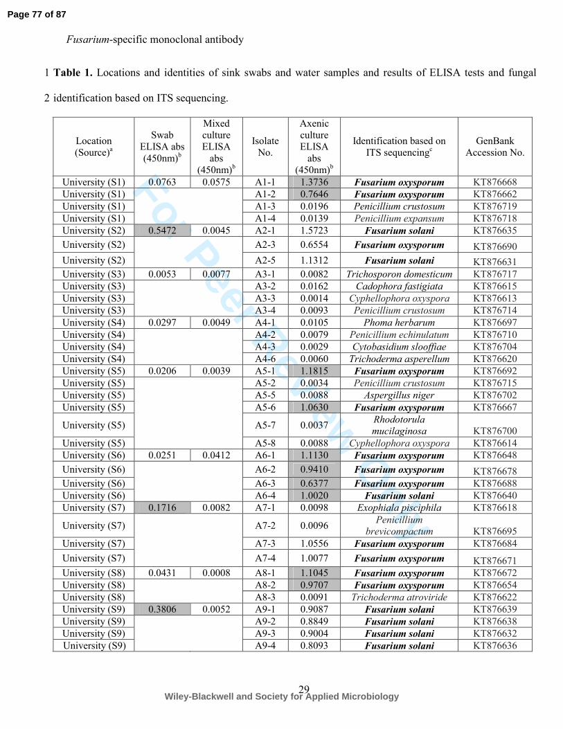
For Peer Review O
nly
Fusarium-specific monoclonal antibody
29
Table 1. Locations and identities of sink swabs and water samples and results of ELISA tests and fungal 1
identification based on ITS sequencing. 2
Location
(Source)a
Swab
ELISA abs
(450nm)b
Mixed
culture
ELISA
abs
(450nm)b
Isolate
No.
Axenic
culture
ELISA
abs
(450nm)b
Identification based on
ITS sequencingc
GenBank
Accession No.
University (S1) 0.0763 0.0575 A1-1 1.3736 Fusarium oxysporum KT876668
University (S1) A1-2 0.7646 Fusarium oxysporum KT876662
University (S1) A1-3 0.0196 Penicillium crustosum KT876719
University (S1) A1-4 0.0139 Penicillium expansum KT876718
University (S2) 0.5472 0.0045 A2-1 1.5723 Fusarium solani KT876635
University (S2) A2-3 0.6554 Fusarium oxysporum KT876690
University (S2) A2-5 1.1312 Fusarium solani KT876631
University (S3) 0.0053 0.0077 A3-1 0.0082 Trichosporon domesticum KT876717
University (S3) A3-2 0.0162 Cadophora fastigiata KT876615
University (S3) A3-3 0.0014 Cyphellophora oxyspora KT876613
University (S3) A3-4 0.0093 Penicillium crustosum KT876714
University (S4) 0.0297 0.0049 A4-1 0.0105 Phoma herbarum KT876697
University (S4) A4-2 0.0079 Penicillium echinulatum KT876710
University (S4) A4-3 0.0029 Cytobasidium slooffiae KT876704
University (S4) A4-6 0.0060 Trichoderma asperellum KT876620
University (S5) 0.0206 0.0039 A5-1 1.1815 Fusarium oxysporum KT876692
University (S5) A5-2 0.0034 Penicillium crustosum KT876715
University (S5) A5-5 0.0088 Aspergillus niger KT876702
University (S5) A5-6 1.0630 Fusarium oxysporum KT876667
University (S5) A5-7 0.0037 Rhodotorula
mucilaginosa KT876700
University (S5) A5-8 0.0088 Cyphellophora oxyspora KT876614
University (S6) 0.0251 0.0412 A6-1 1.1130 Fusarium oxysporum KT876648
University (S6) A6-2 0.9410 Fusarium oxysporum KT876678
University (S6) A6-3 0.6377 Fusarium oxysporum KT876688
University (S6) A6-4 1.0020 Fusarium solani KT876640
University (S7) 0.1716 0.0082 A7-1 0.0098 Exophiala pisciphila KT876618
University (S7) A7-2 0.0096 Penicillium
brevicompactum KT876695
University (S7) A7-3 1.0556 Fusarium oxysporum KT876684
University (S7) A7-4 1.0077 Fusarium oxysporum KT876671
University (S8) 0.0431 0.0008 A8-1 1.1045 Fusarium oxysporum KT876672
University (S8) A8-2 0.9707 Fusarium oxysporum KT876654
University (S8) A8-3 0.0091 Trichoderma atroviride KT876622
University (S9) 0.3806 0.0052 A9-1 0.9087 Fusarium solani KT876639
University (S9) A9-2 0.8849 Fusarium solani KT876638
University (S9) A9-3 0.9004 Fusarium solani KT876632
University (S9) A9-4 0.8093 Fusarium solani KT876636
Page 77 of 87
Wiley-Blackwell and Society for Applied Microbiology

For Peer Review O
nly
Fusarium-specific monoclonal antibody
30
University (S10) 0.0034 0.0028 A10-1 0.0188 Rhodotorula slooffiae
University (S10) A10-2 0.0020 Exophiala pisciphila KT876616
University (S11) 0.0231 0.0005 B1-1 0.6064 Fusarium dimerum KT876625
University (S11) B1-6 0.8138 Fusarium dimerum KT876628
University (S12) 0.0200 0.0020 B2-1 0.5678 Fusarium dimerum KT876626
University (S12) B2-5 0.4827 Fusarium dimerum KT876624
University (S13) 0.0091 0.0023 B3-4 0.0026 Phoma herbarum KT876696
University (S14) 0.0163 0.0008 B4-1 0.6992 Fusarium dimerum KT876627
University (S15) 0.0132 0.0003 B5-1 0.8008 Fusarium oxysporum KT876674
University (S15) B5-2 0.0048 Mucor circinelloides KT876701
University (S15) B5-3 0.8851 Fusarium oxysporum KT876677
University (S16) 0.0229 0.0002 B6-1 0.8193 Fusarium oxysporum KT876676
University (S16) B6-2 0.7582 Fusarium oxysporum KT876661
University (S17) 0.0395 0.0014 B7-1 0.8201 Fusarium oxysporum KT876675
University (S17) B7-6 0.7758 Fusarium dimerum KT876623
University (S18) 0.0133 0.0011 B8-1 0.7347 Fusarium solani KT876637
University (S18) B8-7 0.0095 Rhodosporidium babjevae KT876706
University (S19) 0.0212 0.0048 B9-1 0.0051 Meyerozyma
guilliermondii KT876707
University (S19) B9-2 0.8104 Fusarium oxysporum KT876657
University (S19) B9-3 0.6302 Fusarium solani KT876634
University (S19) B9-5 0.0017 Penicillium crustosum KT876720
University (S20) 0.5467 0.0394 B10-1 0.7467 Fusarium oxysporum KT876680
University (S20) B10-2 0.7667 Fusarium oxysporum KT876659
University (S20) B10-6 0.0071 Candida parapsilosis KT876703
University (S20) B10-7 0.0251 Meyerozyma
guilliermondii KT876711
University (S20) B10-9 0.0031 Cystobasidium slooffiae KT876712
University (S20) B10-12 0.0251 Trichosporon asteroides KT876713
University (S21) 0.0083 0.0269 C1-1 0.0044 Clavispora lusitaniae KT876708
University (S21) C1-3 0.8875 Fusarium oxysporum KT876682
University (S21) C1-4 0.8884 Fusarium dimerum KT876629
University (S21) C1-7 0.0003 Exophiala pisciphila KT876617
University (S22) 0.2977 0.0047 C2-1 0.9443 Fusarium oxysporum KT876658
University (S22) C2-4 0.9825 Fusarium oxysporum KT876694
University (S22) C2-7 0.8677 Fusarium oxysporum KT876687
University (S23) 0.0808 0.0116 C3-1 0.9853 Fusarium oxysporum KT876683
University (S23) C3-2 1.0160 Fusarium oxysporum KT876693
University (S23) C3-4 0.7485 Fusarium oxysporum KT876644
University (S24) 0.1113 0.0202 C4-1 0.8930 Fusarium oxysporum KT876670
University (S24) C4-2 0.0028 Candida intermedia KT876709
University (S24) C4-5 0.9008 Fusarium dimerum KT876630
University (S25) 0.5741 0.0097 C5-1 0.9874 Fusarium oxysporum KT876652
University (S25) C5-2 1.0996 Fusarium oxysporum KT876656
University (S25) C5-3 1.1236 Fusarium oxysporum KT876649
University (S25) C5-4 1.0355 Fusarium oxysporum KT876669
University (S25) C5-5 1.0672 Fusarium oxysporum KT876660
University (S26) 0.0603 0.0348 C6-1 1.0135 Fusarium oxysporum KT876665
Page 78 of 87
Wiley-Blackwell and Society for Applied Microbiology
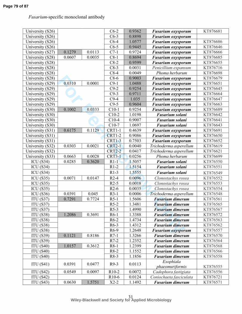
For Peer Review O
nly
Fusarium-specific monoclonal antibody
31
University (S26) C6-2 0.9362 Fusarium oxysporum KT876681
University (S26) C6-3 0.8898 Fusarium oxysporum
University (S26) C6-4 1.0577 Fusarium oxysporum KT876686
University (S26) C6-5 0.9445 Fusarium oxysporum KT876646
University (S27) 0.1279 0.0113 C7-1 0.9724 Fusarium oxysporum KT876666
University (S28) 0.0607 0.0035 C8-1 0.8694 Fusarium oxysporum KT876685
University (S28) C8-2 0.9599 Fusarium oxysporum KT876655
University (S28) C8-3 0.0031 Penicillium expansum KT876716
University (S28) C8-4 0.0049 Phoma herbarum KT876698
University (S28) C8-6 0.9003 Fusarium oxysporum KT876679
University (S29) 0.0310 0.0001 C9-1 1.0488 Fusarium oxysporum KT876651
University (S29) C9-2 0.9254 Fusarium oxysporum KT876645
University (S29) C9-3 0.9711 Fusarium oxysporum KT876664
University (S29) C9-4 1.035 Fusarium oxysporum KT876647
University (S29) C9-5 0.9604 Fusarium oxysporum KT876663
University (S30) 0.1002 0.0333 C10-1 0.9254 Fusarium oxysporum KT876689
University (S30) C10-2 1.0198 Fusarium solani KT876642
University (S30) C10-4 0.9007 Fusarium solani KT876641
University (S30) C10-7 1.0697 Fusarium solani KT876643
University (S31) 0.6175 0.1129 CRT1-1 0.4639 Fusarium oxysporum KT876691
University (S31) CRT1-2 0.9086 Fusarium oxysporum KT876650
University (S31) CRT1-3 0.7503 Fusarium oxysporum KT876652
University (S32) 0.0303 0.0021 CRT2-1 0.0040 Trichoderma asperellum KT876619
University (S32) CRT2-2 0.0417 Trichoderma asperellum KT876621
University (S33) 0.0663 0.0028 CRT3-1 0.0256 Phoma herbarum KT876699
ICU (S34) 0.0285 0.3620 R1-1 1.5057 Fusarium solani KT876550
ICU (S34) R1-2 1.5154 Fusarium solani KT876551
ICU (S34) R1-3 1.5555 Fusarium solani KT876549
ICU (S35) 0.0071 0.0147 R2-4 0.0096 Clonostachys rosea KT876552
ICU (S35) R2-5 0.0018 Clonostachys rosea KT876553
ICU (S35) R2-6 0.0035 Clonostachys rosea KT876554
ICU (S36) 0.0391 0.045 R3-1 0.0006 Trichoderma asperellum KT876548
ITU (S37) 0.7291 0.7724 R5-1 1.5606 Fusarium dimerum KT876561
ITU (S37) R5-2 1.3481 Fusarium dimerum KT876565
ITU (S37) R5-3 1.4990 Fusarium dimerum KT876567
ITU (S38) 1.2086 0.3691 R6-1 1.3388 Fusarium dimerum KT876572
ITU (S38) R6-2 1.4734 Fusarium dimerum KT876563
ITU (S38) R6-3 1.4312 Fusarium dimerum KT876562
ITU (S38) R6-9 1.2648 Fusarium oxysporum KT876557
ITU (S39) 0.1121 0.8186 R7-1 1.3266 Fusarium dimerum KT876570
ITU (S39) R7-2 1.2352 Fusarium dimerum KT876564
ITU (S40) 1.0157 0.3612 R8-1 1.2399 Fusarium dimerum KT876568
ITU (S40) R8-2 1.1552 Fusarium dimerum KT876566
ITU (S40) R8-3 1.1856 Fusarium dimerum KT876558
ITU (S41) 0.0391 0.0477 R9-3 0.0113 Exophiala
phaeomuriformis KT876555
ITU (S42) 0.0549 0.0097 R10-2 0.0072 Cadophora fastigiata KT876556
ITU (S42) R10-6 0.0124 Coniochaeta fasciculata KT876721
ITU (S43) 0.0630 1.5751 X2-2 1.1492 Fusarium dimerum KT876571
Page 79 of 87
Wiley-Blackwell and Society for Applied Microbiology
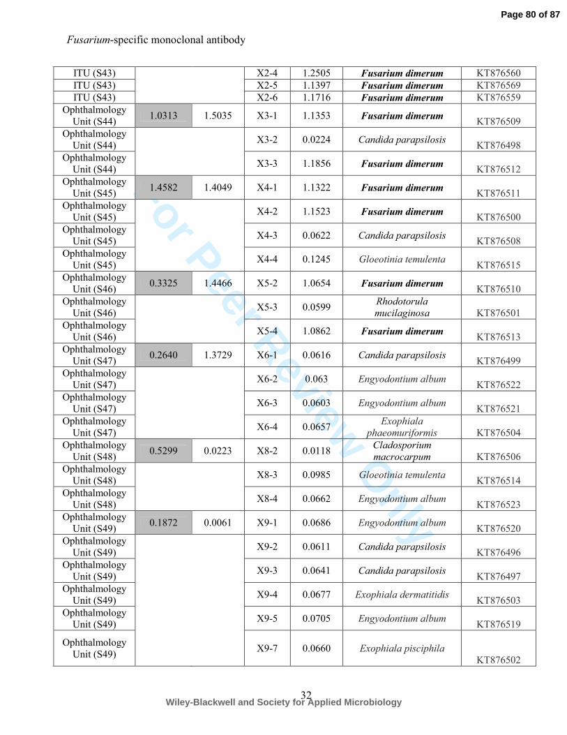
For Peer Review O
nly
Fusarium-specific monoclonal antibody
32
ITU (S43) X2-4 1.2505 Fusarium dimerum KT876560
ITU (S43) X2-5 1.1397 Fusarium dimerum KT876569
ITU (S43) X2-6 1.1716 Fusarium dimerum KT876559
Ophthalmology
Unit (S44) 1.0313 1.5035 X3-1 1.1353 Fusarium dimerum
KT876509
Ophthalmology
Unit (S44) X3-2 0.0224 Candida parapsilosis
KT876498
Ophthalmology
Unit (S44) X3-3 1.1856 Fusarium dimerum
KT876512
Ophthalmology
Unit (S45) 1.4582 1.4049 X4-1 1.1322 Fusarium dimerum
KT876511
Ophthalmology
Unit (S45) X4-2 1.1523 Fusarium dimerum
KT876500
Ophthalmology
Unit (S45) X4-3 0.0622 Candida parapsilosis
KT876508
Ophthalmology
Unit (S45) X4-4 0.1245 Gloeotinia temulenta
KT876515
Ophthalmology
Unit (S46) 0.3325 1.4466 X5-2 1.0654 Fusarium dimerum
KT876510
Ophthalmology
Unit (S46) X5-3 0.0599
Rhodotorula
mucilaginosa KT876501
Ophthalmology
Unit (S46) X5-4 1.0862 Fusarium dimerum
KT876513
Ophthalmology
Unit (S47) 0.2640 1.3729 X6-1 0.0616 Candida parapsilosis
KT876499
Ophthalmology
Unit (S47) X6-2 0.063 Engyodontium album
KT876522
Ophthalmology
Unit (S47) X6-3 0.0603 Engyodontium album
KT876521
Ophthalmology
Unit (S47) X6-4 0.0657
Exophiala
phaeomuriformis KT876504
Ophthalmology
Unit (S48) 0.5299 0.0223 X8-2 0.0118
Cladosporium
macrocarpum KT876506
Ophthalmology
Unit (S48) X8-3 0.0985 Gloeotinia temulenta
KT876514
Ophthalmology
Unit (S48) X8-4 0.0662 Engyodontium album
KT876523
Ophthalmology
Unit (S49) 0.1872 0.0061 X9-1 0.0686 Engyodontium album
KT876520
Ophthalmology
Unit (S49) X9-2 0.0611 Candida parapsilosis
KT876496
Ophthalmology
Unit (S49) X9-3 0.0641 Candida parapsilosis
KT876497
Ophthalmology
Unit (S49) X9-4 0.0677 Exophiala dermatitidis
KT876503
Ophthalmology
Unit (S49) X9-5 0.0705 Engyodontium album
KT876519
Ophthalmology
Unit (S49)
X9-7 0.0660 Exophiala pisciphila
KT876502
Page 80 of 87
Wiley-Blackwell and Society for Applied Microbiology
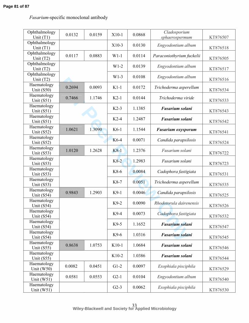
For Peer Review O
nly
Fusarium-specific monoclonal antibody
33
Ophthalmology
Unit (T1) 0.0132 0.0159 X10⁻¹ 0.0868
Cladosporium
sphaerospermum KT876507
Ophthalmology
Unit (T1) X10-3 0.0130 Engyodontium album
KT876518
Ophthalmology
Unit (T2) 0.0117 0.0883 W1-1 0.0114 Paraconiothyrium fuckelii
KT876505
Ophthalmology
Unit (T2) W1-2 0.0139 Engyodontium album
KT876517
Ophthalmology
Unit (T2) W1-3 0.0108 Engyodontium album
KT876516
Haematology
Unit (S50) 0.2694 0.0093 K1-1 0.0172 Trichoderma asperellum
KT876534
Haematology
Unit (S51) 0.7466 1.1746 K2-1 0.0144 Trichoderma viride
KT876533
Haematology
Unit (S51) K2-3 1.1385 Fusarium solani
KT876543
Haematology
Unit (S51) K2-4 1.2487 Fusarium solani
KT876542
Haematology
Unit (S52) 1.0621 1.3090 K6-1 1.1544 Fusarium oxysporum
KT876541
Haematology
Unit (S52) K6-4 0.0071 Candida parapsilosis
KT876524
Haematology
Unit (S53) 1.0120 1.2628 K8-1 1.2376 Fusarium solani
KT876722
Haematology
Unit (S53) K8-2 1.2983 Fusarium solani
KT876723
Haematology
Unit (S53) K8-6 0.0084 Cadophora fastigiata
KT876531
Haematology
Unit (S53) K8-7 0.0051 Trichoderma asperellum
KT876535
Haematology
Unit (S54) 0.9843 1.2903 K9-1 0.0046 Candida parapsilosis
KT876525
Haematology
Unit (S54) K9-2 0.0090 Rhodotorula dairenensis
KT876526
Haematology
Unit (S54) K9-4 0.0073 Cadophora fastigiata
KT876532
Haematology
Unit (S54) K9-5 1.1652 Fusarium solani
KT876547
Haematology
Unit (S54) K9-6 1.0316 Fusarium solani
KT876545
Haematology
Unit (S55) 0.8638 1.0753 K10-1 1.0684 Fusarium solani
KT876546
Haematology
Unit (S55) K10-2 1.0386 Fusarium solani
KT876544
Haematology
Unit (W50) 0.0082 0.0451 G1-2 0.0097 Exophiala pisciphila
KT876529
Haematology
Unit (W51) 0.0581 0.0553 G2-1 0.0104 Engyodontium album
KT876540
Haematology
Unit (W51) G2-3 0.0062 Exophiala pisciphila
KT876530
Page 81 of 87
Wiley-Blackwell and Society for Applied Microbiology
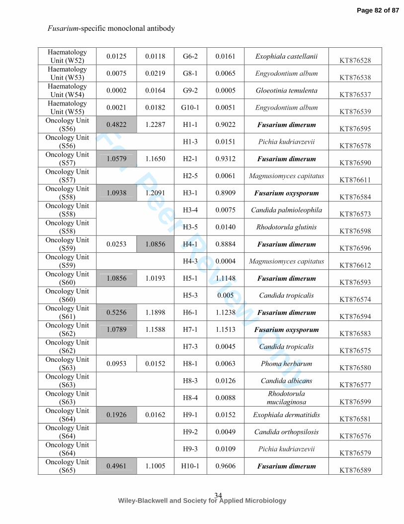
For Peer Review O
nly
Fusarium-specific monoclonal antibody
34
Haematology
Unit (W52) 0.0125 0.0118 G6-2 0.0161 Exophiala castellanii
KT876528
Haematology
Unit (W53) 0.0075 0.0219 G8-1 0.0065 Engyodontium album
KT876538
Haematology
Unit (W54) 0.0002 0.0164 G9-2 0.0005 Gloeotinia temulenta
KT876537
Haematology
Unit (W55) 0.0021 0.0182 G10-1 0.0051 Engyodontium album
KT876539
Oncology Unit
(S56) 0.4822 1.2287 H1-1 0.9022 Fusarium dimerum
KT876595
Oncology Unit
(S56) H1-3 0.0151 Pichia kudriavzevii
KT876578
Oncology Unit
(S57) 1.0579 1.1650 H2-1 0.9312 Fusarium dimerum
KT876590
Oncology Unit
(S57) H2-5 0.0061 Magnusiomyces capitatus
KT876611
Oncology Unit
(S58) 1.0938 1.2091 H3-1 0.8909 Fusarium oxysporum
KT876584
Oncology Unit
(S58) H3-4 0.0075 Candida palmioleophila
KT876573
Oncology Unit
(S58) H3-5 0.0140 Rhodotorula glutinis
KT876598
Oncology Unit
(S59) 0.0253 1.0856 H4-1 0.8884 Fusarium dimerum
KT876596
Oncology Unit
(S59) H4-3 0.0004 Magnusiomyces capitatus
KT876612
Oncology Unit
(S60) 1.0856 1.0193 H5-1 1.1148 Fusarium dimerum
KT876593
Oncology Unit
(S60) H5-3 0.005 Candida tropicalis
KT876574
Oncology Unit
(S61) 0.5256 1.1898 H6-1 1.1238 Fusarium dimerum
KT876594
Oncology Unit
(S62) 1.0789 1.1588 H7-1 1.1513 Fusarium oxysporum
KT876583
Oncology Unit
(S62) H7-3 0.0045 Candida tropicalis
KT876575
Oncology Unit
(S63) 0.0953 0.0152 H8-1 0.0063 Phoma herbarum
KT876580
Oncology Unit
(S63) H8-3 0.0126 Candida albicans
KT876577
Oncology Unit
(S63) H8-4 0.0088
Rhodotorula
mucilaginosa KT876599
Oncology Unit
(S64) 0.1926 0.0162 H9-1 0.0152 Exophiala dermatitidis
KT876581
Oncology Unit
(S64) H9-2 0.0049 Candida orthopsilosis
KT876576
Oncology Unit
(S64) H9-3 0.0109 Pichia kudriavzevii
KT876579
Oncology Unit
(S65) 0.4961 1.1005 H10-1 0.9606 Fusarium dimerum
KT876589
Page 82 of 87
Wiley-Blackwell and Society for Applied Microbiology
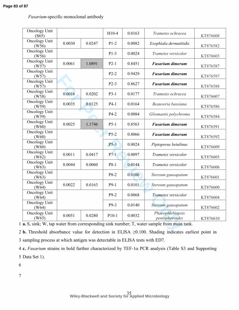
For Peer Review O
nly
Fusarium-specific monoclonal antibody
35
a. S, sink; W, tap water from corresponding sink number; T, water sample from main tank. 1
b. Threshold absorbance value for detection in ELISA ≥0.100. Shading indicates earliest point in 2
sampling process at which antigen was detectable in ELISA tests with ED7. 3
c. Fusarium strains in bold further characterized by TEF-1α PCR analysis (Table S3 and Supporting 4
Data Set 1). 5
6
7
Oncology Unit
(S65) H10-4 0.0163 Trametes ochracea
KT876608
Oncology Unit
(W56) 0.0030 0.0247 P1-2 0.0082 Exophiala dermatitidis
KT876582
Oncology Unit
(W56) P1-3 0.0024 Trametes versicolor
KT876603
Oncology Unit
(W57) 0.0061 1.0891 P2-1 0.8451 Fusarium dimerum
KT876587
Oncology Unit
(W57) P2-2 0.9429 Fusarium dimerum
KT876597
Oncology Unit
(W57) P2-3 0.8627 Fusarium dimerum
KT876588
Oncology Unit
(W58) 0.0016 0.0202 P3-1 0.0177 Trametes ochracea
KT876607
Oncology Unit
(W59) 0.0035 0.0125 P4-1 0.0164 Beauveria bassiana
KT876586
Oncology Unit
(W59) P4-2 0.0084 Gliomastix polychroma
KT876584
Oncology Unit
(W60) 0.0025 1.1746 P5-1 0.8563 Fusarium dimerum
KT876591
Oncology Unit
(W60) P5-2 0.8066 Fusarium dimerum
KT876592
Oncology Unit
(W60) P5-3 0.0024 Piptoporus betulinus
KT876609
Oncology Unit
(W62) 0.0011 0.0417 P7-1 0.0097 Trametes versicolor
KT876605
Oncology Unit
(W63) 0.0044 0.0060 P8-1 0.0144 Trametes versicolor
KT876606
Oncology Unit
(W63) P8-2 0.0106 Stereum gausapatum
KT876601
Oncology Unit
(W64) 0.0022 0.0163 P9-1 0.0101 Stereum gausapatum
KT876600
Oncology Unit
(W64) P9-2 0.0068 Trametes versicolor
KT876604
Oncology Unit
(W64) P9-3 0.0140 Stereum gausapatum
KT876602
Oncology Unit
(W65) 0.0051 0.0280 P10-1 0.0032
Phaeophlebiopsis
peniophoroides KT876610
Page 83 of 87
Wiley-Blackwell and Society for Applied Microbiology
For Peer Review O
nly
Fusarium-specific monoclonal antibody
36
Figure legends 1
Figure 1. Specificity of ED7 determined by Enzyme-Linked Immunosorbent Assay tests of 2
surface washings containing water-soluble antigens from Fusarium species and related and 3
unrelated yeasts and molds. (A) ELISA absorbance values at 450 nm for antigens from F. solani 4
and unrelated yeasts and molds and (B) for antigens from the F. solani teleomorph Haematonectria 5
haematococca and related Fusarium species. Wells were coated with 60 µg protein ml-1
buffer. Bars 6
are the means of three biological replicates ± standard errors and the threshold absorbance value for 7
detection of antigen in ELISA is ≥0.100 (indicated by lines on graphs). Numbers in parentheses 8
after species names denote strain numbers with further details of strains provided in Table S1. 9
10
Figure 2. Characterisation of the ED7 antigen and its epitope and spatial distribution of the 11
antigen in spores and hyphae. (A) Western immunoblot with ED7 using culture fluid from 2-d-old 12
PDB cultures of F. solani CBS224.34 (lane 1) and F. oxysporum f.sp. lycopersici CBS167.30 (lane 13
2). Wells were loaded with 1.6 µg of protein. Mr denotes molecular weight in kDa. Note the major 14
extracellular antigen with molecular weight of ~200 kDa. (B) Absorbance values from ELISA tests 15
with ED7 using surface washings containing water-soluble antigens immobilized to the wells of 16
microtitre plates and treated with trypsin or PBS only (control) at 4oC and 37
oC. Bars are the means 17
of three biological replicates ± standard errors and bars with the same letter are not significantly 18
different at p<0.001 (ANOVA and Tukey-Kramer test). (C) Absorbance values from ELISA tests 19
with ED7 using surface washings containing water-soluble antigens immobilized to the wells of 20
microtitre plates and treated with pronase or Milli-Q H2O only (control) at 4oC and 37
oC. Bars are 21
the means of three biological replicates ± standard errors and bars with the same letter are not 22
significantly different at p<0.001 (ANOVA and Tukey-Kramer test). (D) Stability of the water-23
Page 84 of 87
Wiley-Blackwell and Society for Applied Microbiology
For Peer Review O
nly
Fusarium-specific monoclonal antibody
37
soluble ED7 antigen following heating of surface washings at 100oC over a 70 min period. Treated 1
antigen was subsequently immobilized to the wells of microtitre plates and assayed by ELISA. Bars 2
are the means of three biological replicates ± standard errors and bars with the same letter are not 3
significantly different at p<0.001 (ANOVA and Tukey-Kramer test). (E) Absorbance values from 4
ELISA tests with ED7 using surface washings containing water-soluble antigens immobilized to the 5
wells of microtitre plates and treated with periodate (open circles) or with acetate only control 6
(closed circles) at 4oC over a 20 h period. Each point is the mean of three biological replicates ± 7
standard errors. (F to I) Photomicrographs of F. solani CBS224.34 immunostained with ED7 or 8
TCM control and goat anti-mouse polyvalent Ig fluorescein isothiocyanate (FITC) conjugate. (F) 9
Brightfield image of germinated conidium with hypha probed with ED7 followed by fluorochrome 10
conjugate (scale bar = 6 µm); (G) Same field of view as panel F but examined under 11
epifluorescence. Note intense staining of the cell wall of microconidium and hypha; (H) Brightfield 12
image of germinated conidium with hypha probed with TCM (negative control) followed by FITC 13
conjugate (scale bar = 3 µm); (I) Same field of view as panel H but examined under 14
epifluorescence. Note lack of staining, demonstrating specific binding of ED7 to surface antigen. (J-15
L) Immunogold labeling of sections of conidia and hyphae of F. solani CBS224.34. (J) Transverse 16
section of conidium incubated with ED7 and anti-mouse immunoglobulin 20 nm gold particles, 17
showing antigen in the cell wall and in an extracellular fimbrial matrix surrounding the spore (scale 18
bar = 250 nm); (K) Longitudinal section of hypha incubated with ED7 and anti-mouse 19
immunoglobulin 20 nm gold particles, showing antigen in the cell wall and in an extracellular 20
matrix surrounding the cell (scale bar = 100 nm); (L) Transverse section of a conidium incubated 21
with TCM (negative control) and anti-mouse immunoglobulin 20 nm gold particles, showing lack 22
of staining by the secondary gold conjugate (scale bar = 180 nm). 23
Page 85 of 87
Wiley-Blackwell and Society for Applied Microbiology
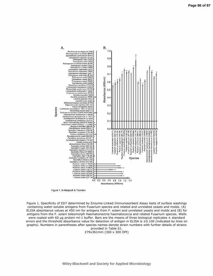
For Peer Review O
nly
Figure 1. Specificity of ED7 determined by Enzyme-Linked Immunosorbent Assay tests of surface washings containing water-soluble antigens from Fusarium species and related and unrelated yeasts and molds. (A) ELISA absorbance values at 450 nm for antigens from F. solani and unrelated yeasts and molds and (B) for
antigens from the F. solani teleomorph Haematonectria haematococca and related Fusarium species. Wells were coated with 60 µg protein ml-1 buffer. Bars are the means of three biological replicates ± standard
errors and the threshold absorbance value for detection of antigen in ELISA is ≥0.100 (indicated by lines on graphs). Numbers in parentheses after species names denote strain numbers with further details of strains
provided in Table S1. 279x361mm (300 x 300 DPI)
Page 86 of 87
Wiley-Blackwell and Society for Applied Microbiology
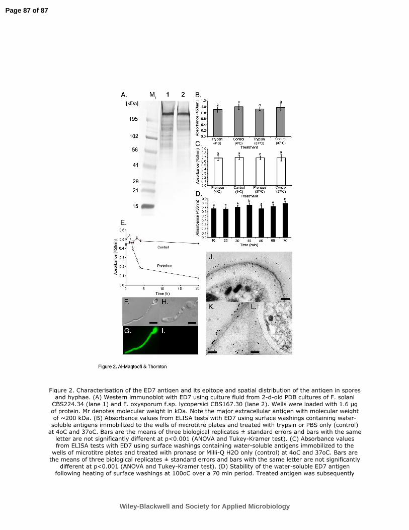
For Peer Review O
nly
Figure 2. Characterisation of the ED7 antigen and its epitope and spatial distribution of the antigen in spores and hyphae. (A) Western immunoblot with ED7 using culture fluid from 2-d-old PDB cultures of F. solani CBS224.34 (lane 1) and F. oxysporum f.sp. lycopersici CBS167.30 (lane 2). Wells were loaded with 1.6 µg
of protein. Mr denotes molecular weight in kDa. Note the major extracellular antigen with molecular weight of ~200 kDa. (B) Absorbance values from ELISA tests with ED7 using surface washings containing water-soluble antigens immobilized to the wells of microtitre plates and treated with trypsin or PBS only (control) at 4oC and 37oC. Bars are the means of three biological replicates ± standard errors and bars with the same letter are not significantly different at p<0.001 (ANOVA and Tukey-Kramer test). (C) Absorbance values from ELISA tests with ED7 using surface washings containing water-soluble antigens immobilized to the wells of microtitre plates and treated with pronase or Milli-Q H2O only (control) at 4oC and 37oC. Bars are the means of three biological replicates ± standard errors and bars with the same letter are not significantly
different at p<0.001 (ANOVA and Tukey-Kramer test). (D) Stability of the water-soluble ED7 antigen following heating of surface washings at 100oC over a 70 min period. Treated antigen was subsequently
Page 87 of 87
Wiley-Blackwell and Society for Applied Microbiology
For Peer Review O
nly
immobilized to the wells of microtitre plates and assayed by ELISA. Bars are the means of three biological replicates ± standard errors and bars with the same letter are not significantly different at p<0.001 (ANOVA
and Tukey-Kramer test). (E) Absorbance values from ELISA tests with ED7 using surface washings containing water-soluble antigens immobilized to the wells of microtitre plates and treated with periodate (open circles) or with acetate only control (closed circles) at 4oC over a 20 h period. Each point is the mean
of three biological replicates ± standard errors. (F to I) Photomicrographs of F. solani CBS224.34
immunostained with ED7 or TCM control and goat anti-mouse polyvalent Ig fluorescein isothiocyanate (FITC) conjugate. (F) Brightfield image of germinated conidium with hypha probed with ED7 followed by fluorochrome conjugate (scale bar = 6 µm); (G) Same field of view as panel F but examined under
epifluorescence. Note intense staining of the cell wall of microconidium and hypha; (H) Brightfield image of germinated conidium with hypha probed with TCM (negative control) followed by FITC conjugate (scale bar = 3 µm); (I) Same field of view as panel H but examined under epifluorescence. Note lack of staining,
demonstrating specific binding of ED7 to surface antigen. (J-L) Immunogold labeling of sections of conidia and hyphae of F. solani CBS224.34. (J) Transverse section of conidium incubated with ED7 and anti-mouse immunoglobulin 20 nm gold particles, showing antigen in the cell wall and in an extracellular fimbrial matrix surrounding the spore (scale bar = 250 nm); (K) Longitudinal section of hypha incubated with ED7 and anti-
mouse immunoglobulin 20 nm g 279x361mm (300 x 300 DPI)
Page 88 of 87
Wiley-Blackwell and Society for Applied Microbiology