foot loading is different in people with and without pincer nails
DESCRIPTION
Background Recent studies suggest that pincer nails are caused by lack of upward mechanical forces on the toe pad. However, clinically significant pincer nails are also often observed among healthy walkers. It was hypothesized that in these cases, the affected toes do not receive adequate physical stimulation from walking and loading.TRANSCRIPT
RESEARCH Open Access
Foot loading is different in people with andwithout pincer nails: a case control studyHitomi Sano1*, Kaori Shionoya2 and Rei Ogawa1
Abstract
Background: Recent studies suggest that pincer nails are caused by lack of upward mechanical forces on the toepad. However, clinically significant pincer nails are also often observed among healthy walkers. It was hypothesizedthat in these cases, the affected toes do not receive adequate physical stimulation from walking and loading. Totest this, the gait characteristics of pincer nail cases were assessed by measuring plantar pressure during walking.
Methods: In total, 12 bilateral pincer nail cases (24 affected feet) and 12 age- and sex-controlled healthy controlsubjects (24 ft) were enrolled in this prospective case–control study. Plantar pressure during free ambulation inboth the barefoot and shod state was assessed using a digital pressure-plate system named S-Plate platform(Medicapteurs Co. France). First toe pressure and the frequencies of peak pressure in the first toe, metatarsal head,or other foot areas were calculated.
Results: In both the barefoot and shod state, the pincer nail group had significantly lower pressure on the first toethan the control group. In both the barefoot and shod state, the peak pressure area was mostly the metatarsalhead area in the pincer nail group, whereas it was mostly the first toe area in the control group. Binomial logisticregression analysis revealed that peak pressure area was a significant risk factor for pincer nail development.
Conclusion: Walking behavior appears to contribute to pincer nail development. Pincer nails of walkers could betreated by correcting the walking behaviour so that more pressure is placed on the toe pad.
Keywords: Pincer nail, Mechanical force, Ingrown nail, Nail deformity
BackgroundFingernails play a key role in protecting the distal pha-langes, enhancing tactile discrimination and performingfine manipulation [1–4]. They are also used for scratchingand grooming. Toenails contribute to pedal biomechanics.During daily life, nails are constantly exposed to physicalstimuli such as counter pressure and shear force. Thesemechanical forces may therefore play a marked role in nailconfiguration and participate in the development of naildeformities.The term pincer nail was introduced by Cornelius and
Shellery in 1968 [5] and is defined as a transverse over-curvature of the nail plate. Another condition is ingrownnail, which is a painful inflammatory condition causedby a nail edge that has usually been rendered sharp byimproper trimming [6]. Both conditions accompany each
other at high rates. Notably, although pincer and in-grown nails are common diseases, their precise etiolo-gies remain unclear. Although poorly fitting shoes havebeen considered to be one of the most common causesof pincer nails, our previous study showed that bedrid-den patients who do not wear shoes or bear their ownweight have a high incidence of pincer nails [7]. Further-more, we showed that the nails of fingers that are sub-ject to little physical stimulation tend to curve inwardwhile the nails of fingers that are subject to a lot ofphysical stimulation tend to have a flat shape [7–9].While it is generally believed that nail shrinkage is anabnormal phenomenon [6], cumulative research at ourhospital has led us to hypothesize that human nails areconstitutively equipped with an automatic shrinkagefunction that allows them to adapt to daily upwardmechanical forces. In line with this, we proposed that apincer nail may be caused by the lack of appropriatemechanical forces on the nail [10]. At face value, the fact
* Correspondence: [email protected] of Plastic, Reconstructive and Aesthetic Surgery, NipponMedical School, 1-1-5 Sendagi, Bunkyo-ku, Tokyo 113-8603, JapanFull list of author information is available at the end of the article
JOURNAL OF FOOTAND ANKLE RESEARCH
© 2015 Sano et al. Open Access This article is distributed under the terms of the Creative Commons Attribution 4.0International License (http://creativecommons.org/licenses/by/4.0/), which permits unrestricted use, distribution, andreproduction in any medium, provided you give appropriate credit to the original author(s) and the source, provide alink to the Creative Commons license, and indicate if changes were made. The Creative Commons Public DomainDedication waiver (http://creativecommons.org/publicdomain/zero/1.0/) applies to the data made available in thisarticle, unless otherwise stated.
Sano et al. Journal of Foot and Ankle Research (2015) 8:43 DOI 10.1186/s13047-015-0100-y
that pincer nails are also often observed among healthywalkers could be seen to contradict the latter hypothesis.However, we speculated that, in these cases, insufficientphysical stimulation of the toe pad during walking or load-ing may be the causative factor. To test this hypothesis,the present study on the gait characteristics of pincer nailcases was performed. For this, the plantar pressure duringwalking was measured.
MethodsIn total, 24 ft of 12 adult participants with bilateralpincer nails and 24 ft of 12 healthy age- and sex-matched participants were enrolled in this prospectivecase–control study (Table 1). All subjects were free ofany ankle and foot pathology. Only participants withpincer nail who were free from any pain after treat-ment with an super elastic wire, an elastic wire usedwidely in Japan for conservative pincer nail correction,were eligible for this study [11]. The pincer nail groupwas compared to the control group for nail configur-ation and plantar pressure. Furthermore, binomiallogistic regression analysis was applied to identify therisk factors for pincer nail development.
Informed consentInformed consent was obtained from all individual partici-pants included in the study. All procedures performed instudies involving human participants were in accordancewith the ethical standards of the institutional and/ornational research committee and with the 1964 Helsinkideclaration and its later amendments or comparable eth-ical standards.
Measurement of plantar pressureA digital pressure-plate system named the S-Plate plat-form (Medicapteurs Co. France) was chosen to measureplantar pressure, because of its portability, accuracy andcost-effectiveness. It was implemented as a device to de-termine plantar pressure profile after pilot studies con-firmed its repeatability. The S-Plate platform can collectdata from the right and left foot at the same time duringnatural walking and is used to measure plantar pressureon both bare and shod feet during free ambulation. The
foot was divided into eight anatomical regions: first toe,small toes, medial metatarsal, middle metatarsals, lateralmetatarsals, medial arch and lateral arch, as reported byBoyd LA et al. [12]. Three trials were sequentially per-formed under each condition on the same day. Theaverage pressure of each first toe area was calculated.Moreover, the peak pressure area, which is defined asthe site that receives the most intense pressure in theplantar, in each foot and in both in the barefoot andshod state, was determined. Peak pressure is defined asthe maximum pressure of the peak pressure area. Thefrequency with which the first toe, the metatarsal head,or the remaining areas of the foot was the peak pres-sure area in the barefoot or shod state was calculatedfor both groups.
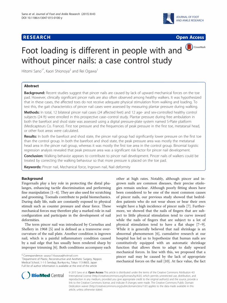
Analysis of first toe nail configurationNail configuration was measured by calculating thecurve index [defined as (nail height/nail width)*100 (%)]and the nail thickness (Fig. 1). The nail height, nail widthand the thickness of the central part of the nail plateswere measured by using a pair of calipers. For the pincernail cases, the pre-treatment configuration served as thenail configuration in this analysis.
Statistical analysisAll data were expressed as means ± SD. Statistical ana-lysis was performed using Microsoft Excel 97–2003(Microsoft, USA) and SPSS statistical software (SPSS,Chicago, IL). Continuous data were compared by usingthe Mann-Whitney U test. Categorical data were com-pared using the chi-square test or Yates chi-square testfollowed by Haberman residual analysis. Bonferroni’scorrection was used to correct for multiple comparisons.The relationship between pincer nail development (thedependent variable) and clinical covariates, includingweight, height, BMI, age, sex, peak pressure area (firsttoe vs. other areas of the foot), and the pressure on thefirst toe were assessed by binomial logistic regressionanalysis. p < 0.05 was considered to indicate statisticalsignificance.
Table 1 Demographic characteristics of the study participants
Pincer nail (n = 12 cases) Control (n = 12 subjects) P value
Gender 6 females, 6 males 6 females, 6 males 1
Age (years) 48.5 ± 17.8 46.8 ± 11.9 0.7
Height (cm) 163.8 ± 9.2 165.2 ± 7.9 0.58
Weight (kg) 61.3 ± 15.3 61.9 ± 10.3 0.86
BMI (kg/m2) 22.6 ± 3.8 22.6 ± 2.7 0.98
The p values were generated by Mann-Whitney U testsThe control consisted of healthy age- and sex-matched participantsBMI body mass index
Sano et al. Journal of Foot and Ankle Research (2015) 8:43 Page 2 of 6
ResultsThe Mann-Whitney U test revealed that the pincer nailgroup had a significantly higher mean first toe nail curveindex than the control group (57.9 ± 24.9 % vs. 13.9 ±5.4 %, p < 0.001).The Mann-Whitney U test also revealed that the pres-
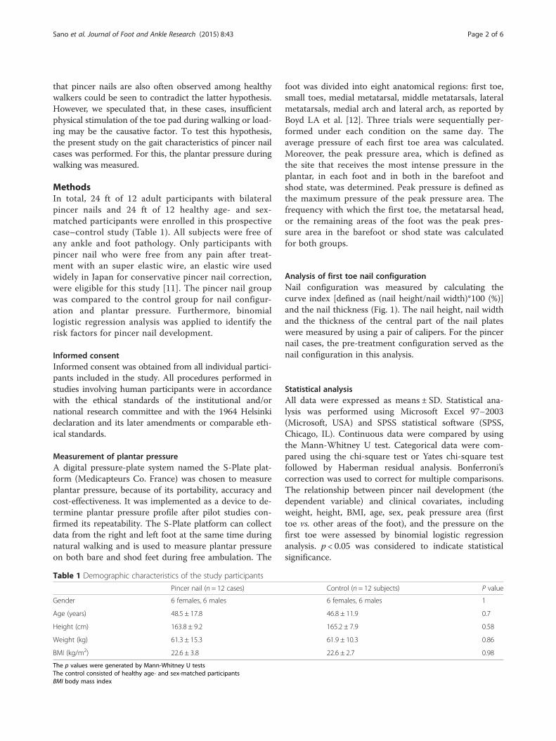
sure on the first toe of the pincer nail group was signifi-cantly lower than that of the control group (bare feet:1010.0 ± 309.0 vs.1318.8 ± 226.2 g/cm2, p < 0.01, shod fee:861.7 ± 240.6 vs.1287.2 ± 322.9 g/cm2, p < 0.01). Moreover,in the pincer nail group, the first toe pressure of shod feetwas significantly lower than that of bare feet (861.7 ±240.6 vs.1010.0 ± 309.0 g/cm2, p < 0.05) This differencewas not observed in the control group (1287.2 ± 322.9vs1318.8 ± 226.2 g/cm2, p = 0.71) (Fig. 2).Yates chi-square analysis revealed that in the barefoot
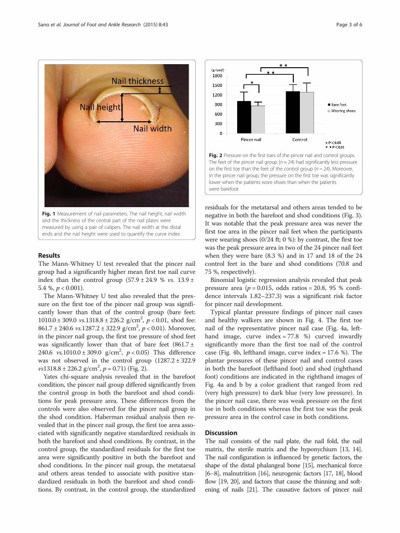
condition, the pincer nail group differed significantly fromthe control group in both the barefoot and shod condi-tions for peak pressure area. These differences from thecontrols were also observed for the pincer nail group inthe shod condition. Haberman residual analysis then re-vealed that in the pincer nail group, the first toe area asso-ciated with significantly negative standardized residuals inboth the barefoot and shod conditions. By contrast, in thecontrol group, the standardized residuals for the first toearea were significantly positive in both the barefoot andshod conditions. In the pincer nail group, the metatarsaland others areas tended to associate with positive stan-dardized residuals in both the barefoot and shod condi-tions. By contrast, in the control group, the standardized
residuals for the metatarsal and others areas tended to benegative in both the barefoot and shod conditions (Fig. 3).It was notable that the peak pressure area was never thefirst toe area in the pincer nail feet when the participantswere wearing shoes (0/24 ft; 0 %): by contrast, the first toewas the peak pressure area in two of the 24 pincer nail feetwhen they were bare (8.3 %) and in 17 and 18 of the 24control feet in the bare and shod conditions (70.8 and75 %, respectively).Binomial logistic regression analysis revealed that peak
pressure area (p = 0.015, odds ratios = 20.8, 95 % confi-dence intervals 1.82–237.3) was a significant risk factorfor pincer nail development.Typical plantar pressure findings of pincer nail cases
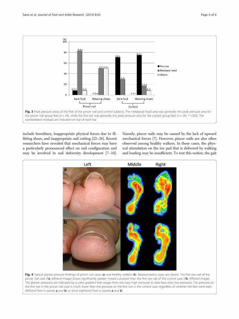
and healthy walkers are shown in Fig. 4. The first toenail of the representative pincer nail case (Fig. 4a, left-hand image, curve index = 77.8 %) curved inwardlysignificantly more than the first toe nail of the controlcase (Fig. 4b, lefthand image, curve index = 17.6 %). Theplantar pressures of these pincer nail and control casesin both the barefoot (lefthand foot) and shod (righthandfoot) conditions are indicated in the righthand images ofFig. 4a and b by a color gradient that ranged from red(very high pressure) to dark blue (very low pressure). Inthe pincer nail case, there was weak pressure on the firsttoe in both conditions whereas the first toe was the peakpressure area in the control case in both conditions.
DiscussionThe nail consists of the nail plate, the nail fold, the nailmatrix, the sterile matrix and the hyponychium [13, 14].The nail configuration is influenced by genetic factors, theshape of the distal phalangeal bone [15], mechanical force[6–8], malnutrition [16], neurogenic factors [17, 18], bloodflow [19, 20], and factors that cause the thinning and soft-ening of nails [21]. The causative factors of pincer nail
Fig. 1 Measurement of nail parameters. The nail height, nail widthand the thickness of the central part of the nail plates weremeasured by using a pair of calipers. The nail width at the distalends and the nail height were used to quantify the curve index
Fig. 2 Pressure on the first toes of the pincer nail and control groups.The feet of the pincer nail group (n = 24) had significantly less pressureon the first toe than the feet of the control group (n = 24). Moreover,in the pincer nail group, the pressure on the first toe was significantlylower when the patients wore shoes than when the patientswere barefoot
Sano et al. Journal of Foot and Ankle Research (2015) 8:43 Page 3 of 6
include hereditary, inappropriate physical forces due to ill-fitting shoes, and inappropriate nail cutting [22–26]. Recentresearchers have revealed that mechanical forces may havea particularly pronounced effect on nail configuration andmay be involved in nail deformity development [7–10].
Namely, pincer nails may be caused by the lack of upwardmechanical forces [7]. However, pincer nails are also oftenobserved among healthy walkers. In these cases, the phys-ical stimulation on the toe pad that is delivered by walkingand loading may be insufficient. To test this notion, the gait
Fig. 3 Peak pressure areas of the feet of the pincer nail and control subjects. The metatarsal head area was generally the peak pressure area forthe pincer nail group feet (n = 24), while the first toe was generally the peak pressure area for the control group feet (n = 24). * < 0.05. Thestandardized residuals are indicated on top of each bar
Fig. 4 Typical plantar pressure findings of pincer nail cases (a) and healthy walkers (b). Representative cases are shown. The first toe nail of thepincer nail case (4a, lefthand image) shows significantly greater inward curvature than the first toe nail of the control case (4b, lefthand image).The plantar pressures are indicated by a color gradient that ranges from red (very high pressure) to dark blue (very low pressure). The pressure onthe first toe in the pincer nail case is much lower than the pressure on the first toe in the control case regardless of whether the feet were bare(lefthand foot in panels a and b) or shod (righthand foot in panels a and b)
Sano et al. Journal of Foot and Ankle Research (2015) 8:43 Page 4 of 6
characteristics of patients with a pincer nail were assessedin the present study.Indeed, plantar pressure analysis of these patients and
healthy control participants revealed that the first toes ofthe pincer nail group experienced significantly less pres-sure during free ambulation than the first toes of thecontrol group, both in the barefoot and shod conditions.Moreover, compared to the barefoot condition, the pres-sure on the first toes of the pincer nail group was signifi-cantly reduced by wearing shoes. Given that poorlyfitting shoes are considered to be a major cause of pin-cer nails, this observation suggests either that patientswith pincer nail tend to wear bigger shoes to avoid painor that pincer nail develops because the first toe naildoes not receive sufficient pressure from the ill-fittingshoes.Based on our hypothesis, it seems plausible that the
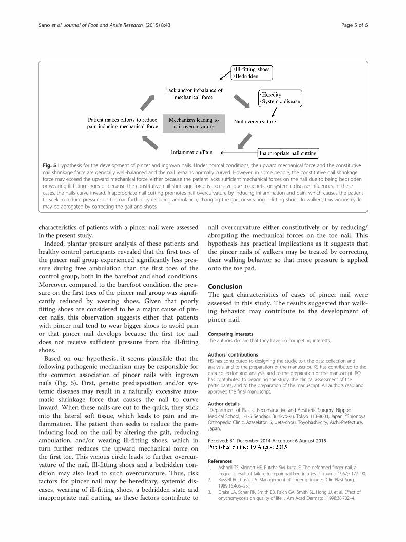
following pathogenic mechanism may be responsible forthe common association of pincer nails with ingrownnails (Fig. 5). First, genetic predisposition and/or sys-temic diseases may result in a naturally excessive auto-matic shrinkage force that causes the nail to curveinward. When these nails are cut to the quick, they stickinto the lateral soft tissue, which leads to pain and in-flammation. The patient then seeks to reduce the pain-inducing load on the nail by altering the gait, reducingambulation, and/or wearing ill-fitting shoes, which inturn further reduces the upward mechanical force onthe first toe. This vicious circle leads to further overcur-vature of the nail. Ill-fitting shoes and a bedridden con-dition may also lead to such overcurvature. Thus, riskfactors for pincer nail may be hereditary, systemic dis-eases, wearing of ill-fitting shoes, a bedridden state andinappropriate nail cutting, as these factors contribute to
nail overcurvature either constitutively or by reducing/abrogating the mechanical forces on the toe nail. Thishypothesis has practical implications as it suggests thatthe pincer nails of walkers may be treated by correctingtheir walking behavior so that more pressure is appliedonto the toe pad.
ConclusionThe gait characteristics of cases of pincer nail wereassessed in this study. The results suggested that walk-ing behavior may contribute to the development ofpincer nail.
Competing interestsThe authors declare that they have no competing interests.
Authors’ contributionsHS has contributed to designing the study, to t the data collection andanalysis, and to the preparation of the manuscript. KS has contributed to thedata collection and analysis, and to the preparation of the manuscript. ROhas contributed to designing the study, the clinical assessment of theparticipants, and to the preparation of the manuscript. All authors read andapproved the final manuscript.
Author details1Department of Plastic, Reconstructive and Aesthetic Surgery, NipponMedical School, 1-1-5 Sendagi, Bunkyo-ku, Tokyo 113-8603, Japan. 2ShionoyaOrthopedic Clinic, Azasekitori 5, Ueta-chou, Toyohashi-city, Aichi-Prefecture,Japan.
Received: 31 December 2014 Accepted: 6 August 2015
References1. Ashbell TS, Kleinert HE, Putcha SM, Kutz JE. The deformed finger nail, a
frequent result of failure to repair nail bed injuries. J Trauma. 1967;7:177–90.2. Russell RC, Casas LA. Management of fingertip injuries. Clin Plast Surg.
1989;16:405–25.3. Drake LA, Scher RK, Smith EB, Faich GA, Smith SL, Hong JJ, et al. Effect of
onychomycosis on quality of life. J Am Acad Dermatol. 1998;38:702–4.
Fig. 5 Hypothesis for the development of pincer and ingrown nails. Under normal conditions, the upward mechanical force and the constitutivenail shrinkage force are generally well-balanced and the nail remains normally curved. However, in some people, the constitutive nail shrinkageforce may exceed the upward mechanical force, either because the patient lacks sufficient mechanical forces on the nail due to being bedriddenor wearing ill-fitting shoes or because the constitutive nail shrinkage force is excessive due to genetic or systemic disease influences. In thesecases, the nails curve inward. Inappropriate nail cutting promotes nail overcurvature by inducing inflammation and pain, which causes the patientto seek to reduce pressure on the nail further by reducing ambulation, changing the gait, or wearing ill-fitting shoes. In walkers, this vicious cyclemay be abrogated by correcting the gait and shoes
Sano et al. Journal of Foot and Ankle Research (2015) 8:43 Page 5 of 6
4. Salazard B, Launay F, Desouches C, Samson P, Jouve JL, Magalon G.Fingertip injuries in children: 81 cases with at least one year follow-up. RevChir Orthop Reparatrice Appar Mot. 2004;90:621–7.
5. Sano H, Ichioka S. Influence of mechanical forces as a part of nailconfiguration. Dermatology. 2012;225:210–4.
6. Sano H, Ogawa R. Role of mechanical forces in hand nail configurationasymmetry in hemiplegia: an analysis of four hundred thumb nails.Dermatology. 2013;226:315–8.
7. Sano H, Shionoya K, Ogawa R. Finger nail configuration is influenced bymechanical forces on finger pads. J Dermatol. 2013;40:1056–7.
8. Cornelius 3rd CE, Shelley WB. Pincer nail syndrome. Arch Surg. 1968;96:321–2.9. Kosaka M, Kusuhara H, Mochizuki Y, Mori H, Isogai N. Morphologic study of
normal, ingrown, and pincer nails. Dermatol Surg. 2010;36:31–8.10. Sano H, Ogawa R. Clinical evidence for the relationship between nail
configuration and mechanical forces. PRS-Go. 2014;2:e216.11. Moriue T, Yoneda K, Moriue J, Matsuoka Y, Nakai K, Yokoi I, et al. A simple
therapeutic strategy with super elastic wire for ingrown toenails. DermatolSurg. 2008;34:1729–32.
12. Boyd LA, Bontrager EL, Mulroy SJ, Perry J. The reliability and validity of thenovel pedar system of in-shoe pressure measurement during freeambulation. Gait Posture. 1997;5:165.
13. Perrin C. The 2 clinical subbands of the distal nail unit and the nail isthmus.Anatomical explanation and new physiological observations in relation tothe nail growth. Am J Dermatopathol. 2008;30:216–21.
14. Johnson M, Shuster S. Determination of nail thickness and length. Br JDermatol. 1994;130:195–8.
15. Tosti A, Piraccini BM. Biology of nails and nail disorders. In: Fitzpatrick TB,Wolff K, editors. Fitzatrick’s dermatology in general medicine. 7th ed. NewYork: McGraw-Hill; 2008. p. 778–94.
16. Al-Dabbagh TQ, Al-Abachi KG. Nutritional koilonychia in 32 Iraqi subjects.Ann Saudi Med. 2005;25:154–7.
17. Horowitz SH. Diabetic neuropathy. Clin Orthop Relat Res. 1993;296:78–85.18. Andersen H. Motor dysfunction in diabetes. Diabetes Metab Res Rev.
2012;28:89–92.19. Alemany M. Regulation of adipose tissue energy availability through blood
flow control in the metabolic syndrome. Free Radic Biol Med. 2012;52:2108–19.20. Loenneke JP, Abe T, Wilson JM, Thiebaud RS, Fahs CA, Rossow LM, et al.
Blood flow restriction: an evidence based progressive model. Acta PhysiolHung. 2012;99:235–50.
21. Baron EB. Nutrition. In: Tierney LM, Mcphee SJ, Papadakis MA, editors.Current medical diagnosis and treatment. 38th ed. Stanford: Appleton andLange; 1999. p. 1182.
22. de Berker D, Carmichael AJ. Congenital alternate nail dystrophy. Br JDermatol. 1995;133:336–7.
23. Chapman RS. Letter: overcurvature of the nails–an inherited disorder. Br JDermatol. 1973;89:317–8.
24. Baran R, Dawber RPR. Diseases of the nail and their management. In: BaranR, de Berker DAR, Holzberg M, Thomas L, editors. Dermatology. 3rd ed.Boston: Blackwell Science; 2001. p. 496–9.
25. Vanderhooft SL, Vanderhooft JE. Pincer nail deformity after Kawasaki’sdisease. J Am Acad Dermatol. 1999;41:341–2.
26. Baran R. Letter: pincer and trumpet nails. Arch Dermatol. 1974;110:639–40.
Submit your next manuscript to BioMed Centraland take full advantage of:
• Convenient online submission
• Thorough peer review
• No space constraints or color figure charges
• Immediate publication on acceptance
• Inclusion in PubMed, CAS, Scopus and Google Scholar
• Research which is freely available for redistribution
Submit your manuscript at www.biomedcentral.com/submit
Sano et al. Journal of Foot and Ankle Research (2015) 8:43 Page 6 of 6