foot - ju medicine · flat foot (pes planus) is a condition in which the medial longitudinal arch...
TRANSCRIPT

Foot
Dr. Heba Kalbouneh
Associate Professor of Anatomy and Histology

Dorsum of the Foot Sole of the Foot
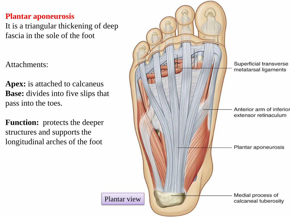
Plantar aponeurosis
It is a triangular thickening of deep
fascia in the sole of the foot
Attachments:
Apex: is attached to calcaneus
Base: divides into five slips that
pass into the toes.
Function: protects the deeper
structures and supports the
longitudinal arches of the foot
Plantar view
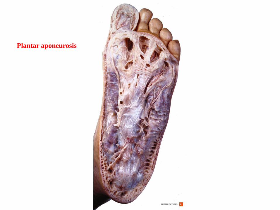
Plantar aponeurosis
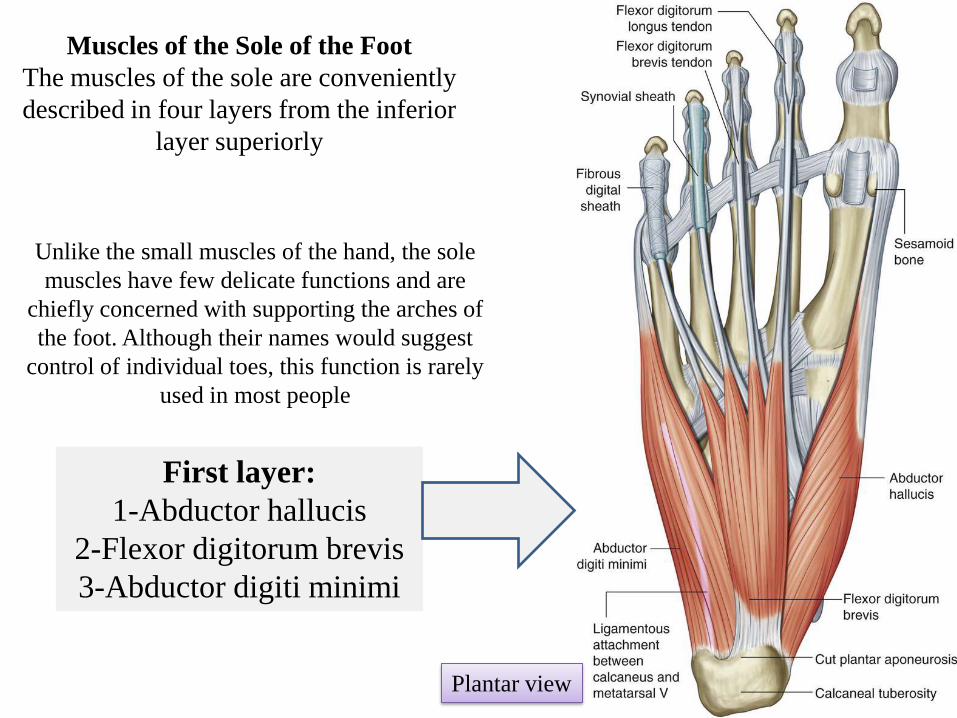
First layer:
1-Abductor hallucis
2-Flexor digitorum brevis
3-Abductor digiti minimi
Muscles of the Sole of the Foot
The muscles of the sole are conveniently
described in four layers from the inferior
layer superiorly
Unlike the small muscles of the hand, the sole
muscles have few delicate functions and are
chiefly concerned with supporting the arches of
the foot. Although their names would suggest
control of individual toes, this function is rarely
used in most people
Plantar view

Second layer:
1-Quadratus plantae
2-Lumbricals
3-Flexor digitorum longus tendon
4-Flexor hallucis longus tendon
Plantar view

Third layer:
1-Flexor hallucis brevis
2-Adductor hallucis
3-Flexor digiti minimi brevis
Plantar view

Fourth layer:
1-Interossei
2- Fibularis longus tendon
3-Tibialis posterior tendon
Plantar view

Medial Plantar Artery
Is the smaller of the terminal branches of the
posterior tibial artery
Ends by supplying the medial side of the big
toe
Lateral Plantar Artery
Is the larger of the terminal branches of
the posterior tibial artery
Forms the plantar arch
At the proximal end of the first
intermetatarsal space joins the dorsalis pedis
artery
Arteries of the Sole of the Foot Posterior
tibial artery
Lateral
Plantar Artery
Medial
Plantar Artery
Plantar view

Dorsalis Pedis Artery
(the Dorsal Artery of the Foot)
Starts as a continuation of anterior tibial
artery
Enters the sole of the foot (between the
two heads of the first dorsal interosseous
muscle) and joins the lateral plantar artery
Dorsal view
Anterior tibial artery
Arteries of the Dorsum of the Foot

Surface anatomy

Tibial nerve
Lateral
Plantar
Nerve
Medial
Plantar
Nerve
Medial Plantar Nerve
Is a terminal branch of the tibial nerve
Supplies the medial 2/3 of sole and medial
three and a half toes
Lateral Plantar Nerve
Is a terminal branch of the tibial nerve
Supplies lateral 1/3 of sole and lateral
one and a half toes
Nerves of the Sole of the Foot
The nerves extend onto the dorsum and
supply the nail beds and the tips of the toes
These nerves supply most intrinsic
muscles of the foot
Plantar view
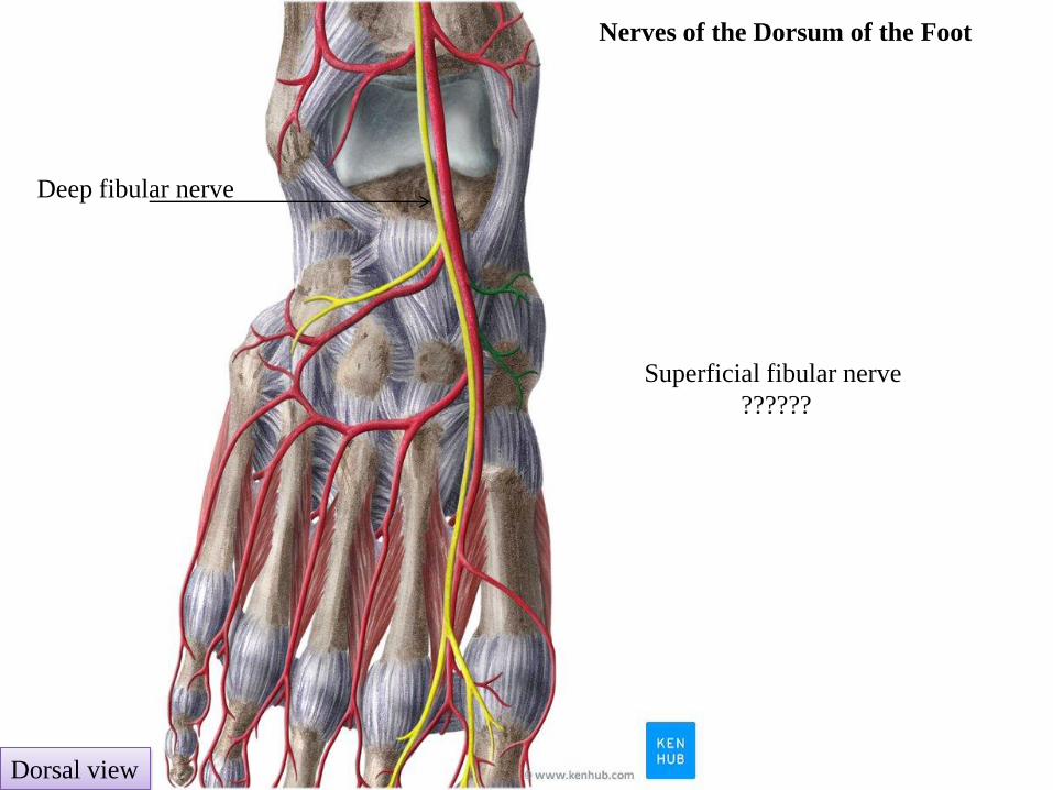
Nerves of the Dorsum of the Foot
Dorsal view
Deep fibular nerve
Superficial fibular nerve
??????

The Arches of the Foot
A segmented structure can hold up weight only if it is built in the form of an arch
The foot has three such arches:
1. Medial Longitudinal Arch
2. Lateral Longitudinal Arch
3.Transverse Arch
Function of arches of the foot
Protect the soft tissues of the sole
Distribution of the body weight

Note
The bones of the foot do not lie in a horizontal
plane. They form longitudinal and transverse
arches relative to the ground, which absorb and
distribute downward forces from the body
during standing and moving on different planes
Normally, the ball of the foot
carries about 40% of the weight and the
heel carries about 60%.
Lateral view

Body weight
50% on
the right
side
50% on
the left
side
The ball
of the foot
The heel
Arches of the Foot
The bones of the foot are arranged in
two arches that are held in position
by ligaments and tendons
Usually, the arches are fully
developed by age 12 or 13.
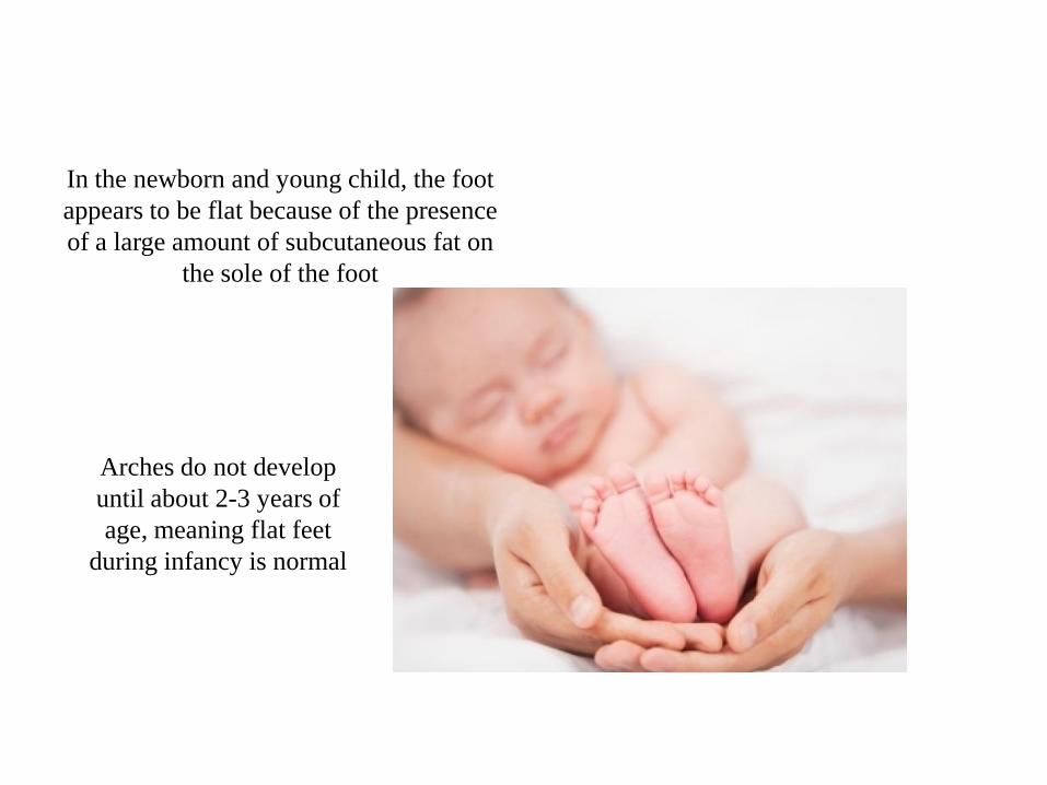
In the newborn and young child, the foot
appears to be flat because of the presence
of a large amount of subcutaneous fat on
the sole of the foot
Arches do not develop
until about 2-3 years of
age, meaning flat feet
during infancy is normal
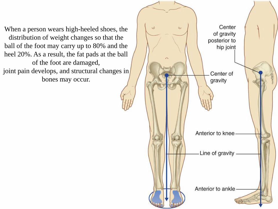
When a person wears high-heeled shoes, the
distribution of weight changes so that the
ball of the foot may carry up to 80% and the
heel 20%. As a result, the fat pads at the ball
of the foot are damaged,
joint pain develops, and structural changes in
bones may occur.

Plantar surface showing
the surface of contact with
the ground when standing

Calcaneus
Talus
Navicular
Cuneiforms Cuboid
1st 2nd 3rd 4th
5th
Plantar view

Plantar view
Medial Longitudinal Arch Lateral Longitudinal Arch
It is formed by the calcaneus, talus,
navicular, three cuneiforms and first
three metatarsal bones
It is formed by the
calcaneus, cuboid and
4th and 5th metatarsal
bones
Note: The
lateral arch is the
flatter and lies on
the ground in the
standing position

Transverse Arch
It is formed by the metatarsal
bases, the cuboid and the three
cuneiform bones

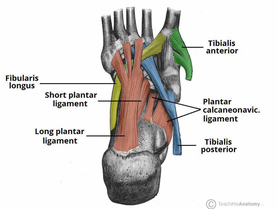
Muscles that provide dynamic
support for the arches during
walking include:
Tibialis anterior
Tibialis posterior
Fibularis longus
Ligaments that support the arches include:
Plantar calcaneonavicular (spring ligament)
Ahort plantar ligament (Plantar calcaneocuboid)
Long plantar ligament
Plantar aponeurosis

Long plantar ligament runs from
calcaneus and cuboid to the bases of
the lateral metatarsal bones
Plantar view
Calcaneus
Cuboid

Long plantar ligament
Plantar view
Calcaneus
Cuboid
Short plantar ligament
(Plantar calcaneocuboid )

Sustentaculum tali
Medial view
Calcaneus
Medial
cuniform
Calcaneus Navicular
Talus

Plantar calcaneonavicular (spring ligament)
Medial view
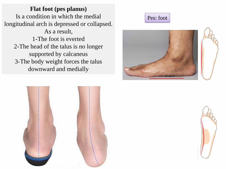
Flat foot (pes planus)
Is a condition in which the medial
longitudinal arch is depressed or collapsed.
As a result,
1-The foot is everted
2-The head of the talus is no longer
supported by calcaneus
3-The body weight forces the talus
downward and medially
Pes: foot
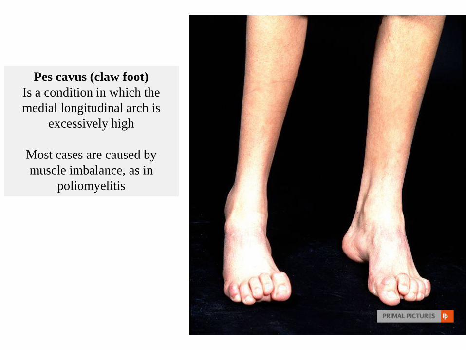
Pes cavus (claw foot)
Is a condition in which the
medial longitudinal arch is
excessively high
Most cases are caused by
muscle imbalance, as in
poliomyelitis

Plantar fasciitis
It happens to person who is standing or
walking for long time
It causes pain and tenderness of the sole
of the foot
Repeated attacks of this condition induce
ossification in the posterior attachment of
the aponeurosis

Tarsal tunnel syndrome
Due to compression of tibial nerve as it
travels through the tarsal tunnel.
Manifestation :
Motor
Sensory

Tarsal tunnel syndrome: distribution of sensory loss

Tarsal tunnel syndrome due to ganglion

Tibial nerve in tarsal tunnel