foot-and-mouth disease (fmd) training … meeting of trainers and co-trainers day 1 – monday...
TRANSCRIPT

FOOT-AND-MOUTH DISEASE (FMD)TRAINING MANUALcourse

PROGRAM- NTC

2011 – EuFMD: NTCs-Approximate program
Day 0 - Sunday Departure for Nakuru -14.00. Short meeting of trainers and co-trainers Day 1 – Monday Welcome; intro of participants, Introduction to Course, organisation of the week. Explanation of the 3 Teams and Roles facilitated by Team Leader or EuFMD Rep Epidemiology /control, of FMD in Kenya What is special about FMD investigations? Relevance of FMD pathogenesis, clinical signs, lesion ageing, kinetics of infectivity and transmission, to tracing FMD spread Sampling and Diagnostic procedures for FMD (include probang sampling in field) Principles of dangerous contact risk assessment If time Lessons learnt in FMD investigation – UK 2007 Afternoon: planning the field investigations Setting up laptops for internet. Demonstration of the EuFMD Training Wikispace- Online Group work exercises Epi-team: decide on roles , main lines of investigation, questions, gathering data on the suspect outbreak circumstances Clinical Team: decide roles, gathering data, decide on forms, roles, organisation Informatics Team:–assist with data required on recent outbreaks, village data Feedback, Output: agree on outbreak to investigate, summarise tasks and roles, forms to be completed Biosecurity talk – both optimal (EU) and pragmatic
Day 2 – Tuesday - Travel to field sites Supervised Clinical team work; Supervised Epi team work Decide on plans for Wedns/facilitated by Team leader Day 3 – Wednesday As decided on Day 2, new investigation or follow up previous one, or work on samples/findings from Day 2; Day 4 – Thursday AM1: lab findings reported followed by Group work – finalising /assembling the Clinical and Epi Report, and Summary AM2: Present and discuss Clinical and Epi Report PM: Case Study or Review Lessons Learnt Facilitated Feedback /evaluation of the Course, tips for improving Final biosecurity talk/declaration/actions Day 5 - Friday Visit to national park Back to Nairobi, arrival time 17.00
1: Monday
2/17/2011
1
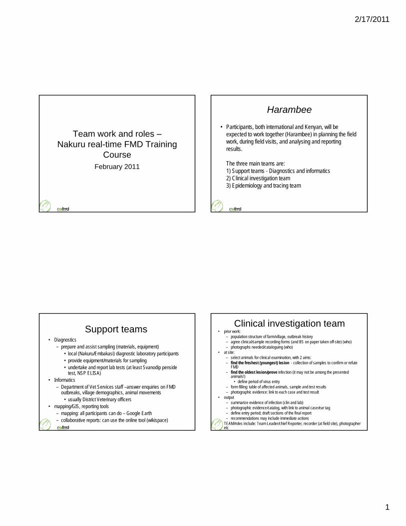
Team work and roles –Nakuru real-time FMD Training
CourseCourseFebruary 2011
Harambee
• Participants, both international and Kenyan, will be expected to work together (Harambee) in planning the field work, during field visits, and analysing and reporting results.
The three main teams are:1) Support teams - Diagnostics and informatics2) Clinical investigation team3) Epidemiology and tracing team
Support teams • Diagnostics
– prepare and assist sampling (materials, equipment)• local (Nakuru/Embakasi) diagnostic laboratory participants• provide equipment/materials for sampling • undertake and report lab tests (at least Svanodip penside
test NSP ELISA)test, NSP ELISA)• Informatics
– Department of Vet Services staff –answer enquiries on FMD outbreaks, village demographics, animal movements
• usually District Veterinary officers• mapping/GIS, reporting tools
– mapping: all participants can do – Google Earth– collaborative reports: can use the online tool (wikispace)
Clinical investigation team• prior work:
– population structure of farm/village, outbreak history– agree clinical/sample recording forms (and BS on paper taken off-site) (who)– photographs needed/cataloguing (who)
• at site:– select animals for clinical examination, with 2 aims: – find the freshest (youngest) lesion - collection of samples to confirm or refute
FMDfind the oldest lesion/prove infection (it may not be among the presented – find the oldest lesion/prove infection (it may not be among the presented animals!)
• define period of virus entry– form filling: table of affected animals, sample and test results– photographic evidence: link to each case and test result
• output– summarize evidence of infection (clin and lab)– photographic evidence/catalog, with link to animal case/ear tag– define entry period; draft sections of the final report – recommendations may include immediate actions
• TEAM/roles include: Team Leader/chief Reporter, recorder (at field site), photographer etc

2/17/2011
2
Clinical Examination Form
Name of the Farmer: Zafer Altay - Room 3 Farm ID: ……………. Epidemiological unit: Yagnurcak village Date: 13. 10. 2009
Animal ID Species Type L/S/M/PD/A Temp. Vesicular Lesions
Y/N
Age of Lesions
Samples Tissue/Blood/Serum
Number of Photos
Comments
TR 25967577, No. 10 bov. cow - n.m. N S (No. 4) Vaccinated in April 09
no eartag, No. 11 bov. calf - n.m. n.e. -
Example: Examination form – records signs, the photograph numbers, lesion age
no eartag, No. 12 bov. calf L/S 40,5° Y (M + F) > 7 d S (No. 3), Saliva No. 3 SANYO676, 678
Respiratory symptoms
no eartag, No. 13 bov. calf - n.m. Y (M) > 7 d
no eartag, No. 14 bov. calf - n.m. Y (M) > 7 d Very small lesions
L: … Lameness, S: … Salivation, M: … Drop of Milk Yield, PD: … Perinatal Death, A: ……Abortion Estimated age of oldest lesion seen: > 7 d
from OLDEST lesion: work out time window when infection entered group
Epidemiology/Tracing Team• tasks• pre-visit:
– identify recent outbreaks of relevance in region (location and time before present)– identify procedure on site (team leader, reporter, questions/questionnaires to use)
• at site:– undertake group and where required individual interviews– use participatory interview methods where needed to identify recent and longer term
FMD events and risk factors/entry/spread routes – sketch out village entry/exit and boundaries; – sketch out village entry/exit and boundaries; – identify animal groups in village that may be at different risk/history
• after return:– make source and spread windows, time-line– identify possible and mostly likely source– identify possible spread– recommend follow-up actions
• output: immediate report (to group), and follow-up written sections for final report
• TEAM/roles include: Team Leader, chief Reporter, interviewer & recorders (at field site), mapper (of field site) and GIS (for report), photographer etc
Example: use of Google Earth to illustrate where the clinical team sampled animals
Example - Timeline of the events identified in Yagmurcuk village, Turkey – based on oldest
lesion found
Most probable period of infection
rodu
ctio
n fro
m
Mar
ker
ubat
ion
perio
d
ship
men
t
ympt
oms
g gr
oup;
10
days
old
e
dete
cted
~19 20 18 19 20 21 22 23 24 25 26 27 28 29 30 1 2 3 4 5 6 7 8 9 10 11 12 13
Sta
rt of
cal
f int
rE
rzur
um
Star
t of t
he in
c u
Last
day
of
Sta
rt o
f sy
Vis
it of
EUF
MD
train
ing
lesi
ons
wer
e
Aug Aug Sept Sept September Sept Sept Sept Oct Oct Oct Oct Oct Oct Oct Oct Oct Oct Oct Oct Oct
Incubation period Period of lesions
Term of shipmentof the approx. 200 calves from Erzurum Market to the village

2/17/2011
3
Timelines of events associated with Yagmurcuk FMD outbreak
Sheading of the FMD virus
Incubation period Lesion age estimation
Example of time-line of events with in red, a schematic representation of how FMD virus shedding by infected animals built up then declined in the village
Incubation period Lesion age estimation 19.09. 20.09. 21.09. 22.09. 23.09. 24.09. 25.09. 26.09. 27.09. 28.09. 29.09. 30.09. 01.10. 02.10. 03.10. 04.10. 05.10. 06.10. 07.10. 08.10. 09.10. 10.10. 11.10. 12.10. 13.10. Legend: Incubation period Most likely date of infection Posible date of outbreak Lesion age estimation

FMD-OVERVIEW

11/11/2010
1
FMD-overview
Kenya, 15th-19th November 2010
Nick Juleff, Institute for Animal Health
Overview• Introduction to FMD
• Pathogenesis
• Kinetics of infection and transmission
• Clinical signs
• Vaccination
Foot-and-mouth disease• highly contagious, acute vesicular disease of cloven-
hoofed animals
• Economically significant– Taiwan 1997: US$1.6 billion– UK 2001: £3.1 billion direct, £3.6b indirect– UK 2007: no official figure yet; ~£100m estimate (Defra)– UK 2007: no official figure yet; ~£100m estimate (Defra)– Japan 2010: ? (~£1.7 billion)
• Direct production losses
• Major barrier to trade in animals and products
• FMD is not considered a public health problem (Acha and Szyfres 1987)
Aetiology• Foot-and-mouth disease virus (FMDV)• Small, non-enveloped RNA virus• Family picornoviridae, genus Aphthovirus
A
OAsia1
C
SAT2SAT3
SAT1
Seven immunologically distinct serotypes
Conjectured status for FMD (2009/10)
Taiwan
Japan
Rep. Korea
Intermediate, sporadic
Endemic FMD ‐Free
Free. Virus present in game parks
Free with vaccination
Countries with multiples zones:FMD‐free, free with vaccination or not free
Don King (IAH)
http://www.wrlfmd.org/fmd_genotyping/
Susceptible species
• FMD affects mainly ruminant animals and pigs, but there are over 70 animal species known to be susceptible, to a greater or lesser degree, to the disease.
• Cattle, water buffalo, pig, sheep and goat.

11/11/2010
2
Other species and wildlife animal- Yaks and wild bovidae are susceptible. Infection is common in
African buffaloes but is subclinical. These animals may be a source of infection for cattle.
- Wild pig species are also susceptible.
- Camelidae (camels, llamas, alpacas, vicunas) have a low order of susceptibility and any infections are likely to be subclinical.
- Many deer and antelope species are susceptible and UK deer can transmit to farm animals. All such species should be regarded to be susceptible until proven otherwise. In Africa, clinical FMD observed in impala and greater kudu.
- Elephants can be experimentally infected.
- Natural disease has been observed in European hedgehogsduring past outbreaks in the United Kingdom. Several other small rodents and other mammals, e.g. coypu and agouti, have been shown experimentally to be susceptible but probably play no part in the epidemiology of the disease.
Overview• Introduction to FMD
• Pathogenesis
• Kinetics of infection and transmission
• Clinical signs
• Vaccination
Pathogenesis of FMD• Incubation periods:
– 2-14 days in individual animals, but dose related
– 2-5 days most common – but may be as short as 24 hours
FMDV O UKG 34/2001 (PanAsia strain)
– Three groups of 4 pigs
Importance of dose – experiment design
– Each group placed in a separate isolation room– Intravenous inoculation
• High inoculation group - 105.9 TCID50
• Medium inoculation group - 104.9 TCID50
• Low inoculation group - 103.9 TCID50
Mid inoculation group
0
2
4
6
8
10
36
37
38
39
40
41Mid Mean Temp Clinical signs
Low inoculation group
0
2
4
6
8
10
36
37
38
39
40
41Low Mean Temp Clinical signs
s (lo
g)/m
l ser
um
Temper
Importance of dose – results
00 24 48 72 96 120 144 168 192
36
High inoculation group
0
2
4
6
8
10
0 24 48 72 96 120 144 168 19236
37
38
39
40
41High Mean Temp Clinical signs
Logarithmic mean of the three inoc groups
0
2
4
6
8
10
0 24 48 72 96 120 144 168 192
High Mean Mid Mean Low Mean
00 24 48 72 96 120 144 168 192
36
FMD
V ge
nom
e co
pie
rature ( °C)
Hours
Routes of Infection
• Cattle• Sheep mainly inhalation• Goats• Pigs - inhalation and/or ingestion
• Through skin and mucosae - all speciesDirect or indirect (mechanical/fomites) contact with infected animals, contaminated animal products, or by airborne virus

11/11/2010
3
Respiratory system- major route of infection in ruminant animals- Cattle and sheep: very small doses of virus can initiate infection. (10-25 TCID50).
Virus entry
- Pigs: require about 600 times more FMDV by this route butare major generators of infected aerosols
NB: aerosols are mainly generated by exhaled breath, but can also result from such things as splashing, exhaust air from milk tanks, use of pressure hoses
Oral infection- Much higher virus doses are required to infect animals by the oral route- Ruminant animals are seldom infected naturally by this route (105 TCID 50 for cattle).Pigs: frequent route
Other routesVirus may also enter through lesions in the skin or mucosa (Concrete, grass seeds, feeding on rough fodder, foot-rot, trauma from milking machines…)AI, vaccines and drugs
- The oropharynx is considered as the usual tissues for initial FMD virus multiplication
Virus spread in the infected animal
p- Virus can be detected in the pharynx for 1-3 days before the onset of viraemia and clinical signs
•The virus is then transported in blood/lymphatics?
- Secondary sites of virus multiplication in cornified, stratified squamousepithelium
– where the vesicular lesions quickly develop– viral amplification– virus also multiplies in the myocardium
Virus excretion– Large quantities in expired air
• Pigs liberate vast quantities of airborne virus in their expired breath. – An infected pig : up to 400 million TCID50 /day,– ruminant animals excrete a maximum of only 120,000 TCID50/day
– In all secretions and excretions • ruptured vesicles (• Lacrymal secretions (• Saliva (cattle: 106 to 108.8/ml ID50)• Urine (cattle: 102.5 to 105.5/ml ID 50)• Faeces (cattle: 102 to 103.3/ml ID50)
• milk (cattle: 103 to 104.5/ml ID50)• Semen
• Excretion of virus can begin up to four days before clinical disease.
• Most virus excretion ceases about 4-5 days after the appearance of virus vesicles, when antibodies develop, except in the oesophogeal-pharyngeal fluid
FMDV in serum, secretions and body fluids
Cattle infected by indirect contacts with pigs
Challenge Clinical signs
Development of immunity against FMDV
• Immunity to FMD is primarily mediated by antibodies
• 3 to 5 days from the first appearance of clinical signs, circulating antibodies detected by ELISA
• High levels of antibodies are reached 2 to 4 days later• High levels of antibodies are reached 2 to 4 days later
• The antibody titre normally stays at a relatively high level for many months after infection and may remain detectable for several years in ruminants
• In pigs, especially in fast-growing young animals, antibodies may only be detectable for a few months
• Antibodies appear rapidly and clear virus from most sites
• FMD virus can persist to 28 days and beyond in the oropharynx of ruminants but not pigs – so called “persistently infected” or “carrier” animals
• up to 50% of ruminant animals become persistently infected after clinical recovery
FMDV persistence
• occurs irrespective of the immune status of the animal
• The length of persistence is species-dependant Cattle up to 3.5 years Sheep up to 9 months Goats up to 4 monthsAfrican buffalo at least 5 years
• Virus “excretion” is intermittent, low level and declines over time (virus recovery from probang/pharynx samples)
11/11/2010
4
Probang sample
Bovine Anatomy, Budras et al
Significance of Persistently Infected Animals
• Controversial. Lack of consensus underlies many disagreements on control policies
• Spread from carriers is certainly rare– it has been documented in the field e.g. in the UK in theit has been documented in the field e.g. in the UK in the
1920’s when total stamping out was suspended and from African buffalo
– It has not been reproduced in cattle under experimental conditions
– Carrier African buffalo can transmit FMDV to naïve cattle under experimental conditions
• FMD-free trading partners adopt the precautionary principle and maintain embargoes because of carriers and their potential risk
Overview• Introduction to FMD
• Pathogenesis
• Kinetics of infection and transmission
• Clinical signs
• Vaccination
Diagnosis of FMDV based on epidemiologal and clinical
findings• Highly contagious• Fever: dull, depressed, anorexia, loss of condition• Reduction in milk yield• Nasal discharge (serous to mucopurulent)• Vesicles:
– Mouth (+/- salivation)• Examination must include gingiva, gums, tongue, buccal cavity and
nares– Feet (+/- lameness)
• Examinations must include coronary band, interdigital space, bulbs of heel, accessory digits and pressure points of the limb (important in pigs)
– Mammary glands• Death in young animals (abortion, myocarditis)
2-3 day old lesion
Stratum corneum
Stratum granulosum EPIDERMIS
Stratum spinosum
Stratum basale
DERMIS

11/11/2010
5
FMDV O Lausanne2 days old lesions

11/11/2010
6
Field UK 20013 day old lesion
Differential Diagnosis• infectious diseases
– Swine vesicular disease – Vesicular stomatitis– vesicular exanthema of swine (calicivirus)– Rinderpest– Bluetongue– Peste des petits ruminants
– BVD/mucosal disease– Bovine papular stomatitis, ORF (parapoxvirus)– Bovine ulcerative mammilitis– Pseudocowpox– Malignant catarrhal fever– Contagious ecthyma (‘scabby mouth’)– Infectious bovine rhinotracheitis/infectious pustular
vulvovaginitis– Dermatophilosus infection
Differential Diagnosis (cont)
• Dermatitis– Scalding, wetting, contact dermatitis, photosensitisation– Phytophotodermatitis-contact with plants containing
furocoumarins
• TraumaTrauma– Rough grazing (sheep!)
• lameness– hoof abscess, footrot, bad flooring, new concrete, mud
?
A
D
B
Epidemiology team Carlisle. Phil Watson VLA Penrith.
? DC
A B
C?
CD

11/11/2010
7
OMAGODS
FMD ? Rough Grazing
Overview• Introduction to FMD
• Pathogenesis
• Kinetics of infection and transmission
• Clinical signs
• Vaccination
• Grown in suspension cultures• Inactivated by binaryethyleneimine• Purified, remove NSP (mostly!)• Adjuvant
– Alhydrogel and saponin – or oils to form a single or double oil emulsion
• Allowed for but unlikely to be used for economic reasons• If vaccination used it takes longer to regain “FMD-free” trading
All licensed FMD vaccines are killed
• If vaccination used, it takes longer to regain FMD-free trading status (3 months vs. 6 months)
• Specific for each strain and short duration of immunity• Does NOT prevent infection• vaccinated animals can be sub clinically and persistently
infected • Policy “uncertainty” is reducing as experience and validation of
NSP antibody assays increases

11/11/2010
8
el, M
eria
l Ani
mal
Hea
lthAbout 3 months inc tests
About 4 days
Slid
e co
urte
sy o
f Ti
m D
oe
FMD Vaccines – Antigenic Diversity
FMD Type Extent of diversity
O 2 main lineages: South America and ‘Old Europe’; Middle East and Asia; several recognised vaccine strains (Manisa, BFS, Campos). The most prevalent serotype
A High antigenic diversity. New antigenic variants emerge frequently. Second most prevalent serotype.
C Limited antigen diversity and very restricted geographical distribution
SAT 1-3 Highly genetically diverse. Limited endemic range with periodic excursions.
ASIA1 Limited antigenic diversity. Middle East and Asia
Acknowledgements
• David Paton• Eoin Ryan• Don King
• Colleagues at the IAH

KINETICS OF INFECTION
AND TRANSMISSION

2/16/2011
1
Kinetics of infection and transmission
Kenya, 15th‐19th November 2010Nick Juleff, Institute for Animal Health
Overview• Infection
• Incubation period
• Kinetics of infection
• Virus emission
• Transmission windows
Routes of Infection
• Cattle• Sheep mainly inhalation• Goats• Pigs ‐ inhalation and/or ingestionPigs inhalation and/or ingestion
• Through skin and mucosae ‐ all species
Direct or indirect (mechanical/fomites) contact with infected animals, contaminated animal products, or by airborne virus
Estimated minimum doses* for various species and routes of exposure
From Alexandersen S. et al. 2003
* Estimated minimum doses reported to cause clinical disease in TCID50
Overview• Infection
• Incubation period
• Kinetics of infection
• Virus emission
• Transmission windows
Incubation period• Incubation period: 1–14 days
– most common 2-5 days
• Dose-related: low dose longer incubation
• Importance of dose – experimental design
FMDV O UKG 34/2001 (PanAsia strain)
– Three groups of 4 pigs– Each group placed in a separate isolation room– Intravenous inoculation
• High inoculation group ‐ 105.9 TCID50
• Medium inoculation group ‐ 104.9 TCID50
• Low inoculation group ‐ 103.9 TCID50
2/16/2011
2
Mid inoculation group
0
2
4
6
8
10
36
37
38
39
40
41Mid Mean Temp Clinical signs
Low inoculation group
0
2
4
6
8
10
36
37
38
39
40
41Low Mean Temp Clinical signs
og)/ml serum
Tempera
Importance of dose – results
0
0 24 48 72 96 120 144 168 192
36
High inoculation group
0
2
4
6
8
10
0 24 48 72 96 120 144 168 192
36
37
38
39
40
41High Mean Temp Clinical signs
Logarithmic mean of the three inoc groups
0
2
4
6
8
10
0 24 48 72 96 120 144 168 192
High Mean Mid Mean Low Mean
0
0 24 48 72 96 120 144 168 192
36
FMDV gen
ome copies (lo ature (
°C)
Hours
Overview• Infection
• Incubation period
• Kinetics of infection
• Virus emission
• Transmission windows
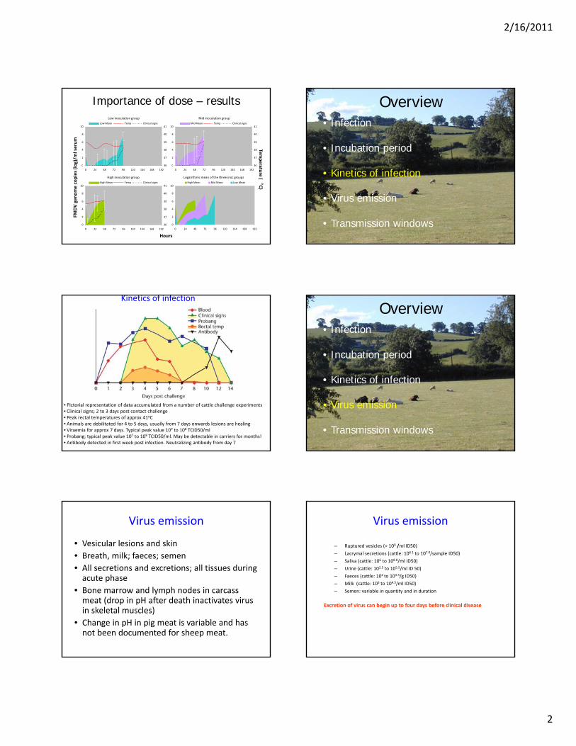
Kinetics of infection
• Pictorial representation of data accumulated from a number of cattle challenge experiments• Clinical signs; 2 to 3 days post contact challenge• Peak rectal temperatures of approx 41oC• Animals are debilitated for 4 to 5 days, usually from 7 days onwards lesions are healing• Viraemia for approx 7 days. Typical peak value 107 to 108 TCID50/ml• Probang; typical peak value 107 to 108 TCID50/ml. May be detectable in carriers for months!• Antibody detected in first week post infection. Neutralizing antibody from day 7
Overview• Infection
• Incubation period
• Kinetics of infection
• Virus emission
• Transmission windows
Virus emission
• Vesicular lesions and skin• Breath, milk; faeces; semen• All secretions and excretions; all tissues during acute phaseacute phase
• Bone marrow and lymph nodes in carcass meat (drop in pH after death inactivates virus in skeletal muscles)
• Change in pH in pig meat is variable and has not been documented for sheep meat.
Virus emission
– Ruptured vesicles (> 109 /ml ID50)
– Lacrymal secretions (cattle: 106.1 to 107.0/sample ID50)
– Saliva (cattle: 106 to 108.8/ml ID50)
– Urine (cattle: 102.5 to 105.5/ml ID 50)
– Faeces (cattle: 102 to 103.3/g ID50)
– Milk (cattle: 103 to 104.5/ml ID50)
– Semen: variable in quantity and in duration
Excretion of virus can begin up to four days before clinical disease

2/16/2011
3
Virus emission
• Airborne virus mainly from the exhaled breath of infected animals as droplets and droplet nuclei (pigs)
• Originates initially from the upper and later from the lower respiratory tract (?)
• The precise mechanism of release has not been determineddetermined
• Also potential for generation of aerosols from virus containing fluids or dried material, for example from milk tankers, urine, faeces
• quantitatively a minor role compared to breath
Virus emission – pigs
John Gloster IAH
Virus emission ‐ cattle
John Gloster IAH
Overview• Infection
• Incubation period
• Kinetics of infection
• Virus emission
• Transmission windows
Defining and quantifying the period of infectiousness in cattle experimentally infected
with type O FMDV
2 days later 4 days later 6 days later48 hrs
inoculates
challenged donors
direct contact recipients
indirect contact recipients
Animals monitored for 14 days post‐challenge
In contact for 24 hrs
Challenge and transmission events in relation to the first clearly visible lesion in oral cavity, nose
or tongueVN47VN89VN90VO75VO76VQ05VQ06VR56day -8 -7 -6 -5 -4 -3 -2 -1 0 1 2 3 4 5
Day 0 – first day clinical signs were observed– day of challenge, ‐ transmission event

2/16/2011
4
Infected premises
Very hard to measure under field conditions
Up to:• 20 weeks on hay/straw• 4 weeks on cow’s hair 18 – 20oC• 14 days dry faeces• 39 days in urine• 6 months in slurry in winter• 6 months in slurry in winter• 3 days on soil in summer & 28 days in autumn• Snow‐covered soil: More than 185 days • survival decreases with increasing heat and decreasing humidity
Bartley LM, Donnelly CA, Anderson RM, Review of foot‐and‐mouth disease virus survival in animal excretions and on fomites. Vet Rec 2002 Nov 30;151(22):667‐9
Acknowledgements
• John Gloster• David Paton• Colleagues at the
IAHIAH

AGEING OF LESIONS

2/17/2011
1
Ageing of lesions
Kenya, 15th-19th November 2010Nick Juleff, Institute for Animal Health, PirbrightNick Juleff, Institute for Animal Health, Pirbright
Why is this important?• Age range of lesions‐particularly the oldest lesion
Essential for case history and epidemiological report
Pre‐requisite to determine origin of infection
Duration and weight of virus excretion
Prediction of further spread
Estimating the age of lesionsDay of Clinical Disease
Appearance of lesion
Day 1 Blanching of epithelium followed by formation of fluid filled vesicle
Day 2 Freshly ruptured vesicles characterised by raw epithelium, a clear edge to the lesion and no deposition of fibrin
http://www.defra.gov.uk/animalh/diseases/fmd/about/clinical.htmState Veterinary Journal, 5,Number 3, October 1995, pages 4 – 8
Day 3 Lesions start to lose their sharp demarcation and bright red colour. Deposition of fibrin starts to occur.
Day 4 Considerable fibrin deposition has occurred and regrowth of epithelium is evident at the periphery of the lesion.
Day 7 Extensive scar tissue formation and healing has occurred. Some fibrin deposition is usually still present.
Estimating the age of lesions• Photographs of contact exposure – field and experimental
Clinical manifestation may vary between virus strains –Different affinityespecially sheep (lesions may be too transient for gauging time of infection)
Complicated by secondary infections
Between day 0 and 5 – one day margin of accuracy

2/17/2011
2
Cattle ‐ Day 1
Unruptured tongue vesicle, fluid filled, blanching of epithelium
Cattle ‐ Day 1
One day old vesicle, ruptured when tongue drawn from mouth
Cattle ‐ Day 2
Note raw epithelium, clear edge to lesion and no deposition of fibrin
Cattle ‐ Day 2
Field example. Note raw epithelium, clear edge to lesion and no deposition of fibrin

2/17/2011
3
Cattle ‐ Day 2
No raw epithelium, clear edge to lesion and no deposition of fibrin
Cattle ‐ Day 3
Lesions start to lose their sharp demarcation, fibrin deposition starts to occur on edge of lesions
Cattle ‐ Day 4
Considerable fibrin deposition has occurred and regrowth of epithelium is evident at edge of lesion
Cattle ‐ Day 5
Note progressive loss of lesion margination and extensive fibrin infilling

2/17/2011
4
Cattle ‐ Day 7
Field example. Extensive scar tissue formation and healing has occurred. Some fibrin deposition is usually still present
Cattle ‐ Day 10
Scarring and indentation at site of lesion, fibrous tissue proliferation, loss of papillae
Cattle ‐ Day 1
Unruptured vesicle, fluid filled, blanching of epithelium
Cattle ‐ Day 2
Ruptured vesicle, note raw epithelium, clear edge to lesion and no deposition of fibrin

2/17/2011
5
Cattle ‐ Day 3
Note friable epithelium, deposition of fibrin starting to occur
Cattle ‐ Day 4
Considerable fibrin deposition
Cattle ‐ Day 4
Considerable fibrin deposition and regrowth of epithelium
Cattle ‐ Day 5
Granulation/regrowth of epithelium
2/17/2011
6
Cattle ‐ Day 7
Healing progressing under the necrotic epithelium
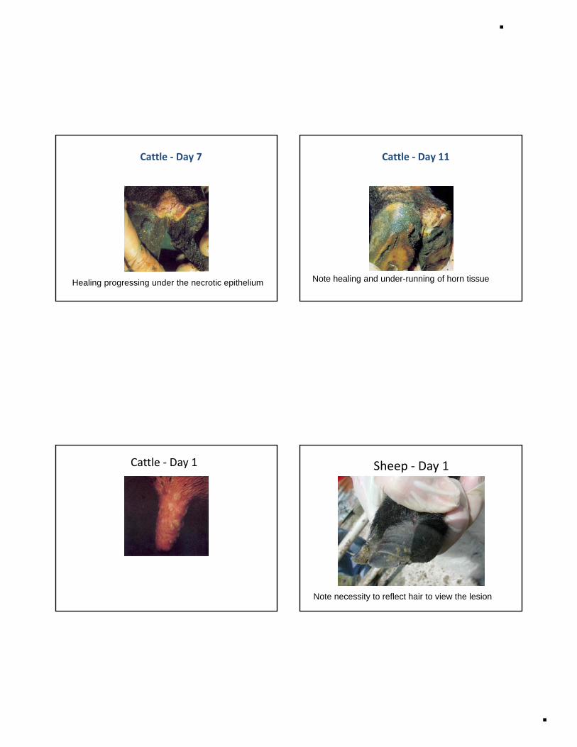
Cattle ‐ Day 11
Note healing and under-running of horn tissue
Cattle ‐ Day 1 Sheep ‐ Day 1
Note necessity to reflect hair to view the lesion

2/17/2011
7
Sheep ‐ Day 2
Ruptured and unruptured coronary band vesicle
Sheep ‐ Day 3
Sero-fibrinous exudate and swelling
Sheep ‐ Day 4
Signs of early healing
Sheep ‐ Day 6
Scab formation and healing
2/17/2011
8
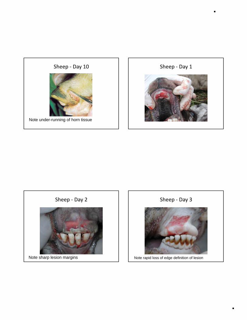
Sheep ‐ Day 10
Note under-running of horn tissue
Sheep ‐ Day 1
Sheep ‐ Day 2
Note sharp lesion margins
Sheep ‐ Day 3
Note rapid loss of edge definition of lesion

2/17/2011
9
Sheep ‐ Day 4 Sheep ‐ Day 6
Sheep ‐ Day 7Pigs
Most information obtained from foot lesions
If lesion is at coronary band < 1 week old(7 days for lesion to mature and new horn growth to begin)
Thereafter meas re distance from coronarThereafter measure distance from coronary band to lesion
horn grows at 1mm per week-adultshorn grows at 2mm per week-weaners

2/17/2011
10
Pigs – Day 1 Pigs – Day 3
Note deposition of fibrin
Pigs – Day 4
Note extensive fibrinous in-filling
Pigs – Day 8
Healing lesion

2/17/2011
11
Pigs – Day 1Pigs – Day 3
Supernumerary digits
Pigs – Day 9 Acknowledgements
David PatonColleagues at the Institute for Animal Health

DIAGNOSTIC AND
SAMPLING PROCEDURES
FOR FMD

11/11/2010
1
Diagnostic and Sampling procedures for FMD
Nick Juleff IAHEuFMD training
Kenya,15th-19th November 2010
EN
T
1 Rapid confirmation of clinical signs
2 Active surveillance for infected animals(including pre-clinical cases)
Diagnostic windows
FMD virus in blood
Clinicallesions
antibody response
3 sero-surveillence for FMDV exposed animals
DAYS
MEA
SUR
EM
14654321 87 ● ● ● ●
Representative “in contact” cattle data from Alexandersen et al., 2003 and unpublished data from IAH
Laboratory diagnosis of FMD
• Confirms clinical diagnosis
• Supports but does not replace the need for accurate clinical diagnosis
• The quality of the diagnosis is only as good as the quality of the sample submitted
• Requires full epidemiological information on samples submitted for rational interpretation
UninfectedUninfected FMD Virus infectedFMD Virus infected Recovered (or Recovered (or vaccinated)vaccinated)
Lesion, swab, probang or Lesion, swab, probang or clotted blood samplesclotted blood samples
Clotted blood samplesClotted blood samples(saliva)(saliva)
Probang samplesProbang samples
Principals of FMD Diagnosis
Live VirusLive Virusby virus isolation by virus isolation in cell culturesin cell cultures
Viral Proteins by LFD or double Viral Proteins by LFD or double antibody sandwich ELISAantibody sandwich ELISA
Viral Nucleic AcidViral Nucleic Acidby RTby RT--PCR PCR
AntiAnti--FMD antibodies can be FMD antibodies can be detected in serum by ELISA detected in serum by ELISA
or VNTor VNT
Virus or viral Virus or viral components can be components can be
detecteddetected
AntiAnti--viral antibodies viral antibodies can be detectedcan be detected
1-4 days 10-30 minutes (LFD)
4 hours (ELISA)
Within a day 5-18 hours (ELISA)
2-3 days (VNT)
FMDV diagnosis: Window of detection by different techniques
clinical signs of FMD
EpitheliumSerumMilk
Cell culture/ Ag ELISA
1 2 3 4 5 6 7 8 9 10 11 12 14 15
Antibodies
RT-PCREpitheliumSerumMilk
ELISA Structural
ELISA Non structural
VNT
Virus isolation
(CTY or IBRS2)
Ag ELISA
1-4 days
~4 hours
Current assays for FMDV detection
Time to report result (hrs)
AutomatedTaqMan®RT-PCR
~5 hours
1 10 100

11/11/2010
2
Virological Samples
• Urgent– send as soon as possible, by most direct route– always give advance warning to lab and ETA – correct external package label to identify urgency– do not package together with other samples of less urgency
Hazardous• Hazardous– package and label properly
• Fragile – keep cool but not frozen, except by prior arrangement, if long
delay– avoid extremes of pH therefore use buffered media
• Adequate quantities• Separate tube for each animal• Correct forms
Sampling from lesions• Lesion material is the richest source of FMDV
and the sample of choice for diagnosis• Samples from ~ 4 animals with obvious lesions
should be sufficient to confirm a diagnosis • The most suitable materials areThe most suitable materials are
– Vesicular epithelium, vesicular fluid, heart muscle from myocarditis cases
– For tissues• At least 2 cm2 of epithelium from unruptured or freshly
ruptured vesicles• Transport medium - equal amounts of glycerine and 0.04 M
phosphate buffer pH 7.2-7.6 – For vesicular fluids
• Plain, small volume tube
Sampling in absence of lesions
• For diagnosis select ~ 6 animals, prioritizing those with suspicious clinical signs– Fever depression lameness hot feetFever, depression, lameness, hot feet
• Collect clotted blood samples to obtain serum to detect viraemia or antibodies
• Collect probang and/or oronasal swabs
Probang samples• Aliquot 2-3 ml 0.08 M PBS with 0.01% BSA, 0.002% phenol red and antibiotics,
adjusted to pH 7.2 per animal to be sampled. • Cattle: pour probang sample from the cup into a wide-necked bottle & examine
visually for quality. Add 2ml, including visible cellular material, to equal volume of buffer and mix. The final pH of a normal sample should be ~pH 7.6.
• Sheep: insert probang cup directly into a disposable container into which has been dispensed 3 ml of buffer solution and gently mix
• Samples taken from some animals may be heavily contaminated with ruminal contents these should be discarded and the animal's mouth should be flushedcontents - these should be discarded and the animal s mouth should be flushed with water before repeat sampling
Different sizes:Sheep
Calf
Cow
3 bucket “system”•Water
•4% Na2CO3 or 0.2% citric acid
•Water
WRL procedure for FMDV antigen detection
Original epithelium/vesicular fluid
Antigen detection ELISAVirus isolation in tissue culture
+ - +48 hours- +
First blind passage
Second blind passage
FMDV detected& serotypedNo Virus Detected
Confirmation
-48 hours +
Lateral Flow Device (LFD)Not serotype specific
• Based on technology used in pregnancy test kits• Similar sensitivity to Ag-ELISA• High specificity• Used to test epithelium or vesicular fluid
‘Pen-side’ test for antigen detection
• Used to test epithelium or vesicular fluid• Result within minutes• Used on-farm in UK 2007• Used in regional lab in Turkey in 2009• Relatively low cost per test
12
11/11/2010
3
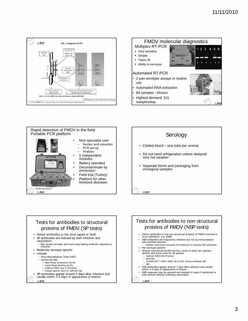
FIG. 1. Diagram of LFA
Oem, J. K. et al. 2009. Clin. Vaccine Immunol. 16(11):1660-1664
FMDV molecular diagnosticsMultiplex RT-PCR
Very sensitiveSimpleTakes 4hAbility to serotype
Automated RT-PCR• 2 pan-serotype assays in routine
use• Automated RNA extraction• 84 samples ~5hours• Highest demand: 311
samples/day
Rapid detection of FMDV in the field: Portable PCR platform
• Non-specialist user– Nucleic acid extraction– PCR set-up– Analysis
• 5 independent modules
Smiths Bio-SeeqTM
• Battery operated• Decontaminate by
immersion• Field trial (Turkey) • Platform for other
livestock diseases
Serology
• Clotted blood – one tube per animal
• Do not need refrigeration unless delayed/ very hot weather
• Separate forms and packaging from virological samples
Tests for antibodies to structural proteins of FMDV (SP tests)
• Detect antibodies to the virus capsid or shell• SP antibodies are induced by both infection and
vaccination– But usually stronger and more long-lasting antibody response to
infectionRelatively serotype specific• Relatively serotype specific
• Include– Virus Neutralisation Tests (VNT)– Various ELISAs
• Solid Phase Competition ELISA• Liquid Phase Blocking ELISA• Ceditest FMDV type O (Prionics)• Isotype-specific tests for IgM and IgA
• SP antibodies appear around 5 days after infection and usually within 2-3 days of appearance of lesions
Tests for antibodies to non-structural proteins of FMDV (NSP tests)
• Detect antibodies to the non-structural proteins of FMDV involved in virus replication, e.g. 3ABC
• NSP antibodies are induced by infection but not by immunisation with purified vaccines– Multiple vaccination increases the likelihood of inducing NSP antibodies
• Pan serotype specific• Several commercial ELISA test kits, some of which are species-
specific and some work for all species– Ceditest FMDV-NS (Prionics)– Bommeli– SVANOVIR™ FMDV 3ABC-Ab ELISA, Svanova Biotech AB – UBI
• NSP antibodies appear around 7 days after infection and usually within 3-4 days of appearance of lesions
• NSP response may be reduced and delayed in case of subclinical or mild clinical infection following vaccination

11/11/2010
4
Sampling in Kenya• Epithelium samples: gly-iso buffer • Vesicular fluid: collect using a syringe and needle. No buffer.•Blood: whole blood in EDTA/Trizol, clotted blood in plain vacutainer•Probang samples: buffer

FMD EPIDEMIOLOGY AND
TRACING DANGEROUS
CONTACTS

1
FMD epidemiology and tracing dangerous contacts
Kenya, 15th-19th November 2010Nick Juleff, Institute for Animal Health • Epidemiology and spread
DEFRA t l li• DEFRA control policy
• Tracing dangerous contacts
• Prioritising: which DCs are the most important?
Routes of infection• Direct contact with infected animals(ban on animal movements)
• Contaminated animal products(ban on meat/milk from infected areas)(ban on meat/milk from infected areas)
• Airborne virus(use wind records to estimate spread)
• Mechanical transmission of virus on people, vehicles, etc BIOSECURITY
Incubation and excretion periods• Incubation period: 1–14 days
– most common 2-5 days
• Dose-related: low dose longer incubation
• Virus excretion: may occur before onset of clinical signs
Virus excretion: range and highest excretion periods relative to appearance of first lesions
Day of first lesions
-8 -7 -6 -5 -4 -3 -2 -1 0 1 2 3 4 5 6 7 8 9 10 11 12 13 14 15
Pigs
Cattle
Sheep
Why is FMD so contagious?
• WIDE host range
• HIGH Morbidity/LOW mortality
• Infection dynamics
• Patterns of viral shedding
• FMD virus can survive in environment for long periods
• Animal movements UK 2001: best estimate is that >100 farms were already infected before first diagnosis
• Role of markets & abattoirs
Key epidemiological factors
UK 2001: 10 infected sheep in Longtown market exposed 24,500 sheep
• Pigs: high level excretion, airborne spread
• Sheep: subclinical infection, easily missed
• Persistence of epidemic through “local spread”: stringent biosecurity reduces risk

2
• Epidemiology and spread
DEFRA t l li• DEFRA control policy
• Tracing dangerous contacts
• Prioritising: which DCs are the most important?
Principles for controlling a FMD outbreakPrevent transmission from infected to susceptible animals
↓↓ production of virusCull infected and in-contact animals, ± dangerous contacts if high-risk
↓↓ potential for direct contact between animals↓↓ pMovement restrictions
↓↓ virus survival time in the environmentBiosecurity, cleansing & disinfection of infected farms
↓↓ number of susceptible animalsEmergency vaccination or contiguous culling if situation v. severe
DEFRA - Initial control measures• Establish temporary control zone
– Stop animal movements– Size – prevent spread of disease– Supplementary movement control zone (2007)
• “Stamping-out” on infected premises (IP) – 24 hours
• Cull dangerous contactsg– 48 hours
• Establish 3km protection and 10 km surveillance zones
• Protection zone (3km radius around IP)– No animal movement except to emergency slaughter under licence– Movement of animal products, feed and bedding under licence – Requirement for increased on/off farm biosecurity– Animal products treated to destroy FMDV– Footpaths closed– census– surveillance - regular inspections, sero-surveillance– tracing at start– For early / index cases, the EU requires tracing of animals and products moved
from the zone since 21 days before the probable introduction of infection
• Surveillance zone (10km radius around IP)– As for protection zone except:
• Footpaths remain open• Licensed animal movements are possible
Additional control strategies
• Scientific and veterinary advice
• Cull other susceptible livestock exposed to disease– e.g. Premises under viral plumes, adjoining premisesg p , j g p– Extensive sampling, clinical examination (2007 pre-clinical
diagnosis)
• Pre-emptive or “firebreak” culling of animals
• Epidemiology and spread
DEFRA t l li• DEFRA control policy
• Tracing dangerous contacts
• Prioritising: which DCs are the most important?

3
Establishing Timelines
• Estimate age of oldest lesion on farmDetermine date lesions first appeared (NB: error margin)
• Subtract incubation period 1-14 days window for introduction of virus
• Prioritise “onto farm” contacts in most likely incubation period of 2-5 days before lesions appeared
The image cannot be displayed. Your computer may not have enough memory to open the image, or t…
The image cannot be displayed. Your computer may not have enough memory to open the image, or the image ma ha
The image cannot be displayed. Your computer may not have enough memory to open the image, or the image ma ha
The image cannot be displayed. Your computer may not have enough memory to open the image, or the image may hav
The image cannot be displayed.
The image cannot be displayed. Your computer may not have enough memory to open the image, or the image ma ha
The image cannot be displayed. Your computer may not have enough memory to open the image, or the image may have been corrupted. Restart your computer, and then open the file again. If the red x still appears, you may have t…
IP2b
IP2c
IP3c
IP4b
IP5
IP1b(2)
IP1b(1)
Timeline example: UK 2007
29-
Jul-
07
19-
Au
g-0
7
12-
Au
g-0
7
05-
Au
g-0
7
26-
Au
g-0
7
02-
Sep
-07
09-
Sep
-07
16-
Sep
-07
23-
Sep
-07
The image cannot be displayed. Your computer may not have enough memory to open the image, or the image ma ha
Your computer may not have enough memory to open the image, or the image may hav
The image cannot be displayed. Your computer may not have enough memory to open the image, or the i…
IP3bIP6b
IP7
22-
Jul-
07
15-
Jul-
07
The image cannot be displayed. Your computer may not have enough memory to open the image, or the image ma ha
30-
Sep
-07
IP8
29-
Jul-
07
19-
Au
g-0
7
12-
Au
g-0
7
05-
Au
g-0
7
26-
Au
g-0
7
02-
Sep
-07
09-
Sep
-07
16-
Sep
-07
23-
Sep
-07
Date
The image cannot be displayed. Your computer may not have enough memory to open the image, or the image ma ha
Your computer may not have enough memory to open the image, or the image may hav
The image cannot be displayed. Your computer may not have enough memory to open the image, or the i…
IP3bIP6b
IP7
22-
Jul-
07
15-
Jul-
07
The image cannot be displayed. Your computer may not have enough memory to open the image, or the image ma ha
30-
Sep
-07
IP8
Data from Cottam et al., 2008
Incubation period (<14 days)Most likely date of infection (2-5 days before clinical disease)
Lesion age estimation
Effect of lesion age and
incubation period on time
of infection Floo
ding
+ lo
rry
mov
es
Airb
orne
spr
ead
day
1stM
eria
l slu
rry
rele
ase
2nd
Mer
ial s
lurr
y re
leas
e
And
lorr
y m
oves
July August
10 d
ay o
ld +
/-2
days
NB Could be further lag between escape of virus and exposure of cattle
9 10 11 12 13 14 15 16 17 18 19 20 21 22 23 24 25 26 27 28 29 30 31 1 2 3 4-15 -14 -13 -12 -11 -10 -9 -8 -7 -6 -5 -4 -3 -2 -1 1 2 3 4 5 6 7 8 9 10 11 12
July August11 12 13 14 15 16 17 18 19 20 21 22 23 24 25 26 27 28 29 30 31 1 2 3 4
-15 -14 -13 -12 -11 -10 -9 -8 -7 -6 -5 -4 -3 -2 -1 1 2 3 4 5 6 7 8 9 10
July August13 14 15 16 17 18 19 20 21 22 23 24 25 26 27 28 29 30 31 1 2 3 4
-15 -14 -13 -12 -11 -10 -9 -8 -7 -6 -5 -4 -3 -2 -1 1 2 3 4 5 6 7 8
Lessons from the field• Epidemiological benefits of lesion ageing, extensive
sampling, sequencing virus isolates in real time– 2nd phase of outbreaks (IP3 – IP8) shares all the unique
changes common to 1st phase– Therefore outbreaks are linked and not due to independent
sources– IP5 (farm with FMD serology positive cattle and sheep)IP5 (farm with FMD serology positive cattle and sheep)
bridges gap between two phases of the outbreak
• Diagnosis of preclinical viraemic animals using real-time PCR
• Hobby/part-time farmers with inadequate handling facilities: implications for neighbouring farmers?
Tracing dangerous contacts (DCs)Methods:• Interview farmer & farm staff• Examine written records (milk tanker, feed deliveries, AI visits,
etc) • Walk boundaries of farm• Estimate age of FMD lesions• Contact dairy vets AI techs etc• Contact dairy, vets, AI techs etc
Key information:• Estimated age of oldest lesion• Animal movements• Personnel visiting farm• Farm personnel visiting other livestock holdings• Vehicle/equipment movements• Security of farm boundaries
Risk periods
• Time of contact is very important• Tracings onto farm: incubation period 1-14 days
before lesions appear; most likely 2-5 days• Tracings off farm: refer to virus excretion period
V irus excretion: range and highest excretion periods relative to appearance of first lesions
Day of first lesions
-8 -7 -6 -5 -4 -3 -2 -1 0 1 2 3 4 5 6 7 8 9 10 11 12 13 14 15
Pigs
Cattle
Sheep
NB: virus can be excreted before clinical signs appear
Milk: can contain virus 4 days before disease appears
4
Risk periods• Levels of environmental FMD contamination increase from first
case until after final case on farm has recovered• Slow decrease in virus levels thereafter, related to temperature
and humidity• Fomite transmission contacts: relationship between time of
contact and degree of risk
Environmental FMD virus
contamination
Time
Vir
us in
en
viro
nmen
t
Animals with FMD on farm
No infected animals left on farm
Type of contact
Animal movements most critical contact• Pigs: high level of virus excretion• Sheep: mild to sub-clinical disease; easy to miss
L l dLocal spread• Check farm boundaries• Nose-to-nose contact with neighbouring herds/flocks
possible?• Fences stock-proof?• Shared streams at boundary – close proximity possible?• Common grazing?
Tracing animal movements
• Trace source of animals moved onto farm during incubation period
•Trace destination of animals moved off farm during virus excretion period (from 4 days prior to disease appearance onwards)
•Identify any markets/stopovers en route, incl. pick-ups at other farms
Method: Use animal movement databases, farmer interview, records, etc
Action: restrict all implicated premises, initiate investigations
Tracing contiguous herds & common grazing
• Determine if neighbouring stock were in fields adjacent to IP; evaluate fencing/waterways/natural features between premises.
• Determine which other farms had stock grazing land also used by IP stock, and relevant timelines.
Method: Maps of contiguous farms, VI to walk farm boundary, telephone enquiries, local knowledge
Action: Issue restriction notices and initiate investigations on any farms where stock grazed commonage at same time or after stock from IP, subsequent to estimated earliest date of infection introduction, and any contiguous farms.
Type of contact:Personnel
• Close interaction with animals? Vet, AI tech, shared labourer, foot-parers, sheep shearers, etc
• Interaction with affected stock or non-affected stock?
• Visits by farmer & staff to other livestock holdings: contact with animals at other premises?
Tracing personnel contacts• Identify workers, especially those in contact with animals, on
the IP who also work on other farms• Identify itineraries of vets, AI techs, other technicians who were
on IP during risk periods
Method: • Interview farmer and farm staff, any other relevant personnelte e a e a d a sta , a y ot e e e a t pe so e• Interview vets, AI techs etc, examine calls book• Relate farmer names to herd numbers, identify on map• Relate contact dates to risk periods, prioritise accordingly
Action:• Evaluate degree of risk of each contact• Prioritise accordingly; restrict highest risk ones• As a minimum, first 3 farms visited by vets/AI techs after IP
require investigation as a priority

5
Type of contact:Vehicles/equipment
• Milk tankers: can leak virus-containing milk at pipe connectionTrailers which contained animals: gross• Trailers which contained animals: gross contamination with virus possible
• All vehicles: wheels, wheel arches, undercarriage contaminated with faeces/muck with virus
• Key question: was vehicle/equipment in contact with animals or in yard/lane contaminated with faeces?
Tracing milk tankers
• Identify premises visited following collection of milk from IP during risk period.
Method:• Contact dairy by phone, request records of tanker itineraries Contact dairy by phone, request records of tanker itineraries
during risk period; verify with driver (may vary route)• For tracings off the IP, the risk period is from 4 days prior to the
appearance of clinical signs, although tracings which occurred during clinical disease should be prioritised
Action:• Identify premises visited• Prioritise farms with large numbers of stock and farms visited
immediately after IP.
Tracing other vehicles/equipment contacts• Identify nature of contacts• Estimate time in relation to risk period• Ascertain origin or destination of vehicles/equipment • Ascertain degree of contact with infected animals on IP • Evaluate risk level and prioritise contact investigation
accordingly
Method: • Interview farmer and farm workers and other relevant
personnel. • Phone premises which were origin/destination of contact
vehicles and establish number and species of stock on farms • Establish dates of contacts and relate to risk period
Action: investigate priority contacts first
Risk of airborne spread
• Virus emission profile estimated by number of animals with FMD and age range of lesions
• Meteorological agency (e.g. Met Office) will combine this with meteorological data
• Model output: map with areas showing relative risks of airborne FMD spread
• Farms in “high risk” category: investigate first
• Virus can spread long distances by air, but this is not as common as other methods of spread (animal movement, fomites)
Summary:
• Latest UK Met Office model, including GIS output in operational service
Assessment of Biosecurity on IP
• Level of biosecurity in operation on IP prior to diagnosis can hugely influence onward spread
• Difficult to assess after diagnosis• General level of farm hygiene can provide
rough estimation
Biosecurity on dangerous contact: also critical risk factor, but impossible to assess without inspection, unless prior knowledge available
• Epidemiology and spread
DEFRA t l li• DEFRA control policy
• Tracing dangerous contacts
• Prioritising: which DCs are the most important?

6
Reasons for prioritisation
• The number of contacts to be traced can become very large
• Resources for investigations not unlimited• Time can be critical
Need to prioritise “hot” contacts
Prioritising Dangerous Contacts
Three factors to consider:1. Species: pigs > cattle > sheep2. Type of contact: animal movement > people in
direct contact with FMD animals > vehicles in direct contact etccontact etc.
3. Time of contact in virus excretion windowNB: may get multiple waves of infection in herd/flock
continual virus excretion as new animals fall ill V irus excretion: range and highest excretion periods relative to appearance of first lesions
Day of first lesions
-8 -7 -6 -5 -4 -3 -2 -1 0 1 2 3 4 5 6 7 8 9 10 11 12 13 14 15
Pigs
Cattle
Sheep
Prioritising DCs: Likely result of virus spread
• Pigs: if infected, will generate huge amount of virus tracings to pig farms a priority
• Number of animals on DC: as numbers i d h f i f tiincrease, so does chance of infection and significance of outbreak
• Type of enterprise:- Markets- Abattoirs- Farms owned by dealers
Categorising risk posed by DCs
Very high risk:• Animal movements during risk period• Farms owned/worked on by workers from IP• Contiguous herds with possibility of nose-to-nose g p y
contact• Animals grazing common land with IP stock• Any market or abattoir connected to IP during risk
period
Categorising risk posed by DCs
High risk:• Farms visited by vets/AI techs after IP during risk
periodperiod• Farms visited by milk tanker after collection at IP• Contiguous farms where nose-to-nose contact is less
likely but stock are in adjacent fields
Categorising risk posed by DCs
Medium risk• Shared equipment/trailers/vehicles in direct contact
with infected animals on IPwith infected animals on IP• Neighbouring/nearby farms with some distance
between animals on IP and DC• Personnel in contact with animals on IP and DC

7
Categorising risk posed by DCs
Low risk:• Vehicles/equipment shared between farms but not in
contact with animals• Personnel shared between farms but not in contact
with animals• Personnel visiting the IP and then other farms but
not in contact with animals
Acknowledgements
• Eoin Ryan, Dept of Agriculture, Ireland• Dónal Sammin, Dept of Agriculture, Ireland, p g ,• Colleagues in the Institute for Animal Health,
Pirbright, UK

THE 2007 UK FMD
OUTBREAK

11/11/2010
1
The 2007 UK FMD outbreak:The 2007 UK FMD outbreak:field investigation perspectivefield investigation perspective
Nick Juleff, Institute for Animal Health
Slides prepared by Eoin Ryan
UK 2007 FMD EpidemicUK 2007 FMD EpidemicVirus escape from Pirbright site (IAH and Merial vaccine plant)8 infected premises, most consisting of multiple holdingsMainly extensive beef productionMainly extensive beef productionMost farms in semi-urban areasPart-time/hobby farmers1578 animals on IPs culled278 animals infected
IP1: 3 holdingsIP1: 3 holdings29th July: farmer notices an animal “off colour”
2nd August: several cattle lame & drooling. PVP tells farmer to contact Defra directly.
3rd August: Defra vet examines & takes samples; +ve for FMDV
4th August: cattle at all 3 holdings killed; virus identified as O1 BFS 1860 (used at Pirbright site)
IP1: main holdingIP1: main holding•38 cattle, all infected
•Lesion ages: 3 to 10 days old
•Beef store cattle grazing on open pasture 4km from Pirbright site
•No handling facilities: Defra•No handling facilities: Defra brought in gates & straw bales to make corral
•Shot with rifles then pithed
IP1: two other holdingsIP1: two other holdings
4 cattle at home farm: no FMDV
22 cattle on open pasture: no FMD lesions; one animal PCR +ve (viraemic)
Only link between premises: farmerOnly link between premises: farmer
First time preclinically viraemic animals detected using PCR in an outbreak
Logistical problems again: straw bales, rifles
Carcases transported by sealed lorry to incinerator 70 miles away
IP2: three holdingsIP2: three holdings49 cattle 1km from IP1; 58 at home farm; 12 on third holding
6th August: farmer notifies Defra, samples taken from holding with 49 cattle: +ve
25 killed that night, the rest on next day
Cattle examined & bled post-mortem

11/11/2010
2
IP2IP249 cattle: 44 had lesions, 2-7 days old
- Logistical probs: no handling facilities, no water, rifles, journalists
58 cattle on 2nd farm: no FMD lesions BUT
- 15 of 58 viraemic by PCR/VI
12 cattle at 3rd holding: no FMD
Phase 1: AugustPhase 1: August
3 Aug: IP1 (3 locations)
6 Aug: IP2 (3 locations)
3 t t h d ll d3 contact herds culled
24 Aug: PZs lifted
8 Sep: SZ lifted
Origin: contamination
from Pirbright site
Likely times of lab escape of virus from Likely times of lab escape of virus from Pirbright site in 2007Pirbright site in 2007
20th July: flooding23rd July: best airborne spread day
22nd July: 1st centrifuge waste discharged25th July: 2nd centrifuge waste discharged
20th July: 4 lorries via Westwood Lane25th July: 2 lorries via Westwood Lane
Effect of lesion age and
incubation period on time
of infection Floo
ding
+ lo
rry
mov
es
Airb
orne
spre
ad d
ay1st
Mer
ial s
lurr
y re
leas
e
2nd
Mer
ial s
lurr
y re
leas
e
And
lorr
y m
oves
July August
10 d
ay o
ld +
/-2
days
NB Could be further lag between escape of virus and exposure of cattle
9 10 11 12 13 14 15 16 17 18 19 20 21 22 23 24 25 26 27 28 29 30 31 1 2 3 4-15 -14 -13 -12 -11 -10 -9 -8 -7 -6 -5 -4 -3 -2 -1 1 2 3 4 5 6 7 8 9 10 11 12
July August11 12 13 14 15 16 17 18 19 20 21 22 23 24 25 26 27 28 29 30 31 1 2 3 4
-15 -14 -13 -12 -11 -10 -9 -8 -7 -6 -5 -4 -3 -2 -1 1 2 3 4 5 6 7 8 9 10
July August13 14 15 16 17 18 19 20 21 22 23 24 25 26 27 28 29 30 31 1 2 3 4
-15 -14 -13 -12 -11 -10 -9 -8 -7 -6 -5 -4 -3 -2 -1 1 2 3 4 5 6 7 8
DifficultiesDifficulties
Assuming lesion of 12 days and longish incubation then likely release times too lateFlooding period fits best as time of release most
i i h l i d i b iconsistent with lesions and incubation But flooding and first lorry movements precede likely virus release through drainsSecond period of lorry movements more likely associated with viral contamination, but too late to have infected farm
EGHAM
WINDSOR
WOKING
GUILDFORDIP1a
IP2a
IP2b
IP5
IP7
IP6
IP3cIP4
IP3bEGHAM
WINDSOR
WOKING
GUILDFORDIP1b
IP2b
IP2c
IP5
IP7
IP6
IP3cIP4b
IP3b
PIRBRIGHT SITE
5 km
EGHAM
WINDSOR
WOKING
GUILDFORDIP1a
IP2a
IP2b
IP5
IP7
IP6b
IP3cIP4
IP3bEGHAM
WINDSOR
WOKING
GUILDFORDIP1b
IP2b
IP2c
IP5
IP7IP3c
IP4b
IP3b
PIRBRIGHT SITE
5 km IP8
EGHAM
WINDSOR
WOKING
GUILDFORDIP1a
IP2a
IP2b
IP5
IP7
IP6
IP3cIP4
IP3bEGHAM
WINDSOR
WOKING
GUILDFORDIP1b
IP2b
IP2c
IP5
IP7
IP6
IP3cIP4b
IP3b
PIRBRIGHT SITE
5 km
EGHAM
WINDSOR
WOKING
GUILDFORDIP1a
IP2a
IP2b
IP5
IP7
IP6b
IP3cIP4
IP3bEGHAM
WINDSOR
WOKING
GUILDFORDIP1b
IP2b
IP2c
IP5
IP7IP3c
IP4b
IP3b
PIRBRIGHT SITE
5 km IP8
Phase 2: Phase 2: SeptemberSeptember
IP3IP3
Confirmed 12th Sept8 l ti 2 FMD8 locations: 2 FMD+ve36/47 cattle had FMD lesions, 1-5 days old9/29 cattle had lesions at 2nd holding, 1-5 days oldOther holdings all FMD negativeCulling completed 16 Sep (4 days later)Logistical probs on out-farms:
No handling facilities, escapees, police

11/11/2010
3
IP4IP4Separated from IP3 by narrow streamDiagnosed Sept 13th (farmer report)54/54 beef cattle had FMD lesions 5 10 dayslesions, 5-10 days800 pigs kept 500m away: no FMDFarmer had been on holidays for 10 days – unclear who checked cattleExcellent handling facilities –fast cull
IP5: the missing linkIP5: the missing linkAfter IP3 & 4, 3km PZ implementedAll sheep bled, cattle & pigs “clinically inspected” – often just observed over fence17th Sep: IP5 sheep tested seropositive IP5: 16 sheep, 22 cattle, 2 pigs Farmer retired & unwell; animals were petsFMD lesions ~ 3 weeks old (large error margin)Virus detected in probangs – 10/16 sheep carriers
Crown Estates
Cook
Berryman
Regan
Whitty
Salmon
Bovington
IPA ongoingIPA culled
Raynen
IPA ongoing sheepWiggins
Wallace
Robertson
HarveyIntensive patrol areas:Intensive patrol areas:••Animals examined every 2 days (in theory)Animals examined every 2 days (in theory)
••Blood samples: real time PCR to detect Blood samples: real time PCR to detect viraemic preclinicals viraemic preclinicals
••Small no. of farmsSmall no. of farms
••Some premises: clinical examination very Some premises: clinical examination very difficult (no facilities)difficult (no facilities)
IP621st Sept, Defra inspection34 cattle, 2 locations2/32 cattle with 3-4 day lesionsAnother 5 viraemic
IP724th Sept, Defrainspection
ALDERSHOT
HEATHROWWINDSOR
XXXXWOKING
XX
M4
XXGUILDFORD
EGHAM
XX
XXXX
M3
M25
GODALMING
1b
2b
2c2a
Pirbright site
8
7
6b3b
3c
54b 3a
3d3e
3f
3g3h
ALDERSHOT
HEATHROWWINDSOR
XXXXWOKING
XX
M4
XXGUILDFORD
EGHAM
XX
XXXX
M3
M25
GODALMING
1b
2b
2c2a
Pirbright site
8
7
6b3b
3c
54b 3a
3d3e
3f
3g3h
16 Dexter cattle, wild & unhandledHobby farmer: retired architect14 with 1-5 day lesionsAll FMDV +veUse of LFD on farmLogistical probs: xylazinedarts, rifles; urban area
IP829th Sept, Defra inspection54 beef cattle at infected site 1 with lesions, 2 days; 15 viraemic134 cattle, 16 sheep4 locations, 134 cattle, 16 sheepOther sites FMDV -ve
XX 1c1aXX 1c1a
Shared IAH / Merial FacilityShared IAH / Merial FacilitySource of virus: Pirbright siteSource of virus: Pirbright site
Virus: O1 BFS1860 (1967 UK outbreak)
3 strains used on Pirbright site:IAH1 (v. small quantities, Institute for Animal
Health research)IAH2 (v small quantities Institute for AnimalIAH2 (v. small quantities, Institute for Animal
Health research)MAH (6,000 litre batches for vaccine production,
Merial Animal Health)
Molecular epidemiology reveals transmission pathways
(Cottam et al, 2008, PLoS Pathogens 4(4): e1000050 )

11/11/2010
4
Genealogy and infection profilesGenealogy and infection profiles
Cottam et al, 2008, PLoS Pathogens
Incubation period (<14 days)Most likely date of infection (2-5 days before clinical disease)
Lesion age estimation
Merial vaccine waste inactivation method not validated for large volumes
Permitted by Defra (UK Govt) – MAH did not breach their licence
Di t ibilit f iDispute over responsibility for pipes
Unresolved questions:How did virus get from Pirbright to IP1?How credible is the pipe/soil/lorry theory?How did it get from IP1/2 to IP5?
Lessons from the fieldLessons from the fieldEpidemiological benefits of lesion ageing, extensive sampling, sequencing virus isolates in real time– 2nd phase of outbreaks (IP3 – IP8) shares all the unique
changes common to 1st phase– Therefore outbreaks are linked and not due to independent
sources– IP5 (farm with FMD serology positive cattle and sheep)IP5 (farm with FMD serology positive cattle and sheep)
bridges gap between two phases of the outbreak
Diagnosis of preclinical viraemic animals using real-time PCR
Hobby/part-time farmers with inadequate handling facilities: implications for neighbouring farmers?
AcknowledgmentsAcknowledgmentsEoin RyanDavid PatonDonald King Bryan CharlestonColleagues at the IAH

BIOSECURITY AND FMD

11/11/2010
1
Biosecurity and FMD
EuFMD trainingKenya,
15th-19th November 2010
Nick Juleff, Institute for Animal Health
Biosecurity & FMD• Real risk of transmission associated with personnel
• Veterinary surveillance, farm visits etc High risk? (2001 UK outbreak - 2 day veterinary surveillance 3km protection zone)
• New sequencing techniques allow detailed analysis of transmission pathways “nowhere to hide”
• Vital to “lead by example”; if vets do not observe biosecurity properly, very difficult to persuade other staff & farm visitors
Adapting to Circumstances
• Achievable level of biosecurity depends on the circumstances
• Apply general principles using veterinary judgement• During the EuFMD training course: may not always• During the EuFMD training course: may not always
be possible to achieve highest levels of biosecurity• Every effort should be made to maximise biosecurity
even if local farmers are not observing it• Essential to avoid any feeling among farmers that we
may be spreading disease
Biosecurity talk
• General biosecurity principles
• Biosecurity & containment protocols used in y pUK
• Specific instructions for Kenya
Why is FMD so contagious?
• WIDE host range: cattle, sheep, pigs, goats, other cloven-hoofed animals
• HIGH Morbidity/LOW mortality: lots of animals affected but• HIGH Morbidity/LOW mortality: lots of animals affected but few deaths in adults
• Infection dynamics: rapid, high levels of virus/transmission
• Patterns of viral shedding: FMD virus shed in saliva, breath, milk, vesicles, urine, faeces (all excretions and secretions)
Routes of infection• Direct contact with infected animals(ban on animal movements)
• Contaminated animal products(ban on meat/milk from infected areas)(ban on meat/milk from infected areas)
• Airborne virus(use wind records to estimate spread)
• Mechanical transmission of virus on people, vehicles, etc BIOSECURITY

11/11/2010
2
Physical and chemical properties of the virus
• Relatively stable/resistant in the environment⇒ importance of cleaning and disinfection in control⇒ importance of cleaning and disinfection in control
• Particularly susceptible to small pH changes⇒ the use of “mild” acidic or alkaline reagents as
disinfectants
Virus survival• Inactivated below pH 6.5 or above pH 9, most stable at pH
7.2-7.6
Documented examples in the UK:• Survival: 14 days in dry faeces, 39 days in urine and up to
6 months in slurry in winter
• 3 days on soil in summer and 28 days in autumn
• Up to 20 weeks on hay/straw or up to 4 weeks on cow’s hair at 18 to 20oC
• Survival is dependant on pH, temperature, humidity and initial concentration
Biosecurity principles• Minimise contact between farms
• Do not enter or bring anything onto or off a farm unless necessary
• Carry out cleansing and disinfection before and after visiting any farm
• Strict segregation between “dirty” and “clean” areas are essential
• Quarantine period: if you have been in a “dirty” area, avoid any premises with livestock for at least 5 days
• NB: Risk reduction at every step
Disinfection principles:• Clean before disinfection
- dirt and organic matter can protect virus from disinfectant
• Disinfect surfaces fully and completely- splashing disinfectant on something is not enough
• Ensure adequate contact time- disinfectants need time to work
• Use approved disinfectant (http://www.defra.gov.uk/animalh/diseases/control/testing_disinfectants.htm)
Virus
WashingSoda
Na2CO3
Citric acid+ 0.005%
NP40 FAM 30 Virkon
Foot-and-mouth disease 4% 0.2% 1:240 1%
Personal Biosecurity 1
Inner layer:• Tyvek suit underneath
waterproof outer-wear• Inner layer of gloves in case
outer layer tears
Outer layer:• Latex/nitrile Gloves • Wellingtons with plastic over-
boots to reduce dirt on boots, facilitating cleaning
• Waterproof outer-wear• Hat/hood
Personal Biosecurity 2
When leaving farm via disinfection point:• Clean and disinfect items to be removed, e.g. mobile
phones in ziplock bags, samples, etc, and place in bag in clean area
• Clean and disinfect outer layer of clothing• Discard boot covers and outer gloves in bag on dirty
side; waterproof overalls in bag on clean sideside; waterproof overalls in bag on clean side• Clean and disinfect wellies, then place in bag on clean
side with overalls• Discard paper suit and inner gloves in bag on dirty
side• Clean and disinfect hands, arms, fingernails, etc.• Double-bag everything being removed: samples,
wellies, outer waterproof suit.

11/11/2010
3
Personal Biosecurity 3
• Do not bring lighter/cigarettes onto farm, unless prepared to leave them there.
• If you wear glasses these must be submerged in• If you wear glasses, these must be submerged in disinfectant when leaving
• Remember: if something is exposed on farm, it must be disinfected prior to removal
Vehicle biosecurity for visits
• Empty all non-essential items from car
• Arrange a “clean” area (e.g. back seat) and a “dirty” area (e.g. boot); line both with plastic bags
• Do not drive onto farm; park outside premises
• Put all necessary kit into bag to take onto farm; (returning to car will require a change of outer-wear-scrubs)
Vehicle biosecurity: Cleaning and disinfecting vehicles
• 2001 example• Apply principles of cleaning and disinfection• Clean exterior using power-washer or hose and
disposable sponge remove all visible dirtdisposable sponge, remove all visible dirt• NB: wheel arches and tyres• Spray with disinfectant over exterior• Interior: dispose of all rubbish, clean all dirt• Wipe steering wheel, gearstick, pedals, handbrake,
footwell, etc with cloth dipped in disinfectant• Assess risk in rest of vehicle and act accordingly
Establishing a disinfection point at entrance to farm: 1
• Requires bucket/container, supply of water, disinfectant, brush, plastic sheeting, plastic bags
• Best location is at gate/fence: visible divide between “clean” and “dirty” sides is importantand “dirty” sides is important
• Lay down plastic sheeting on clean side
• Plastic bag for contaminated rubbish on dirty side
• Plastic bags for wellies, samples etc on clean side
Establishing a disinfection point at entrance to farm: 2
• Bucket/container with disinfectant and brush on clean side; a second disinfectant bucket with brush on dirty side
• NB – disinfectant must be kept fresh, replenished and at proper concentration
• Water supply essential: if not available near disinfection point, arrange for supplies to be delivered
• Dirty side: wellies, bags containing samples & paper records, mobile phones in bags etc cleaned and disinfected
• Clean side: second disinfection of above; C&D of hands, arms, glasses etc
Establishing a disinfection point at entrance to farm: 3
When clearing up disinfection point
• Have all items on dirty side put in plastic bag, sealed d t i f l ( h d) f fand put in safe place (e.g. shed) on farm for
subsequent disposal
• C&D items (e.g. brush, bucket) on clean side, put in plastic bag, seal, and put in “dirty” area of your vehicle.
11/11/2010
4
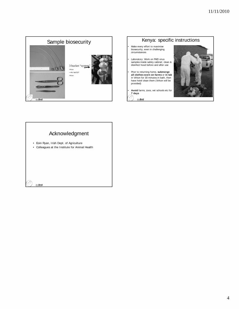
Sample biosecurity
3 bucket “system”•Water
• 4% Na2CO3
•Water
Kenya: specific instructions• Make every effort to maximise
biosecurity, even in challenging circumstances
• Laboratory: Work on FMD virus samples inside safety cabinet; clean & disinfect hood before and after use
• Prior to returning home, submerge all clothes worn on farms or in lab in Virkon for 30 minutes in bath, then have hotel clean them (Virkon will be provided)
• Avoid farms, zoos, vet schools etc for 7 days
Acknowledgment
• Eoin Ryan, Irish Dept. of Agriculture• Colleagues at the Institute for Animal Health

EMERGING TRENDS IN
FMD SAT1 OCCURRENCE
IN KENYA

1
EMERGING TRENDS IN FOOT AND MOUTH DISEASE SAT1 OCCURRENCE IN
KENYA
Wekesa, S. N1., Sangula, A.K
1.
1National Foot and Mouth Disease Laboratory, Embakasi, Kenya, P.O Box 18021-00500, N airobi, Kenya
Corresponding author; Email: [email protected]
Introduction
In the recent past, Kenya has witnessed an upsurge of Foot and mouth Disease (FMD) serotype SAT1 outbreaks, with grave consequences including mortalities among adult cattle herds and severe teat lesions that had not been observed before. The epidemic has swept across the country indiscriminately including farms that have had regular and stringent vaccination regimes using trivalent vaccines incorporating SAT1. The first witnessed cases were in June 2009 in Trans-mara district located in the south-western region of the country. This later spread to the Rift valley, Central and Eastern provinces and seems to be spreading further across the country. FMD SAT1 had been having low and sporadic incidences in Kenya for several years since 1962 when it was first recorded. The highest reported outbreaks in the history of the serotype in the country was in 1979, where 46 outbreaks occurred in 8 different districts. From 1980 to 1999, incidences had reduced, but up-surged in 1999 and since then has remained low until 2009 and 2010 (DVS Kenya reports). The overall trends of reported FMD outbreaks during the last 15 years in Kenya are illustrated in Figure 1 while trends for SAT1 over the last 48 years are shown in figure 2. SAT1 outbreaks have successfully been controlled since the 1979 epidemic through ring vaccinations using the current vaccine strain. Following the observed alarming spread and apparent failure of ring vaccinations, we report records of SAT1 outbreaks observed in the recent past in Kenya.

2
Materials and Methods
Epithelial samples from clinical cases were routinely submitted to the National FMD laboratory, Embakasi by various District Veterinary Officers (DVOs) across the country for serotype identification. The samples were accompanied by forms that gave information on morbidity and mortality rates among herds affected. A total of 184 samples had reached the laboratory from various districts by the end of August 2010 compared to 105 by the same period in 2009.They were tested using virus isolation on tissue culture and antigen detection ELISA (Anonymous, 2004). Parallel samples were submitted to the World Reference Laboratory (WRL), Pirbright for comparative serotyping and sequencing. Furthermore, investigation teams comprising of officers from the National laboratory and the field veterinary services visited some of the affected farms in response to reports by farmers and field staff of suspected FMD outbreaks characterized by severe clinical signs in previously vaccinated herds. Clinical examinations were undertaken and photos of lesions recorded. Various samples including epithelial tissue were taken for laboratory diagnosis at Embakasi and WRL.
Results
Serotyping results by the Embakasi laboratory and the WRL showed that most of the outbreaks were caused by FMDV SAT1. By the end of August 2010, SAT1 outbreaks comprised 70% (91/130) of the total positively confirmed laboratory submissions and only 30% (39/130) were due to the other serotypes (O and SAT2). So far, all the eight provinces have reported FMD outbreaks with higher occurrences recorded in the Rift valley, Central and Eastern regions. Out of the 80 districts countrywide that have so far reported the disease, 52 have had SAT1.
FIG1: FMD Outbreaks in Kenya from 1995 to 2010 FIG2: FMD SAT1 Outbreaks in Kenya from 1962 to 2010

3
All the farms affected by SAT1 reported typical FMD clinical signs as described in Kitching, (2002). The most prominent signs observed in affected cattle are represented by the photos in Figure3. A morbidity rate of 100% was observed in all the visited farms. Mortality rates in some farms went as high as 50%, but on average, 15% across all ages. One model farm with very stringent vaccination schedule had an average of 15.8% (90/570) mortalities of which 64.4% (58/90) were adult milkers. Genotypic analysis by WRL revealed that these 2009/2010 Kenyan SAT1 FMDV field strains were genetically divergent from the vaccine strain by as much as 10% nucleotide difference within topotype I (NWZ), (Figure 4).
FIG 3: Severe FMD Clinical signs in one of the dairy farms: 1-frothing and recumbency; 2-Ulceraions and peeling muzzle; 3- Teat blisters and Ulcerations; 4 – Death of an adult animal
1 2
3 4

4
Fig. 4: Neighbour-joining tree of FMDV SAT1 viruses (© Institute for Animal Health) Discussion
The serotyping reports have shown that SAT1 has early this year emerged as the most prevalent serotype in Kenya. In the recent past, the most prevalent serotypes have been O and SAT2 meaning that most of the readily available vaccine stocks have been against the prevailing strains of these two serotypes (DVS Kenya reports). The emergence of SAT1 as the prevalent serotype implies that sufficient stocks of vaccine relevant to the current strains had to be availed to the farming community within a short time. This can proof to be a challenge where routine characterization of strains is not being undertaken and there is only a single vaccine strain in use (Kitching et al., 1989). The clinical picture of 100% morbidity rates and as high as 50% mortality rates gave an indication of naivety of the populations affected by this SAT1 strains. This was further evidenced by the WRL revelation of genetically divergent strains to the current vaccine strain (WRL report). It is interesting that although belonging to the same topotype, it is apparent

5
that there was significant antigenic difference between these outbreak strains and the vaccine strain as demonstrated by the lack of protection observed in the field. The Kenyan 2009/2010 SAT1 outbreak strains were also closely related to viruses causing outbreaks in neighboring Tanzania implying regional spread of these strains. Though the genetic diversity may not necessarily imply that there is significant antigenic difference with the vaccine strains, it is an indicator of a change that calls for immediate antigenic matching and enhanced vaccine performance monitoring. Effective vaccine matching should be part of routine FMD vaccine production and quality control processes.
References
1. Anonymous, (2004). Manual of Diagnostic Tests and Vaccines for Terrestrial Animals. Office International des Epizooties. World organization for animal health, 111-136.
2. DVS Annual Reports (unpublished): Annual reports for the Director of Veterinary Services Kenya, generated by FMD lab, Embakasi.
3. Kitching R.P. 2002. Clinical variation in foot and mouth disease: cattle. OIE Scientific and
technical Review, 21(3): 513-517.
4. Kitching R.P., Knowles N.J., Samuel A.R. & Donaldson A.I. 1989. Development of foot and mouth disease virus strain characterisation. A review. Tropical Animal Health
Production, 21: 153-166.
5. World Reference Laboratory, Pirbright: (WRL FMD/2010/00026): FMD detection and serotyping results reports by the Institute of Animal Health Pirbright. http://www.wrlfmd.org/
Acknowledgements
The authors thank the Director of Veterinary Services (DVS), Kenya for facilitating this work, all the District Veterinary Officers and the Kenya Wildlife Services for providing the samples. Much gratitude goes to all laboratory staff members at the FMD Embakasi laboratory for their support and hard work. The WRL is thanked for genetic characterization and serotyping reports.

VADEMECUM FOR FMD
OUTBREAK DETECTION
AND INVESTIGATION

Vade mecum for FMD outbreak detection and investigation
Example (prepared for Lebanon, 12/2009)
Foreword:
This document is prepared by the EuFMD Secretariat and is intended to assist veterinary services with the investigation
and response to suspect FMD outbreaks. It should be adapted for application by field officers, and updated after
changes in diagnostic capacity or experience.
Protocol for outbreak detection:
In case of confirmation of the suspicion, suitable samples should be taken in order to confirm infection.
The investigation of suspicions should be made by competent persons under authority of the vet services, who
understand the type of samples required for laboratory confirmation.
Penside tests (Svanodip – FMD) can be used on site or at the laboratory, but only with suitable samples from early cases.
A range of samples, including whole blood for serum, are advisable if early lesions (<6 days) are not abundant.
The veterinarians collecting samples should complete tables for each animal, supplying information (table 1).
Table 1. Information collected on animals examined and sampled.
no animal ID
species
and
sex1
age1
clinical signs type of lesions
sampl
es
taken5
vacci-
nation
status1
estimated
age of the
oldest
lesions
lam
en
ess
fever2
sali
vati
on
foo
t3
mo
uth
4
teats
inta
ct
vesic
le
recen
tly r
up
-tu
red
vesic
le
raw
ero
de
d a
rea
ulc
er
wit
h f
ibri
no
us
scab
ulc
er
wit
h f
ibro
sis
ind
isti
nc
t b
reak c
oro
-
na
ry b
an
d

The flow of activities to arrive at a confirmation of FMDV is given in the following decision tree:
Event information from farmers
Not confirmed suspicion confirmed by veterinary services
< 6 days > 6 days
sampling Swab, epithelium
blood
sampling Swab (as epithelium may
not be possible) Probang
blood (for serum)
Laboratory: - antigen detection ELISA (often negative over 6 days) - PCR (needed) - serology (NSP – can confirm exposure to FMDV)
In situ rapid test pen-side
result (-) result (+)
further
investigations Laboratory
Ag detection ELISA
immediate measures identification
confirmed
identification of Ag
not identified
sample to WRL for genotyping
use of ring emergency vaccination
Decision tree for outbreak detection and investigation
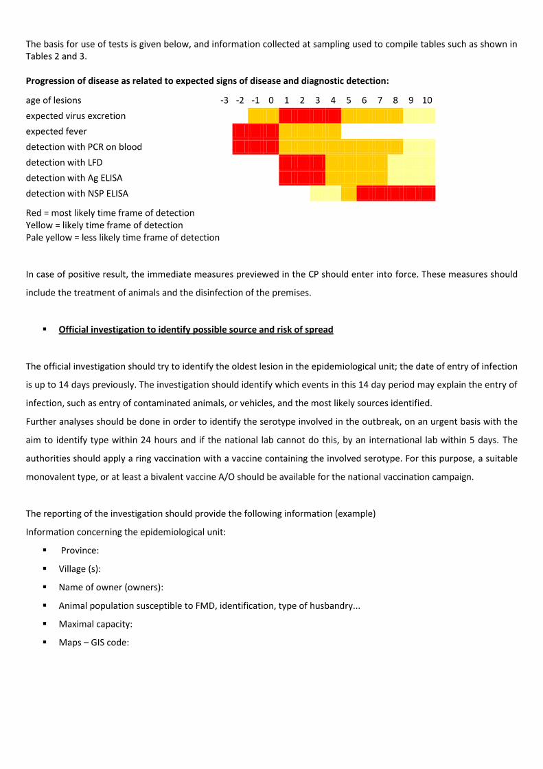
The basis for use of tests is given below, and information collected at sampling used to compile tables such as shown in Tables 2 and 3. Progression of disease as related to expected signs of disease and diagnostic detection:
age of lesions -3 -2 -1 0 1 2 3 4 5 6 7 8 9 10
expected virus excretion
expected fever
detection with PCR on blood
detection with LFD
detection with Ag ELISA
detection with NSP ELISA
Red = most likely time frame of detection Yellow = likely time frame of detection Pale yellow = less likely time frame of detection In case of positive result, the immediate measures previewed in the CP should enter into force. These measures should
include the treatment of animals and the disinfection of the premises.
Official investigation to identify possible source and risk of spread
The official investigation should try to identify the oldest lesion in the epidemiological unit; the date of entry of infection
is up to 14 days previously. The investigation should identify which events in this 14 day period may explain the entry of
infection, such as entry of contaminated animals, or vehicles, and the most likely sources identified.
Further analyses should be done in order to identify the serotype involved in the outbreak, on an urgent basis with the
aim to identify type within 24 hours and if the national lab cannot do this, by an international lab within 5 days. The
authorities should apply a ring vaccination with a vaccine containing the involved serotype. For this purpose, a suitable
monovalent type, or at least a bivalent vaccine A/O should be available for the national vaccination campaign.
The reporting of the investigation should provide the following information (example)
Information concerning the epidemiological unit:
Province:
Village (s):
Name of owner (owners):
Animal population susceptible to FMD, identification, type of husbandry...
Maximal capacity:
Maps – GIS code:

Information concerning the outbreaks (or epizootic):
Timeline (from earliest to last event)
1. Start of possible window of introduction of FMDV (date and possibly risk factor of introduction of
FMDV, dissemination of the disease) (1-14 days before first signs)
2. First observation of clinical signs (date)(=day 0)
3. Notification of suspicion to veterinary services (date)
4. Date of sampling
5. Date of official investigation
6. Date confirmed
Clinical examination and sampling of the animals
1. Describe (table 3)
2. Clinical signs (table 3)
3. Aging of lesions (table 1)
Sampling
1. Blood sampling
2. Tissue sampling
3. Pen side test
4. Bio-security & disinfection
5. Sending samples to the lab
Vaccination history: Describe (date, vaccine, animals...)
Results of the laboratory analysis (table 2)
Source and spread of infection:
1. most likely event that introduced infection in the unit
2. time period when spread of infection most likely to occur from the epi-unit (= from 2-4 days before
signs depending on species)

Table 2. Results of the laboratory investigation of the samples taken in the village (example of compilation)
animal ID LFD NSP Ag ELISA clinical
signs conclusion3
+1 + inconclusive + (2-3 d) infected
1234567 +1 + type O + (2-3 d) infected
6789234 +2 + - + (2-3 d) infected
1 epithelium and saliva
2 epithelium and vesicle fluid
3 conclusion based on references in table 3
Table 3: Animals examined and sampled in the village (example of compilation).
no animal ID species
and sex
1 age
1
clinical signs type of lesions
samples taken
5
vacci-nation status
1
estimated age of the
oldest lesions
lam
en
ess
feve
r2
saliv
atio
n
foo
t3
mo
uth
4
teat
s
inta
ct v
esi
cle
rece
ntl
y ru
p-
ture
d v
esi
cle
ra
w e
rod
ed
are
a u
lce
r w
ith
fib
rin
ou
s sc
ab
ulc
er
wit
h
fib
rosi
s in
dis
tin
ct
bre
ak c
oro
-
nar
y b
and
1 12344567 bov / M
7 months
- NT - - - - - - - - - - B - S not reported
2 12324567 bov / M
not known
- NT - - - - - - - - - - B - S not reported
3 12234567 bov / M
7 months
- NT - - - - - - - - - - B not reported
4 12234567 bov / F 1.5 year
- NT + - LTD - - - - + + - B not reported
6 to 7 days
5 12334567 bov / F 1.3 year
+ NT + C G - - - - + - B 10.2008 5 to 6 days
6 12344567 bov / F 1.3 year
+ NT + I LG - - + (I) - +
(M) - B - E 10.2008 7 days
7 12345567 bov / F 1.5 year
+ NT + - MLD - - - - + - B - S - E not reported
5 to 6 days
8 12345667 bov / F 1.3 year
+ NT + I L - - - - - + - B 10.2008 7 days
9 12345677 bov / F 1.3 year
- NT + - ML - - - - - + - B 10.2008 7 days
10 12345678 bov / F 1.3 year
+ NT + I LTD - - - + (I) +
(T) +
(D) - B 10.2008 7 days
11 not identified
bov / M
not known
- NT - - - - - - - - +? - B - P not reported
7 to 10 days
1 information retrieved from the livestock information system
2 NT: not tested (animals did not appear to have fever)
3 foot: Coronary band – Inter-digital space /
4 mouth: Muzzle - Lips - Gums - Tongue - Dental pad /
5 samples: Blood - Saliva -
Vesicle fluid - Epithelium - Probang sample

NTC2-NOVEMBER2010

EuFMD training course – Nakuru, Kenya – November 2010

Executive Summary
A five day course was convened in Nakuru, Kenya supported by FAO and the EuFMD Commission.
Participants included veterinarians from Kenya, France, Latvia, Switzerland, Australia, New Zealand
and United States. The course was organized with a one day symposium in Nakuru led by FAO and
an FMD technical expert from the Institute on Animal Health Pirbright. Instruction was provided on
aging of FMD lesions, biosecurity, sample collection and investigation technique. This was followed
by two days of field visits in which three outbreaks were investigated and samples were collected
and tested immediately in the Regional Veterinary Laboratory. The remainder of the course
consisted of report writing, discussion of laboratory results and the epidemiological information that
had been gathered.
Laboratory results yielded serotype O from Farm 2 and 3 visited. Non-structural protein antibody
ELISA results were also obtained during the course. These results are presented in Appendix A and
discussed in the relevant sections. This document contains laboratory results as of December 1,
2010.
Appendix B displays a map of farms visited and areas discussed in the epidemiology investigation.
Participants had the opportunity to observe clinical cases of FMD, perform sample collection,
conduct interviews with farmers, collate epidemiological information, practice biosecurity principles
and asses laboratory results. This unique opportunity was made possible by the laboratory staff in
Nakuru and Nairobi, farmers, district veterinarians and the EuFMD team.

Background
Veterinary services The Department of Veterinary Services (VS) in Kenya undertakes disease control, investigation and
disease surveillance. Kenya is divided into 8 provinces (Figure 1) each headed by a Provincial Director
of Veterinary Services. The private sector performs the clinical services and artificial insemination.
Figure 1: Map of Kenya showing the 8 provinces
KEY:
1. Central 2. Coast 3. Eastern 4. Nairobi 5. North Eastern 6. Nyanza 7. Rift Valley 8. Western
The Rift Valley Province has 64 districts. Nine of these districts (Nakuru, Naivasha, Nakuru North,
Rongai, Molo, Njoro, Kuresoi, Subukia and Gilgil) form the Nakuru county where the investigation
took place. Each district is headed by a District Veterinary Officer (DVO) who performs the
departmental mandate of disease control, vaccination, meat inspection and extension service. The
DVO is supported by livestock officers and animal health technicians who are based in the divisions
and locations. The regional veterinary laboratory in Nakuru performs disease surveillance and
submits samples to the national laboratory in Embakasi, Nairobi where all FMD analysis is
performed.

FMD Situation FMD is endemic in Kenya and most outbreaks are reported in the Rift Valley Province which is the
largest of the 8 provinces. To date, serotypes O, A, C, SAT1, and SAT2 have been detected in Kenya.
Most common are serotype O and SAT2, however since 2008 SAT1 outbreaks have increased
dramatically. Serotype C was last documented in 2004. The situation in wildlife is not well studied,
but buffalos are known to harbour SAT serotypes. A random countrywide survey was conducted in
2009/2010 to obtain a better picture of FMD prevalence in the different animal species. Results will
be available soon.
In recent years sporadic FMD outbreaks have been reported in all districts. In 2010 FMD, outbreaks
appear to be more frequent and spreading faster than before. For the current calendar year, twenty
three FMD outbreaks have been confirmed by the national FMD laboratory (Table 1). The main
serotypes responsible for these outbreaks are SAT1 and O. Information of number of infected farms
and species are not available, thus morbidity and mortality in the Rift Valley province cannot be
estimated.
Table 1: FMD outbreaks confirmed by national FMD laboratory in Nakuru county and close
neighbour districts from 01.01.2010 to 15.11.2010
serotype date District
O 02.08.2010 Nakuru Central
O 02.08.2010 Nakuru Central
O 30.08.2010 Laikipia West
SAT1 18.06.2010 Laikipia North
SAT1 24.06.2010 Laikipia Central
SAT1 28.06.2010 Laikipia East
SAT1 10.07.2010 Nakuru North
SAT1 16.07.2010 Njoro
SAT1 26.08.2010 Koibatek
SAT1 26.08.2010 Koibatek
SAT1 10.09.2010 Naivasha
SAT1 10.09.2010 Nakuru Central
SAT1 30.09.2010 Gilgil
SAT1 01.10.2010 Mogotio
SAT1 07.10.2010 Marigat
SAT1 08.10.2010 Nakuru North
SAT2 16.08.2010 Naivasha
NVR 15.01.2010 Naivasha
NVR 08.06.2010 Naivasha
NVR 22.06.2010 Naivasha
NVR 22.09.2010 Nakuru Central
NVR 30.09.2010 Nakuru North
NVR 30.09.2010 Gilgil
NVR= No virus recovered

Livestock distribution and movement In Kenya there are 1.5 million beef cows, 1.25 million dairy cows, 9 million sheep, 12 million goats, 1
million camels and 30000 pigs. The numbers of animals in Nakuru County can be seen in Table 2.
a) Animal movements associated with trade:
1. Laikipia → Rongai → Baringo
2. Baringo → Nakuru → Naivasha → Nairobi
b) Animal movements associated with grazing:
1. Naivasha → Nakuru → Laikipia;
2. Narok/Kajiado → Navaisha → Nakuru → Rongai /Laikipia
Table 2: Livestock population in Nakuru county (data from 18.11.2010)
District Cattle Sheep Goat
Naivasha 86161 120667 48915
Nakuru North 53000 23000 17000
Molo 23550 12580 1756
Njoro 32970 33550 23564
Rongai 126950 50847 70155
Nakuru 11500 2830 5480
Subukia 10000 14000 12000
Gilgil n.a. n.a. n.a.
Kuresoi n.a. n.a. n.a.
In Nakuru county there are 4 main livestock routes and about 10 livestock markets where animals
move for trade or pasture (nomadic herds) from one district to another.
FMD diagnostics FMDV antigen is detected using Ag ELISA , virus isolation using baby Hamster kidney cells (BHK) or
calf thyroid cells (CTY) or the lateral flow device LFD as a penside test. All 5 serotyes (O, A, C, SAT1,
SAT2) circulating in Kenya are screened. For PCR, samples for FMDV antigen detection are sent to
the FMD world reference laboratory in IAH, Pirbright, UK. However, it would be useful to establish
the PCR method in the local lab in the future to strengthen the diagnostic power in the affected
region.
FMDV antibodies are detected using a test for structural proteins (SPs) for example virus
neutralisation test (VNT) or liquid phase blocking ELISA (LBPE) which cannot differentiate between
vaccinated and naturally infected animals. To be able to differentiate between those two antibodies
non-structural proteins tests (the 3ABC and 3D ELISA) are used.
Probang, tissue, saliva and vesicular fluid samples are analysed using Ag ELISA and its results
confirmed with samples which show a cytopathogenic effect (CPE) when passaged on BHK or CTY
cells.

Serum is analysed using mostly VNT or otherwise LBPE. Animals which are known to be vaccinated,
animals which come from an area where vaccination takes place and animals which are going to be
exported are tested using 3-ABC or 3-D.
The most likely time for detection of virus is within the first 4 days after clinical lesions appeared, but
could be possible up to 7 days and for PCR detection up to 10 days after the first clinical signs.
Antibodies can be detected most likely after 7 days post infection (p.i.), and possibly already after 5
days p.i. The figure below represents the window of detection for various techniques or tissues.
Figure 2: This figure depicts the window of detection by different techniques or tissues. From: IAH
FMD control history and vaccination Until the early 1980s, FMD was controlled in designated areas of Kenya, including parts of the Rift
Valley province with compulsory and free vaccination for cattle. Small ruminants were not
vaccinated. This vaccination service achieved 80% vaccination coverage which enabled Kenya to
access international trade markets. However, a change in government policy from 1986 introducing
cost sharing with the farmers and an unavailability of government subsidised vaccines meant the
vaccination coverage decreased below 10%. This resulted in more frequent FMD outbreaks.
Currently, farmers can access more expensive vaccines from the Kenya Veterinary Vaccines
Production Institute (KEVEVAPI) to supplement the DVO’s supply. Vaccinations available for use in
Kenya are multivalent formulations and contain either 3 or 4 of the serotypes O, A, SAT1 and SAT2.

When an outbreak is declared, quarantine is imposed on a location level. Movement may occur
within the location but is restricted into and out of the location. Restrictions are placed on milk and
other animal products. Ring vaccination around the outbreak is performed and nearby markets is
closed. There are also restrictions on movement of animal feed out of the affected farm. The official
in charge of the administrative unit assists with enforcement of quarantine.
Recommendations for further control and investigations of the outbreaks are provided below for
each farm. These recommendations are based on the local situation and farm-level biosecurity
measures. In contrast to an endemic country, control measures in an FMD free country vary per
country and could range from culling all infected animals to vaccination to live based on rate of
disease spread and other economic and political factors. Furthermore, all animal movements would
cease and all possible movements of animals, animal products, humans and vehicles in the risk
period would be traced back and forward to find all farms were the disease might have spread to.
Ring vaccination, whenever possible, as well as a vaccination to live strategy, should be considered
to control the outbreak. Policy varies per country for disease control measures yet the ultimate goal
is to become disease free by OIE standards as rapidly as possible.
The investigation team The NTC2 EU-FMD team was supervised by Nick Juleff (UK), Keith Sumption (FAO), Nadia Rumich
(FAO) and consisted of local veterinarians (Eunice Chepkwony, Sabenzia Wekesa, Veronica Wanjohi,
Abraham Sangula, Gitnui Kaba, Bernard Rono, William Birgen and Fredrick Mukendi), as well as
veterinarians from other countries (Mary Vanandel , NZ; Jennifer Davis, AU; Laurie Fromberg, US;
Mara Uzule-Springe, LA; Yann Villagi, FR; Silke Bruhn, CH). Roles were switched between a clinical
investigation team taking care of the sampling on the farm as well as aging lesions and an
epidemiological team who did interviews of the farmers and DVOs to gather epidemiological
relevant information. Animals to sample were chosen based on looking for the oldest lesions on the
farm to be able to trace back the highest risk period of FMDV introduction as well as for the
youngest lesions to be able to still detect virus. Selection took place relying on the information
provided by the owner as well as the appearance of clinical signs.
The epidemiological investigation was performed using a questionnaire developed by the group
(Appendix C) aiding in the establishment of the probable timeline of important events. The possible
period of introduction of the virus was estimated by subtracting 14 days from the day of the oldest
known lesion. The high risk period was estimated at 2-5 days within the incubation period. The
period of highest shedding was determined to be day 1 to 4 after the first and last animal displayed
clinical signs.
Biosecurity measures On arrival at each of the 3 farms visited, the investigating team vehicles were parked outside the
infected area near the farm entrance. Protective clothing was donned by all team members. This
consisted of disposable overalls, latex gloves (double gloving with taping at the wrist to the
protective suit) and rubber boots.

On departure, the team cleaned the boots thoroughly with a brush with 0.5% Sodium Lauryl Ethyl
Sulphate and a second washing with Dettol®. Finally the boots were disinfected by dipping them into
4% sodium bicarbonate solution. The car tyres of the vehicles were washed with 4% sodium
bicarbonate solution.
At the end of each day the clothes worn on the farms were soaked overnight in a 0.2% citric acid
solution and then laundered by the hotel.

Case farm 1 – Tere Estate
Farm history
Location
The farm is located in the Nakuru North district, Bahati division, Bahati location and Wanyororo B
sublocation.
Production system
There were five cattle on the farm, along with approximately 15-20 chickens and 10 dogs. Cattle are
dairy cattle and milk is sold.
Animals graze on the farm only. They do not share grazing or water with any other animals.
However, they are fed a supplementary feed, a compounded product purchased from Mairu kumi
shopping centre. The feed is purchased monthly and carried home from the shop in a bag. The last
feed was purchased in late October. Mairu kumi is also in the Bahati division and has been
experiencing outbreaks of FMD.
Farm biosecurity
The farm is relatively isolated with coffee plantations surrounding the area where the cattle are
kept. However, no biosecurity measures were taken on the farm.
According to the owner of the cattle, there is no contact with wildlife.
Clinical examination and sampling Only two animals are reported by the farmer to be showing clinical signs—Animal A and Animal C.
Four animals were sampled- one cow, one bull, and two calves. Animals displayed clinical signs such

as salivation, lameness, and decreased milk production. All cattle displayed mouth lesions while
three of the four cattle had feet lesions, one of which had a secondary infection. One cow also
displayed possible scarring on the teats. Age of the mouth lesions ranged from 7 days to greater
than 10 days. Serum samples were taken from all four of the cattle. A probang sample was taken on
one of the calves. Epithelial tissue was taken from one of the cows and a lateral flow device (penside
test) was conducted. The epithelial sample was too old yielding negative results to the penside test.
All animals sampled were treated with multivitamins and antibiotics. Further details are provided in
Appendix A: Clinical examination form – Farm 1. Serotype for this farm at the time the report was
written was unknown.
Preliminary laboratory results yielded negative antigen ELISA results from the epithelial tissue from
Animal A as well as negative cell culture from the epithelial tissue from Animal A. These results
should be interpreted with caution as the epithelial sample obtained was likely too old. NSP test
results yielded negative results from Animal A and weak positive results from Animal B, Animal C and
Animal D. Since these lesions were dated between 7-10 days, the weak positives could possibly be
due to this outbreak of FMD or due to previous vaccination (impure vaccine containing NSPs),
previous exposure to any serotype of FMD or improperly aging the oldest lesions.
Top Left: Animal A (cow) dental pad lesion aged to about 8 days old
Top Right: Animal C (calf) tongue lesion aged to about 7 days old
Bottom Left: Animal C (calf) foot lesion
Bottom Right: Animal D (calf) lower lip and lingual periodontal lesion aged to greater than 10 days old.

Epidemiological findings
FMD & vaccination history
The animals have no history of vaccination as far as the farmer is aware. Two animals—Animal A and
B—were purchased in July 2009 whilst two others were born on the property. The district
veterinarian reported that vaccination had not been practiced in this area as there had been no
outbreaks in the area.
Current outbreak
The farmer reported only two animals showing clinical signs.
Animal A, the first animal to show clinical signs, was observed to be limping on Wednesday 10th
November. By Friday November 12th she was salivating and the farmer reported the potential
outbreak to a private veterinarian. The outbreak was reported to the district veterinarian on Sunday
November 14th; the district veterinarian visited the property on Monday 15th November. A second
animal, Animal C, was seen to be limping on Saturday 13th November.
Animal movements and other contacts
There have been no recent animal movement on or off the farm. No animal products have been
moved onto the farm. Milk was moved off the farm prior to the outbreak however since quarantine
has been imposed it has not been longer sold (also decreased volume due to production drop).
Temporary workers enter the farm daily and walk along a path shared by cattle. Workers come from
surrounding neighbourhoods some of which have been experiencing FMD disease outbreaks.
The last visit by a veterinarian was in April/ May 2010 when a private veterinarian was called out to
treat mastitis.
The monthly feed was last purchased in late October from Mairu kumi, where there have been FMD
outbreaks in the surrounding area.

Timeline
infection timeline -15 -14 -13 -12 -11 -10 -9 -8 -7 -6 -5 -4 -3 -2 -1 0 1 2 3 4 5 6 7 8 9 10 11 12 13 14 15
date (Oct/Nov) 22 23 24 25 26 27 28 29 30 31 1 2 3 4 5 6 7 8 9 10 11 12 13 14 15 16 17 18 19 20 21
most likely time of introduction
possible time of introduction
highest period of shedding
Farm summary Most likely source of introduction:
The purchased feed is considered a highly likely source of introduction due to the known FMD
outbreaks in the area that it came from and the direct pathway to the animals.
Introduction by manure/ infectious material on shoes of workers coming onto the farm is considered
possible but not the most likely.
Recommendations:
More information is required on farms in the locality. Although FMD has not been reported this does
not rule out possible cases.
It is recommended that the farm remains under quarantine until the cases are resolved. Animals
should be moved off the pathway shared with workers to try and prevent spread to other farms.
The farmer could implement basic biosecurity measures such as separating pathways for workers
and trucks coming onto the farm from animals. Routine vaccination of animals should also be
practiced against known circulating serotypes.
It is also recommended that feed retailers take measures to minimise spread of disease by avoiding
purchase of feed from known infected areas. Farmers purchasing feed should also be aware and
purchase feed locally or from areas not experiencing outbreaks of FMD.
Additional information that could be of value in this instance would include a map of the farm
showing fences, farm boundaries, extent of resident animal ranges and location of water points. A
graphical representation of roads and paths used by workers and stock could clarify local conditions
and this information could clarify entry and exit pathways for disease.

Tracing information needs to be obtained if available for milk collection and feed purchases. This
could allow neighbouring properties to be prioritised for examination.
Estimated risk of spread:
The farm is isolated and very small, so that virus shedding as well as possible routes to spread
disease are minimal.
Case farm 2 – Solai Arus
Farm history
Location
The farm was located in Ruiyabei location and Arusito sublocation in Solai division.
The owner of the farm was Wilbert Mameti. The owner was not present for the visit and his wife
answered the questions of the EuFMD team.
Production system
Farm had mixed beef and dairy production with approximately 50 cattle (30 adults and 20
calves/yearlings). There were also dogs and chickens on the farm.
The whole herd grazes together on the farmer’s property only. The animals share a common
watering point, on the property, with animals from neighbouring farms.

Farm biosecurity
There is no biosecurity on the farm. Animals from approximately six neighbouring farms enter the
property daily for water from the common watering point. Most neighbouring farms have 30-50
cattle and some also have sheep and goats. However, the sheep and goats do not share the watering
point.
Clinical examination and sampling Animals displayed clinical signs such as salivation, nasal discharge, swollen protruding tongue, and
lameness. Two animals were sampled including one cow (Animal E) and one bull (Animal F). As most
of the herd was pregnant, the farmer was concerned that the stress of examination and sample
collection would further compromise the animal’s condition. As typical lesions were noted in the
examined animals and LFD penside testing yielded a positive result, it was decided that further
examination was not in the best interest of the herd.
The farmer gave information that first clinical signs were two weeks ago thus it was not possible to
examine the oldest lesions.
All animals sampled displayed mouth lesions. Due to being in a crush, foot lesions were not able to
be safely visualized. Age of the lesions was estimated to be around one day old without including a
margin of error. Serum samples were taken from all animals sampled. The cow sampled had a
swollen protruding tongue with a large ruptured blister. While trying to grasp the tongue, the distal
end was exfoliated and an epithelial sample was obtained. A Penside test was run with this epithelial
tongue sample and yielded positive results. A probang sample was taken from the bull. An epithelial
sample was also taken from the bull, but a penside test was not conducted. All animals were treated
with multivitamins and antibiotics. Further details are provided in Appendix A: Clinical examination
form – Farm 2.
Preliminary laboratory results yielded positive antigen ELISA results from the epithelial tissue from
Animal E and Animal F as well as positive cell culture from the epithelial tissue from Animal E and
Animal F. The virus was typed as serotype O. NSP test results yielded weak positive results from
Animal E and Animal F. Since these lesions were aged at Day 1, the weak positives could possibly be
due to previous vaccination with vaccine containing NSPs, previous exposure to any serotype of FMD
or improperly aging of the oldest lesions.

Top Left: Animal E (cow) with one day old ruptured vesicles on the tongue
Top Right: Animal E (cow) with raw epithelium after outer layers of epidermis was removed during examination
Bottom Left: Positive Penside Test from Animal E
Bottom Right: Salivation and blanched vesicle on dental pad of Animal F (bull) aged to one day.
Epidemiological findings
FMD and vaccination history
The animals were vaccinated against serotypes O, SAT1 and SAT2 on the 11th September 2010 due to
an outbreak of SAT1 in Nakuru and the proximity of this farm to a livestock market (approximately 1
km). Animals from many areas, including Nakuru come to the market to be sold.
Prior to this the last routine vaccination was approximately 18 months ago.
Some animals on affected neighbouring farms had been vaccinated and did not show clinical signs of
FMD.
Current outbreak
Disease was first noticed in neighbouring farms approximately 3 weeks ago.
The first case on this farm was noticed by the farmer two weeks ago. One calf showed lameness and
salivation. Disease then spread through the herd with the majority of cases noticed on Saturday 13th
November. The farmer estimates approximately 20 animals appear to be affected.
The date that the outbreak was reported is unknown.
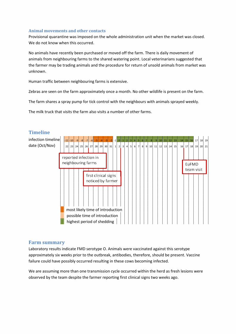
Animal movements and other contacts
Provisional quarantine was imposed on the whole administration unit when the market was closed.
We do not know when this occurred.
No animals have recently been purchased or moved off the farm. There is daily movement of
animals from neighbouring farms to the shared watering point. Local veterinarians suggested that
the farmer may be trading animals and the procedure for return of unsold animals from market was
unknown.
Human traffic between neighbouring farms is extensive.
Zebras are seen on the farm approximately once a month. No other wildlife is present on the farm.
The farm shares a spray pump for tick control with the neighbours with animals sprayed weekly.
The milk truck that visits the farm also visits a number of other farms.
Timeline
infection timeline -11 -10 -9 -8 -7 -6 -5 -4 -3 -2 -1 0 1 2 3 4 5 6 7 8 9 10 11 12 13 14 15 16 17 18 19
date (Oct/Nov) 22 23 24 25 26 27 28 29 30 31 1 2 3 4 5 6 7 8 9 10 11 12 13 14 15 16 17 18 19 20 21
most likely time of introduction
possible time of introduction
highest period of shedding
Farm summary Laboratory results indicate FMD serotype O. Animals were vaccinated against this serotype
approximately six weeks prior to the outbreak, antibodies, therefore, should be present. Vaccine
failure could have possibly occurred resulting in these cows becoming infected.
We are assuming more than one transmission cycle occurred within the herd as fresh lesions were
observed by the team despite the farmer reporting first clinical signs two weeks ago.
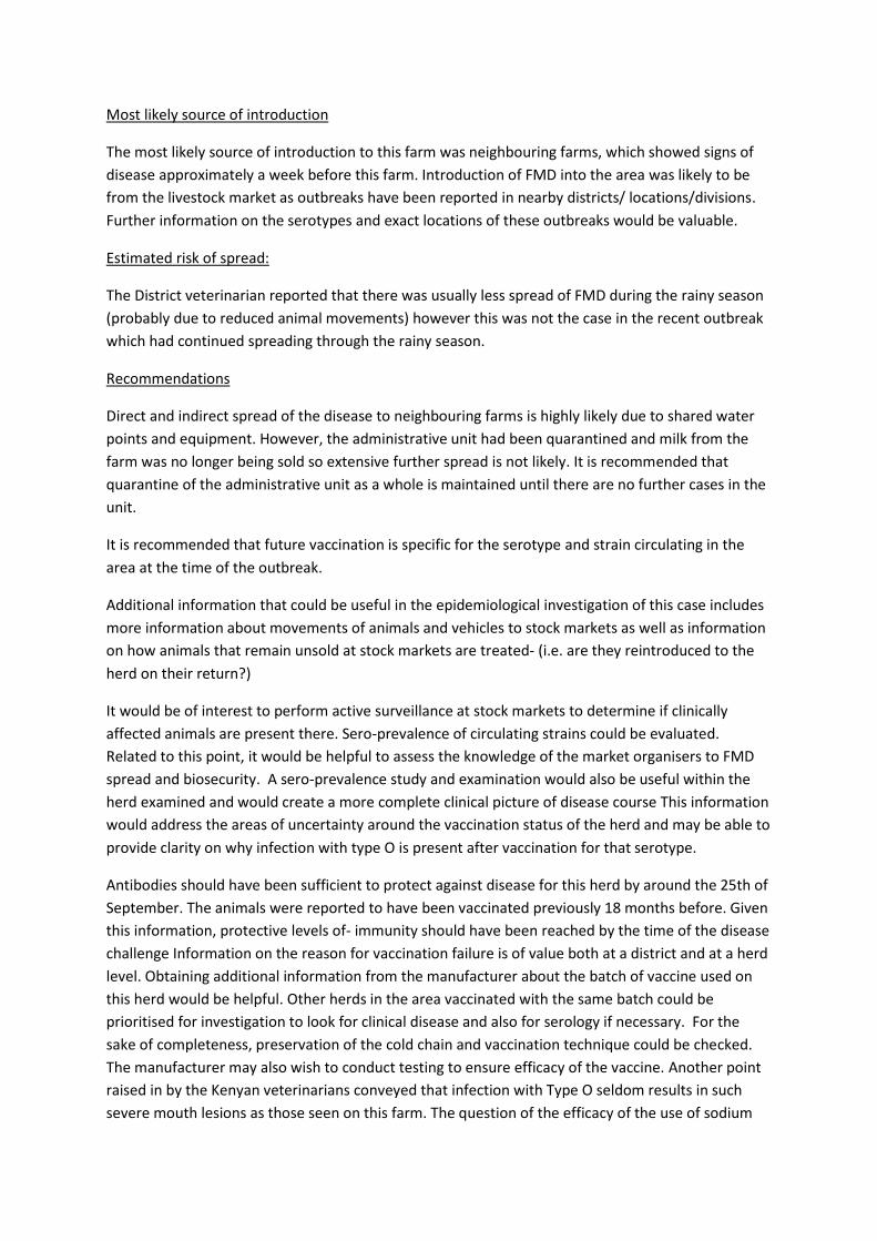
Most likely source of introduction
The most likely source of introduction to this farm was neighbouring farms, which showed signs of
disease approximately a week before this farm. Introduction of FMD into the area was likely to be
from the livestock market as outbreaks have been reported in nearby districts/ locations/divisions.
Further information on the serotypes and exact locations of these outbreaks would be valuable.
Estimated risk of spread:
The District veterinarian reported that there was usually less spread of FMD during the rainy season
(probably due to reduced animal movements) however this was not the case in the recent outbreak
which had continued spreading through the rainy season.
Recommendations
Direct and indirect spread of the disease to neighbouring farms is highly likely due to shared water
points and equipment. However, the administrative unit had been quarantined and milk from the
farm was no longer being sold so extensive further spread is not likely. It is recommended that
quarantine of the administrative unit as a whole is maintained until there are no further cases in the
unit.
It is recommended that future vaccination is specific for the serotype and strain circulating in the
area at the time of the outbreak.
Additional information that could be useful in the epidemiological investigation of this case includes
more information about movements of animals and vehicles to stock markets as well as information
on how animals that remain unsold at stock markets are treated- (i.e. are they reintroduced to the
herd on their return?)
It would be of interest to perform active surveillance at stock markets to determine if clinically
affected animals are present there. Sero-prevalence of circulating strains could be evaluated.
Related to this point, it would be helpful to assess the knowledge of the market organisers to FMD
spread and biosecurity. A sero-prevalence study and examination would also be useful within the
herd examined and would create a more complete clinical picture of disease course This information
would address the areas of uncertainty around the vaccination status of the herd and may be able to
provide clarity on why infection with type O is present after vaccination for that serotype.
Antibodies should have been sufficient to protect against disease for this herd by around the 25th of
September. The animals were reported to have been vaccinated previously 18 months before. Given
this information, protective levels of- immunity should have been reached by the time of the disease
challenge Information on the reason for vaccination failure is of value both at a district and at a herd
level. Obtaining additional information from the manufacturer about the batch of vaccine used on
this herd would be helpful. Other herds in the area vaccinated with the same batch could be
prioritised for investigation to look for clinical disease and also for serology if necessary. For the
sake of completeness, preservation of the cold chain and vaccination technique could be checked.
The manufacturer may also wish to conduct testing to ensure efficacy of the vaccine. Another point
raised in by the Kenyan veterinarians conveyed that infection with Type O seldom results in such
severe mouth lesions as those seen on this farm. The question of the efficacy of the use of sodium

bicarbonate as a topical treatment was also raised. Some felt that application of sodium bicarbonate
may have led to more severe mouth lesions.
Case farm 3 – Eden Farms
Farm history
Location
Kampimoto
Production system
This is a dairy operation with 44 bovines on the property. The cattle run as one herd. Currently the
bull is kept in a separate camp. Two heifers and two steers are also separated.
There are 11 Dorper-cross sheep on the property. The sheep and cattle co-graze and share a water
source.
Dairy meal is bought in weekly from Nakuru for the calves. Other feed for the animals is produced on
the farm.
Dogs and poultry are present on the property.
Farm workers are resident on the premises.
Farm biosecurity
The farm is well contained with an electric fence and a hedge. There is only one entrance to the
property on a well travelled road. The farm workers live on the premises, therefore, human
movements are limited compared with some production units.
The neighbouring farm boundary is approximately 600m away and animal contact with the
neighbouring property is unlikely. The neighbouring farm has in excess of 100 bovines and has
experienced a severe acute FMD outbreak with reported mortality within the last 3 weeks.
There is no contact with wildlife.
Clinical examination and sampling Seven animals were examined; five cattle and three sheep. Clinical signs observed were salivation,
nasal discharge and limping. Lesions were observed on four cattle and one sheep. The age of lesions
on the cattle ranged from three days to more than seven days. The lesions observed on the sheep
were approximately ten days old. Serum was collected from all sheep and four of the cattle.
Epithelium was collected from three cattle and penside tests were conducted. Two tests were
positive for foot and mouth disease virus antigen while one was negative. A probang sample of
orophangeal fluid was collected from one cow. All animals sampled were treated with multivitamin
and antibiotics. Further details are provided in Appendix A: Clinical examination form – Farm 3.
Preliminary laboratory results yielded positive antigen ELISA results from the epithelial tissue from
Animal 2 and Animal 3 as well as positive cell culture from the epithelial tissue from Animal 2 and
Animal 3. The virus was typed as serotype O. NSP test results yielded a strong positive from Animal 1
and Animal 3, weak positives for Animal 4, Animal 5 and Animal 6 (sheep). NSP test was negative for
Animal 2, Animal 7 (sheep) and Animal 8 (sheep). Since these lesions were aged between day 3 to
greater than 10 days, the positives could possibly be due to previous vaccination with vaccine
containing NSPs, previous exposure to any serotype of FMD or improperly aging of the oldest
lesions.

Top Left: Animal 2 (bull) salivating.
Top Right: Note blanching of epithelium on the tongue representing an unruptured vesicle (bull).
Bottom Left: Animal 3 with lesions along the dental pad aged at 5 days old (cow).
Bottom Right: Animal 7 (ewe) with lesions on the tongue aged to >10 days.
The animals kept separate from the main herd, included the bull, two heifers and two steers also
displayed clinical signs of FMD despite of being kept separate from the herd.
Epidemiological findings
FMD & vaccination history
Bovines on this farm were vaccinated by the district veterinarian as a routine measure last year. The
vaccination took place more than 6 months ago. The vaccination was performed with a trivalent
vaccination containing O, A and SAT1 serotypes.
Good biosecurity measures have prevented any previous outbreaks on this property.

Current outbreak
The farmer reported the first clinical signs appeared on the farm on the 15th of November (2 days
before the EuFMD team visit). The clinical signs noted were salivation, anorexia and depression. Only
two days after the appearance of the first clinical signs, 37 of the 44 bovines present on the property
were affected.
The sheep on the property were reported to be limping.
Animal movements and other contacts
Movements on and off the property were well documented by the farmer.
As previously noted, farm workers reside on the property. Some visitors to the farm workers may
have gone unrecorded.
Milk is collected daily by Brookside, a local dairy purchaser.
Hay is purchased by neighbouring farmers on the farm. This is a daily occurrence.
Hay was harvested starting on the 29th of October and the harvesting is ongoing at this time.
Harvesting is performed by a contractor who also performs this service on neighbouring farms.
Cattle were moved onto the field where hay was harvested on the 13th of November.
Weather conditions two weeks ago were cool and humid.
A veterinary practitioner visits this farm every fortnight. No artificial insemination services have been
used on this property in the last 9 months. The bull which was examined was brought onto the
property in place of the artificial insemination between 9 months and one year ago.
Heifers were sold off the property on the 3rd of November and collected by a livestock transport
company. These animals were taken to the Karichu district.
There is no record of any new animals being brought onto the farm in the last month.

Timeline
infection timeline -16 -15 -14 -13 -12 -11 -10 -9 -8 -7 -6 -5 -4 -3 -2 -1 0 1 2 3 4 5 6 7 8 9 10 12 13 14 15
date (Oct/Nov) 22 23 24 25 26 27 28 29 30 31 1 2 3 4 5 6 7 8 9 10 11 12 13 14 15 16 17 18 19 20 21
most likely time of introduction
possible time of introduction
highest period of shedding
Farm summary Potential sources of introduction are listed in order of likelihood.
The livestock transport truck is considered to be high risk both because of the temporal location of
the visit and because of the high levels of exposure to large numbers of animals. From the age of the
lesions examined and the history given by the farmer, it seems that the sheep were infected first.
However it is possible that older and less severe lesions have gone unnoticed in the cattle herd.
The vehicles used to purchase hay from the property are also possible in the disease pathway.
Introduction by manure/ infectious material on the wheels of the vehicles and the shoes of people
coming onto the farm is considered possible.
The contractor who harvested the hay from the property is also a possible point of infection.
Equipment used for cutting and baling hay may have been a mechanical vector for infectious
material.
The milk collection vehicle is a possible vector of disease though less likely.
Human movements onto the property are not logged and are a possible entry pathway.
The neighbouring farm is reported to be involved in an acute, severe outbreak with some cows
having died. This property should be regarded as a possible source of aerosol transmission. In the
preceding 3 weeks, the weather has been humid and rainy. These conditions could be consistent
with aerosol transmission whilst the likelihood of infection to this farm by aerosol transmission is
low

Recommendations:
More information is required on farms in the locality. It would be beneficial to know the strain
present on the neighbouring farm. Further tracing is required to clarify the disease state of the
heifers transported off the property and the other animals present on the farm that they were taken
to.
It is recommended that the farm remain under quarantine until the cases are resolved. Contact with
any outside animals should be eliminated and animal-human contact limited as much as possible.
Ideally sales of hay and milk should be halted although this may not be feasible. The sale of hay from
the farm is a high risk pathway of spread to further properties.
To prevent future outbreaks the farmer could implement further biosecurity measures such as a
vehicle disinfection station on the single access road. Additionally, routine vaccination of all bovines
at 6 month intervals with a quadri-valent vaccination containing O, A, SAT1 and SAT2 would provide
a further level of protection. However, tri-valent vaccines are much more common and less
expensive in Kenya.
It is also recommended that feed retailers take measures to minimise spread of disease by avoiding
purchase of feed from known infected areas. Farmers purchasing feed should also be aware and
purchase feed locally or from areas not experiencing outbreaks of FMD.
Additional information that would be of benefit in this case includes accurate information on the
locations that the heifers that were moved off the farm were relocated to. Heifers were sold to
Kerichu and they should be traced to investigate if the outbreak spread from Farm 3 to other farms.
The truck which picked the heifers up on Farm 3 may have introduced FMD on Farm 3. This truck
came from Kipkelion, a city close to the area Kerichu which has been experiencing a type O FMD
outbreak. Sequencing the isolates obtained on Farm 3 and from the outbreak in the Kerichu area
would provide evidence of a link between the outbreaks.
Estimated risk of spread:
The timelines show that farm 3 was infected before farm 2. Although the two farms were infected
with serotype O it is not likely that farm 3 infected farm 2. Further tracing information would be
needed to try to link these farms, if possible.
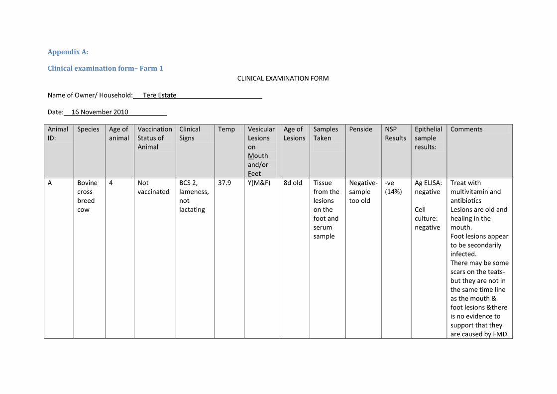
Appendix A:
Clinical examination form– Farm 1
CLINICAL EXAMINATION FORM
Name of Owner/ Household: Tere Estate
Date: 16 November 2010
Animal ID:
Species Age of animal
Vaccination Status of Animal
Clinical Signs
Temp Vesicular Lesions on Mouth and/or Feet
Age of Lesions
Samples Taken
Penside NSP Results
Epithelial sample results:
Comments
A
Bovine cross breed cow
4 Not vaccinated
BCS 2, lameness, not lactating
37.9 Y(M&F) 8d old Tissue from the lesions on the foot and serum sample
Negative- sample too old
-ve (14%)
Ag ELISA: negative Cell culture: negative
Treat with multivitamin and antibiotics Lesions are old and healing in the mouth. Foot lesions appear to be secondarily infected. There may be some scars on the teats- but they are not in the same time line as the mouth & foot lesions &there is no evidence to support that they are caused by FMD.

B
Bovine cross breed bull
2 Not vaccinated
BCS 3.5 38.1 Y (M) 8d Plus Serum sample
Not done + (79%) Small lesions filling up already and healing
C
Bovine cross breed calf
18mo Not vaccinated
BCS 3 Lame and salivating
38.9 Y(M&F) 7d mouth 7 d foot
Serum sample, probang ++
Not done + (74%) 2 notable lesions on the tongue, one small on the tip and one large on the dorsum of the tongue, concurrent joint effusion hind limb at the level of the hock
D
Bovine cross breed calf
1yoa Not vaccinated
BCS 2 39.4 Y (F&M) 10d plus
Serum sample
Not done + (79%) Healing lesions in the mouth, this animal has a history of eating plastic packets and appears to have an impacted rumen.

Clinical examination form – Farm 2
CLINICAL EXAMINATION FORM
Name of Owner/ Household: Solai Arus
Date: 16 November 2010
Animal ID:
Species Age of animal
Vaccination Status of Animal
Clinical Signs
Temp Vesicular Lesions on Mouth and/or Feet
Age of Lesions
Samples Taken
Penside NSP Results
Epithelial tissue results
Comments
E (Pembe)
Bovine cross breed cow
3 years
serotypes O, SAT1 and SAT2 on the 11th September 2010
BCS 2.5 Salivation, nasal discharge, painful, protruding tongue
41.5 Y(M) D1 Tissue from the lesions in the mouth serum sample
Positive result
+ (75%)
AgELISA: positive Cell culture: positive
On exam, the tongue has a blister ruptured leaving the most distal part of the tongue without epidermis. The owner reports that this animal is gravid.
F
Bovine cross
4 years
serotypes O, SAT1
BCS 3.5, salivating,
40.5 Y (M) D1 Serum sample Probang
Not done
+ (79%)
AgELISA: Positive
Lesions present on

(Chetakampuni)
breed bull
and SAT2 on the 11th September 2010
lame sample of oropharyngeal fluid Tissue sample from the mouth
Cell culture: positive
the dental pad Fluid aspirated from the vesicle and tissue samples collected from the the region.

Clinical examination form – Farm 3
CLINICAL EXAMINATION FORM
Name of Owner/ Household: Eden Farms
Date: 17 November /11/2010
Animal ID:
Species Age of animal
Vaccination Status of Animal
Clinical Signs
Temp Vesicular Lesions on Mouth and/or Feet
Age of Lesions
Samples Taken
Penside NSP Results
Epithelium Test Results
Comments
1
Bovine, Holstein cross Cow
Older than 5 years old
Serotypes O, A and SAT1 about 1 year ago
No lesions 40.94 No lesions
Serum Not done
++ (83%)
2
Bovine Holstein cross bull
3 yoa Unknown BCS 3 Salivation, nasal discharge
40.39 Y (M) D3 Serum sample Epithelial sample
positive -ve (44%)
AgELISA: positive Cell Culture: positive
Mouth treated with sodium bicarbonate yesterday – lateral flow device penside test still positive. Lesions on the tongue are about 1 day old and those on the dental pad about 3d old

3
Bovine Holstein cross breed cow
Serotypes O, A and SAT1 about 1 year ago
BCS 2 Nasal discharge
39.36 Y(M) D5 Epithelium negative ++ (87%)
AgELISA: positive Cell culture: positive
Lesion present on the dental pad granulating lesion with fibrin still present
4
Bovine Holstein cross cow
Serotypes O, A and SAT1 about 1 year ago
39.1 Y(M) D7+ Serum Orophangeal fluid collected with probang
Not done
+ (64%)
It is reported by the farmer that this animal was the first affected and started with clinical signs on the 15th of November
5 Bovine Holstein cross calf
8 months
Unknown Salivating, limping
40.3 Y(M&F) D5 Serum, Epithelial tissue from the mouth lesions (used for Penside only)
positive + (59%)
Lesions on the upper lip, the dental pad and the tip of the tongue, fibrin filled and starting to granulate. Granulating foot lesions between the cleats.
6
Dorper cross ovine, ram
Not vaccinated
Nasal discharge, limping
NT No lesions
Serum Not done
+ (59%)
Abscess on the foot, no mouth lesions
7
Dorper cross ovine, ewe
Not vaccinated
Nasal discharge
NT Y (M) D10+ Serum Not done
-ve (37%)
No foot lesions noted. The mouth lesions are present on

the dorsum of the tongue, with no lesions on the dental pad (expected location for sheep)
8
Dorper cross ovine ewe
Not vaccinated
Nasal discharge
NT No lesions
Serum Not done
-ve (42%)
No lesions on the feet or in the mouth

Appendix B:
Overview map of farms visited (Arus Farm, Tere Farm and Eden Farm). Kipkelion is where the truck which obtained heifers from Farm 3 originated. The
heifers were delivered in Kerichu. Outbreaks have been confirmed as serotype O in Kerichu many weeks prior to the outbreak on Farm 3.

Appendix C:
People to interview
District official veterinarian farmer of outbreak farm animal technician (animal health assistance) of this village – reported outbreak private livestock officer History
FMD (last year and last outbreak) what serotypes? Levels: Province, District, village, Farm
Vaccination (incl. what serotypes?)
Routine vaccination: Outbreak vaccination:
Levels: District, village, Farm Animal movements
- In and out a) Levels: District(in the last 3 months), village (in the last month), Farm(in the last 3 weeks,
depending on age of lesions)
Map
number of farms incl. number of susceptible species in the 1km zone
A) number of farms incl. number of susceptible species in the 3km zone
B) number of farms incl. number of susceptible species in the 10km zone
C) markets, coming water places, common grazing, common dippings
D) wildlife (national parks)?
Case history:
Report from clinical team How many animals (species, age distribution) on farm, date of first clinical signs (what kind) Source of feed (for the 1 week before day of most likely introduction) Other farmers with ill animals, when? Shared equipment with other farmers Animal products traded Biosecurity measures on farm (in general and after outbreak, especially vermins) Other animals on farm and are they moving? Where? Contact to wildlife (how often, what species)? Personnel movements (especially those who have direct contact to animals) – 2 weeks before first clinical sign
- In and out Animal products (milk, meat, by-products (incl. dead animals)) – 2 weeks before first clinical signs
- In and out


infection timeline -53 -52 -51 -50 -49 -48 -47 -46 -45 -44 -43 -42 -41 -40 -39 -38 -37 -36 -35 -34 -33 -32 -31 -30 -29 -28 -27 -26 -25 -24 -23 -22 -21 -20 -19 -18 -17 -16 -15 -14 -13 -12 -11 -10 -9 -8 -7 -6 -5 -4 -3 -2 -1 0 1 2 3 4 5 6 7 8 9 10 11 12 13 14 15 16 17 18 19
date (Sep/Oct/Nov) 11 12 13 14 15 16 17 18 19 20 21 22 23 24 25 26 27 28 29 30 31 1 2 3 4 5 6 7 8 9 10 11 12 13 14 15 16 17 18 19 20 21 22 23 24 25 26 27 28 29 30 31 1 2 3 4 5 6 7 8 9 10 11 12 13 14 15 16 17 18 19 20 21
most likely time of introduction
possible time of introduction
highest period of shedding
reported infection in neighbouring farms
first clinical signs noticed by farmer
EuFMD team visitvaccinated against serotypes O, SAT1 and SAT2
expect sufficient antibody response to provide immunity

ESTIMATING AGE OF
LESIONS

© Nick Juleff, IAH


PROTOCOL FOR
PROTECTIVE CLOTHING

DIRTY
CLEAN
CLEAN
DIRTY
15. Tape outer glove to sleeve of disposable suit
DIRTY
CLEAN
PPE
EquipmentWaste
Equipment
Waste
11. Pull down legs of waterproof suit outside boots
16. Pull down sleeves of waterproof suit outside gloves
17. Carry equipment onto site
18. When on site, do not remove mobile phone from zip-lock bag
12. Put on boot covers 13. Put on two pairs of disposable gloves
14. Pull sleeve of disposable suit down between the two pairs of gloves
7. Prepare clean & dirty disinfection points at hygiene barrier 8. Put on disposable suit 9. Put on waterproof suit 10. Put on boots – putting legs of
disposable suit inside boots
4. Place mobile phone in zip-lock bag
5. Carry equipment to site in plastic bag
6. Identify suitable location for clean/dirty separation
1. Prepare equipment & PPE (protective clothing) before travelling
3. Remove watch & jewellery before leaving car
2. Prepare “clean” & “dirty” areas in car
PROTOCOL FOR PUTTING ON PROTECTIVE CLOTHING (FMD)
Equipment
HYGIENE BARRIER
NDCC April 2009

5. Clean soles of boots thoroughly & disinfect
10. Remove second outer glove by hooking thumb under cuff - place in waste bag
6. Disinfect remainder of waterproof suit
7. Remove waterproof suit 8. Place suit in PPE bag on “clean” side
12. Remove disposable suit & place in waste bag on “dirty” side
11. Step onto clean side & disinfect boots
16. Disinfect outside of bucket
9. Remove first outer glove –touching only the outer surface
20. Place equipment & PPE bags in “dirty” area of car
19. Pour disinfect over plastic mat & place mat in waste bag
18. Wipe face with wet-wipes17. Disinfect hands using
6-step procedure
13. Remove first inner glove –touching only the outer surface
15. Double bag & seal the equipment & PPE bags
PROTOCOL FOR TAKING OFF PROTECTIVE CLOTHING (FMD)
1. Disinfect equipment & outside of sample boxes/bags
2. Place equipment & samples in equipment bag on clean side
3. Remove boot covers & place in waste bag for disposal
4. Disinfect legs of waterproof suit
14. Remove second inner glove by hooking thumb under cuff - place in waste bag
Equipment
PPE
PPEPPE
PPE
Waste
Waste Waste
Waste
Equipment & PPE
Waste
Waste
Waste
PPEPPE
Equipment
Waste
PPE
NDCC April 2009

CLINICAL EXAMINATION
FORM

Clinical Examination Form (can be adapted)
Name of the Owner/Household: ................ Farm ID: ……………. Epidemiological unit:
Date:
Animal ID Species Type L/S/M/PD/A Temp. Vesicular Lesions on Mouth and
Feet Y/N
Age of Lesions
Samples Tissue/Blood/Serum
Number of Photos
Comments
if no eartag, give order of animals examined..No. 1, 2 etc
bov. calf - n.m. Y (M + F) > 7d PA 130073, PA 130077
L: … Lameness, S: … Salivation, M: … Drop of Milk Yield, PD: … Perinatal Death, A: ……Abortion
Estimated age of oldest lesion seen:


…My Notes…