foot amputation by the moche of ancient peru: osteological ... · along with the moche culture in...
TRANSCRIPT

International Journal of OsteoarchaeologyInt. J. Osteoarchaeol. 10: 177–188 (2000)
Foot Amputation by the Moche of AncientPeru: Osteological Evidence andArchaeological ContextJOHN W. VERANOa,*, LAUREL S. ANDERSONa AND REGULO FRANCOb
a Department of Anthropology, Tulane University, 1021 Audubon St., New Orleans,LA 70118, USAb Fundacion Augusto N. Wiese, Jiron Carabaya No. 501, Lima, Peru
ABSTRACT Three probable cases of foot amputation, with healing, in skeletal remains associated with theMoche culture (AD 100–750) of northern coastal Peru are described. Each case exhibitsnon-functional tibio-talar joints with proliferative bone occupying the normal joint space. Therobusticity of the tibiae and fibulae suggest renewed weight-bearing and mobility followingrecovery. The osteological evidence is consistent with details shown in Moche ceramicdepictions of footless individuals. A footless Moche skeleton with wooden prostheses, describedin 1913 by Peruvian physician Velez Lopez, appears to represent a fourth example of thisprocedure. The Moche surgical approach was similar to a technique that would be pioneered inwestern medicine by the Scottish surgeon Sir James Syme some 1500 years later. Copyright© 2000 John Wiley & Sons, Ltd.
Key words: amputation; Moche; palaeopathology; Peru; surgery; Syme amputation
Introduction
Amputation and trephination are the two mostcommon forms of ancient surgery described inthe palaeopathological literature (Steinbock,1976; Ortner and Putschar, 1981). Trephinedskulls are known from many parts of the world,and there is no question that the practice hasconsiderable antiquity in both the Old Worldand the Americas (Aufderheide and Rodrıguez-Martin, 1998). Evidence for the practice ofamputation in prehistoric times is less convinc-ing. Bloom et al. (1995), Mays (1996), andAufderheide and Rodrıguez-Martin (1998)provide recent reviews of possible cases of am-putation in the archaeological record, emphasiz-ing some of the problems in diagnosis. In theOld World, written descriptions of amputationdate back as early as the sixth century BC, andthe practice is well-described by Celsus and
other Roman physicians of the first century AD
(Meade, 1968). Evidence that amputations wereperformed in the pre-contact New World ismore illusory, as it is based strictly on archaeo-logical evidence. Possible examples of amputa-tion have been reported in skeletal remains fromNorth, Central, and South America (Morse,1969; Hurtado, 1970; Saul, 1972; Friedmann,1973; Stewart, 1974; Merbs, 1989), althoughsome of these probably represent non-union offractures rather than amputation (Stewart,1974). Pre-hispanic artistic depictions of indi-viduals missing feet, hands, or entire limbs havealso been used to suggest that amputations wereperformed in the Americas before Europeancontact (Tello, 1924; Donnan, 1978; Urteaga-Ballon, 1991).
This report describes three recently-discov-ered cases of what appears to be surgical ampu-tation of the foot by disarticulation of the anklejoint in skeletal remains associated with theMoche culture (AD 100–750) of northerncoastal Peru. The remains were recovered in
* Correspondence to: Department of Anthropology, Tulane Uni-versity, 1021 Audubon St., New Orleans, LA 70118, USA. Tel.:+1 504 8623049; fax: +1 504 8655338; e-mail: [email protected]
Copyright © 2000 John Wiley & Sons, Ltd. Received 6 April 1999Revised 10 November 1999
Accepted 14 November 1999

J.W. Verano et al.178
1995 and 1998 at the archaeological sites of ElBrujo and Mocollope in the lower Chicama rivervalley (Figure 1). The bony response in eachcase is remarkably similar, and seems to reflect acommon pattern of healing followed by re-newed adaptation to weight-bearing andlocomotion.
Case descriptions
Cases I and II were excavated at the archaeolog-ical complex of El Brujo, which has been thefocus of a major Peruvian archaeological projectco-directed by archaeologists from the Univer-sity of Trujillo, National Institute of Culture,and the Augusto N. Wiese Foundation of Perusince 1991 (Franco et al., 1994, 1996). One ofthe authors (RFJ) is a co-director of the El Brujoproject and supervises the field research. An-other author (JWV) has been an affiliated re-searcher with the El Brujo Project since 1995,directing the analysis of human remains recov-ered from excavations. Case III was discovered
and collected by Thomas Wake, a zooarchaeol-ogist for the Mocollope Archaeological Project,directed by Glenn Russell of the Institute ofArchaeology, University of California, Los An-geles. Analyses of the human remains describedin this report were conducted by JWV and theother author (LSA).
Case I
El Brujo Complex, West Sector
Tomb 4
Tomb 4, excavated in 1995 from a unit on thewest side of one of the two major adobe plat-forms at El Brujo (Huaca Cao), contained thecomplete skeleton of an adult male, with theexception of the bones of the feet. The burialwas one of four individuals—two adult and twoadolescent males—found in simple graves
Figure 2. Articulated leg bones of Case I (anterior view).Figure 1. Map of the north coast of Peru.
Copyright © 2000 John Wiley & Sons, Ltd. Int. J. Osteoarchaeol. 10: 177–188 (2000)

Moche Foot Amputation 179
lar surface, as well as cranial suture closure(following guidelines presented in Buikstra andUbelaker, 1994) suggest an age at death ofapproximately 35–39 years. Male sex is indi-cated by the morphology of the os coxae andskull.
All foot bones are absent. The distal tibiaeand fibulae show non-functional joint surfaces,
Figure 3. Distal ends of tibiae and fibulae, Case I (anterioroblique view).
Figure 5. Left tibia and fibula, Case II (posterior view).
placed in architectural fill below an intact layerof adobe bricks. Few grave offerings were foundwith these individuals, suggesting that theywere of relatively low status, despite their loca-tion close to the platform. No offerings werefound in Tomb 4 (Franco et al., 1995).
Multiple skeletal age indicators, includingmorphology of the pubic symphysis and auricu-
Figure 4. Radiograph of tibiae and fibulae, Case I.
Copyright © 2000 John Wiley & Sons, Ltd. Int. J. Osteoarchaeol. 10: 177–188 (2000)
J.W. Verano et al.180
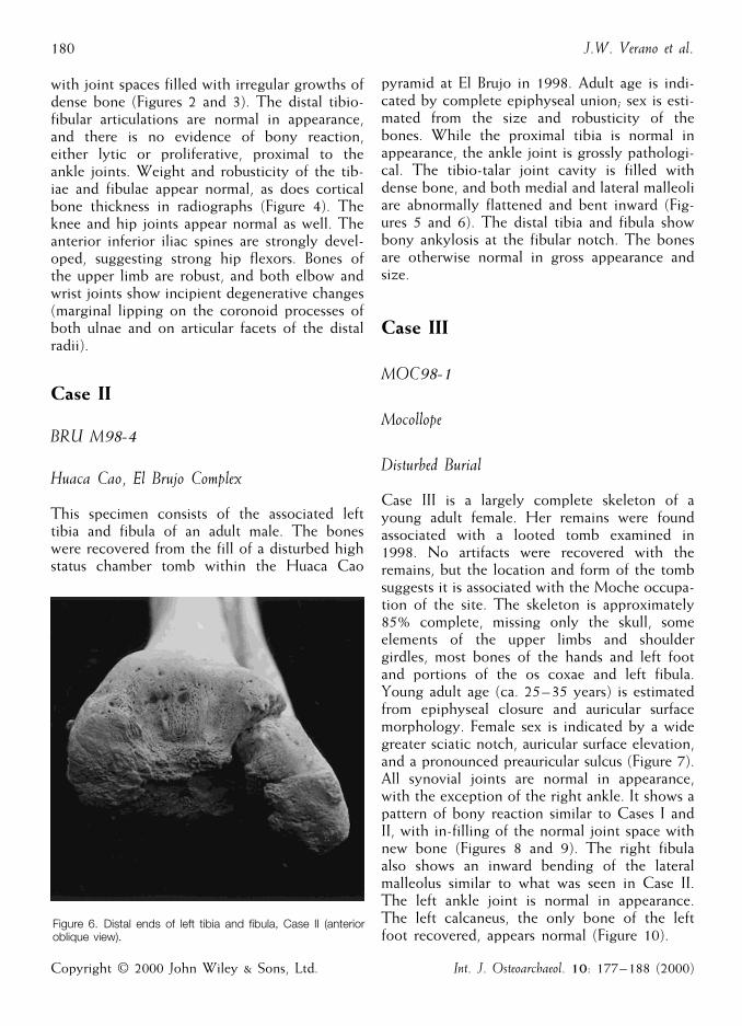
pyramid at El Brujo in 1998. Adult age is indi-cated by complete epiphyseal union; sex is esti-mated from the size and robusticity of thebones. While the proximal tibia is normal inappearance, the ankle joint is grossly pathologi-cal. The tibio-talar joint cavity is filled withdense bone, and both medial and lateral malleoliare abnormally flattened and bent inward (Fig-ures 5 and 6). The distal tibia and fibula showbony ankylosis at the fibular notch. The bonesare otherwise normal in gross appearance andsize.
Case III
MOC98-1
Mocollope
Disturbed Burial
Case III is a largely complete skeleton of ayoung adult female. Her remains were foundassociated with a looted tomb examined in1998. No artifacts were recovered with theremains, but the location and form of the tombsuggests it is associated with the Moche occupa-tion of the site. The skeleton is approximately85% complete, missing only the skull, someelements of the upper limbs and shouldergirdles, most bones of the hands and left footand portions of the os coxae and left fibula.Young adult age (ca. 25–35 years) is estimatedfrom epiphyseal closure and auricular surfacemorphology. Female sex is indicated by a widegreater sciatic notch, auricular surface elevation,and a pronounced preauricular sulcus (Figure 7).All synovial joints are normal in appearance,with the exception of the right ankle. It shows apattern of bony reaction similar to Cases I andII, with in-filling of the normal joint space withnew bone (Figures 8 and 9). The right fibulaalso shows an inward bending of the lateralmalleolus similar to what was seen in Case II.The left ankle joint is normal in appearance.The left calcaneus, the only bone of the leftfoot recovered, appears normal (Figure 10).
with joint spaces filled with irregular growths ofdense bone (Figures 2 and 3). The distal tibio-fibular articulations are normal in appearance,and there is no evidence of bony reaction,either lytic or proliferative, proximal to theankle joints. Weight and robusticity of the tib-iae and fibulae appear normal, as does corticalbone thickness in radiographs (Figure 4). Theknee and hip joints appear normal as well. Theanterior inferior iliac spines are strongly devel-oped, suggesting strong hip flexors. Bones ofthe upper limb are robust, and both elbow andwrist joints show incipient degenerative changes(marginal lipping on the coronoid processes ofboth ulnae and on articular facets of the distalradii).
Case II
BRU M98-4
Huaca Cao, El Brujo Complex
This specimen consists of the associated lefttibia and fibula of an adult male. The boneswere recovered from the fill of a disturbed highstatus chamber tomb within the Huaca Cao
Figure 6. Distal ends of left tibia and fibula, Case II (anterioroblique view).
Copyright © 2000 John Wiley & Sons, Ltd. Int. J. Osteoarchaeol. 10: 177–188 (2000)

Moche Foot Amputation 181
Figure 7. Bones of the pelvic area of Case III, showing age and sex indicators.
Patterning of osseous reaction
The three cases described above show individ-ual differences, but strong similarities in theoverall patterning of bony reaction in the anklejoint(s). The reaction is one of bony prolifera-tion limited to the tibio-talar joint space, withno involvement of the metaphyses or diaphysesof the tibia or fibula. The proliferative boneappears well organized and remodelled in allcases. In one example (Case I) both ankles areinvolved; in another (Case III) only one ankle isaffected; laterality in Case II cannot be deter-mined, as only an isolated tibia and fibula wererecovered. In all three individuals, the relativelynormal size, weight, and robusticity of the af-fected bones suggests that weight was placed onthe distal ends of the affected limbs followinghealing. Weight bearing is further indicated bythe plastic deformation of the medial and lateralmalleoli seen in Cases II and III.
Differential diagnosis
The bilateral symmetry demonstrated by Case Iand the lack of any indication of infection orinflammation of the long bone metaphyses or
diaphyses argues against loss of the feet as adirect result of infection—although surgical re-moval of a diseased or traumatized foot is apossibility. Congenital absence of the feet isunlikely, since this is not a reported congenitaldefect. Moreover, major developmental malfor-mation of the foot or ankle would be expectedto lead to changes in the robusticity and mor-phology of the tibia and fibula—something thatis not seen in these cases. What best fits theobserved pattern of bony changes, in our opin-ion, is intentional amputation of the footthrough disarticulation of the ankle joint, fol-lowed by healing and renewed weight-bearingon the affected limb. This scenario draws sup-port from details shown in representations offootless individuals in Moche art.
Amputation in Moche art
Moche art is well known for ceramic vesselsdepicting individuals with physical defects andpathological conditions (Donnan, 1978). Indi-viduals with missing limbs, hands, and feet arecommonly represented in this group (Weiss,1984; Urteaga-Ballon, 1991). In a review ofpublished collections we have found nearly 100
Copyright © 2000 John Wiley & Sons, Ltd. Int. J. Osteoarchaeol. 10: 177–188 (2000)

J.W. Verano et al.182
sel depicting a footless individual. Of particularinterest is a readily visible median groove on theend of each leg stump. Such grooves are com-monly shown in Moche depictions of footlessindividuals (see also Figure 12), and appear tomark a depressed area in between the lateral andmedial malleoli. Such an anatomical featurewould be expected if a foot were amputated bydisarticulation at the ankle, leaving the malleolias marginal protuberances.
Many individuals with missing feet are shownwith cup-like objects placed over the terminalends of leg stumps, or being held in the hand asif in the act of placing or removing the object(Figure 12). These appear to be prostheses de-signed to permit weight-bearing and locomotionfollowing loss of the foot. Some Moche ceram-ics show individuals wearing the objects andstanding upright (Urteaga-Ballon, 1991).
There has been much speculation about whatthe Moche were communicating in their depic-tion of amputees. Most scholars suggest thatMoche amputations were a form of punishmentrather than an attempt to treat infection orother disease (Velez Lopez, 1913; Tello, 1924;Urteaga-Ballon, 1991). Support for this argu-ment comes from the observation that manyMoche representations of individuals with miss-ing limbs also show what appears to be inten-tional mutilation of the nose and lips. Indeed, inthe ceramic sample we examined, nose and lipdeformities are visible in 63% of amputees(Table 1; Figure 11). Not all cases of lip andnose deformities in Moche art can be attributedto intentional mutilation, however. Some appearto illustrate congenital defects like cleft lip,while others show lesions more suggestive of aninfectious disease such as mucocutaneous leish-maniasis, which is endemic in portions of Perutoday (Urteaga-Ballon, 1991). The relationshipbetween missing limbs and facial deformities inMoche art thus is not a simple one. Leprosy,which can affect the oral and nasal mucosadirectly, and the hands and feet secondarily (asa result of trauma and secondary infection),does not appear to have been present in theNew World before European contact (Stein-bock, 1976; Ortner and Putschar, 1981; Auf-derheide and Rodrıguez-Martin, 1998), andLeishmaniasis does not affect the hands or feet.
Figure 8. Right tibia and fibula compared with left tibia, CaseIII (anterior view).
ceramic vessels showing persons with missinglimbs or extremities. The most common areindividuals missing both feet (\50%), followedby those missing a single foot (26%). Lesscommon are those missing arms, arms distal tothe elbow, and hands (Table 1). Figure 11 is arepresentative example of a Moche ceramic ves-
Copyright © 2000 John Wiley & Sons, Ltd. Int. J. Osteoarchaeol. 10: 177–188 (2000)

Moche Foot Amputation 183
Figure 9. Comparison of right and left ankle joints, Case III (anterior oblique view).
Frostbite damage to the feet as a motive foramputation has been suggested to us, althoughthis seems unlikely in a coastal Peruvian culturesuch as the Moche.
All Moche artistic representations of am-putees that we have examined appear to bemales, although identifying gender in Moche artis difficult in some cases (Lyon, 1978). ObviousMoche gender signifiers such as prominentbreasts or braided hair were not observed, how-ever. It is significant, therefore, that Case III isof female sex, indicating that amputations wereperformed on women as well as men.
Amputees in Moche art are commonly shownwearing a head cloth and a tunic; a few have earspools or necklaces, but none wear elaborateheaddresses or other signifiers of high rank.Most individuals are shown seated in a cross-legged position, kneeling, or lying prone; someare depicted standing (usually holding a longstick for support) or seated on the back of acamelid (probably a llama). Some individuals areshown playing a drum, while others have anoutstretched hand as if begging (Figure 11).
Examples are also known in Moche art ofskeletal figures wearing a prosthesis over a miss-ing foot in scenes that appear to represent aprocession or dance. One recent study of theiconography of Moche footless individuals hy-pothesizes that amputation may have been a
form of ritual mutilation that marked certainindividuals as special attendants to the nobilityin Moche society, and in the afterlife as well(Arsenault, 1993). Whatever the function ormeaning of amputation in Moche society, untilrecently there was little skeletal evidence tosuggest that surgical amputation was actuallyperformed.
Figure 10. Right and left lower leg bones with left calcaneus,Case III.
Copyright © 2000 John Wiley & Sons, Ltd. Int. J. Osteoarchaeol. 10: 177–188 (2000)

J.W. Verano et al.184
Table 1. Frequency of missing feet, hands, and other bodyparts in a sample of 99 Moche ceramic vessels showingapparent amputation or mutilation
Number ofBody part missingcases
26Single foot55Both feet
Arm 26Both arms6Forearm and hand
Both forearms 50One hand2Both hands
Both hands and both feet 1
Individuals with prostheses 2462Individuals with facial mutilation
Previous osteological evidence for
amputation in pre-hispanic Peru
In 1913, Peruvian physician Velez Lopez pub-lished a description of a footless Moche skele-ton found at the site of Mocollope in theChicama River Valley (Velez Lopez, 1913). Theskeleton was found with wooden prosthesesover the distal ends of the tibiae and fibulae.Based on his examination of the material, heconcluded that the skeleton was a double am-putee who had used the prostheses for someperiod of time, as evidenced by wear on theirinferior surfaces. He described these objects as
Figure 11. Moche ceramic vessel showing an individual with missing feet and mutilation of the nose and lips. Courtesy of theSan Diego Museum of Man (Catalog c 1981-14-17).
Copyright © 2000 John Wiley & Sons, Ltd. Int. J. Osteoarchaeol. 10: 177–188 (2000)

Moche Foot Amputation 185
Figure 12. Moche ceramic vessel showing an individual with acup-like prosthesis. Courtesy of the American Museum ofNatural History (Catalog c B4919).
Hand and foot bones are notorious for beinglost in archaeological excavations, as anyonewho has worked with museum collections canattest. Cut marks (indicating perimortem re-moval) or bony reaction to antemortem trauma(as in Cases I–III described above) should bepresent if a hand or foot was in fact amputated.Several cases of Moche burials with either ‘extra’parts or ‘missing’ parts illustrate the problem ofconfident identification. Christopher Donnanand Carol Mackey described an adult maleMoche burial from Huanchaco in the MocheRiver Valley that included extra articulatedhands as grave offerings (Donnan and Mackey,1978). A female skeleton buried in a nearbytomb was missing both hands. Donnan andMackey suggest that the hands may have beenremoved from the occupant of one tomb andplaced with the other. Unfortunately, neitherhand nor forearm bones were examined for cutmarks, the presence of which might have re-solved the question.
Several retainer burials recently excavatedfrom royal Moche tombs at Sipan in the Lam-bayeque River Valley present similar problems.The skeletons of two young adult males, inter-preted as ‘guards’ for the tombs (Alva and Don-nan, 1993) lacked bones of the feet. One of us(JWV) examined these skeletons, but found thatthe preservation of the distal ends of tibiae andfibulae was too poor to identify cut marks orother features that might confirm amputation(Verano, 1997). Two articulated human feetwere found in an adjacent room, however, indi-cating that some activity involving dismember-ment was going on at the site (Alva andDonnan, 1993). If these feet belonged to one ofthe footless guards, this suggests some activityrelated to mortuary ritual, since both individualswere sacrificed to accompany a high status indi-vidual. Unfortunately, the ankle bones werefragmentary and cut marks were not identified.
Another example of a possible healed ampu-tation from Peru is an isolated proximal humeruscollected by Ales Hrdlicka in 1913 from adisturbed communal tomb near Huarochiri inthe central highlands. The bone terminates atthe proximal third of the diaphysis, and showsobvious healing in the form of closure of themedullary cavity and smoothing of the distal
simple wooden cups padded with wool. Thetibiae were reported to be normal in appear-ance, with no evidence of swelling, infection, orother pathology. Velez Lopez concluded thatthe feet were not lost as a result of disease, butwere intentionally amputated—probably as aform of punishment. Unfortunately, VelezLopez did not publish photographs of the skele-ton or prostheses. The material appears to havebeen in a private collection, and its presentwhereabouts are unknown. Nevertheless, theskeleton described by Velez Lopez is of greatinterest because it appears to present a casesimilar to those described in this report. More-over, it was reportedly excavated at the archaeo-logical site of Mocollope, where Case III (thisreport) was found in July of 1998.
While other possible cases of amputation inpre-hispanic Peruvian skeletal remains havebeen described, they are more problematic.
Copyright © 2000 John Wiley & Sons, Ltd. Int. J. Osteoarchaeol. 10: 177–188 (2000)

J.W. Verano et al.186
skeletal elements makes this case problematic, asdoes the issue of surgical technique. It is diffi-cult for us to imagine sectioning a humerusthrough the diaphysis without using a saw, atool that was unknown in Peru prior to Eu-ropean contact. While repeated grooving with asharp knife could theoretically cut through along bone diaphysis, the degree of angulation ofthe terminal end (visible in Figure 13) alsoseems inappropriate if the bone was intention-ally sectioned. In our opinion, fracture withnon-union seems a more likely explanation inthis case.
Moche foot amputation: surgicalapproach
While metal saws date back to before the eighthcentury BC in the Old World (Symes et al.,1998), they were unknown in the Americasprior to European contact. Thus, trans-diaphyseal amputations of limbs would not beexpected to be found in the New World priorto the European introduction of metal saws. Theamputation of a hand or foot by disarticulationat the wrist or ankle joint, however, could bedone with simple cutting tools. In 1842, Scot-tish surgeon Sir James Syme popularized a tech-nique for amputating feet by disarticulation atthe ankle joint, demonstrating that it providedsuperior results to traditional above-ankle trans-tibial amputations in terms of healing and pa-tient mobility (Wagner, 1992; Wilson, 1992).While in the classic Syme procedure the medialand lateral malleoli are subsequently sawed offto provide a more stable support surface for thepatient, the foot itself is removed by surgicaldisarticulation of the tibio-talar joint using ascalpel. Skeletal evidence from Cases I–III pre-sented here, along with Moche artistic depic-tions of amputees showing the protrudingmalleoli of the tibia and fibula, suggests that theMoche developed a technique similar to theSyme method some 1500 years ago. Moreover,the degree of healing seen in Cases I–III indi-cates that they were able to perform this proce-dure successfully. The retention of the medialand lateral malleoli may have made standing
Figure 13. Proximal third of left humerus collected by Hrdlickanear Huarochiri, Peru (posterior aspect). Courtesy of the SanDiego Museum of Man (Catalog c 1915-2-668).
end (Figure 13). The specimen has been studiedand described by several researchers (Rogers,1973; Merbs, 1980), who agree that it may bean amputation, although a healed fracture withnon-union is considered an alternative diagno-sis. The lack of secure dating and associated
Copyright © 2000 John Wiley & Sons, Ltd. Int. J. Osteoarchaeol. 10: 177–188 (2000)

Moche Foot Amputation 187
and walking difficult even with prostheses likethose described by Velez Lopez, although re-cent clinical studies have demonstrated that insome cases a superior outcome can be achievedby performing a Syme ankle disarticulationwithout resection of the malleoli (Pavot, 1973).
Conclusions
We believe that the three cases presented hererepresent the first well-documented skeletal evi-dence that the Moche of ancient Peru per-formed successful amputations of the feet.Amputation by disarticulation of the ankle jointwould have been a simple and logical approach,given the tools available at the time. Osteologi-cal evidence for amputations of hands or armshas not been found to date, but Moche artsuggests that examples may exist. Why theMoche performed amputations will no doubtremain a subject of speculation. Artistic depic-tions of individuals with missing feet disappearalong with the Moche culture in the late eighthcentury AD. A few ceramic vessels showingindividuals with missing arms are known fromthe subsequent Lambayeque culture of northernPeru (ca. AD 900–1200), but these may simplyrepresent continuities of Moche artistic themes.The actual practice of amputation may havedisappeared with the Moche as well. Spanishchroniclers who observed Inca culture firsthandin the sixteenth century do not describe ampu-tation, nor have any unequivocal examples ofamputation been found in Inca period skeletalremains.
Acknowledgements
The authors are grateful to Thomas Wake andGlenn Russell of the Institute of Archaeology,University of California, Los Angeles, for per-mission to study the skeletal remains from Mo-collope, and to Alana Cordy-Collins and RoseTyson of the San Diego Museum of Man forpermission to photograph the ceramics and os-teological specimen in Figures 11–13. Figure 1is courtesy of Donald McClelland of the FowlerMuseum of Cultural History, Los Angeles. Fig-
ure 12 was photographed with permission of theAmerican Museum of Natural History, NewYork. Project funding for the El Brujo excava-tions is generously provided by the Augusto N.Wiese Foundation. The authors are particularlygrateful to the late Dr Guillermo Wiese deOsma, Chairman of the Wiese Foundation, forhis dedication to Moche archaeology and forhis friendship and wise counsel. The WieseFoundation and the research staff of the El BrujoArchaeological Project provided generous logis-tical and research support for JWV and LSAduring their fieldwork in Peru, for which theirare most grateful. Travel and research supportfor this study was provided by grants from theTulane University Committee on Research(JWV) and the Roger Thayer Stone Center forLatin American Studies at Tulane University(JWV, LSA).
References
Alva W, Donnan CB. 1993. Royal Tombs of Sipan.Fowler Museum of Natural History: Los Angeles,CA.
Arsenault D. 1993. El personaje del pie amputado enla cultura Mochica del Peru: un ensayo sobre laarqueologıa del poder. Latin American Antiquity 4:225–245.
Aufderheide AC, Rodrıguez-Martin C. 1998. TheCambridge Encyclopedia of Human Paleopathology. Cam-bridge University Press: Cambridge.
Bloom AI, Bloom RA, Kahila G, Eisenberg E, SmithP. 1995. Amputation of the hand in the 3600-year-old skeletal remains of an adult male: the firstcase reported from Israel. International Journal ofOsteoarchaeology 5: 188–191.
Buikstra JE, Ubelaker DH (eds). 1994. Standards forData Collection From Human Skeletal Remains. ArkansasArchaeological Survey: Fayetteville.
Donnan CB. 1978. Moche Art of Peru: Pre-ColumbianSymbolic Communication. Museum of Cultural His-tory: Los Angeles, CA.
Donnan CB, Mackey CJ. 1978. Ancient Burial Patternsof the Moche Valley, Peru. University of Texas Press:Austin, TX.
Franco R, Galvez C, Vasquez S. 1994. Arquitectura yDecoracion Mochica en La Huaca Cao Viejo,Complejo El Brujo: Resultados Preliminares. InMoche: Propuestas y Perspectivas, Actas del PrimerColoquio Sobre la Cultura Moche, Uceda S, Mujica E
Copyright © 2000 John Wiley & Sons, Ltd. Int. J. Osteoarchaeol. 10: 177–188 (2000)

J.W. Verano et al.188
(eds). Universidad Nacional de La Libertad: Tru-jillo, Peru; 147–180.
Franco R, Galvez C, Vasquez S. 1995. ProgramaArqueologico Complejo ‘El Brujo’, Informe 1995. Funda-cion A.N. Wiese: Lima.
Franco R, Galvez C, Vasquez S. 1996. Los Descu-brimientos Arqueologicos en la Huaca Cao ViejoComplejo ‘El Brujo’. Arkinka 1: 82–94.
Friedmann LW. 1973. Amputation in Pre-ColumbianAmerica. Archives of Physical Medicine and Rehabilita-tion 54: 323–325.
Hurtado ED. 1970. Pre-hispanic osteopathology.In Handbook of Middle American Indians, vol. 9, Stew-art TD (ed.). University of Texas: Austin, TX;68–81.
Lyon PJ. 1978. Female supernaturals in ancient Peru.Nawpa Pacha 16: 95–140.
Mays SA. 1996. Healed limb amputations in humanosteology and their causes: a case study fromIpswich, UK. International Journal of Osteoarchaelogy6: 101–113.
Meade RH. 1968. An Introduction to the History ofGeneral Surgery. W.B. Saunders: Philadelphia, PA.
Merbs CF. 1980. Pathologies from the Hrdlicka Collection,Slide Set II. San Diego Museum of Man: San Diego.
Merbs CF. 1989. Trauma. In Reconstruction of Life fromthe Skeleton, Iscan MY, Kennedy KAR (eds). Alan R.Liss: New York; 161–189.
Morse DF. 1969. Ancient Disease of the Midwest. IllinoisState Museum Reports of Investigations No. 15:Springfield.
Ortner DJ, Putschar WGJ. 1981. Identification ofPathological Conditions in Human Skeletal Remains,Smithsonian Contributions to Anthropology, No.28. Smithsonian Institution Press: Washington,DC.
Pavot AP. 1973. Ankle-disarticulation: a definitivetype of amputation in adults. Archives of PhysicalMedicine and Rehabilitation 54: 307–310.
Rogers SL. 1973. A case of surgical amputation fromaboriginal Peru. In Ethnic Technology Notes No. 11.San Diego Museum of Man: San Diego, CA.
Saul FP. 1972. The human skeletal remains of Altarde Sacrificios: an osteobiographic analysis. Papers
of the Peabody Museum of American Archaeology andEthnology, Harvard University 63: 1–123.
Steinbock RT. 1976. Paleopathological Diagnosis andInterpretation. Charles C. Thomas: Springfield.
Stewart TD. 1974. Nonunion of fractures in antiq-uity, with descriptions of five cases from the NewWorld involving the forearm. Bulletin of the NewYork Academy of Medicine 50: 875–891.
Symes SA, Berryman HE, Smith OC. 1998. Sawmarks in bone: introduction and examination ofresidual kerf contour. In Forensic Osteology, Advancesin the Identification of Human Remains (2nd edn),Reichs KJ (ed.). Charles C. Thomas: Springfield;389–409.
Tello JC. 1924. Arte antiguo peruano. Revista deEstudios Antropologicos, Lima. vol. II. In Catalogueof the Hrdlicka Paleopathology Collection, Tyson RA,Alcauskas ESD (eds). San Diego Museum of Man:San Diego.
Urteaga-Ballon O. 1991. Medical ceramic represen-tation of nasal Leishmaniasis and surgical amputa-tion in ancient Peruvian civilization. In HumanPaleopathology, Current Syntheses and Future Options,Ortner DJ, Aufderheide AC Jr. (eds). SmithsonianInstitution Press: Washington, DC; 95–101.
Velez Lopez LR. 1913. Las mutilaciones en los vasosantropomorfos del antiguo Peru. XVIII Session ofthe International Congress of Americanists: Lon-don; 267–275.
Verano JW. 1997. Human skeletal remains fromTomb I, Sipan (Lambayeque river valley, Peru);and their social implications. Antiquity 71: 670–682.
Wagner FW Jr. 1992. The Syme ankle disarticula-tion. In Atlas of Limb Prosthetics: Surgical, Prosthetic,and Rehabilitation Principles, Bowker JH, Michael JW(eds). Mosby Year Book: St. Louis, MO; 413–430.
Weiss P. 1984. Paleopatologıa Americana. Boletin deLima 33: 17–52.
Wilson AB. 1992. History of amputation surgery andprosthetics. In Atlas of Limb Prosthetics: Surgical, Pros-thetic, and Rehabilitation Principles, Bowker JH,Michael JW (eds). Mosby Year Book: St. Louis,MO; 3–15.
Copyright © 2000 John Wiley & Sons, Ltd. Int. J. Osteoarchaeol. 10: 177–188 (2000)