food safety testing: rapid molecular methods for chemical and
TRANSCRIPT
TURUN YLIOPISTON JULKAISUJAANNALES UNIVERSITATIS TURKUENSIS
SARJA - SER. A I OSA - TOM. 397
ASTRONOMICA - CHEMICA - PHYSICA - MATHEMATICA
TURUN YLIOPISTOTurku 2009
Food Safety Testing: Rapid Molecular Methods for
Chemical and Biological Hazards
by
Virve Hagren

From the Department of Biochemistry and Food Chemistry / Biotechnology University of Turku Turku, Finland Supervised by Mika Tuomola, Ph.D. Department of Biochemistry and Food Chemistry / Biotechnology University of Turku Turku, Finland and Professor Timo Lövgren, Ph.D. Department of Biochemistry and Food Chemistry / Biotechnology University of Turku Turku, Finland Reviewed by Geoff Barnard, Ph.D. Department of Veterinary Medicine University of Cambridge Cambridge, UK and Professor Hannu Korkeala, DVM, Ph.D., Ms.Soc.Sci. Department of Food and Environmental Hygiene University of Helsinki Helsinki, Finland Opponent Professor Aldert Anthonie Bergwerff, Ph.D. Department of Veterinary Public Health and Food Safety Ghent University Ghent, Belgium ISBN 978-951-29-3915-2 (PRINT) ISBN 978-951-29-3916-9 (PDF) ISSN 0082-7002 Painosalama Oy – Turku, Finland 2009

To my family

Contents
4
CONTENTS
CONTENTS ...................................................................................................................4
LIST OF ORIGINAL PUBLICATIONS ....................................................................6
ABBREVIATIONS........................................................................................................7
ABSTRACT ...................................................................................................................8
1 INTRODUCTION....................................................................................................9
2 REVIEW OF THE LITERATURE......................................................................10
2.1 VETERINARY RESIDUES AND FOOD SAFETY ...............................................10 2.1.1 Coccidiosis and anticoccidials.........................................................................10 2.1.2 Veterinary residue control in the EU...............................................................12 2.1.3 Methods for the detection of anticoccidial residues in food............................14 2.1.4 Future trends in residue control .......................................................................20
2.2 FOODBORNE PATHOGENS AND FOOD SAFETY ...........................................21 2.2.1 Salmonella spp. as a foodborne pathogen .......................................................21 2.2.2 Salmonella control in the EU ..........................................................................23 2.2.3 Methods for the detection of Salmonella in food ............................................23 2.2.4 Future trends in foodborne pathogen testing ...................................................33
3 AIMS OF THE STUDY.........................................................................................35
4 SUMMARY OF MATERIALS AND METHODS..............................................36
4.1 IMMUNOASSAYS.....................................................................................................36 4.1.1 Antibodies (I-III) .............................................................................................36 4.1.2 Preparation of labels (I-III)..............................................................................36 4.1.3 Dry chemistry wells (I, II) ...............................................................................37 4.1.4 Sample preparation (I-III)................................................................................38 4.1.5 Assay procedures (I-III) ..................................................................................38
4.2 PCR ASSAY ...............................................................................................................39 4.2.1 Oligonucleotides (IV, V) .................................................................................39 4.2.2 Labelling of probes (IV, V) .............................................................................40 4.2.3 Dry chemistry vessels (IV, V) .........................................................................41 4.2.4 Sample preparation (V) ...................................................................................41 4.2.5 Assay protocol (IV, V) ....................................................................................42 4.2.6 Reference method (V) .....................................................................................42
Contents
5
5 SUMMARY OF RESULTS AND DISCUSSION................................................43
5.1 IMMUNOASSAY CHARACTERISTICS ...............................................................43 5.1.1 Assay format (I-III) .........................................................................................43 5.1.2 Labels and time-resolved fluorescence detection (I-III)..................................44 5.1.3 Dry chemistry (I, II) ........................................................................................46 5.1.4 Sample preparation (I-III)................................................................................47 5.1.5 Assay performance (I-III)................................................................................48
5.2 PCR PLATFORM AND ASSAY CHARACTERISTICS.......................................50 5.2.1 Principle of the GenomEra platform (IV)........................................................50 5.2.2 Internal amplification control (IV, V) .............................................................51 5.2.3 Labels and time-resolved fluorescence detection (IV, V) ...............................52 5.2.4 Dry chemistry (IV, V) .....................................................................................53 5.2.5 Sample preparation (V) ...................................................................................54 5.2.6 Assay performance (V) ...................................................................................57
6 CONCLUSIONS.....................................................................................................58
ACKNOWLEDGEMENTS ........................................................................................60
REFERENCES ............................................................................................................62
ORIGINAL PUBLICATIONS ...................................................................................73

List of Original Publications
6
LIST OF ORIGINAL PUBLICATIONS
This thesis is based on the following original publications, referred to in the text by their Roman numerals (I-V): I Virve Hagren, Steven R. H. Crooks, Christopher T. Elliott, Timo Lövgren, and
Mika Tuomola (2004) An all-in-one dry chemistry immunoassay for the screening of coccidiostat nicarbazin in poultry eggs and liver. J Agric Food
Chem 52:2429-2433. II Virve Hagren, Lisa Connolly, Christopher T. Elliott, Timo Lövgren, and Mika
Tuomola (2005) Rapid screening method for halofuginone residues in poultry eggs and liver using time-resolved fluorometry combined with the all-in-one dry chemistry assay concept. Anal Chim Acta 529:21-25.
III Virve Hagren, Pekka Peippo, Mika Tuomola, and Timo Lövgren (2006) Rapid
time-resolved fluoroimmunoassay for the screening of monensin residues in eggs. Anal Chim Acta 557:164-168.
IV Virve Hagren, Piia von Lode, Anniina Syrjälä, Tero Soukka, Timo Lövgren,
Hannu Kojola, and Jussi Nurmi (2008) An automated PCR platform with homogeneous time-resolved fluorescence detection and dry chemistry assay kits. Anal Biochem 374:411-416.
V Virve Hagren, Piia von Lode, Anniina Syrjälä, Teemu Korpimäki, Mika
Tuomola, Otto Kauko, and Jussi Nurmi (2008) An 8-hour system for Salmonella detection with immunomagnetic separation and homogeneous time-resolved fluorescence PCR. Int J Food Microbiol 125:158-161.
In addition, some unpublished data are included. The original publications have been reproduced with the permission of the copyright holders.
Abbreviations
7
ABBREVIATIONS
B/B0 value calculated by dividing the signal of the sample/standard by the signal of the zero sample/standard
BPW buffered peptone water BSA bovine serum albumin CCα decision limit CCβ detection capability CFU colony forming unit DCC N,N'-dicyclohexylcarbodiimide DELFIA dissociation-enhanced lanthanide fluoroimmunoassay DNA deoxyribonucleic acid DNC 4,4'-dinitrocarbanilide EFSA European Food Safety Authority ELISA enzyme-linked immunosorbent assay EU European Union EVIRA Finnish Food Safety Authority FAO Food and Agriculture Organization of the United Nations HACCP Hazard Analysis and Critical Control Point HPLC high performance liquid chromatography IAC internal amplification control IgG immunoglobulin G IMS immunomagnetic separation ISO International Organization for Standardization LC liquid chromatography LOD limit of detection LOQ limit of quantification MRL maximum residue limit mRNA messenger RNA MS mass spectrometry NASBA nucleic acid sequence-based amplification NHS N-hydroxysuccinimide PCR polymerase chain reaction RNA ribonucleic acid rRNA ribosomal RNA RT room temperature RT-PCR reverse transcription PCR S/N signal-to-noise ratio SPE solid phase extraction SPR surface plasmon resonance TRF time-resolved fluorometry WHO World Health Organization

Abstract
8
ABSTRACT
The central goal of food safety policy in the European Union (EU) is to protect consumer health by guaranteeing a high level of food safety throughout the food chain. This goal can in part be achieved by testing foodstuffs for the presence of various chemical and biological hazards. The aim of this study was to facilitate food safety testing by providing rapid and user-friendly methods for the detection of particular food-related hazards. Heterogeneous competitive time-resolved fluoroimmunoassays were developed for the detection of selected veterinary residues, that is coccidiostat residues, in eggs and chicken liver. After a simplified sample preparation procedure, the immunoassays were performed either in manual format with dissociation-enhanced measurement or in automated format with pre-dried assay reagents and surface measurement. Although the assays were primarily designed for screening purposes providing only qualitative results, they could also be used in a quantitative mode. All the developed assays had good performance characteristics enabling reliable screening of samples at concentration levels required by the authorities. A novel polymerase chain reaction (PCR)-based assay system was developed for the detection of Salmonella spp. in food. The sample preparation included a short non-selective pre-enrichment step, after which the target cells were collected with immunomagnetic beads and applied to PCR reaction vessels containing all the reagents required for the assay in dry form. The homogeneous PCR assay was performed with a novel instrument platform, GenomEra™, and the qualitative assay results were automatically interpreted based on end-point time-resolved fluorescence measurements and cut-off values. The assay was validated using various food matrices spiked with sub-lethally injured Salmonella cells at levels of 1-10 colony forming units (CFU)/25 g of food. The main advantage of the system was the exceptionally short time to result; the entire process starting from the pre-enrichment and ending with the PCR result could be completed in eight hours. In conclusion, molecular methods using state-of-the-art assay techniques were developed for food safety testing. The combination of time-resolved fluorescence detection and ready-to-use reagents enabled sensitive assays easily amenable to automation. Consequently, together with the simplified sample preparation, these methods could prove to be applicable in routine testing.

Introduction
9
1 INTRODUCTION
Food safety has a major impact on public health, and therefore, food safety issues have gained considerable attention. The recent high profile food-related crises such as bovine spongiform encephalopathy, dioxin contamination outbreaks, and epidemics caused by foodborne pathogens have shaken consumer confidence in the ability of the food industry and the authorities to guarantee food safety. To respond to consumers concerns, the EU introduced an integrated "from farm to fork" strategy, which became the cornerstone of the EU’s food safety policy (Anonymous, 2000). The aim of this policy was to provide the same high level of health protection to consumers all over Europe. Food safety was designed to rest on the following pillars: reviewed and updated legislation, appropriate scientific advice for decision making, data collection and analysis, consumer information, and official enforcement and control measures covering all aspects of the food chain from raw materials to food consumption. The basis for food legislation was set in Regulation 178/2002, which presented the general principles and requirements of food law and provided the framework for other related legislation (Anonymous, 2002b). Following that, several steps were taken towards to improved food safety control. For example, the European Food Safety Authority (EFSA) was established, the aim of which is to scientifically assess and communicate on risks associated with the food chain (Anonymous, 2002b). Furthermore, the control of chemical and biological hazards in food, which was traditionally based on reactive and enforcement-orientated approach relying mainly on regulatory testing of end products, was updated with the introduction of the Hazard Analysis and Critical Control Point system (HACCP) (Anonymous, 2004a, 2004b). The application of HACCP provided a more effective and comprehensive control approach by relying on the prevention of hazards and it also placed more responsibility for food hygiene and safety on food operators. In addition, the introduction of a harmonized approach for official controls which are performed by the authorities to verify the compliance with feed and food law, animal health, and animal welfare rules, further strengthened the realization of the food safety policy (Anonymous, 2004c, 2004d). Altogether, several important steps have been taken at the legislative level to improve food safety. Nevertheless, due to globalization of food production and distribution, food-related hazards can spread fast in the food chain. To confront this challenge, combined and continuous efforts, including the development and implementation of novel methods of analysis for food control, are required to achieve a high level of food safety now and in the future.
Review of the Literature
10
2 REVIEW OF THE LITERATURE
The Food and Agriculture Organization of the United Nations (FAO) and the World Health Organization (WHO) define food control as "… a mandatory regulatory activity of enforcement by national or local authorities to provide consumer protection and ensure that all foods during production, handling, storage, processing, and distribution are safe, wholesome and fit for human consumption; conform to quality and safety requirements; and are honestly and accurately labelled as prescribed by law." (FAO/WHO, 2003). Food is considered to be unsafe if it is injurious to health or unfit for human consumption (Anonymous, 2002b). The potential sources of hazards that can compromise food safety include biological, chemical, and physical agents. The hazards can enter the food chain, e.g. due to improper agricultural practices, poor hygiene, misuse of chemicals, lack of preventive controls in food processing and preparation, improper storage, and contaminated raw materials, ingredients, and water (FAO/WHO, 2003). This literature review focuses on selected chemical and biological hazards present in food, namely a group of veterinary residues and a particular foodborne pathogen, respectively, and introduces control activities and methods that are used in food safety testing to detect these hazards in various foodstuffs.
2.1 VETERINARY RESIDUES AND FOOD SAFETY
There is a wide variety of potential chemical hazards such as veterinary drugs, feed additives, growth promoters, dioxins, heavy metals, and pesticides that can pose a risk to food safety. Anticoccidial feed additives represent one group of substances which are targeted within the national residue control programmes according to Directive 96/23/EC (Anonymous, 1996). The following sections describe measures and methods used in the residue control of a selected group of veterinary residues, namely anticoccidials, or coccidiostats.
2.1.1 Coccidiosis and anticoccidials
Coccidiosis is a protozoal disease affecting a number of domestic animals. The symptoms of coccidiosis in poultry vary, e.g. from diarrhoea, reduced growth rate, and loss of egg production to death. Thus, even a mild form of this disease has a negative impact on the poultry industry. Because most of the damage caused by the infection occurs before clinical signs become apparent, the prevention of this disease is considered to be even more important than the treatment. The protozoa responsible for coccidiosis in chickens mainly belong to the genus Eimeria. Flocks are usually infected with several Eimeria species, of which E. acervulina, E. brunetti, E. maxima, E. mitis, E. necatrix, E. praecox, and E. tenella
Review of the Literature
11
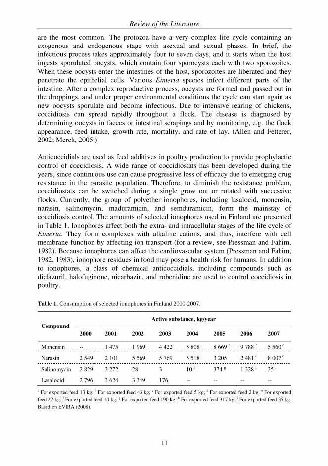
are the most common. The protozoa have a very complex life cycle containing an exogenous and endogenous stage with asexual and sexual phases. In brief, the infectious process takes approximately four to seven days, and it starts when the host ingests sporulated oocysts, which contain four sporocysts each with two sporozoites. When these oocysts enter the intestines of the host, sporozoites are liberated and they penetrate the epithelial cells. Various Eimeria species infect different parts of the intestine. After a complex reproductive process, oocysts are formed and passed out in the droppings, and under proper environmental conditions the cycle can start again as new oocysts sporulate and become infectious. Due to intensive rearing of chickens, coccidiosis can spread rapidly throughout a flock. The disease is diagnosed by determining oocysts in faeces or intestinal scrapings and by monitoring, e.g. the flock appearance, feed intake, growth rate, mortality, and rate of lay. (Allen and Fetterer, 2002; Merck, 2005.) Anticoccidials are used as feed additives in poultry production to provide prophylactic control of coccidiosis. A wide range of coccidiostats has been developed during the years, since continuous use can cause progressive loss of efficacy due to emerging drug resistance in the parasite population. Therefore, to diminish the resistance problem, coccidiostats can be switched during a single grow out or rotated with successive flocks. Currently, the group of polyether ionophores, including lasalocid, monensin, narasin, salinomycin, maduramicin, and semduramicin, form the mainstay of coccidiosis control. The amounts of selected ionophores used in Finland are presented in Table 1. Ionophores affect both the extra- and intracellular stages of the life cycle of Eimeria. They form complexes with alkaline cations, and thus, interfere with cell membrane function by affecting ion transport (for a review, see Pressman and Fahim, 1982). Because ionophores can affect the cardiovascular system (Pressman and Fahim, 1982, 1983), ionophore residues in food may pose a health risk for humans. In addition to ionophores, a class of chemical anticoccidials, including compounds such as diclazuril, halofuginone, nicarbazin, and robenidine are used to control coccidiosis in poultry. Table 1. Consumption of selected ionophores in Finland 2000-2007.
Active substance, kg/year
Compound
2000 2001 2002 2003 2004 2005 2006 2007
Monensin -- 1 475 1 969 4 422 5 808 8 669 a 9 788 b 5 560 c
Narasin 2 549 2 101 5 569 5 769 5 518 3 205 2 481 d 8 007 e
Salinomycin 2 829 3 272 28 3 10 f 374 g 1 328 h 35 i
Lasalocid 2 796 3 624 3 349 176 -- -- -- --
a For exported feed 13 kg; b For exported feed 43 kg; c For exported feed 5 kg; d For exported feed 2 kg; e For exported feed 22 kg; f For exported feed 10 kg; g For exported feed 190 kg; h For exported feed 317 kg; i For exported feed 35 kg. Based on EVIRA (2008).
Review of the Literature
12
Broilers are normally fed with anticoccidials almost throughout their lives because even subclinical infections are detrimental to the growth and feed conversion rate of the birds. If a correct level of medication is used, no cross-contamination and improper use of medicated feed occurs, and withdrawal periods prior to slaughter are observed, coccidiostat residues should not be found in excess in the food chain. Egg laying hens are not allowed to be fed with anticoccidials during the laying period at all because of the potential risk for accumulation of unwanted residues in eggs. However, studies have confirmed that eggs and poultry are still commonly contaminated with coccidiostat residues, although the awareness of the industry has increased and feed formulations have been improved (Kennedy et al., 1998; Cannavan et al., 2000; Mortier et al., 2005b; Danaher et al., 2008). At the EU level, the most commonly found anticoccidials in 2006 in poultry and eggs were nicarbazin, lasalocid, robenidine, and diclazuril (Anonymous, 2008). The main reasons for the incidence of residues have been reported to be the improper use of anticoccidials and feed contamination (Kennedy et al., 2000; McEvoy, 2002). The use of coccidiostats in the EU will be phased out in the near future (Anonymous, 2003b), and therefore, other means of controlling coccidiosis are required. The first line of defence is the application of high hygiene standards in poultry farms to reduce the number of oocysts in the environment, but the actual control of coccidiosis requires chemoprophylaxis and/or vaccination strategies. Laying hens obtain immunity within the first few months of their lives either naturally (with the help of anticoccidials) or through vaccination (Chapman, 1999). Because protective immunity after natural infection takes several weeks to develop, it is not a feasible option for broilers with a short lifetime. Therefore, although vaccines have not yet been widely utilized in broiler production, early vaccination schemes may prove to be a noteworthy option (Williams, 2002). However, the antigenic variation of Eimeria strains and the cost issues involved in the development of various types of vaccines still pose challenges to coccidiosis control (Dalloul and Lillehoj, 2006).
2.1.2 Veterinary residue control in the EU
Veterinary medicinal products are substances used in animal husbandry for treating or preventing disease in animals (Anonymous, 2001). Improper use of veterinary medicinal products can lead to the occurrence of residues of these substances in foodstuffs, which has raised concerns among the public. To protect human health against possible harmful effects resulting from exposure to these residues, the EU has addressed food safety issues at several levels especially during the last decade. The key elements of legislation providing the framework for residue control in the EU are briefly presented here. Prior to authorization for use in food-producing animals, the safety of veterinary medicinal products has to be assessed (Anonymous, 1990, 2001). After the safety evaluation, the necessity of establishing maximum residue limits (MRLs), i.e. the maximum concentration of residues legally permitted to be present in food, for that
Review of the Literature
13
particular substance is estimated. Feed additives have not been included in this MRL evaluation until recently (Anonymous, 2003b). In addition to the safety file with the proposed acceptable daily intake based on the no observed (adverse) effect level, the establishment of MRLs for a particular substance requires, e.g. the selection of a marker residue and consideration of other related issues such as the ratio of the marker residue with regard to total residues and the residue depletion kinetics. Moreover, the MRLs should cover each edible tissue (or product) of each target species for which the substance is intended based on the tissue residue distribution pattern. For example, the edible tissues for poultry consist of muscle, liver, kidney, and fat and skin in natural proportions. The evaluated substances are categorized under the Annexes of the Regulation 23 7/90 (MRL Regulation) as follows: substances, for which final MRLs have been established (Annex I), substances, for which it is not considered necessary to establish MRLs (Annex II), substances with provisional MRLs (Annex III), substances, for which no MRL could be established because residues of these substances, at any concentration level, in foodstuffs of animal origin create a health hazard, and thus, their use is prohibited (Annex IV) (Anonymous, 1990). Currently, MRL Regulation is under review with the aim of simplifying the existing legislation and improving the availability of veterinary medicinal products (Anonymous, 2007). The MRLs provide the basis for the establishment of reference points for residue control purposes (Anonymous, 1996) and of withdrawal periods, which is the time required to pass between the last administration of the drug (or the feed additive) to animals and the production of foodstuffs from such animals ensuring that the residue levels in tissues or products have fallen below the stated MRL. Thus, assuming that the substance is used properly and according to regulations, after the withdrawal period the food should be safe to consume. (Anonymous, 2005b.) Residue control is defined in Directive 96/23/EC, which describes the system for monitoring of substances and residues thereof in live animals and animal products (Anonymous, 1996). The Directive also determines specific sampling levels and frequencies, as well as the groups of substances (Table 2), to be monitored. The minimum number of samples to be analyzed for each food commodity is linked to the production figures for the preceding year. To fulfil the requirements of the Directive, the Member States have established national residue control programmes, the results of which are reported annually to the Commission. The Finnish Food Safety Authority (EVIRA) is responsible for implementing the programme in Finland. As an example, EVIRA tested more than 10 000 samples in 2005 for the presence of 80 different chemical compounds and 99.7% of the samples were negative or below the legally permitted concentrations or action levels (EVIRA, 2006a). The implementation of coccidiostat residue monitoring to the national residue control programmes has been complicated by the fact that although coccidiostats are included in the MRL evaluation, many of the compounds do not yet have official MRLs. Therefore, Member States have set national action levels to guide the monitoring task. For instance, in Finland the action level for ionophore residues in eggs has been 10 µg/kg and in poultry any positive result has resulted in an investigation (National Food Agency et al., 2005).
7

Review of the Literature
14
Table 2. Substances to be monitored in residue control according to Directive 96/23/EC.
Group A - Substances having anabolic effect and unauthorized substances
− Stilbenes, stilbene derivatives, and their salts and esters
− Antithyroid agents
− Steroids
− Resorcylic acid lactones including zeranol
− β-agonists
− Compounds included in Annex IV to Council Regulation (EEC) No 2377/90
Group B - Veterinary drugs and contaminants
− Antibacterial substances including sulphonamides and quinolones
− Other veterinary drugs (anthelmintics, anticoccidials including nitroimidazoles, carbamates and pyrethroids, sedatives, non-steroidal anti-inflammatory drugs, and other pharmacologically active substances)
− Other substances and environmental contaminants (organochlorine compounds including PCBs, organophosphorus compounds, chemical elements, mycotoxins, dyes, and others)
Abbreviations: PCB, polychlorinated biphenyl.
In contrast to other areas of food safety testing, the methods used in residue control can be freely selected as long as they comply with the requirements set in the Decision 2002/657/EC (Anonymous, 2002a). The Decision relates to Directive 96/23/EC and outlines the criteria for the performance of screening and confirmatory methods and for the interpretation of results. It aims to guarantee the quality and comparability of test results, which are generated by laboratories approved for official residue control. Moreover, to ensure harmonised implementation of Directive 96/23/EC the concept of a minimum required performance limit was introduced for methods which are used to detect substances with no set permitted limit, substances with no authorization, or prohibited substances. The main advantage of Decision 2002/657/EC is its flexible adaptation to emerging problems and technical developments, which enables easier implementation of novel analytical methods in routine use (Stolker and Brinkman, 2005).
2.1.3 Methods for the detection of anticoccidial residues in food
The methods of analysis used in residue control are generally divided into two categories: screening and confirmatory methods. Screening methods are defined as "…methods that are used to detect the presence of a substance or class of substances at the level of interest. These methods have the capability for a high sample throughput and are used to sift large numbers of samples for potential non-compliant results. They are specifically designed to avoid false compliant results.", and confirmatory methods as "…methods that provide full or complementary information enabling the substance
Review of the Literature
15
to be unequivocally identified and if necessary quantified at the level of interest." (Anonymous, 2002a). In principle, by combining screening and confirmatory methods a cost-effective system for residue control can be achieved. The initial screening, which is performed with a rapid and inexpensive qualitative method, classifies samples as negative or potentially positive. The suspect samples have to be further analyzed with a confirmatory method to obtain unequivocal identification and quantification of the substance. Nevertheless, it seems that confirmatory methods are often used for both purposes and the development and validation of screening methods has received less attention. To be truly applicable for routine use, high throughput screening methods should be inexpensive, rapid, robust, and reliable, as well as being able to detect preferably multiple residues simultaneously at the required levels. Consequently, it can be very challenging to combine all these favourable characteristics in a single assay. The first assays to detect the presence of coccidiostats were based on growth inhibition of Eimeria in cell culture and were mainly used to study the effects of anticoccidial activity (McDougald and Galloway, 1973; Strout and Ouellette, 1973). Moreover, various thin-layer chromatography methods combined with bioautography were developed for ionophores, which have antibiotic effects and can inhibit the growth of certain bacteria (Weiss and MacDonald, 1985). The following sections introduce current screening and confirmatory methods and sample preparation procedures for selected anticoccidials, mainly ionophores, halofuginone, and nicarbazin, in food samples.
2.1.3.1 General principles of sample preparation
Sample preparation forms an integral part of the method of analysis and its role is even more pronounced when more sensitive detection of residues is required. Typically a combination of sample preparation techniques is used for complex sample matrices such as food. Therefore, regardless of the analyte or method of analysis, sample preparation for residue control using food matrices tends to be slow, and thus, it is the limiting factor in the overall sample throughput. Consequently, solutions for simplifying, automating, and speeding up sample preparation without compromising the assay performance would be particularly beneficial because they could provide savings in time and money and eventually lead to an increased testing rate. In particular, screening methods aiming to minimize the cost and analysis time may settle for a simpler sample preparation, although sometimes at the expense of analyte recovery. Foodstuffs of animal origin used in residue control commonly require extensive sample preparation. The sample preparation can include several steps: sampling, homogenization, extraction, clean-up, and concentration. The aim is to provide a homogeneous, representative sample for analysis where the analyte in question is recovered and concentrated to enable detection of residue concentrations typically at the level of µg/kg. In addition, interfering substances, which may co-extract with the analyte, should be excluded. The extraction procedure is dependent on the analyte and

Review of the Literature
16
its concentration, the sample matrix, and the method of analysis. Organic solvents such as acetonitrile, ethyl acetate or methanol are normally used in extraction, although some approaches using only aqueous extraction have been reported (Elissalde et al., 1993; Beier et al., 1998). After extraction, further purification steps, e.g. another liquid extraction step or solid phase extraction (SPE) may be necessary, if sample matrix effects still cause interference in the assay. A final evaporation step can be included in the procedure to concentrate the extract, and thus increase the sensitivity of the assay. Further information regarding sample preparation techniques can be found, for example, in an article by Ridgway et al. (2007).
2.1.3.2 Screening methods
Screening methods for coccidiostats mainly rely on antibody-based techniques. Coccidiostats form a heterogeneous group in terms of chemical structures and sensitivity requirements. Therefore, multi-analyte approaches using broad-specificity antibodies where the entire class of drugs sharing similar chemical structures and MRLs or action levels can be screened simultaneously (Korpimäki et al., 2004), cannot be utilized in coccidiostat screening. However, two members of the ionophore group, salinomycin and narasin, provide a minor exception to that rule, as their structures differ only by one methyl group enabling 100% cross-reactivity (Kennedy et al., 1995; Peippo et al., 2004). Coccidiostats are small molecular weight analytes, often referred to as haptens, and they have to be conjugated to a carrier protein such as apo-transferrin (Crooks et al., 1997), human serum albumin (Connolly et al., 2002), or keyhole limpet hemocyanin (Rowe et al., 1994; Beier and Stanker, 2001) to evoke antibody production. In general, the challenge in antibody production for coccidiostats is to obtain antibodies that recognize hapten and not parts of the carrier protein. The antibodies may also detect the metabolites of the parent compound, which cannot usually be tested because of the lack of reference standards (Elliott et al., 1998). Occasionally the hapten has to be modified to facilitate conjugation to carriers. For example, the structure of 4,4'-dinitrocarbanilide (DNC), the marker residue for nicarbazin, is not particularly suitable for conjugation, and therefore, a hydrazone derivative and a DNC mimic (4'-nitrosuccinanilic acid) containing a carboxyl group for conjugation have been suggested as alternatives to conjugation instead of the less reactive parent compound (Beier and Stanker, 1998). However, the monoclonal antibodies generated with the hydrazone derivative did not recognise free DNC (Beier and Stanker, 1998). The mimic, on the other hand, was successfully employed to create polyclonal antibodies for nicarbazin (Connolly et al., 2002), but the corresponding monoclonal counterparts did not perform as well in terms of sensitivity (Beier and Stanker, 2001). The selection of screening methods for coccidiostats is still limited. The reported antibody-based methods for coccidiostats are mainly based on a heterogeneous competitive immunoassay format with enzyme labels (enzyme-linked immunosorbent assay, ELISA) (Kennedy et al., 1995; Crooks et al., 1997; Watanabe et al., 2001; Huet

Review of the Literature
17
et al., 2005). The assays are usually performed in a standard microtiter plate format allowing easy handling of samples and high assay throughput. In addition to capacity issues, immunoassays are easy to perform, relatively rapid, and amenable to automation. Immunoassays relying on detection technologies such as time-resolved fluorometry (TRF) (Crooks et al., 1998; Peippo et al., 2004) and other assay formats such as biosensors (McCarney et al., 2003; Danaher et al., 2008) have also been reported for use in coccidiostat analysis. Biosensors form a heterogeneous group of compact analytical devices comprising two distinct elements: a biological recognition element (e.g. enzyme, antibody, microbial cell) either integrated with or in close contact (at least in principle) with a signal transduction element (e.g. optical, electrochemical, piezoelectric), which converts the signal from the biological element to a quantifiable signal (for reviews, see Patel, 2002 and Baeumner, 2003). Immunosensors, which use antibodies as recognition elements, represent the form of biosensors commonly utilized in veterinary residue analysis (Ricci et al., 2007). Because biosensors also provide possibilities for portable systems, analysis of turbid samples, on-line monitoring, and sensitive detection, there is continuous interest in the development of biosensors not only for the food safety market, but also for the field of medical, military, and environmental applications (Alocilja and Radke, 2003). However, there are still some general technical barriers, cost considerations, and performance issues that need to be solved, e.g. by utilizing advances in transducers and recognition elements together with the possibilities offered by nanotechnology before large-scale commercialization of biosensors for food safety can occur (Luong et al., 2008; Palchetti and Mascini, 2008). Some examples of different types of screening assays for coccidiostat residues are presented here. Huet et al. (2005) described a competitive ELISA for nicarbazin and halofuginone residues in eggs and chicken muscle. The same sample preparation procedure using acetonitrile extraction and hexane wash could be applied to both analytes. The assay performance in terms of detection capability (CCβ) was good, but the practicability of the assay suffered from the overnight incubation step. McCarney et
al. (2003) reported the development of a regenerable optical immunosensor based on surface plasmon resonance (SPR) for nicarbazin residues in poultry liver and eggs. The sample preparation included acetonitrile extraction and liver samples were further purified with hexane wash. The SPR assay cycle took only 7 min to complete, providing results in nearly real-time. The same SPR assay was later modified for poultry samples and used in a large-scale survey to investigate the incidence of DNC residues in Ireland (Danaher et al., 2008). In summary, the current role of antibody-based methods in official residue control is in the screening of samples and potentially positive samples have to be re-analyzed with a confirmatory method. At best, antibody-based assays can offer good sensitivity, robustness, inexpensive analysis, high throughput, and speed of analysis with generally less complicated sample preparation compared to confirmatory methods. To be applicable to routine testing, these methods, regardless of the chosen assay format, have to be validated with appropriate sample matrices and have adequate detection

Review of the Literature
18
limits for screening purposes. The remaining issues to be considered are the complexity of sample preparation and preferred assay time. The level of sample preparation depends on the analyte, sample matrix, and overall performance of the assay, which is partly affected by the choice of antibody, other assay parameters, and detection chemistry. Unfortunately, the development and application of either in-house or commercial antibody-based methods to the field of coccidiostat analysis is still limited. For instance, there are several commercial assays available for other residues such as antimicrobials and growth promoters, but the screening tests for coccidiostats are only now emerging on the market.
2.1.3.3 Confirmatory methods
The combination of liquid chromatography (LC) with mass spectrometry (MS) represents the method of choice for residue control for most classes of veterinary drugs (for reviews, see Balizs and Hewitt, 2003 and Stolker and Brinkman, 2005). Although LC-MSn methods provide good sensitivity and specificity and enable quantification and confirmation, they often require more extensive sample preparation than antibody-based assays. Thus, the overall analysis time and assay throughput, which is also limited by the sequential nature of the analysis as opposed to the batch-mode commonly used in immunoassays, have been considered to be limiting factors in LC-MSn. LC-MSn equipment is also expensive to acquire and operate, and therefore, the cost of analysis is higher than with screening methods. Previously, LC was used in combination with ultraviolet absorbance detection systems (Anderson et al., 1981; Schenck et al., 1992; Draisci et al., 1995; Matabudul et al., 1999), but currently most methods rely on the use of (tandem) MS with electrospray ionization interface. Recently published methods based on LC-MS-MS for coccidiostats are presented in Table 3. In general, these methods fulfil the sensitivity requirements, i.e. have a proper decision limit (CCα) and CCβ, and most have been validated according to Decision 2002/657/EC. Many methods employ sample preparation procedures, which are capable of recovering residues of multiple coccidiostats. The level of sample preparation varies and is partly dependent on the selected analytes and matrices. The use of organic solvents in sample preparation is still common practice, although the volumes have been reduced as the sample preparation methods have improved. An example of these improvements is the introduction of SPE, which has become a routine tool in sample preparation, although the selection of an optimal SPE column and extraction conditions can require several attempts (Rosén, 2001). The separation of coccidiostats in LC is mostly performed with non-polar C18 columns and the complexity of the mobile phase is dependent on the application. For example, the mobile phase can vary from a simple combination of acetonitrile and ammonium acetate solution (Rosén, 2001) to a complex mixture of acetonitrile, water, methanol, tetrahydrofuran, and trifluoroacetic acid (Matabudul et
al., 2002). Many of the LC-MSn methods are used for both screening and confirmation purposes and the screening aspect can be strengthened, e.g. by pooling of the samples (Rosén, 2001).

Review of the Literature
19
Table 3. Recent LC-MS-MS methods and related sample preparation procedures for anticoccidials in chicken tissues and eggs.
Analyte Sample matrix Main steps in sample preparation Reference
LAS, MON, NAR, and SAL
Muscle and egg
Weighing of the homogenized sample, mixing with anhydrous sodium sulfate, acetonitrile extraction with shaking, centrifugation, SPE, evaporation to dryness, reconstitution, and filtration
Rokka and Peltonen (2006)
LAS, MON, NAR, and SAL
Egg
Weighing of the homogenized sample, acetonitrile extraction with vortexing and sonication, centrifugation, evaporation to dryness, reconstitution, sonication, and filtration
Mortier et al. (2005a)
DIC, HAL, LAS, MAD, MON, NAR, NIC, ROB, and SAL
Muscle and egg
Weighing of the minced/mixed sample, mixing with anhydrous sodium sulfate, acetonitrile extraction with shaking, centrifugation, SPE, evaporation to dryness, and reconstitution
Dubois et al. (2004)
DIC, DIM, HAL, NIC, and ROB
Egg
Weighing of the homogenized sample, acetonitrile extraction with vortexing and sonication, centrifugation, evaporation, and filtration
Mortier et al. (2003)
HAL Liver and egg
Weighing of the minced/homogenized sample, trypsin digestion o/n with shaking, 2x ethyl acetate extraction with shaking followed by centrifugation, 2x ammonium acetate extraction with shaking, hexane wash, SPE, evaporation to dryness, and reconstitution
Yakkundi et
al. (2003)
LAS, MON, NAR, NIC, and SAL
Liver and egg
Weighing of the sample, mixing with anhydrous sodium sulfate, acetonitrile extraction with shaking, centrifugation, SPE, evaporation, and filtration
Matabudul et
al. (2002)
NIC Liver and egg
Weighing of the minced/homogenized sample, homogenization + extraction with acetonitrile, centrifugation, evaporation to dryness, hexane wash, and centrifugation
Yakkundi et
al. (2001)
Abbreviations: DIC, diclazuril; DIM, dimetridazole; HAL, halofuginone; LAS, lasalocid; MAD, maduramicin; MON, monensin; NAR narasin; NIC, nicarbazin; o/n, overnight; ROB, robenidine; SAL, salinomycin; SPE, solid phase extraction.
In summary, the continuous development of MS techniques together with improved sample preparation procedures is expected to enhance the performance of these systems even more and enable truly multi-residue analysis (Stolker and Brinkman,
MON, NAR, and SAL
Liver and egg Weighing of the
Rosén (2001) centrifugation, and SPE homogenization + extraction with methanol,
homogenized/mixed sample,
,

Review of the Literature
20
2005; Stolker et al., 2007). Thus, LC-MSn methods will continue to have a major role in residue control as confirmatory methods and they may also retain a permanent role in screening, if antibody-based methods do not respond to that challenge.
2.1.4 Future trends in residue control
Future developments in the methods used in residue control are likely to be comparable with those occurring in clinical diagnostics. The key requirements for testing methods include speed, simplicity, and robustness of analysis with adequate detection limits and minimal sample preparation. Currently, the complex sample preparation represents a bottleneck in residue analysis. Solutions for easier sample preparation could include the use of automated sample pre-treatment systems allowing multi-residue extractions, higher throughput, and reduced consumption of organic solvents. Sample preparation could also be simplified by using other sample matrices than foodstuffs. For example, liquid samples such as plasma and urine are homogeneous by nature and do not require extensive clean-up prior to analysis. However, correlation between residue levels in these samples and the corresponding tissue, as well as depletion kinetics, have to be established before this kind of approach (i.e. predictive indicators) would be feasible. As an example, the analysis of serum or plasma for monensin residues has been suggested to be an indicator of the residue levels in liver (Atef et al., 1993; Crooks et
al., 1998). Moreover, testing could be more directed to the beginning of the production chain, namely to feedstuffs, as feed contamination, due to, e.g. cross-contamination in feed mills or farms, is a common reason for anticoccidial residues in foods (Kennedy et
al., 1998; Cannavan et al., 2000; Cannavan and Kennedy, 2000; Kennedy et al., 2000; McEvoy, 2002; Yakkundi et al., 2002). For example, Campbell et al. (2007) developed a portable and rapid lateral flow device for on-site testing of nicarbazin in feedstuffs where the sample was simply ground in methanol and diluted in assay buffer prior to analysis. The assay was sensitive enough for screening at concentrations at or above 2 mg/kg, which was adequate based on previous data (Cannavan and Kennedy, 2000). With regard to methods of analysis, multi-residue confirmatory methods based on LC-MSn will remain at the core of residue control, but hopefully also rapid and user-friendly antibody-based screening methods in new assay formats will find their way into routine residue testing. In addition, techniques such as transcriptomics and proteomics based on the measurement of effect rather than of target compound concentrations may create entirely novel concepts for detecting residues in food. These techniques are currently under investigation in the EU-funded project "BioCop" (www.biocop.org). In conclusion, because veterinary medicinal products are commonly used in animal husbandry, residue control plays a significant role in ensuring food safety. Therefore, advances in sample preparation and analytical methods used in residue control will in their part strengthen the overall level of food safety.
Review of the Literature
21
2.2 FOODBORNE PATHOGENS AND FOOD SAFETY
The biological safety of food can be compromised for several reasons such as bacteria, viruses, prions, toxins, and parasites. These agents can cause foodborne illnesses, typically either infectious or toxic in nature, through the ingestion of contaminated food or water. Foodborne illnesses caused by micro-organisms such as Salmonella spp., Campylobacter spp., Listeria monocytogenes, and Escherichia coli O157:H7 are a large and growing public health problem, representing a significant financial burden to countries in terms of medical care and lost productivity. For example, the annual cost of all cases of salmonellosis in the USA has been rated to be over $2 500 million (in 2007 dollars) (Economic Research Service, 2008). The following sections concentrate on one of the most commonly occurring foodborne pathogens, Salmonella, and the measures and methods used in Salmonella control.
2.2.1 Salmonella spp. as a foodborne pathogen
The genus Salmonella, one of the leading causes of foodborne illness (Tirado and Schmidt, 2001; EFSA, 2007), belongs to the Enterobacteriaceae family and consists of gram-negative, facultatively anaerobic, motile (except for rare non-motile serovars), rod-shaped bacteria. Salmonella taxonomy and nomenclature is complicated and various approaches, including biochemical and serological characteristics and deoxyribonucleic acid (DNA) homology, have been suggested as the basis for classification. The WHO Collaborating Centre for Reference and Research on Salmonella recommends a classification system according to the Kauffmann-White scheme where the genus is divided into two type species, S. bongori and S. enterica, and the latter is further divided into six subspecies designated by names (and Roman numerals) as follows, enterica (I), salamae (II), arizonae (IIIa), diarizonae (IIIb), houtenae (IV), and indica (VI) (Grimont and Weill, 2007). Serovars belonging to subspecies I are named and others are only designated by formula. After preliminary identification of Salmonella, further confirmation is done with biochemical and serological tests where the latter comprises the agglutination of bacterial antigens such as somatic (O) lipopolysaccharides on the external surface of the bacterial outer membrane, flagellar (H) antigens associated with the peritrichous flagella, and capsular (Vi) antigen present in only a few serovars with Salmonella-specific antibodies. In addition, phage typing can be used for further subtyping of some serovars mainly for epidemiological purposes. Currently, the Kauffmann-White scheme contains over 2 500 serovars of Salmonella (Grimont and Weill, 2007). (D’Aoust et al., 2001.) The widespread occurrence of Salmonella spp. in the environment together with intensive animal husbandry creates a serious threat for food safety throughout the food chain. Moreover, the pathogen can adapt to extreme conditions, and thus, is very resilient to various stresses utilized in food processing. (D’Aoust et al., 2001.) Mead et
al. (1999) estimated that of all Salmonella infections the rate of foodborne transmission is 95%, although the organism may also be transmitted through direct contact with

Review of the Literature
22
infected animals or humans. Salmonella infection is most frequently acquired through foodstuffs contaminated with animal faeces, and poultry, pig meat, and eggs have been reported to be the major sources of human infections (EFSA, 2007). At later stages of food processing, contamination can occur through infected humans, unclean food preparation areas, or cross-contamination with other raw foodstuffs, if appropriate hygiene measures are not followed. Consequently, the risk of contracting Salmonella from foods can be reduced by proper storage of foods, thorough cooking and good hygiene throughout the food production chain. The disease caused by Salmonella is called salmonellosis. The reported infectious dose has varied from a small number of cells (D’Aoust, 1985; Kapperud et al., 1990) to over 105 cells (Kothary and Babu, 2001) depending on the particular Salmonella serovar, type of food, and physical condition of the infected individual (D’Aoust, 1985; Waterman and Small, 1998; Kothary and Babu, 2001). In general, the outcome from the exposure to foodborne pathogens depends on a number of host factors such as pre-existing immunity, state of nutrition, age, and ability to elicit immune response. Thus, the incidence, severity, and lethality of foodborne diseases are higher in certain vulnerable segments of the population. The incubation period for the most common form of salmonellosis, gastroenteritis, can vary from a few hours up to three days, and the acute symptoms, nausea, vomiting, diarrhoea, abdominal pain, and fever, usually last for four to ten days. The infection can also result in more serious conditions including enteric fever (caused by serovars Typhi and Paratyphi), bacteremia or reactive arthritis. After the onset of clinical symptoms, the diagnosis is based on the isolation of the bacteria from stool. The treatment of gastroenteritis caused by Salmonella is based on resting and prevention of dehydration, but in severe cases antibiotics may be required. However, the prophylactic use of antibiotics in animal farming has contributed to the emergence of multidrug-resistant Salmonella strains, which can complicate the treatment of salmonellosis (Rabsch et al., 2001). The Centers for Disease Control and Prevention has estimated that the annual incidence of foodborne illness caused by pathogens in the USA is approximately 76 million cases in total, resulting in 325 000 hospitalizations and 5 000 deaths (Mead et al., 1999), and approximately 1.4 million cases with 15 000 hospitalizations and 400 deaths are caused by non-typhoidal salmonellosis (Voetsch et al., 2004). In the EU, zoonoses (diseases that are naturally transmissible directly or indirectly between animals and humans) affected more than 350 000 citizens in 2006 (EFSA, 2007). Salmonella was the second most commonly reported zoonotic agent and the leading cause of foodborne outbreaks; in 2006 approximately 160 000 confirmed human cases were reported and over 3 100 outbreaks with serovar Enteritidis being the predominant cause were registered that affected almost 23 000 people and resulted in 23 deaths (EFSA, 2007). In Finland during the past ten years the number of reported cases of Salmonella infections has approximately been between 2 000 and 3 000 annually (National Public Health Institute, 2008), and the majority of the cases are acquired from abroad (EVIRA, 2006b; EFSA, 2007). Nevertheless, because many mild cases

Review of the Literature
23
are not reported, the actual number of Salmonella infections is likely to be much higher.
2.2.2 Salmonella control in the EU
Food industry and authorities share the responsibility for controlling the microbiological safety of food. While the industry follows legislation and self-control plans according to HACCP, the authorities ensure industry compliance with the legislation and monitor food safety through national control programmes. As discussed earlier, traditional food safety control has relied on general sanitation inspections and end product testing, thus being a reactive rather than a preventive approach. Together with the EU’s integrated "from farm to fork" policy and good manufacturing and hygiene practices, HACCP aims to create a prevention-based food safety system reaching throughout the food chain. HACCP relies on the identification of risks, integration of appropriate control measures into the design of the process, and implementation and monitoring of these controls. Therefore, the testing focus can, in principle, be gradually shifted from the end products towards the raw materials combined with adequate in-process monitoring. In the EU, the microbiological safety of food has been thoroughly addressed at legislative level. The measures include, for instance, general microbiological criteria, which function as tools for assessing the safety and quality of foods and that of the processes for manufacturing, handling, and distribution (Anonymous, 2005a), the obligation of the Member States to annually monitor and collect data on zoonoses, zoonotic agents, antimicrobial resistance, and foodborne outbreaks (Anonymous, 2003a), and the specific control of Salmonella (Anonymous, 2003c). In Finland, the National Salmonella Control Programme has been applied since 1995 to protect consumers against Salmonella infections spread through foods of animal origin (Anonymous, 1994). The implementation of the programme has generally kept the annual occurrence of Salmonella in production animals and foodstuffs originating from them at levels of less than 1% (EVIRA, 2006b). However, the recent Salmonella crisis in Finland, which was due to contaminated feed, further highlighted the importance of monitoring of the entire food chain starting from primary production and feedstuffs. Moreover, collaboration projects at international level have been established to advance microbiological food safety worldwide. For example, the Global Salm-Surv programme coordinated by the WHO promotes the capacity and quality of Salmonella
surveillance, serotyping, and antimicrobial resistance testing, and the FAO together with the WHO have performed microbiological risk assessments for Salmonella in eggs and broilers (FAO/WHO, 2002).
2.2.3 Methods for the detection of Salmonella in food
The detection of foodborne pathogens is challenging for several reasons: target bacteria are often present in low numbers in foods, bacteria may be sub-lethally injured due to

Review of the Literature
24
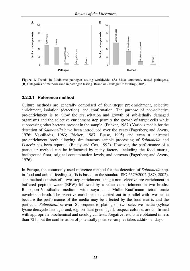
food processing, and the high amount of other bacteria and food matrix can further complicate the analysis. Thus, most methods use sample enrichment to increase the number of live target bacteria in the matrix to detectable levels prior to analysis. Moreover, the legislative demand, the absence of Salmonella cells in 25 g (or 10 g) of food, sets strict performance requirements for methods of analysis (Anonymous, 2005a). In general, the methods used in foodborne pathogen detection can be divided into traditional (also referred to as conventional methods) and alternative methods (also referred to as rapid methods). The traditional methods include culture methods, which still represent the golden standard in the testing of microbiological quality of food. Some culture methods have gained an internationally accepted reference method status in foodborne pathogen detection such as the methods published by the International Organization for Standardization (ISO). Although culture-based methods are selective, sensitive, and detect only viable cells, they are also labour-intensive and time-consuming requiring several days or even weeks to obtain a confirmed result. The terms rapid methods or alternative methods are synonyms referring to a vast array of novel testing methods, which can significantly reduce the analysis time compared to traditional methods. However, because of lengthy sample preparation, most alternative methods cannot be considered to be truly rapid in the original sense of the word. Alternative methods should have characteristics such as speed of analysis, ease of use and/or automation, improved analytical performance (e.g. sensitivity, specificity), possibility for miniaturization, and reduction of total cost, in order to be noteworthy rivals to traditional methods (ISO, 2003). In addition, the success of an alternative method depends on other factors such as robustness, reliability, throughput, overall convenience, and the level of validation and standardization, which all determine the true applicability of the method to routine use. Alternative methods for Salmonella provide qualitative information on whether the pathogen is present or not in the sample. If the alternative method gives a presumptive positive test result, confirmation, i.e. further identification and typing of the particular strain using the reference method, is required for epidemiological surveillance and national control programmes. In 2005, the food industry was estimated to perform nearly 110 million tests for foodborne pathogens and Figure 1 presents the categories of tested pathogens and the methods used (Strategic Consulting, 2005). Traditional culture methods still dominate the field of pathogen testing, although alternative methods are steadily increasing their market share as novel and better methods appear.
Review of the Literature
25
Salm
onella
Lis
teria
Escherichia
coli
O157
Cam
pylo
bacte
r
Oth
ers
0
20
40
60
80
100
Pathogen
% o
f all p
ath
og
en
tests
Method
Culture
-based
Antibody-b
ased
Nucle
ic a
cid
-based
Oth
ers
0
20
40
60
80
100
% o
f all p
ath
og
en
tests
A B
Figure 1. Trends in foodborne pathogen testing worldwide. (A) Most commonly tested pathogens. (B) Categories of methods used in pathogen testing. Based on Strategic Consulting (2005).
2.2.3.1 Reference method
Culture methods are generally comprised of four steps: pre-enrichment, selective enrichment, isolation (detection), and confirmation. The purpose of non-selective pre-enrichment is to allow the resuscitation and growth of sub-lethally damaged organisms and the selective enrichment step permits the growth of target cells while suppressing other bacteria present in the sample. (Fricker, 1987.) Various media for the detection of Salmonella have been introduced over the years (Fagerberg and Avens, 1976; Vassiliadis, 1983; Fricker, 1987; Busse, 1995) and even a universal pre-enrichment broth allowing simultaneous sample processing of Salmonella and Listeria has been reported (Bailey and Cox, 1992). However, the performance of a particular method can be influenced by many factors, including the food matrix, background flora, original contamination levels, and serovars (Fagerberg and Avens, 1976). In Europe, the commonly used reference method for the detection of Salmonella spp. in food and animal feeding stuffs is based on the standard ISO 6579:2002 (ISO, 2002). The method consists of a two-step enrichment using a non-selective pre-enrichment in buffered peptone water (BPW) followed by a selective enrichment in two broths: Rappaport-Vassiliadis medium with soya and Muller-Kauffmann tetrathionate novobiocin broth. The selective enrichment is carried out in parallel with two media because the performance of the media may be affected by the food matrix and the particular Salmonella serovar. Subsequent to plating on two selective media (xylose lysine deoxycholate agar and, e.g. brilliant green agar), suspect colonies are confirmed with appropriate biochemical and serological tests. Negative results are obtained in less than 72 h, but the confirmation of potentially positive samples takes additional days.
Review of the Literature
26
2.2.3.2 Alternative methods
The following sections present the main techniques and most prominent advances in the field of alternative methods for the detection of Salmonella in food matrices with the emphasis on nucleic acid techniques. All the methods described are used to analyze samples taken directly from the enrichment broth (or sometimes even directly from the food matrix) without the need of isolating and obtaining a pure culture of the pathogen.
Antibody-based methods
The use of antibodies in the detection of Salmonella started with agglutination tests forming the basis for serotyping and fluorescent antibody techniques (Thomason et al., 1957; Haglund et al., 1964; Sperber and Deibel, 1969). Krysinski and Heimsch (1977) reported one of the first enzyme immunoassays for Salmonella. Since then various types of immunoassays have been routinely applied to the detection of this foodborne pathogen. In addition, anti-Salmonella antibodies have been employed in immunomagnetic separation (IMS) where antibody-coated magnetic beads are used to capture target cells from the sample (Skjerve and Olsvik, 1991). In general, immunoassays for Salmonella are based on the heterogeneous non-competitive ELISA format (Mattingly, 1984; Cudjoe et al., 1995; Holt et al., 1995; Valdivieso-Garcia et al., 2003; Fukuda et al., 2005; Magliulo et al., 2007) where the target cells are "sandwiched" between antibodies typically directed against somatic and flagellar antigens. Although the use of enzyme labels is common practice, other types of labels have also been reported for Salmonella immunoassays (Tu et al., 2002; Gehring et al., 2008). The detection limits of Salmonella immunoassays with food matrix are usually in the range of 104-106 cells/mL, but with careful selection of detection method and antibody the sensitivity can be further improved. Although the samples have to be enriched prior to immunoassay to allow the target organism to reach a detectable level, the following sample preparation steps are minimal because of the inherent robustness of antibody-based methods. Generally, the enriched samples are used as such or boiled (to inactivate pathogens and release antigens) before addition to the assay. The cross-reactivity of antibodies with antigens in closely related bacteria is considered to be the main disadvantage of antibody-based methods. There is a vast array of commercial antibody-based assay kits available for the detection of Salmonella. Several extensive evaluations have been conducted to show that the performance of the kits is comparable to the reference method to promote the certification of the kits (Curiale et al., 1997; Bird et al., 1999; Hughes et al., 2001; Feldsine et al., 2008). However, as the sample preparation always includes pre-enrichment, which is usually followed by selective enrichment and even post-enrichment depending on the sample matrix and kit, these methods still take at least 24 h to perform. The level of sophistication of these assays ranges from a simple, manual lateral flow device (dipstick) with visual detection to automated systems. The main benefits of the dipstick are the ease of use and speed of analysis because after enrichment the user only adds a drop of broth to the test and the result is ready in a few
Review of the Literature
27
minutes without washing or further manipulations. Lateral flow tests for Salmonella are available, e.g. from BioControl Systems (Bellevue, WA, USA), DuPont Qualicon (Wilmington, DE, USA), Strategic Diagnostics (Newark, DE, USA), and Neogen (Lansing, MI, USA). Examples of immunoassays amenable to automation include, e.g. microtiter plate based Assurance EIA Salmonella and TRANSIA PLATE Salmonella Gold from BioControl Systems, and bioMérieux (Marcy l'Etoile, France) offers a fully automated immunoanalyzer, the VIDAS system, for the detection of Salmonella. Immunosensors are a commonly used biosensor type in the detection of foodborne pathogens (Patel, 2002; Ricci et al., 2007; Palchetti and Mascini, 2008). In particular, optical immunosensors such as the label-free SPR-based systems have raised interest in this field (Koubová et al., 2001; Bokken et al., 2003; Bergwerff and van Knapen, 2006; Homola, 2008), partly because of the commercially available systems, e.g. Biacore (GE Healthcare, Uppsala, Sweden). In the scientific literature, immunosensors for the detection of Salmonella in food matrix have utilized various transducer elements with detection limits typically in the range of 103-105 cells/mL and assay times of up to 90 min (Seo et al., 1999; Varshney et al., 2003; Taitt et al., 2004; Ko and Grant, 2006; Mazumdar et al., 2007). In general, sample matrix effects tend to impair sensor performance in terms of sensitivity, and therefore, the use of proper sample matrices is essential during method development to be able to evaluate the applicability of the method for routine use. Unfortunately, many biosensor methods described for the detection of Salmonella in food have not been validated properly, which makes it difficult to estimate the true potential of these methods. The principles and future challenges of biosensors, which were briefly discussed in 2.1.3.2, are also valid for applications for foodborne pathogens. Consequently, although the potential of biosensors in food safety applications has long been recognised (Oh, 1993), the biosensors are only now beginning to fulfil expectations and require further improvements in performance to be truly applicable to food safety testing at the levels required by the authorities. In conclusion, there are validated and robust antibody-based methods with good performance characteristics available for the detection of Salmonella in food. However, in order to achieve the detection limit of 1 CFU/25 g of food, sample enrichment has to be employed prior to assay to increase the number of target cells. Thus, even in the best-case scenario, the time to result approaches 24 h. Although this cannot be considered to be particularly rapid, the assay time is still considerably improved compared to the time required by the reference method.
Nucleic acid-based methods
Following the early hybridization assays for Salmonella (Fitts et al., 1983; Olsen et al., 1995), in vitro amplification techniques emerged enabling more sensitive detection. Currently, PCR represents the most well-known and established technique (Saiki et al., 1985), although some novel approaches such as the isothermal nucleic acid sequence-based amplification (NASBA) using ribonucleic acid (RNA) targets have

Review of the Literature
28
also been applied to foodborne pathogen detection (Cook, 2003). NASBA has the advantage over PCR of having the potential to detect only viable cells. Nevertheless, this novel technique requires further development to be able to follow in the footsteps of PCR.
PCR: advantages and disadvantages
PCR-based methods rely on genes, and thus, are not influenced by the growth state or the environment of the bacteria. Therefore, PCR is considered to be more reliable than the traditional culture-based methods, which use physiological and morphological criteria for identification. Furthermore, with nucleic acid amplification non-cultivable and slow-growing organisms can be detected, which can give a negative result with a culture method. In addition to improved sensitivity, specificity, and rapidity, state-of-the-art PCR techniques also enable automation and high throughput. Despite these advantages, nucleic acid-based methods have not yet been able to shake the reference status of culture methods. The implementation of PCR into routine use has not been easy because of several disadvantages associated with PCR. These include the high expertise in molecular biology required for performing PCR, the laborious procedures for both sample preparation and PCR, the false positive results due to lack of discrimination power between viable and non-viable cells if DNA is used as a target, the lack of robustness, proper validation and standardization of the methods, and contamination problems. However, there are solutions to all of the above mentioned issues. For example, commercial ready-to-use PCR kits and sample preparation systems for DNA purification easing up the manual work in PCR have been introduced. The problem of false positive results can be circumvented by sample enrichment, by using, e.g. ethidium monoazide, which has been suggested to irreversibly bind to the DNA of damaged cells and prevent amplification (Nogva et al., 2003; Guy et al., 2006), or by reverse transcription (RT)-PCR. In RT-PCR RNA, either messenger RNA (mRNA) or ribosomal RNA (rRNA), is used as a template for amplification because it is a better measure of cell viability than DNA (McKillip et al., 1998; Sheridan et al., 1998; Rijpens et al., 2002). Nevertheless, because RNA, and especially mRNA, is labile compared to DNA, the handling and processing of samples for RT-PCR is challenging and may lead to less sensitive detection. In addition, the physiological state of bacteria can affect the level of transcription, and thereby, the sensitivity of RT-PCR (Szabo and Mackey, 1999). To enhance the status of PCR in foodborne pathogen testing, several PCR methods have recently undergone a thorough validation process (Lübeck et al., 2003; Malorny et al., 2003a; Malorny et al., 2003b; Abdulmawjood et al., 2004; D’Agostino et al., 2004; Malorny et al., 2007) with the ultimate aim of having standardized PCR methods available as is the case with traditional culture methods (Malorny et al., 2003c). The contamination problem was especially highlighted when the detection of amplification products was based on post-PCR processing of samples, using techniques such as gel electrophoresis or heterogeneous hybridization. However, the emergence of homogeneous assay concepts where the detection of the amplicon is

Review of the Literature
29
performed with intercalating dyes (Higuchi et al., 1992) or sequence-specific labelled probes or primers (Marras et al., 2006), has provided safer and simpler means for detection. In addition, the use of labelled probes has further increased the specificity of PCR.
General principles of food sample preparation for PCR
Laborious sample preparation is considered to be one of the main reasons why nucleic acid-based methods have not yet gained wider acceptance. Given the variety and complex nature of foods and the range of micro-organisms to be detected, a universal sample preparation procedure would be difficult to design. Thus, various methods for the separation and concentration of foodborne pathogens from complex sample matrices have been developed (for reviews, see Lantz et al., 1994, Benoit and Donahue, 2003, and Stevens and Jaykus, 2004a). Moreover, the inherent sensitivity of PCR to various inhibitors (from sample matrix and/or sample preparation), which interfere with polymerase activity or cell lysis or cause degradation or capture of nucleic acids, makes sample preparation for PCR even more challenging (Rossen et al., 1992; Wilson, 1997). The use of enrichment can be considered as standard procedure in food sample preparation prior to PCR. The purpose of enrichment is to increase the number of target pathogens, which are often present in low numbers in the sample, and therefore, easily lost among the indigenous microflora. The increase in cell number is particularly important because of the large initial sample volume (typically 250 mL) compared to the small volume (< 50 µL) used in PCR. Consequently, the required cell density for reliable and repeatable amplification in PCR is typically in the range of 103 cells/mL. In addition, during enrichment the number of dead cells, which otherwise can generate false positive results in PCR, is diluted. To further improve detection limits, the sample can be subjected to various types of separation and concentration procedures after enrichment with the goal of providing a homogeneous sample with a small volume and high recovery of viable target cells for PCR. If required, the effect of PCR inhibitors can be reduced by dilution or by DNA extraction, using, e.g. commercially available kits producing highly purified DNA. Nevertheless, the performance of the kits can vary in terms of recovery and DNA purity depending on the sample matrix and the manufacturer (Amagliani et al., 2007). PCR has the potential, at least in principle, to replace cultural enrichment through specific amplification. Few approaches have been reported where the detection of foodborne pathogens is performed directly (without enrichment) from a representative food sample using various concentration techniques (Stevens and Jaykus, 2004b; Wolffs et al., 2006). However, the selection of a suitable technique is dependent on the matrix (and organism) in question (Stevens and Jaykus, 2004b). Despite the significant time-savings, the sensitivity of these direct methods requires improvement to be comparable with PCR methods using enrichment. To sum up, sample preparation continues to be the most labour-intensive step in the detection of foodborne pathogens with PCR. Therefore, efficient sample preparation

Review of the Literature
30
methods are required to fully exploit the potential of PCR. It remains to be seen, whether a method will be developed that would either eliminate the need for food samples to be enriched, or at least decrease the enrichment time considerably prior to PCR detection without loss of assay sensitivity. The shortening of the total assay time and simplified sample preparation procedures would certainly enhance the acceptance of PCR in food diagnostics.
PCR assays for Salmonella
Target genes for PCR amplification in the detection of foodborne pathogens include, e.g. rRNA genes, protein genes coding for toxins and enzymes, and repetitive elements. The rRNA genes are prominent targets because the widely available information on rRNA sequences facilitates the design of probes and primers. Furthermore, rRNA sequences contain a mix of conserved and variable segments allowing differentiation at various levels. However, PCR performance in terms of selectivity and sensitivity can vary considerably based on the selected target and primers, even within the same gene (He et al., 1994). For example, some years ago Malorny et al. (2003a) evaluated the performance of four previously published primer pairs for Salmonella, targeting genes oriC (Widjojoatmodjo et al., 1991), ompC (Kwang et al., 1996), invA (Rahn et al., 1992), and random fragment (Aabo et al., 1993). The primer pair for the invA gene located on the pathogenicity island 1 of Salmonella encoding proteins of the type III secretion system (Collazo and Galán, 1997) was found to have the best selectivity. This primer set was extensively tested with various Salmonella and non-Salmonella strains in a collaborative study (Malorny et al., 2003a), followed by a thorough validation with food matrices (Malorny et al., 2003b). In addition to selecting proper targets, as well as primers (and probes), and optimizing the PCR assay, other challenges include the choice and optimization of sample preparation to obtain the required performance level. Shortened pre-enrichment times for Salmonella have been used with varying degrees of success depending on the sample matrix, type of contamination, following sample preparation, and detection method (Soumet et al., 1994; Gouws et al., 1998; Ferretti et al., 2001; Ellingson et al., 2004; Myint et al., 2006; Notzon et al., 2006; Josefsen et al., 2007; Warren et al., 2007). For example, Ellingson et al. (2004) described a 12-h method for the detection of Salmonella in raw and ready-to-eat beef products using real-time PCR. The sample preparation consisted of a 6-h pre-enrichment step followed by centrifugation of a 15 mL sub-sample and subsequent DNA extraction. Although the results were in 100% agreement with the reference method, the spiking levels were considerably higher than the legislative demand. Warren et al. (2007) on the other hand used appropriate spiking levels and obtained a detection limit of approximately 10 CFU/25 g with only a 5-h pre-enrichment. This exceptionally short pre-enrichment time was possible because of a flow-through immunocapture system (Pathatrix, Matrix MicroScience, Newmarket, UK) that allowed the analysis of the entire sample volume of 250 mL. However, during food processing bacteria are exposed to various stresses, which may slow down the

Review of the Literature
31
recovery of the cells during pre-enrichment. Accordingly, naturally contaminated samples may require a longer incubation time than their artificially contaminated counterparts. Therefore, naturally contaminated samples or, in the case of artificial inoculation, sub-lethally injured cells should be used in assay validation in order not to overestimate the sensitivity of the assay (Rijpens et al., 1999). Unfortunately, this type of approach is rarely reported. In scientific literature, numerous PCR methods based on either homogeneous or heterogeneous assay concepts have been described for the detection of Salmonella in foods. Nevertheless, in many cases the methods have not been validated properly against the reference method. Moreover, the assays can have significant variations in performance, in terms of specificity and detection limit depending on the overall PCR design, reagent selection, sample matrix, and sample preparation. However, there have also been many excellent reports published over the years and a brief overview of recently published methods with particular points of interest is presented in Table 4. The trend in PCR is clearly towards real-time methods, which are expected to gradually replace traditional PCR methods. Recently, real-time PCR methods enabling multiplexing have emerged in the field of foodborne pathogen detection (Jothikumar et
al., 2003; Wang et al., 2004; Wolffs et al., 2007; Elizaquível and Aznar, 2008). Unfortunately, the advantages of multiplexing do not always extend to the sample preparation phase because different pathogens usually require separate enrichment procedures. Examples of commercial PCR kits for Salmonella include the BAX System (DuPont Qualicon), iQ-Check (Bio-Rad, Marnes-la-Coquette, France), Foodproof (Biotecon Diagnostics, Potsdam, Germany), R.A.P.I.D. LT Food Security System (Idaho Technology, Salt Lake City, UT, USA), TaqMan (Applied Biosystems, Foster City, CA, USA), and Assurance GDS (Biocontrol Systems). These state-of-the-art PCR kits provide results in approximately 24 h, which includes the time required for sample preparation consisting of standard pre-enrichment followed by at least a cell lysis step. The kits are generally based on homogeneous PCR with real-time detection and the sensitivities are in the range of 103-104 cells/mL enabling detection down to 1-10 CFU/25 g of food. The commercial kits have been thoroughly validated against the reference method by a third party to provide objective evidence on the performance characteristics of the kits in various food matrices. In summary, properly validated PCR assays are fit for purpose and enable specific and sensitive detection of Salmonella in food at levels required by the legislation. In general, the homogeneous assay concept (with labelled probes) improves the performance of the assays, and therefore, the application of real-time PCR methods with the capability for quantification is expected to increase in the field of foodborne pathogen detection. Although food samples still have to be enriched, future advances in sample preparation combined with the amplification power of PCR will probably shorten the total assay time considerably, which further strengthens the competitive advantage of PCR compared to antibody-based methods.

Review of the Literature
32
Table 4. Recently published PCR methods for the detection of Salmonella in food.
Sample
matrix
Sample
preparation
Assay
description Detection limit Merits Reference
Pork and poultry neck skin
Pre-enrichment (8 h, 37 °C in BPW without shaking) and DNA extraction
Real-time PCR with TaqMan probes
1-10 CFU/25 g
Results in 12 h and homogeneous assay concept
Josefsen et al. (2007)
Tomatoes, potato salad, and beef
Pre-enrichment (5 h, 37 °C in TSYE with shaking), IMS, and DNA extraction
Real-time PCR with TaqMan probes
Approaching level of < 10 CFU/25 g
Pathatrix IMS system, results in 8 h, and homogeneous assay concept
Warren et
al. (2007)
Milk, beef, and fish
Pre-enrichment (18 h, 37 °C in BPW without shaking) and DNA extraction
Real-time PCR with hybridization probes
< 5 CFU/25 g
Large-scale validation and homogeneous assay concept
Perelle et
al. (2004)
Yogurt and cheese
Centrifugation and DNA extraction
End-point PCR with electrophoresis
< 25 CFU/25 g No enrichment
Stevens and Jaykus (2004b)
Beef and chicken carcass rinse
Pre-enrichment (18 h, 37 °C in BPW without shaking) and DNA extraction
End-point PCR with electrophoresis
< 5 CFU/25 g (or 100 mL)
Large-scale collaborative study using naturally contaminated samples
Malorny et al. (2003b)
Abbreviations: BPW, buffered peptone water; CFU, colony forming unit; DNA, deoxyribonucleic acid; IMS, immunomagnetic separation; PCR, polymerase chain reaction; TSYE, tryptic soy broth supplemented with 0.5% yeast extract.
Other methods
Some technologies, being either at the early stages of development or less employed than the previously mentioned mainstream methods because of cost, complexity, or shortcomings in performance, are briefly introduced here. Impedance microbiology relies on the direct or indirect measurement of electrical changes in culture media or reaction solution, respectively, caused by bacterial growth. The technique has the advantage of detecting only viable cells and it has been applied to the detection of microbial contamination and foodborne pathogens. (Silley and Forsythe, 1996; Wawerla et al., 1999.) However, the classical technique is neither particularly rapid nor sensitive because the changes in impedance can be registered only when the number of bacteria is in the range of 106-107 cells/mL (Yang and Bashir,

Review of the Literature
33
2008). Further, the enrichment medium has to be designed carefully to provide optimal signals for the direct measurement and to support the selective growth of the target bacteria, unless other means of selectivity such as IMS are employed (Yang and Li, 2006). Recently, impedance methods using improved technical solutions and novel assay formats have appeared for the detection of Salmonella (Yang et al., 2004; Mantzila et al., 2008; Yang and Bashir, 2008). Nevertheless, more work is required to enhance the performance of impedance-based techniques and to provide assays suitable for routine testing. Although the number of reported applications is limited, bacteriophages have been used as tools in the detection of pathogens in food. For instance, Lakshmanan et al. (2007) utilized phages instead of antibodies as the recognition element in a biosensor construct to detect Salmonella directly in spiked milk samples. Favrin et al. (2003) described an assay where Salmonella cells were captured from enrichment broth using IMS and infected with bacteriophages. The resulting progeny phages were detected indirectly, after they had infected another population of Salmonella cells. In general, the sensitivities of phage-based methods are catching up with PCR and the methods based on, e.g. reporter bacteriophages have the additional benefit of enabling the differentiation between live and dead cells (Goodridge and Griffiths, 2002; Hagens and Loessner, 2007). Flow cytometry, a technique for sorting cells based on their optical properties, has been applied to some extent to the detection of foodborne bacteria. However, the cost and complexity of the instrumentation have limited its use in routine applications in the past (Veal et al., 2000). Because most micro-organisms are too similar to be distinguished optically, the discrimination of specific targets can be done, e.g. by using fluorescently labelled antibodies. The technique has been applied to the detection of Salmonella in liquid food samples such as eggs, milk, and chicken carcass rinse (McClelland and Pinder, 1994; Wang and Slavik, 1999). In the future, DNA microarray technology, which can provide detailed genetic information on pathogens and, e.g. their virulence factors and antibiotic resistance profiles, may extend outside research applications and find use in food safety testing. Nevertheless, there are several problems relating to sample matrix interference, target cell concentration, assay functionality, and cost of data analysis that need to be addressed before this technique can be of value in food diagnostics. (Olsen, 2000; Kostrzynska and Bachand, 2006.) Altogether, DNA microarrays may ultimately be more suited for identification and subtyping purposes than for routine foodborne pathogen detection.
2.2.4 Future trends in foodborne pathogen testing
A common theme for all assay development done in the field of foodborne pathogens is the pursuit for robust, simple, and rapid methods with good detection limits. The methods should be properly validated and provide possibilities for high throughput,

Review of the Literature
34
multiplexing, and on-site testing. The next-generation methods in the field of alternative methods include biosensors and nucleic acid assays. Biosensor technology has raised a lot of interest and when the problems associated with the technology and cost issues are solved biosensor applications may overtake traditional immunoassays (Lazcka et al., 2007). The significant growth already seen in the use of nucleic acid-based assays is expected to continue in the future (Strategic Consulting, 2005). Although culture methods have retained the status of reference methods so far, the situation may change especially as the standardization of PCR methods progresses. In general, alternative methods are more expensive than traditional culture methods and their performance should therefore be significantly better to be a favourable cost/performance trade-off. The reduction in time-to-results is one of the most valued properties for the food industry. However, because of the lengthy sample preparation involved, alternative methods rarely provide results within the same day. Thus, future efforts should also concentrate on solving sample preparation issues. Although alternative methods still need to prove their value to the market, the future for these methods looks bright, especially when the time issues relating to sample preparation are solved.
Aims of the Study
35
3 AIMS OF THE STUDY
The general aim of the present study was to provide rapid and user-friendly molecular methods for food safety testing with regard to chemical and biological hazards in foods. The study relied on two separate techniques: immunoassays and PCR, which were used for the detection of selected veterinary residues (I-III) and a commonly occurring foodborne pathogen (IV, V), respectively. More specifically, the aims were:
1. To develop and validate rapid, competitive time-resolved fluoroimmunoassays for selected anticoccidial residues in food.
2. To demonstrate the applicability of the dry chemistry assay concept for
competitive immunoassays and PCR.
3. To explore the potential of a novel, automated PCR instrument platform with a built-in fluorometer for rapid nucleic acid testing, and to evaluate the performance characteristics of the platform.
4. To develop and validate a homogeneous time-resolved fluorescence PCR
assay for the detection of Salmonella spp. in food using the automated PCR platform.

Summary of Materials and Methods
36
O
ON
N
O
O
Eu3+
O
N
O
OO
NS
O
ON
N
O
O
Eu3+
O
N
O
OO
NS
4 SUMMARY OF MATERIALS AND METHODS
The materials and methods are described in the original publications I-V. Only a brief summary and some additional information, in particular regarding publications IV and V, are presented here.
4.1 IMMUNOASSAYS
4.1.1 Antibodies (I-III)
The polyclonal rabbit antibodies against nicarbazin (I) (Connolly et al., 2002) and halofuginone (II) were produced and supplied by the partners of the "Poultry-check" project (QLK1-CT-1999-00313) funded by the European Commission. The polyclonal rabbit antibody against monensin (III) was kindly provided by Prof. Elliott (Queen’s University Belfast, Belfast, UK).
4.1.2 Preparation of labels (I-III)
The labels consisted of protein carriers coupled with europium chelates and analytes (or their derivatives). The intrinsically fluorescent 7-dentate europium chelate (Takalo et al., 1994), kindly provided by Jaana Rosenberg (Department of Biotechnology, University of Turku, Turku, Finland) and Innotrac Diagnostics (Turku, Finland), was used for the labelling of the carrier proteins, ovalbumin (I, II) and apo-transferrin (III) (both from Sigma, St. Louis, MO, USA). The structure of the chelate is presented in Figure 2.
The selected analytes were DNC (I), the marker residue for nicarbazin, halofuginone (II), and monensin (III) (Figure 3). For the first two analytes, derivatives, a DNC mimic (4'-nitrosuccinanilic acid, Sigma) and a succinyl derivative of halofuginone (a gift from Prof. Elliott, Queen’s University Belfast) were used in the labelling instead of the less reactive parent compounds. Monensin (Sigma) could be used as such because it has an intrinsic carboxyl group.
Figure 2. Structure of the 7-dentate europium chelate, {2,2',2'',2'''-{[4-[(4-isothiocyanatophenyl)ethynyl]-pyridine-2,6-diyl]bis(methylenenitrilo)}tetrakis(acetato)}europium(III). The broken lines indicate the non-covalent bonds between the ligand and the lanthanide ion.

Summary of Materials and Methods
37
H3C
OH3CO CH2OH
CH3
H3CH3C
CH2CH3CH3
HO
H3C
CH3
COOH
OO O
O
H
H H H
H
OH
NH
O
NH
O2N NO2Br
Cl
N
N
O
HO
NH
O
H3C
OH3CO CH2OH
CH3
H3CH3C
CH2CH3CH3
HO
H3C
CH3
COOH
OO O
O
H
H H H
H
OH
H3C
OH3CO CH2OH
CH3
H3CH3C
CH2CH3CH3
HO
H3C
CH3
COOH
OO O
O
H
H H H
H
OH
NH
O
NH
O2N NO2
NH
O
NH
O2N NO2Br
Cl
N
N
O
HO
NH
O
Br
Cl
N
N
O
HO
NH
O
A B
C
Figure 3. Chemical structures of the anticoccidials (A) DNC, (B) halofuginone, and (C) monensin.
Two strategies were employed in the preparation of the labels: either proteins were first labelled with lanthanide chelates, after which activated analytes were coupled to the labelled proteins (I, II) or the analyte coupling was performed prior to labelling (III). The carboxyl group present in the analytes (or their derivatives) was activated in organic solution using a 1.1-fold molar excess of N,N'-dicyclohexylcarbodiimide (DCC) and N-hydroxysuccinimide (NHS) (both from Acros Organics, Geel, Belgium) in order to facilitate the formation of an amide bond between the analyte and the protein (Langone and Van Vunakis, 1982). The coupling reaction was performed in 50 mM phosphate buffer, pH 7.0-7.2, with optimized amounts of activated analyte (a 200-fold molar excess (I) or a 100-fold molar excess (II, III)). The protein carrier was labelled with a 60-fold molar excess of europium chelate in 50 mM carbonate buffer, pH 9.2-9.8. All labels were purified by gel filtration and stored at 4 °C.
4.1.3 Dry chemistry wells (I, II)
The dry chemistry assay concept was used as the basis in the preparation of ready-to-use assay wells for nicarbazin (I) and halofuginone (II) (Lövgren et al., 1996). First, analyte-specific antibodies (50 µL/well) were attached to single microtiter wells, which were pre-coated with goat anti-rabbit immunoglobulin G (IgG) directed against heavy chains of rabbit IgG (BiosPacific, Emeryville, CA, USA). After a washing step, a 50 µL aliquot of optimized buffer solution containing, e.g. sugar, detergent, and protein was dispensed and dried in the wells to form an insulation layer. The label was carefully added on top of the layer in a volume of 1 µL and dried. The dry wells were packed into barcode-labelled pens designed to be used in the Aio! immunoanalyzer (Innotrac Diagnostics) and stored at 4 °C in sealed packages with desiccant for routine use.
Summary of Materials and Methods
38
4.1.4 Sample preparation (I-III)
The sample matrices, eggs and chicken liver, were obtained from local supermarkets and Broilertalo (Eura, Finland), respectively. In brief, homogenized and weighed samples were spiked when required with appropriate concentrations of standard solutions, DNC (I) (Sigma), halofuginone hydrobromide (II) (a gift from Prof. Elliott, Queen’s University Belfast) or monensin (III). The extraction was performed with acetonitrile combined with sonication (I, II) or shaking (III). After centrifugation, the supernatants were collected and evaporated to dryness in a heating block under a stream of nitrogen. Liver samples were further purified using a hexane and methanol-water mixture (I, II), and subsequent to incubation, the liver samples were centrifuged and the aqueous layer was collected for evaporation. All dried samples were reconstituted in water (II, III) or methanol-water mixture (I).
4.1.5 Assay procedures (I-III)
The immunoassays were performed as heterogeneous competitive assays in a one-step format, i.e. all immunoreagents were incubated simultaneously. The details of assay procedures are summarized in Table 5. The assays for nicarbazin (I) and halofuginone (II) were carried out in the fully automated Aio! immunoanalyzer with a custom-made assay protocol employing the ready-to-use dry chemistry wells and surface measurement. The monensin assay (III) was performed manually in a microtiter plate format starting with the attachment of the analyte-specific antibody to the anti-rabbit IgG-coated wells and ending with signal measurement using dissociation-enhanced lanthanide fluoroimmunoassay (DELFIA) technology and a Victor 1420 Multilabel Counter (PerkinElmer Life and Analytical Sciences, Turku, Finland).
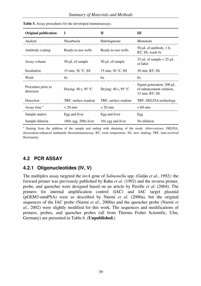
Summary of Materials and Methods
39
Table 5. Assay procedures for the developed immunoassays.
Original publication I II III
Analyte Nicarbazin Halofuginone Monensin
Antibody coating Ready-to-use wells Ready-to-use wells 50 µL of antibody, 1 h, RT, SS, wash 4x
Assay volume 50 µL of sample 50 µL of sample 25 µL of sample + 25 µL of label
Incubation 15 min, 36 °C, SS 15 min, 36 °C, SS 30 min, RT, SS
Wash 6x 6x 6x
Procedure prior to detection
Drying: 40 s, 95 °C Drying: 40 s, 95 °C Signal generation: 200 µL of enhancement solution, 15 min, RT, SS
Detection TRF, surface readout TRF, surface readout TRF, DELFIA technology
Assay time a < 20 min < 20 min < 60 min
Sample matrix Egg and liver Egg and liver Egg
Sample dilution 100x egg, 200x liver 10x egg and liver No dilution
a Starting from the addition of the sample and ending with obtaining of the result. Abbreviations: DELFIA, dissociation-enhanced lanthanide fluoroimmunoassay; RT, room temperature; SS, slow shaking; TRF, time-resolved fluorometry.
4.2 PCR ASSAY
4.2.1 Oligonucleotides (IV, V)
The multiplex assay targeted the invA gene of Salmonella spp. (Galán et al., 1992): the forward primer was previously published by Rahn et al. (1992) and the reverse primer, probe, and quencher were designed based on an article by Perelle et al. (2004). The primers for internal amplification control (IAC) and IAC target plasmid (pGEM3-mmPSA) were as described by Nurmi et al. (2000a), but the original sequences of the IAC probe (Nurmi et al., 2000a) and the quencher probe (Nurmi et
al., 2002) were slightly modified for this work. The sequences and modifications of primers, probes, and quencher probes (all from Thermo Fisher Scientific, Ulm, Germany) are presented in Table 6. (Unpublished.)

Summary of Materials and Methods
40
Table 6. Oligonucleotide primers, probes, and quencher probes used in the PCR assay.
Oligonucleotide Sequence from 5' to 3' end Modifications Nucleotide
position
Salmonella forward GTG AAA TTA TCG CCA CGT TCG GGC AA -- 371-396
Salmonella reverse CVG CRA CRC GYT CTG AAC CTT T -- 475-496
Salmonella probe ACY GTS GTY CAG TTT ATC GTT ATT AC 5' Aminolink C6 3' Phosphate
448-473
Salmonella quencher ACG ATA AAC TGR ACS ACR GT 3' Dabcyl 448-467
IAC forward TGA ACC AGA GGA GTT CTT GCA -- 523-543
IAC reverse CCC AGA ATC ACC CGA GCG A -- 667-685
IAC probe CCT TCT GAG GGT GAT TGC G 5' Aminolink C6 3' Phosphate
597-601, 604-617
IAC quencher AAT CAC CCT CAG AAG G 3' Dabcyl 600-601, 604-617
Nucleotide positions refer to GenBank accession numbers M90846 (for Salmonella) and X05332 (for IAC). Codes for wobble bases are as follows: R = A and G; S = G and C; V = A and G and C; Y = C and T. Underlined letters denote bases that are inverted in the IAC sequence compared to X05332. Missing nucleotide positions in the IAC probe and quencher denote a deletion of two base pairs in the original sequence (Ylikoski et al., 1999). Dabcyl, 4-((4-(dimethylamino)phenyl)azo)benzoic acid.
4.2.2 Labelling of probes (IV, V)
The amino groups of the probes were labelled with intrinsically fluorescent 9-dentate GenomEra chelates (Abacus Diagnostica, Turku, Finland): terbium chelate (Takalo et
al., 2008) was employed in the detection of the Salmonella target sequence and europium chelate (von Lode et al., 2003) was used for the IAC. The structures of the chelates are shown in Figure 4. Probes were labelled with an excess of chelate at a DNA concentration of 2 µg/µL in 50 mM carbonate buffer, pH 9.8. After an overnight incubation at 37 °C probes were purified with reversed-phase high performance liquid chromatography (HPLC) using a µRPC C2/C18 ST 4.6/100 column (GE Healthcare) and a 16-minute linear gradient of 14-30% acetonitrile in 50 mM triethylammonium acetate buffer (Fluka, Buchs, Switzerland) at a flow rate of 1 mL/min. The probes were dried, dissolved in 10 mM Tris-HCl, pH 8.3 containing 50 mM KCl, quantified spectrophotometrically, and stored in small aliquots at −20 °C. (Unpublished.)

Summary of Materials and Methods
41
O
NN
N
N N
N
O
OO
O
O
O O
O
Eu3+O
OH
HO
HO
HO
O
O
OH
HO
HO
HOS
MeO OMe
O O
OOOO
O
O
O
O
O O
OO
NN N
N N
N
S
Tb3+
O
NN
N
N N
N
O
OO
O
O
O O
O
Eu3+O
OH
HO
HO
HO
O
O
OH
HO
HO
HOS
O
NN
N
N N
N
O
OO
O
O
O O
O
Eu3+O
OH
HO
HO
HO
O
O
OH
HO
HO
HOS
MeO OMe
O O
OOOO
O
O
O
O
O O
OO
NN N
N N
N
S
Tb3+
MeO OMe
O O
OOOO
O
O
O
O
O O
OO
NN N
N N
N
S
Tb3+
A B
Figure 4. Structures of the GenomEra chelates. The broken lines indicate the non-covalent bonds between the ligand and the lanthanide ion.
(A) {2,2',2'',2'''-{[2-(4-isothiocyanatophenyl)ethylimino]bis(methylene)bis{4-{[4-(α-galactopyranoxy)phenyl]- ethynyl}pyridine-6,2-diyl}bis(methylenenitrilo)}tetrakis(acetato)}europium(III)
(B) {2,2',2'',2'''-{[2-(4-isothiocyanatophenyl)ethylimino]bis(methylene)bis{4-[4-(carboxymethoxy)-2-methoxy-phenyl]-pyridine-6,2-diyl}bis(methylenenitrilo)}tetrakis(acetato)}terbium(III)
4.2.3 Dry chemistry vessels (IV, V)
The ready-to-use PCR kits (Abacus Diagnostica) for the detection of Salmonella were a part of the Magda™ CA Salmonella system (Raisio Diagnostics, Turku, Finland). The PCR kits consisted of GenomEra reaction vessels containing all the reagents for the Salmonella assay in dry form (Korpimäki et al., 2005; Nurmi et al., 2005). The preparation of the dry chemistry vessels started with the application of the master mix and polymerase as separate drops in small volumes to the reaction chamber of the vessel. After an overnight drying in a vacuum desiccator at room temperature (RT), the lid and the back of the vessel were heat-sealed with foil and the vessel was barcoded. The vessels were packed in aluminium foil packages with desiccant and stored at 4 °C for routine use. (Unpublished.)
4.2.4 Sample preparation (V)
The selected sample matrices were raw meat (sliced or minced beef, pork, and poultry) and ready-to-eat meals (sandwiches, meatballs, and chicken pasta). The sample preparation was performed with the Magda CA Salmonella system, which contained reagents and consumables for sample pre-enrichment and IMS. In brief, food samples were mixed, weighed (25 g) and when required, artificially contaminated with various strains of Salmonella spp. (obtained from Raisio Diagnostics) at a level of approximately 1-10 CFU/25 g. The samples were diluted 1:10 in BPW containing the

Summary of Materials and Methods
42
Magda CA enrichment supplement and homogenized. After a 6-h incubation at 37 °C with shaking (300 rpm), a 10 mL sub-sample of the broth was collected for IMS. During the IMS step (1 h, 37 °C, 300 rpm), the target cells were captured using superparamagnetic particles coated with Salmonella-specific antibodies followed by the collection of the particles with a PickPen magnetic tool (Bio-Nobile, Turku, Finland). Subsequently, the particles were washed twice with Magda CA wash buffer and once with Magda CA elution buffer prior to suspension in a 60 µL volume of the same elution buffer for PCR analysis.
4.2.5 Assay protocol (IV, V)
The homogeneous Magda CA Salmonella PCR assay was performed with a novel PCR instrument platform, GenomEra (Abacus Diagnostica). The assay was started by manually adding a 30 µL aliquot of the sample to the ready-to-use dry chemistry vessel. Thus, the amplification reaction contained 10 mM Tris-HCl, pH 8.3, 50 mM KCl, 5.5 mM MgCl2, 0.32 mM dNTPs, 0.50 µM Salmonella primers, 0.015 µM Salmonella probe, 0.10 µM Salmonella quencher, 0.15 µM IAC primers, 0.030 µM IAC probe, 0.11 µM IAC quencher, 10 000 copies of IAC template, 2 U of polymerase, 2 g/L bovine serum albumin (BSA), 25 g/L trehalose, and 0.01% NaN3 (unpublished). The vessel was immediately inserted into the instrument where the correct assay protocol was identified and initiated based on the barcode on the vessel. The vessel was automatically and irreversibly sealed with heat inside the instrument before thermal cycling. The PCR consisted of 45 cycles, during which the vessel was transferred between blocks maintained at constant temperatures. For the end-point time-resolved fluorescence measurement, all oligonucleotides in the reaction vessel were denaturated and the vessel was then transferred to the measurement block. The fluorescence signal was measured twice at selected time intervals in order to record the signals after denaturation (D) and after hybridization (H) and the results (R) for each label were calculated with the following formula R = H/D*100 (Nurmi et al., 2008). The qualitative results were automatically reported by the instrument software based on the comparison of the result to the pre-determined cut-off value embedded in the lot-specific barcode. The total assay time was 45 min.
4.2.6 Reference method (V)
All food samples analyzed with PCR were also tested with the slightly modified culture-based reference method ISO 6579:2002 (ISO, 2002).

Summary of Results and Discussion
43
5 SUMMARY OF RESULTS AND DISCUSSION
5.1 IMMUNOASSAY CHARACTERISTICS
5.1.1 Assay format (I-III)
Immunoassay is an analytical technique employed to detect and quantify biomolecules using specific antigen-antibody interactions (for a comprehensive review, see Price and Newman, 1997). The assays can be classified as heterogeneous or homogeneous based on the requirement for separation of bound and unbound reagents prior to detection; the homogeneous assay format relies on a simple "mix and measure" principle without any separation steps, whereas the heterogeneous assay format relies on separation, which is typically achieved by immobilizing one of the reagents (either antibody or antigen) to a solid phase and by removing the unbound reagents with washing. Consequently, the separation and washing steps slow down the heterogeneous assays, but with the benefit of improved detection limits. The homogeneous assays on the other hand are rapid because of fast reaction kinetics and lack of separation. Furthermore, the simplicity of the assay concept makes homogeneous assays particularly amenable to automation. However, the dynamic range and sensitivity of many homogeneous assays is restricted by matrix interferences. (Hemmilä, 1985; Hemmilä and Webb, 1997.) Immunoassays can be further divided into two major categories: competitive (limited reagent) and non-competitive (excess reagent or sandwich) assays based on the measurement of antibody binding site occupancy by the analyte (Ekins, 1997). In the non-competitive assay format two antibodies are used to bind the analyte and the signal detected is directly proportional to the concentration of the analyte, leading to improved sensitivity, precision, and specificity, faster kinetics, and a wider working range (Ekins and Dakubu, 1985). The competitive assay format is usually applied to the analysis of haptens where the simultaneous binding of two antibodies is not possible because of the small size of the molecule. Although some innovative non-competitive assays for small molecules have been described (Barnard and Kohen, 1990; Self et al., 1994), these approaches have not been widely employed. In the competitive assay system the signal is inversely proportional to the analyte concentration, and consequently, the maximum signal level is obtained in the absence of the analyte. The main factors influencing sensitivity are the affinity and amount of the antibody, and to some extent the specific activity of the label. In general, the performance of competitive assays is limited in many ways compared to their non-competitive counterparts (Ekins and Dakubu, 1985). For example, competitive assays have a narrower working range, are usually less sensitive, and require longer incubation times than non-competitive assays. All the developed heterogeneous immunoassays were used for the detection of haptens, and therefore, the assays were based on the competitive assay format where a fixed
Summary of Results and Discussion
44
amount of labelled analyte (or labelled derivative) competed with the unlabelled analyte present in the sample for the limited number of antibody binding sites.
5.1.2 Labels and time-resolved fluorescence detection (I-III)
A wide range of lanthanide chelates has been developed over the years to suit various heterogeneous and homogeneous assay applications and to improve the time-resolved fluorescence detection (for reviews, see Hemmilä and Mukkala, 2001 and Hemmilä and Laitala, 2005). The unique fluorescence properties of lanthanide chelates, e.g. sharp emission lines, well separated excitation and emission wavelengths (i.e. large Stokes’ shift), and long and distinct fluorescence lifetimes enable spectral and temporal resolution in the measurement. As a result, a significant reduction in the background fluorescence mainly caused by autofluorescence from biological sample material and light scattering is achieved, leading to improved signal-to-noise ratios (S/N), and subsequently, increased assay sensitivity. (Siitari et al., 1983; Soini and Lövgren, 1987.) Consequently, TRF in combination with lanthanide chelate labels has been regarded as one of the most sensitive non-isotopic label technologies (Madersbacher et
al., 1993). The success of TRF began with DELFIA technology, which was introduced in the 1980s (Hemmilä et al., 1984; Soini and Lövgren, 1987). The defining characteristic of DELFIA is the use of practically non-fluorescent chelates for labelling and completing the bioaffinity reaction, after which lanthanide ions are dissociated into an acidic aqueous solution (enhancement solution) to form a strongly fluorescent secondary complex prior to signal measurement. DELFIA is a strictly heterogeneous assay technology enabling highly sensitive assays with detection limits almost down to the femtomolar range (Hemmilä et al., 1984). Nevertheless, it has some disadvantages such as the final liquid handling step, which slows down the assay and makes the assay susceptible to contamination from external lanthanide ions, and the loss of spatial localization of the signal. Assays based on DELFIA principle have been utilized in various fields, including veterinary residue analysis (Elliott et al., 1994; Crooks et al., 1998; Tuomola et al., 2002; Korpimäki et al., 2004) and clinical diagnostics (Lövgren et al., 1984; Qin et al., 1997; Eriksson et al., 2000; Nurmikko et al., 2001). The emergence of intrinsically fluorescent chelates such as the 7- and 9-dentate europium chelates by Takalo et al. (1994) and von Lode et al. (2003), respectively, provided the possibility to direct surface measurement (Lövgren et al., 1996; Meriö et
al., 1996; Pettersson et al., 2000). However, the 7-dentate lanthanide chelate was sensitive to aqueous quenching, i.e. water molecules deteriorated the signal intensity, and therefore, a drying step was required prior to surface readout (von Lode et al., 2003). Nevertheless, the simplified surface readout requiring no separate signal enhancement facilitated the automation of heterogeneous assays and enabled the development of more rapid assay protocols (Pettersson et al., 2000). The combination of surface measurement and intrinsically fluorescent chelates has been utilized
Summary of Results and Discussion
45
particularly in research and clinical diagnostics (Pettersson et al., 2000; Eriksson et al., 2003; von Lode et al., 2004; Ylikotila et al., 2005; Wittfooth et al., 2006). In all the immunoassays developed in the current work, one of the underlying themes was the use of TRF and intrinsically fluorescent 7-dentate europium chelates. All the selected analytes had only one reactive group available and the direct labelling of this group did not provide adequate signal levels for the surface measurement (unpublished). However, the labelling degree could be increased by using carrier molecules with several reactive groups. This type of multiple labelling has been shown to enhance the sensitivity of the assays despite some unwanted side effects such as increased non-specific binding (Diamandis, 1991; Qin et al., 2001). Therefore, to obtain a better labelling degree, protein carriers were used in the preparation of the labels. The analytes (or their derivatives) and intrinsically fluorescent lanthanide chelates were sequentially conjugated to these carriers. Because both the isothiocyanate group of the chelate and the DCC/NHS activated carboxyl group of the analyte reacted with free amino groups on the protein carrier, the amounts of the analyte and the chelate were roughly optimized for the reaction. In particular, the amount of lanthanide chelates had to be high enough to obtain a sufficient signal level for the surface measurement. Differences in assay performance characteristics, i.e. in signal level and inhibition profile, were observed between label batches due to inherent variation in the sequential coupling reaction. Thus, the particular label concentration used in the assay was re-optimized every time a new label batch was taken into use. If surface measurement is used together with conventional antibody coating procedures, only a fraction of the bound label is actually situated within the area of the excitation beam as opposed to dissociation-enhanced measurement where the signal is measured from solution covering the entire area of the bound label. Thus, surface readout can be more susceptible to local variations in antibody coating. (Soukka et al., 2001.) The shortcomings of surface measurement can in part be compensated by optimizing washing and shaking procedures, by producing a dense and correctly orientated binding surface preferably with (site-specifically oriented) antibody fragments instead of monoclonal or polyclonal antibodies, and/or by confining the capture antibody to the area of the excitation beam, i.e. spot coated wells, to further concentrate the signal and improve assay sensitivity (Soukka et al., 2001; Peluso et al., 2003; Ylikotila et al., 2006; Välimaa et al., 2008). All the assays developed relied on the use of polyclonal antibodies mainly because of the simplified, economical, and rapid production scheme. The assays were performed in microtiter wells pre-coated with anti-rabbit IgG. This secondary antibody was treated with acidic solution to increase the hydrophobicity of the Fc portion of the molecule, and thus, to direct its attachment to the solid surface (Conradie et al., 1983; Kakabakos et al., 1990; van Erp et al., 1992). Accordingly, the analyte-specific rabbit antibody was correctly orientated with reduced loss of antibody conformation or activity. The intra-assay variation of the assays was typically below 10%, whether the surface readout (I, II) or dissociation-enhanced measurement (III) was used, which indicated that the applied

Summary of Results and Discussion
46
coating procedure with polyclonal antibodies was feasible in terms of variation despite the measurement mode. Although the monensin assay (III) was performed manually using DELFIA technology, it could have been performed in the same format as the other assays because of the intrinsically fluorescent label. The effect of the measurement mode, dissociation-enhanced measurement and surface readout on assay performance was tested with various incubation times in a buffer system. The signal level and S/N were approximately two times higher with DELFIA, which resulted in an improved limit of detection (LOD). However, the IC50 (concentration that inhibits 50% of the maximum signal) of the assay was not significantly affected by the measurement mode (unpublished). Because the matrix effects might further lower the signal levels, the measurement based on DELFIA principle was selected for the monensin assay. Nevertheless, the benefits of surface readout such as shorter and simpler assay protocols would generally favour its use.
5.1.3 Dry chemistry (I, II)
The aim of the all-in-one dry chemistry immunoassays was to facilitate and speed up straightforward one-step assays by drying all assay-specific reagents into microtiter wells in advance (Lövgren et al., 1996). In the dry chemistry assays developed, an insulation layer consisting mainly of buffer components and optimized amounts of sugar and protein was required to prevent the premature binding of the label to the antibody. The assay performance was significantly deteriorated if the labelled analyte leaked through the layer during storage. This effect might partly be derived from the differences in the affinity of the antibody for the analyte and the labelled analyte-protein conjugate and the incubation time used in the assay. Consequently, the insulation layer was designed to be dense: 50 µL of buffer containing trehalose (6-8%) and casein (0.2%) was dried in each well to cover the entire bottom of the well. Furthermore, the label was applied on the edge of the well where the layer was thicker than in the middle. The careful optimization of the components of the insulation layer ensured that on the one hand, the performance characteristics of the assay were not compromised due to the reagent composition, and on the other hand, the wells remained stable even during long-term storage (Lövgren et al., 1996). The stability of the all-in-one dry chemistry wells at RT was monitored during a period of one year at selected time points by analyzing standard solutions at three different concentration levels. The data from the stability study for halofuginone is presented in Figure 5. During the study no significant changes in the performance of the assays, with regard to signal level and inhibition profile were observed. Thus, the wells required no special storage conditions and were stable for at least 12 months at RT. This compares well with the stability reported for other immunoassays using the same dry chemistry concept (Pettersson et al., 2000; von Lode et al., 2004).

Summary of Results and Discussion
47
0
20
40
60
80
100
0 1 2 3 4 6 8 10 12
0.5 ng/mL 5 ng/mL
Time (months)
B/B
0 (
%)
Figure 5. Real-time stability study for the halofuginone assay. The wells containing the dried reagents were stored at RT. The columns represent the B/B0 values (calculated by dividing the fluorescence signal of the standard by the signal of the zero standard) for each time point analyzed with two or four replicates. (Unpublished.)
5.1.4 Sample preparation (I-III)
The purpose of sample preparation was to remove interfering substances, to concentrate the amount of target analyte, and to produce a homogeneous, representative sample for analysis. The selected sample matrices were poultry liver (I, II) and eggs (I-III). Liver was preferred as a test material over muscle because muscle generally contains lower concentrations of residues than liver. For example, nicarbazin residue content in liver has been found to be approximately 10 to 30 times higher than the concentration in the corresponding muscle samples (Veterinary Residues Committee, 2007). A relatively straightforward sample preparation procedure was developed for all assays using standard sample preparation techniques for residue analysis (Stolker and Brinkman, 2005). The sample preparation for both matrices started with sample homogenization, after which residues were extracted from the sample matrix with acetonitrile. Shaking (III) or sonication (I, II) was used to improve the efficiency of the extraction. In the case of liver samples, additional purification, i.e. hexane wash, was required to reduce the matrix effects (I, II). The dilution of the samples prior to assay adjusted the assay sensitivity to the appropriate concentration range and further reduced the amount of interfering substances (I, II). The sample preparation also included evaporation steps, which considerably slowed down the procedure. Nevertheless, the evaporation step prior to reconstitution could be omitted if the assay sensitivity was adequate to analyze more dilute samples. Accordingly, the sample preparation procedure could be shortened if the extract was simply diluted to eliminate the harmful effects of organic solvents on antibodies and applied directly to the assay. This approach might prove to be useful at least for the nicarbazin assay (I) where significant sample dilution is used.
Summary of Results and Discussion
48
5.1.5 Assay performance (I-III)
At the time of the assay development in the current work, there were no official MRLs in the EU for the residues of monensin, halofuginone, and nicarbazin used as feed additives in poultry production. Consequently, the sensitivity requirements for the assays, that is the concentration levels the assays should be able to detect, were selected based on existing guidelines. In the monensin assay (III), the target was based on the 10 µg/kg action level used in the Finnish national residue control programme (National Food Agency et al., 2005). An MRL of 30 µg/kg for halofuginone residues in bovine liver was used as a guideline in the development of the halofuginone assay (II) for both sample matrices (European Medicines Agency, 2001). For the nicarbazin assay (I), the targets for DNC residues were set to 200 µg/kg (Joint FAO/WHO Expert Committee on Food Additives, 1999) and 100 µg/kg (Veterinary Medicines Directorate, 2000) in liver and eggs, respectively. In addition to the sample preparation, the assay parameters, including buffer composition, concentration of the antibody and label, and assay incubation time and temperature were optimized in order to achieve the required detection limits. All assays were performed in a simple one-step format where the label and sample were incubated simultaneously in the antibody-coated wells. The dry chemistry assays (I, II) were performed with the fully automated immunoanalyzer, which enabled a user-friendly assay with a total turn-around-time of less than 20 min. The incubation time of the monensin assay (III) was set to 30 min, although the washing, enhancement, and measurement steps somewhat lengthened the procedure. As discussed earlier, the monensin assay could also have been transferred to the automated assay format. The shortening of the incubation time from 30 min to 15 min as with the other analytes should also be feasible because of the elevated incubation temperature in the immunoanalyzer speeding up the reaction rate. Moreover, additional assay optimization could further compensate the different measurement mode. The monensin assay was carried out in a conventional microtiter plate format, whereas the automated dry chemistry assays relied on the use of single microtiter wells. Therefore, although monensin assay was slower to perform, it actually had a better assay throughput than the automated assays. Although the assays were designed to function primarily as screening assays providing qualitative results, quantitative data could also be obtained. The standards used in both approaches were prepared in negative sample matrix and treated and diluted similarly to unknowns (Tuomola, 2000). In the qualitative assay approach, a positive control spiked at a selected concentration level provided the threshold signal, which was used to classify the unknown samples as negative or potentially positive (Figure 6). In the quantitative approach, a standard curve was established.

Summary of Results and Discussion
49
CUT-OFF
Negative
samples
Potentially
positive
samplesACTION LEVEL
Flu
ore
scence s
ignal
Figure 6. Principle of sample classification in the qualitative assay approach. The positive control provides the signal level (cut-off), to which unknown samples are compared. If the signal is higher, the sample is negative (compliant). If the signal is lower, then the sample is potentially positive (non-compliant) and should be subjected to confirmatory analysis. The safety marginal (arrow) between the cut-off and actual action level (such as MRL) can be adjusted accordingly.
The LOD (I, II) and CCα (III) of the assays were based on the mean concentration of blank samples + 3 SD. The limit of quantification (LOQ) corresponded to a particular B/B0 ratio, which was selected to be between 0.80 and 0.85 (I, II). The CCβ (I-III) was determined according to Commission Decision 2002/657/EC based on the rate of false compliant results (Anonymous, 2002a). A summary of assay performance characteristics is presented in Table 7. In general, the sensitivities obtained compared well to the current requirements. Recently, EFSA was given the responsibility for the MRL evaluation of feed additives (Anonymous, 1990, 2003b). Thus, if the sensitivity requirements were to change in the future, the LOQ/CCβ of the assays could be
of all assays was typically below 10% and the interassay variation was less than 15%. Furthermore, all assays had a good recovery of added standards; the recoveries varied between 87% and 116% depending on the analyte and matrix. To properly assess whether true unknown samples behave similarly to their spiked counterparts, incurred samples should be included in the validation process. During the current study, however, the acquirement of incurred samples was difficult, and therefore, only a small number of samples containing DNC residues could be tested (I). The preliminary data indicated that the spiked and incurred samples performed alike in the assay, which was also in line with the recovery results. Moreover, the results from the immunoassay were in agreement with an LC-MS-MS method used to analyze the same samples (Yakkundi et al., 2001). Nevertheless, a larger panel of incurred samples should be analyzed prior to any further conclusions.
re-adjusted, e.g. by modifying the dilution factor of the samples. The intra-assay variation
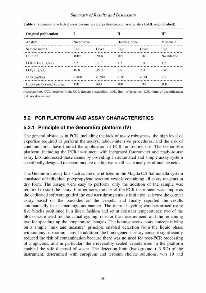
Summary of Results and Discussion
50
Table 7. Summary of selected assay parameters and performance characteristics (I-III, unpublished).
Original publication I II III
Analyte Nicarbazin Halofuginone Monensin
Sample matrix Egg Liver Egg Liver Egg
Dilution 100x 200x 10x 10x No dilution
LOD/CCα (µg/kg) 3.2 11.3 1.7 1.0 1.2
LOQ (µg/kg) 10.0 35.0 2.5 2.0 n.d.
CCβ (µg/kg) < 100 < 200 < 30 < 30 < 2
Upper assay range (µg/kg) 150 400 100 100 100
Abbreviations: CCα, decision limit; CCβ, detection capability; LOD, limit of detection; LOQ, limit of quantification; n.d., not determined.
5.2 PCR PLATFORM AND ASSAY CHARACTERISTICS
5.2.1 Principle of the GenomEra platform (IV)
The general obstacles in PCR, including the lack of assay robustness, the high level of expertise required to perform the assays, labour-intensive procedures, and the risk of contamination, have limited the application of PCR for routine use. The GenomEra platform, including the PCR instrument with integrated fluorometer and ready-to-use assay kits, addressed these issues by providing an automated and simple assay system specifically designed to accommodate qualitative small-scale analysis of nucleic acids. The GenomEra assay kits such as the one utilized in the Magda CA Salmonella system consisted of individual polypropylene reaction vessels containing all assay reagents in dry form. The assays were easy to perform: only the addition of the sample was required to start the assay. Furthermore, the use of the PCR instrument was simple as the dedicated software guided the end user through assay initiation, selected the correct assay based on the barcodes on the vessels, and finally reported the results automatically in an unambiguous manner. The thermal cycling was performed using five blocks positioned in a linear fashion and set at constant temperatures; two of the blocks were used for the actual cycling, one for the measurement, and the remaining two for speeding up the temperature changes. The homogeneous assay concept relying on a simple "mix and measure" principle enabled detection from the liquid phase without any separation steps. In addition, the homogeneous assay concept significantly reduced the risk of contamination because there was no need for post-PCR processing of amplicons, and in particular, the irreversibly sealed vessels used in the platform enabled the safe disposal of waste. The detection limit (background + 3 SD) of the instrument, determined with europium and terbium chelate solutions, was 19 and
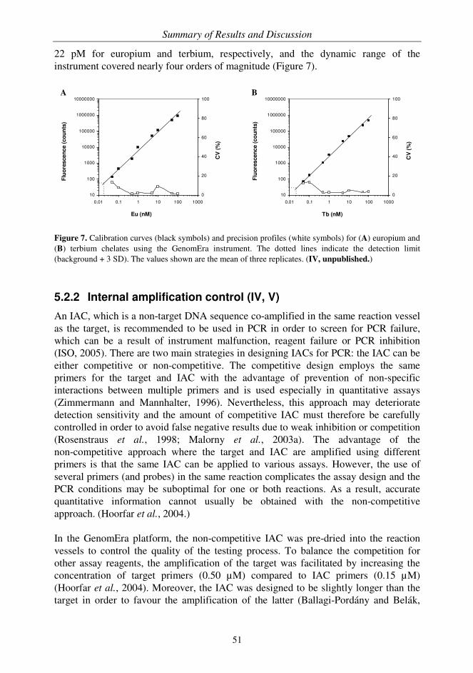
Summary of Results and Discussion
51
10
100
1000
10000
100000
1000000
10000000
0.01 0.1 1 10 100 1000
0
20
40
60
80
100
Eu (nM)C
V (
%)
Flu
ore
scen
ce (
co
un
ts)
10
100
1000
10000
100000
1000000
10000000
0.01 0.1 1 10 100 1000
0
20
40
60
80
100
Tb (nM)
Flu
ore
scen
ce (
co
un
ts)
CV
(%
)
22 pM for europium and terbium, respectively, and the dynamic range of the instrument covered nearly four orders of magnitude (Figure 7). A B
Figure 7. Calibration curves (black symbols) and precision profiles (white symbols) for (A) europium and (B) terbium chelates using the GenomEra instrument. The dotted lines indicate the detection limit (background + 3 SD). The values shown are the mean of three replicates. (IV, unpublished.)
5.2.2 Internal amplification control (IV, V)
An IAC, which is a non-target DNA sequence co-amplified in the same reaction vessel as the target, is recommended to be used in PCR in order to screen for PCR failure, which can be a result of instrument malfunction, reagent failure or PCR inhibition (ISO, 2005). There are two main strategies in designing IACs for PCR: the IAC can be either competitive or non-competitive. The competitive design employs the same primers for the target and IAC with the advantage of prevention of non-specific interactions between multiple primers and is used especially in quantitative assays (Zimmermann and Mannhalter, 1996). Nevertheless, this approach may deteriorate detection sensitivity and the amount of competitive IAC must therefore be carefully controlled in order to avoid false negative results due to weak inhibition or competition (Rosenstraus et al., 1998; Malorny et al., 2003a). The advantage of the non-competitive approach where the target and IAC are amplified using different primers is that the same IAC can be applied to various assays. However, the use of several primers (and probes) in the same reaction complicates the assay design and the PCR conditions may be suboptimal for one or both reactions. As a result, accurate quantitative information cannot usually be obtained with the non-competitive approach. (Hoorfar et al., 2004.) In the GenomEra platform, the non-competitive IAC was pre-dried into the reaction vessels to control the quality of the testing process. To balance the competition for other assay reagents, the amplification of the target was facilitated by increasing the concentration of target primers (0.50 µM) compared to IAC primers (0.15 µM) (Hoorfar et al., 2004). Moreover, the IAC was designed to be slightly longer than the target in order to favour the amplification of the latter (Ballagi-Pordány and Belák,

Summary of Results and Discussion
52
1996; Hoorfar et al., 2004). However, in retrospect the reduction of the IAC copy number, which was designed to be high to compensate for the losses during storage, might be an even more efficient way to limit the competition for general reagents with the additional benefits of enhanced assay sensitivity and easier identification of false negative results. This type of approach could be useful particularly for assay kits stored in cold conditions where reagent losses are reduced compared to storage at RT (Trapmann et al., 2004). (Unpublished.)
5.2.3 Labels and time-resolved fluorescence detection (IV, V)
The emergence of stable fluorescent lanthanide chelates requiring no ion dissociation provided potential for homogeneous assays based on TRF. Early reports of homogeneous immunoassays using environmentally sensitive fluorescent lanthanide chelates exhibiting different signal intensity depending on the immediate chemical environment were published in the late 1980s (Hemmilä et al., 1988; Barnard et al., 1989) and later these types of chelates were applied to nucleic acid assays (Nurmi et
al., 2000b). Subsequently, chelate structures such as 9-dentate chelates forming a nine-coordinate complex with the lanthanide ion to protect the lanthanide ion from aqueous quenching (von Lode et al., 2003; Hakala et al., 2005; Takalo et al., 2008), and even 10-dentate chelates (Takalo and Rosenberg, 2006), have been developed that are particularly suitable for homogeneous assay concept. In the developed nucleic acid assay, intrinsically fluorescent 9-dentate chelates were used for labelling of the oligonucleotide probes. The designing of the probes was relatively simple because only one label moiety without any special secondary structures such as in Molecular Beacon probes and Scorpion primers was required. After labelling, the probes were purified with HPLC in order to separate the non-reacted label and non-labelled oligonucleotides from the labelled product. Thorough HPLC purification decreased the non-specific background, which is particularly important in homogeneous assays with no separation. The Salmonella
assay utilized two different sets of lanthanide chelate labelled probes and their complementary quencher probes; the target-specific probe was labelled with terbium, the IAC-specific probe with europium, and both the quenchers with a non-fluorescent quencher, dabcyl. The probes were designed not to hybridize to the template during thermal cycling. The time-resolved fluorescence detection in the homogeneous Salmonella assay was performed as end-point measurement based on the competitive hybridization principle (Morrison et al., 1989), which has also been used successfully in combination with TRF previously (Kiviniemi et al., 2003; Ylikoski et al., 2004; Kiviniemi et al., 2005; von Lode et al., 2007). The principle was simple: if no PCR products were present during the measurement, the fluorescent probes were hybridized to their complementary quenchers giving only a low level of signal. However, if a target was present and amplified, the proportion of labelled probes bound to the target were not quenched and caused an increase in the fluorescence signal. Thus, the generated signal was dependent on the amount of the amplified product at the end of the PCR. The results for Salmonella and IAC amplification were calculated based on
Summary of Results and Discussion
53
two sequential end-point time-resolved fluorescence measurements where the signal was recorded first after denaturation and then again after hybridization at a constant temperature (Nurmi et al., 2008). The first measurement provided the maximum obtainable signal with all probes free in solution and during the second measurement the signal level depended on the amount of amplified product present. The results calculated for both Salmonella and IAC were compared to lot-specific cut-off values in order to classify the sample as positive, negative or inconclusive. For the sample to be positive for Salmonella, only a successful amplification of the Salmonella target providing a result above the cut-off was required.
5.2.4 Dry chemistry (IV, V)
The labour-intensive phase in PCR reagent setup can be avoided by using ready-to-use mixtures, which facilitate automation and offer speed and reproducibility to the assay. Furthermore, the risk of pipetting errors and contamination is minimized and the amount of waste due to unused materials is reduced compared to bulk reagent analyses. In particular, the possibility to store reagents at RT, which can be accomplished with pre-dried reagents, can be advantageous for the end user. In general, the commercial PCR kits for Salmonella, as discussed in the literature review, still require several pipetting steps before the sample can be added to the assay. Thus, most kits cannot be characterized as truly ready-to-use systems, although exceptions exist, such as the kits by DuPont Qualicon and Idaho Technology. The dry chemistry assay developed in the current work provided a genuinely ready-to-use system because only the addition of the sample was required to start the assay. Freeze-drying (lyophilization) is a commonly used method for drying PCR components. During freeze-drying, reagents are significantly concentrated and protective reagents are, therefore, recommended to be included in the mixture (Franks, 1990). Trehalose has proven to be especially efficient in protecting and stabilizing proteins (Roser, 1991; Colaço et al., 1992; Sola-Penna and Meyer-Fernandes, 1998; Kaushik and Bhat, 2003). Accordingly, trehalose has been included in freeze-dried PCR mixtures (Klatser et al., 1998; Tomlinson et al., 2005; Aziah et al., 2007), which have been stable even for 12 months at RT (Klatser et al., 1998). However, because freeze-drying is a rather cumbersome and expensive technique, other approaches to produce dried components for PCR have been reported; reagents have been embedded in wax and dried using conventional methods with or without protective reagents (Kaijalainen et al., 1993; Blair et al., 1994; Wolff et al., 1995; Nurmi et al., 2001). The dry chemistry concept in combination with TRF, first described in the immunoassay field (Lövgren et al., 1996) and later in PCR (Nurmi et al., 2001), has been further modified to suit the GenomEra system (Korpimäki et al., 2005). In the current work the master mixture and enzyme were also dried as separate spots in order to avoid unwanted side effects during drying and immediately after adding the sample. This separation of key PCR components mimicked a hot-start like property even if a standard DNA polymerase was used. In addition to its stabilizing effect, trehalose is

Summary of Results and Discussion
54
0
10
20
30
40
50
60
0 2 3 4 5
NTC PTC100 PTC500 cut-off
0
10
20
30
40
50
60
0 3 6 9 12
NTC PTC100 PTC500 cut-off
Time (months)
Calc
ula
ted
resu
lt
Time (months)
Calc
ula
ted
resu
lt
recommended, e.g. by Cepheid (Sunnyvale, CA, USA) to be included in PCR to improve the productivity of amplification. Furthermore, proteins such as BSA have been shown to decrease non-specific binding of reagents and relieve PCR inhibition (Kreader, 1996; Khandurina et al., 2000; Erill et al., 2003; Kricka and Wilding, 2003). Therefore, protein and sugar were added to the reaction mixture with the aim of protecting the reagents, enhance the stability and assay performance, and also aid in the dissolution of the reagents upon addition of the sample (unpublished). Stability testing was performed with dried reagents stored at three temperatures (−20 °C, 4 °C, and RT). The results for the kits stored at 4 °C and RT are shown in Figure 8. In general, the results were similar for assay kits whether they were frozen or cold stored. The performance of the assay kits stored at 4 °C did not significantly change during the one year stability study. However, the performance of the kits stored at RT started to weaken as early as at four months. Thus, the stability of the reagents was highly dependent on the storage conditions. A B
Figure 8. Real-time stability study for the Salmonella dry chemistry assay kits stored at (A) 4 °C and (B) RT. The data shown is based on the measurement of terbium fluorescence from the analyte-specific probe. The performance of the assays was tested at various time points with three types of controls: no template control (NTC) and positive template controls (PTC) containing 100 and 500 copies of Salmonella DNA, using at least four replicates. Three different lots with individual lot-specific cut-off values, indicated by lines, were used throughout the study. The cut-offs were used to classify the samples as Salmonella positive or negative based on the time-resolved fluorescence measurements. (IV,
unpublished.)
5.2.5 Sample preparation (V)
As discussed in the literature review, most PCR-based methods used in food diagnostics still rely on traditional sample enrichment prior to detection. PCR-based methods are particularly sensitive to inhibitory effects resulting from substances in the sample material, from sample preparation or both (Rossen et al., 1992; Wilson, 1997). Therefore, proper sample preparation provides the key to the robustness and overall performance of PCR. Consequently, the aim of sample preparation is to produce a homogeneous, representative sample for PCR analysis containing a high amount of target and a minimal amount of interfering substances. In the Magda CA Salmonella
Summary of Results and Discussion
55
system, sample preparation was based on a short non-selective pre-enrichment in supplemented BPW followed by IMS of target cells. Because bacterial cells in naturally contaminated food samples are frequently damaged due to adverse conditions during food processing, sub-lethally injured cells were used in the current work for spiking to mimic the real situation. Different types of stress treatments causing sub-lethal injury and their effect on the recovery and analysis of Salmonella and other foodborne pathogens have been reported (Mansfield and Forsythe, 1996; Rijpens et al., 1999; Brashears et al., 2001; Wuytack et al., 2003; Jasson et al., 2007). The selected stress treatments in this work were heat (30 min, 55 °C) and freezing (24 h, −20 °C). Despite the use of sub-lethally injured cells, the pre-enrichment step used in the Magda CA system could be considerably shortened compared to the time (18±2 h) utilized in the reference method (ISO, 2002). This was achieved by optimizing the protocol with regard to broth composition and aeration. In contrast to our studies, shaking during pre-enrichment has been reported not to have a beneficial effect on the growth of Salmonella during short-term incubation (Josefsen et
al., 2007). However, the shaking speed in the Magda CA Salmonella system was faster than reported by Josefsen et al. (2007), which might have improved the accessibility of nutrients, and thus, increased the growth rate of Salmonella along with other bacteria. IMS is based on the use of antibody-coated paramagnetic particles or beads, which enable the separation and specific collection of the target bacteria from the sample, the reduction in the amount of sample components, background micro-organisms and PCR inhibitors, and the concentration of the sample by reducing the sample volume (Olsvik et al., 1994; Šafařík and Šafaříková, 1999). Anti-Salmonella antibody-coated beads can be tailor-made or obtained in ready-to-use format, e.g. from Invitrogen (Carlsbad, CA, USA). In general, the choice of antibody depends on the application and whether the aim is to capture one or multiple species from a particular genus. In the case of genus Salmonella, the situation is particularly challenging because the number of identified serovars is already over 2 500 (Grimont and Weill, 2007). Early publications describing the use of antibody-coated magnetic particles in the detection of Salmonella utilized both monoclonal (Mattingly, 1984; Mattingly and Gehle, 1984) and polyclonal antibodies (Skjerve and Olsvik, 1991). The magnetic particles employed in the Magda CA Salmonella system were coated with polyclonal antibodies against common structural antigens of Salmonella (unpublished). Due to batch-to-batch variation of the antibodies, every new batch was tested before use to confirm the performance of the antibody. Generally, IMS is carried out in a volume of 1 mL or less in order to allow easy handling of samples in microtiter wells or microtubes. Nevertheless, the use of larger volumes for IMS can be beneficial, i.e. improve the recovery of target cells, and thereby enhance the sensitivity of the assay (Skjerve and Olsvik, 1991; Fluit et al., 1993; Ogden et al., 2000; Rotariu et al., 2005; Notzon et al., 2006), although the amount of background flora and PCR inhibitors also tend to increase with the sample volume. An interesting commercial example of automated IMS is the Pathatrix system

Summary of Results and Discussion
56
(Matrix MicroScience) where the entire sample volume of 250 mL is re-circulated through a magnetic capture phase using a high flow rate, which in combination with high volume washing is claimed to efficiently reduce non-specific binding (Yuk et al., 2006; Warren et al., 2007). Compared to the normal type of IMS, where only a small fraction of the total sample volume is used for IMS, Pathatrix enables more sensitive assays with shorter enrichment times. Ogden et al. (2000) used a traditional IMS system and reported that the use of a 10 mL sample in IMS provided a 9-fold improvement in the recovery of target cells compared to a 1 mL sample. The recovery was not further improved, however, with larger sample volumes (50 mL) due to less effective collection of beads and less probable collision of the beads with the target cells. These drawbacks could partly be compensated for by using a more powerful magnet and by increasing the mixing time or the amount of magnetic particles. Nevertheless, prolonging the time of immunoreaction tends to concurrently increase the amount of non-specific binding due to sample matrix and background flora (Skjerve et al., 1990; Fratamico et al., 1992; Vermunt et al., 1992). This non-specific binding in IMS can be reduced, for example, with detergents and optimized washing procedures (Morgan et al., 1991; Vermunt et al., 1992). Because of the short pre-enrichment step in the Magda CA system, it was particularly important to have good recovery at the IMS phase, which replaced the selective enrichment. Therefore, a sub-sample volume of 10 mL was used for IMS to increase the recovery of target cells without substantial negative effects from the accumulation of inhibitory material compared to a 1 mL sample. The selected incubation time for the beads was based on the optimized combination of assay time, sample volume, amount of beads, recovery, and amount of non-specific binding. Several authors have reported IMS-PCR assays for various matrices where a separate lysis step (Widjojoatmodjo et al., 1991; Widjojoatmodjo et al., 1992; Fluit et al., 1993) or even DNA extraction (Notzon et al., 2006; Yuk et al., 2006) has been performed prior to PCR. However, in the Magda CA system with robust time-resolved fluorescence detection the immunomagnetic beads could be added to PCR as such without any additional steps and the DNA from the cells was released into the solution during the initial denaturation. As discussed, there are several factors affecting IMS, e.g. duration, composition, and temperature of the enrichment phase, number of background micro-organisms, sample matrix, amount, size, and surface area of the antibody-coated magnetic particles, specificity and affinity of the antibody, and the volume, duration, temperature, and mixing rate of the immunoreaction. IMS has been considered to be laborious, not suitable for high throughput applications, and expensive because of the antibodies and disposable beads. However, the use of the PickPen magnetic tool has been shown to increase the sample throughput and improve the efficiency of the IMS procedure (Nou et al., 2006). In addition, the cost-effectiveness of the assay can in part be targeted by optimizing the amount of beads required per reaction.

Summary of Results and Discussion
57
5.2.6 Assay performance (V)
According to legislation, there should be no Salmonella cells present in 25 g of food (or 10 g depending on the food category) (Anonymous, 2005a). Consequently, this places considerable demands on assay performance. The PCR assay developed was optimized by selecting proper reagents, reaction conditions, and sample preparation system. In addition to the primers targeting the invA gene, the specificity of PCR was further increased with probes recognizing correctly amplified target DNA. Based on the results of the inclusivity and exclusivity tests, the selectivity of the Magda CA assay system was 100%. The inherent sensitivity of PCR to inhibition was taken into account in the sample preparation where a final sample volume of 60 µL was obtained. Therefore, in the case of an inconclusive result (neither target nor IAC is amplified), the test could easily be repeated with different dilutions of the sample, although at the cost of assay sensitivity. The validation of the Magda CA Salmonella system was performed with various food samples (n=107), which were artificially contaminated with Salmonella cells approximately at a level of 1-10 CFU/25 g of food. Unfortunately, the use of naturally contaminated samples was not possible because Salmonella contamination is very rare in Finland (EVIRA, 2006b). The overall relative accuracy, sensitivity, and specificity of the system were 99.1%, 98.4%, and 100%, respectively. Further studies should, however, be conducted to assess the assay performance more thoroughly with a larger sample panel using various sample matrices and naturally contaminated samples before the interlaboratory study, which is required to complete the validation process, can be started (ISO, 2003). Nevertheless, the validation data obtained from the method comparison study holds a promise that performance comparable to commercial assays and other reported assays for foodborne pathogens can be achieved (Malorny et al., 2003b; Abdulmawjood et al., 2004; Malorny et al., 2004; Malorny et al., 2007). The main advantage of the system was the exceptional rapidity enabling the entire process to be completed within one workshift of 8 hours. This compares favourably with the best commercial PCR assays, which still require approximately 24 h for obtaining the result, not to mention the time required by the reference method. Consequently, the system developed could be of particular interest to the food industry where prompt reactions to contamination and decreased storage times are valued.
Conclusions
58
6 CONCLUSIONS
Food safety policy in the EU relies on the integrated "from farm to fork" approach aiming to provide a high level of food safety throughout the food chain. As part of this effort, foodstuffs are routinely monitored for various chemical and biological hazards, and therefore, reliable and rapid methods of analysis are required. In this study, rapid and user-friendly molecular methods using TRF as the detection technology were developed for two groups of food-related hazards; immunoassays were employed in the screening of selected veterinary residues and a novel PCR platform suitable for rapid nucleic acid testing was developed for the detection of Salmonella. In addition, the applicability of the dry chemistry assay concept to both types of methods was evaluated. The main conclusions based on the original publications are as follows:
− The combination of lanthanide chelates and time-resolved fluorescence detection provided a robust assay concept applicable to immunoassays and PCR with the possibility for multiplexing by using the europium and terbium chelates. The 7-dentate intrinsically fluorescent europium chelates utilized in the heterogeneous immunoassays allowed signal measurement either using DELFIA technology or simple surface readout after a drying step. The detection could be further simplified with the 9-dentate chelates enabling the homogeneous assay concept with signal measurement directly from the liquid phase.
− The dry chemistry assay concept based on ready-to-use, stable, dried reagents
provided a simple and user-friendly assay system for immunoassays and PCR alike. The concept facilitated automation, which further simplified and accelerated the assays.
− Sample preparation was the most laborious step in all assays, although the
procedures were designed to be simpler than those traditionally used. Sample preparation for the immunoassays was based on the extraction of coccidiostat residues from eggs and chicken liver with organic solvents. Further time-savings could be achieved by replacing the final evaporation step in the procedure with simple dilution, although at the expense of assay sensitivity. The sample preparation for the Salmonella assay was comprised of the non-selective pre-enrichment of food samples followed by the IMS step. Compared to the reference method, significant reductions in the sample preparation time were achieved as the pre-enrichment time was shortened to 6 h and IMS replaced selective enrichment. However, the basic need for sample enrichment still represents a major bottleneck in sample preparation.

Conclusions
59
− The competitive immunoassays for the selected anticoccidials were based on the heterogeneous assay format. The automated assays for nicarbazin and halofuginone with a turn-around-time of less than 20 min consisted of five steps: sample addition, incubation, washing, drying, and signal measurement from the dried surface. The assays represented the first competitive time-resolved fluoroimmunoassays for residue analysis utilizing the full potential of the dry chemistry concept and the automated immunoassay platform with surface measurement. The monensin assay was performed manually using DELFIA technology. Although the actual assay procedure was slower because of the additional signal generation step, the microtiter plate format provided a higher throughput than the single wells used in the
mode with acceptable performance characteristics. − The multiplex PCR assay for Salmonella was performed with a novel PCR
instrument platform using homogeneous end-point detection based on the competitive hybridization principle and the use of TRF. The assay developed provided qualitative assay results for Salmonella spp. with a detection limit of 1-10 CFU/25 g of food. The entire process could be performed within 8 h. Thus, the system which included rapid sample preparation together with a sensitive PCR assay offered significant time-savings compared to other methods available for the detection of Salmonella.
Altogether, the main issues to be solved in the field of food safety testing relate to sample preparation, assay concepts, and performance requirements, as well as instrumentation and automation where small-scale systems for field testing and high throughput systems with the possibility for multi-analyte analysis are required. In particular, simplicity, sensitivity, and speed of sample preparation and assays are of key importance. In this study, several steps were taken towards these goals. The sample preparation procedures were simplified in order to reduce the total assay times and the combination of dried reagents and TRF provided a sensitive, rapid, and easy-to-use assay concept amenable to automation. The general performance characteristics of the assays were good and the target levels set for detection were achieved. All in all, current and future advances in food diagnostics could be expected to lead to an increased testing rate, which would in turn give us a more comprehensive picture of the state of food safety and the opportunity to improve it further.
automa ed assays. All assays could be used either in qualitative or quantitative t

Acknowledgements
60
ACKNOWLEDGEMENTS
This study was carried out at the Department of Biotechnology, University of Turku and at Abacus Diagnostica Ltd. Financial support from the European Commission, the Ministry of Agriculture and Forestry, and the Finnish Funding Agency for Technology and Innovation is gratefully acknowledged. The Academy of Finland is thanked for the grant "Doctoral studies of employed persons". I want to express my sincere gratitude and respect to my supervisor Professor Timo Lövgren for giving me the opportunity to work in the inspiring atmosphere at the Department of Biotechnology. The good foundation obtained at the Department of Biotechnology enabled a smooth transition from academia to industry. I am deeply grateful to Dr. Mika Tuomola, the main supervisor of my thesis, who has guided me through the world of science and given excellent advice during the years regarding the mysteries of food safety testing. I highly appreciate his support and encouragement during this journey. I am grateful to Professor Hannu Korkeala and Dr. Geoff Barnard for reviewing this thesis and for providing constructive and valuable comments. I also want to thank Dr. Mike Nelson for revising the language of the thesis. I warmly thank the co-authors of the original publications: Professor Christopher Elliott, Dr. Lisa Connolly, and Dr. Steven Crooks, the participants of the "Poultry-check" project, for their contributions in the antibody development and sample preparation and for the opportunity to visit their laboratory in Belfast; Pekka Peippo for sharing the world of coccidiostats; Dr. Tero Soukka for being an endless source of good ideas; Hannu Kojola for giving technical advice regarding instrumentation issues; Dr. Teemu Korpimäki for the significant input in the development of the Magda CA system at Raisio Diagnostics together with Dr. Mika Tuomola; and Dr. Piia von Lode, Anniina Syrjälä, Otto Kauko, and Dr. Jussi Nurmi for giving me the opportunity to work at Abacus Diagnostica in such an enthusiastic atmosphere. In particular, I am deeply grateful to Piia and Anniina for their excellent contribution to this work. I owe my thanks to the people who have worked with me over the years for shorter or longer terms in this project. In particular, I wish to acknowledge Pauliina Helo, Marjo Haanperä, Heidi Hynninen, Minna Jantunen, and Ulla Helmi for their contributions and all the partners of the "Poultry-check" project for good collaboration. In addition, I am grateful to Dr. Harri Takalo from Innotrac Diagnostics and Elina Tienari and Jaana Rosenberg from the Department of Biotechnology who have provided valuable insight on the labelling issues. Moreover, the entire personnel of the Department of Biotechnology, post and present, are thanked for creating a relaxed and friendly working atmosphere, and in particular, Marja-Liisa Knuuti, Pirjo Laaksonen, Mirja Jaala, Marja Maula, and Martti Sointusalo, are acknowledged for their kind help in

Acknowledgements
61
various situations. Special thanks go to Dr. Katri Kuningas, Jaana Rosenberg, Saara Wittfooth, Dr. Susann Eriksson, Dr. Johanna Ylikotila, and Dr. Piia von Lode for their friendship. Furthermore, I am grateful to Dr. Piia von Lode and Dr. Mika Tuomola for the valuable feedback they have given me regarding this thesis and to Jaana Rosenberg for her assistance in drawing the chemical structures of the chelates. I wish to thank my not-work-related friends for their delightful and relaxing company. The times spent together playing board games, cooking, travelling, and taking part in various outdoor activities have provided a welcome escape from work. I warmly thank my aunt Helvi, cousin Eva-Kaisa and her fiancé Jussi, who always manage to cheer me up with their positive thinking. I am deeply grateful to my parents, Eila and Veikko, for their love, encouragement, and unlimited support. Finally, my warmest thanks go to my beloved husband, Esko, for his love, understanding, and confidence in me. You mean everything to me. Ilmarinen, April 2009 Virve Hagren

References
62
REFERENCES
Aabo S, Rasmussen OF, Rossen L, Sørensen PD, and Olsen JE (1993) Salmonella identification by the polymerase chain reaction. Mol Cell Probes 7:171-178.
Abdulmawjood A, Bülte M, Roth S, Schönenbrücher H, Cook N, D’Agostino M, Malorny B, Jordan K, Pelkonen S, and Hoorfar J (2004) Toward an international standard for PCR-based detection of foodborne Escherichia coli
O157: validation of the PCR-based method in a multicenter interlaboratory trial. J AOAC Int 87:856-860. Allen PC and Fetterer RH (2002) Recent advances in biology and immunobiology of Eimeria species and in diagnosis
and control of infection with these coccidian parasites of poultry. Clin Microbiol Rev 15:58-65. Alocilja EC and Radke SM (2003) Market analysis of biosensors for food safety. Biosens Bioelectron 18:841-846. Amagliani G, Giammarini C, Omiccioli E, Brandi G, and Magnani M (2007) Detection of Listeria monocytogenes
using a commercial PCR kit and different DNA extraction methods. Food Control 18:1137-1142. Anderson A, Goodall E, Bliss GW, and Woodhouse RN (1981) Analysis of the anti-coccidial drug, halofuginone, in
chicken tissue and chicken feed using high-performance liquid chromatography. J Chromatogr 212:347-355. Anonymous (1990) Council Regulation (EEC) No 2377/90 of 26 June 1990 laying down a Community procedure for
the establishment of maximum residue limits of veterinary medicinal products in foodstuffs of animal origin. Official Journal L 224:1-8.
Anonymous (1994) 94/968/EC: Commission Decision of 28 December 1994 approving the operational programme for the control of salmonella in certain live animals and animal products presented by Finland. Official Journal L 371:36-37.
Anonymous (1996) Council Directive 96/23/EC of 29 April 1996 on measures to monitor certain substances and residues thereof in live animals and animal products and repealing Directives 85/358/EEC and 86/469/EEC and Decisions 89/187/EEC and 91/664/EEC. Official Journal L 125:10-32.
Anonymous (2000) White Paper on Food Safety. World Wide Web URL: http://ec.europa.eu/dgs/health_consumer/ library/pub/pub06_en.pdf. Accessed August 15, 2008.
Anonymous (2001) Directive 2001/82/EC of the European Parliament and of the Council of 6 November 2001 on the Community code relating to veterinary medicinal products. Official Journal L 311:1-66.
Anonymous (2002a) 2002/657/EC: Commission Decision of 12 August 2002 implementing Council Directive 96/23/EC concerning the performance of analytical methods and the interpretation of results. Official Journal L 221:8-36.
Anonymous (2002b) Regulation (EC) No 178/2002 of the European Parliament and of the Council of 28 January 2002 laying down the general principles and requirements of food law, establishing the European Food Safety Authority and laying down procedures in matters of food safety. Official Journal L 31:1-24.
Anonymous (2003a) Directive 2003/99/EC of the European Parliament and of the Council of 17 November 2003 on the monitoring of zoonoses and zoonotic agents, amending Council Decision 90/424/EEC and repealing Council Directive 92/117/EEC. Official Journal L 325:31-40.
Anonymous (2003b) Regulation (EC) No 1831/2003 of the European Parliament and of the Council of 22 September 2003 on additives for use in animal nutrition. Official Journal L 268:29-43.
Anonymous (2003c) Regulation (EC) No 2160/2003 of the European Parliament and of the Council of 17 November 2003 on the control of salmonella and other specified food-borne zoonotic agents. Official Journal L 325:1-15.
Anonymous (2004a) Corrigendum to Regulation (EC) No 852/2004 of the European Parliament and of the Council of 29 April 2004 on the hygiene of foodstuffs (OJ L 139, 30.4.2004). Official Journal L 226:3-21.
Anonymous (2004b) Corrigendum to Regulation (EC) No 853/2004 of the European Parliament and of the Council of 29 April 2004 laying down specific hygiene rules for food of animal origin (OJ L 139, 30.4.2004). Official
Journal L 226:22-82. Anonymous (2004c) Corrigendum to Regulation (EC) No 854/2004 of the European Parliament and of the Council of
29 April 2004 laying down specific rules for the organisation of official controls on products of animal origin intended for human consumption (OJ L 139, 30.4.2004). Official Journal L 226:83-127.
Anonymous (2004d) Corrigendum to Regulation (EC) No 882/2004 of the European Parliament and of the Council of 29 April 2004 on official controls performed to ensure the verification of compliance with feed and food law, animal health and animal welfare rules (OJ L 165, 30.4.2004). Official Journal L 191:1-52.
Anonymous (2005a) Commission Regulation (EC) No 2073/2005 of 15 November 2005 on microbiological criteria for foodstuffs. Official Journal L 338:1-26.
Anonymous (2005b) The rules governing medicinal products in the European Union. Volume 8. Notice to applicants and Guideline. Veterinary medicinal products. Establishment of maximum residue limits (MRLs) for residues of veterinary medicinal products in foodstuffs of animal origin. World Wide Web URL: http://ec.europa.eu/enterprise/pharmaceuticals/eudralex/vol-8/pdf/vol8_10-2005_.pdf. Accessed October 15, 2008.

References
63
Anonymous (2007) Proposal for a Regulation of the European Parliament and of the Council laying down Community procedures for the establishment of residue limits of pharmacologically active substances in foodstuffs of animal origin, and repealing Regulation (EEC) No 2377/90. World Wide Web URL: http://eur-lex.europa.eu/LexUriServ/site/en/com/2007/com2007_0194en01.pdf. Accessed November 4, 2008.
Anonymous (2008) Commission staff working document on the implementation of national residue monitoring plans in the Member States in 2006 (Council Directive 96/23/EC). World Wide Web URL: http://ec.europa.eu/food/food/chemicalsafety/residues/workdoc_2006_en.pdf. Accessed November 19, 2008.
Atef M, Ramadan A, and El-Sooud KA (1993) Pharmacokinetic profile and tissue distribution of monensin in broiler chickens. Br Poult Sci 34:195-203.
Aziah I, Ravichandran M, and Ismail A (2007) Amplification of ST50 gene using dry-reagent–based polymerase chain reaction for the detection of Salmonella typhi. Diagn Microbiol Infect Dis 59:373-377.
Baeumner AJ (2003) Biosensors for environmental pollutants and food contaminants. Anal Bioanal Chem 377:434-445. Bailey JS and Cox NA (1992) Universal preenrichment broth for the simultaneous detection of Salmonella and Listeria
in foods. J Food Prot 55:256-259. Balizs G and Hewitt A (2003) Determination of veterinary drug residues by liquid chromatography and tandem mass
spectrometry. Anal Chim Acta 492:105-131. Ballagi-Pordány A and Belák S (1996) The use of mimics as internal standards to avoid false negatives in diagnostic
PCR. Mol Cell Probes 10:159-164. Barnard G, Kohen F, Mikola H, and Lövgren T (1989) Measurement of estrone-3-glucuronide in urine by rapid,
homogeneous time-resolved fluoroimmunoassay. Clin Chem 35:555-559. Barnard G and Kohen F (1990) Idiometric assay: noncompetitive immunoassay for small molecules typified by the
measurement of estradiol in serum. Clin Chem 36:1945-1950. Beier RC, Dutko TJ, Buckley SA, Muldoon MT, Holtzapple CK, and Stanker LH (1998) Detection of halofuginone
residues in chicken liver tissue by HPLC and a monoclonal-based immunoassay. J Agric Food Chem 46:1049-1054.
Beier RC and Stanker LH (1998) 4,4'-Dinitrocarbanilide – hapten development utilizing molecular models. Anal Chim
Acta 376:139-143. Beier RC and Stanker LH (2001) An antigen based on molecular modeling resulted in the development of a monoclonal
antibody-based immunoassay for the coccidiostat nicarbazin. Anal Chim Acta 444:61-67. Benoit PW and Donahue DW (2003) Methods for rapid separation and concentration of bacteria in food that bypass
time-consuming cultural enrichment. J Food Prot 66:1935-1948. Bergwerff AA and van Knapen F (2006) Surface plasmon resonance biosensors for detection of pathogenic
microorganisms: strategies to secure food and environmental safety. J AOAC Int 89:826-831. Bird CB, Miller RL, and Miller BM (1999) Reveal for Salmonella test system. J AOAC Int 82:625-633. Blair P, Ramanujam R, and Burdick BA (1994) Wax-embedded PCR reagents. PCR Methods Appl 4:191-194. Bokken GCAM, Corbee RJ, van Knapen F, and Bergwerff AA (2003) Immunochemical detection of Salmonella group
B, D and E using an optical surface plasmon resonance biosensor. FEMS Microbiol Lett 222:75-82. Brashears MM, Amezquita A, and Stratton J (2001) Validation of methods used to recover Escherichia coli O157:H7
and Salmonella spp. subjected to stress conditions. J Food Prot 64:1466-1471. Busse M (1995) Media for salmonella. Int J Food Microbiol 26:117-131. Campbell K, Fodey T, Flint J, Danks C, Danaher M, O’Keeffe M, Kennedy DG, and Elliott C (2007) Development and
validation of a lateral flow device for the detection of nicarbazin contamination in poultry feeds. J Agric Food
Chem 55:2497-2503. Cannavan A, Ball G, and Kennedy DG (2000) Nicarbazin contamination in feeds as a cause of residues in eggs. Food
Addit Contam 17:829-836. Cannavan A and Kennedy DG (2000) Possible causes of nicarbazin residues in chicken tissues. Food Addit Contam
17:1001-1006. Chapman HD (1999) Anticoccidial drugs and their effects upon the development of immunity to Eimeria infections in
poultry. Avian Pathol 28:521-535. Colaço C, Sen S, Thangavelu M, Pinder S, and Roser B (1992) Extraordinary stability of enzymes dried in trehalose:
simplified molecular biology. Biotechnology 10:1007-1011. Collazo CM and Galán JE (1997) The invasion-associated type-III protein secretion system in Salmonella – a review.
Gene 192:51-59. Connolly L, Fodey TL, Crooks SRH, Delahaut P, and Elliott CT (2002) The production and characterisation of
dinitrocarbanilide antibodies raised using antigen mimics. J Immunol Methods 264:45-51. Conradie JD, Govender M, and Visser L (1983) ELISA solid phase: partial denaturation of coating antibody yields a
more efficient solid phase. J Immunol Methods 59:289-299. Cook N (2003) The use of NASBA for the detection of microbial pathogens in food and environmental samples.
J Microbiol Methods 53:165-174. Crooks SRH, Traynor IM, Elliott CT, and McCaughey WJ (1997) Detection of monensin residues in poultry liver using
an enzyme immunoassay. Analyst 122:161-163. Crooks SRH, Fodey TL, Gilmore GR, and Elliott CT (1998) Rapid screening for monensin residues in poultry plasma
by a dry reagent dissociation enhanced lanthanide fluoroimmunoassay. Analyst 123:2493-2496.

References
64
Cudjoe KS, Hagtvedt T, and Dainty R (1995) Immunomagnetic separation of Salmonella from foods and their detection using immunomagnetic particle (IMP)-ELISA. Int J Food Microbiol 27:11-25.
Curiale MS, Gangar V, and Gravens C (1997) VIDAS enzyme-linked fluorescent immunoassay for detection of Salmonella in foods: collaborative study. J AOAC Int 80:491-504.
D’Agostino M, Wagner M, Vazquez-Boland JA, Kuchta T, Karpiskova R, Hoorfar J, Novella S, Scortti M, Ellison J, Murray A, Fernandes I, Kuhn M, Pazlarova J, Heuvelink A, and Cook N (2004) A validated PCR-based method to detect Listeria monocytogenes using raw milk as a food model - Towards an international standard. J Food Prot 67:1646-1655.
Dalloul RA and Lillehoj HS (2006) Poultry coccidiosis: recent advancements in control measures and vaccine development. Expert Rev Vaccines 5:143-163.
Danaher M, Campbell K, O’Keeffe M, Capurro E, Kennedy G, and Elliott CT (2008) Survey of the anticoccidial feed additive nicarbazin (as dinitrocarbanilide residues) in poultry and eggs. Food Addit Contam 25:32-40.
D’Aoust JY (1985) Infective dose of Salmonella typhimurium in cheddar cheese. Am J Epidemiol 122:717-720. D’Aoust JY, Maurer J, and Bailey JS (2001) Salmonella species. In: Food microbiology: fundamentals and frontiers,
Doyle MP, Beuchat LR, and Montville TJ (eds.) pp. 141-178. ASM Press, Washington, DC. Diamandis EP (1991) Multiple labeling and time-resolvable fluorophores. Clin Chem 37:1486-1491. Draisci R, Lucentini L, Boria P, and Lucarelli C (1995) Micro high-performance liquid chromatography for the
determination of nicarbazin in chicken tissues, eggs, poultry feed and litter. J Chromatogr A 697:407-414. Dubois M, Pierret G, and Delahaut P (2004) Efficient and sensitive detection of residues of nine coccidiostats in egg
and muscle by liquid chromatography-electrospray tandem mass spectrometry. J Chromatogr B 813:181-189. Economic Research Service (2008) Foodborne illness cost calculator: Salmonella. World Wide Web URL:
http://www.ers.usda.gov/data/foodborneillness/salm_Intro.asp. Accessed August 26, 2008. EFSA (2007) The Community summary report on trends and sources of zoonoses, zoonotic agents, antimicrobial
resistance and foodborne outbreaks in the European Union in 2006. The EFSA Journal 130:1-352. Ekins RP and Dakubu S (1985) The development of high sensitivity pulsed light, time-resolved fluoroimmunoassays.
Pure Appl Chem 57:473-482. Ekins RP (1997) Immunoassay design and optimisation. In: Principles and practice of immunoassay, Price CP and
Newman DJ (eds.) pp. 173-207. Macmillan, London. Elissalde MH, Beier RC, Rowe LD, and Stanker LH (1993) Development of a monoclonal-based enzyme-linked
immunosorbent assay for the coccidiostat salinomycin. J Agric Food Chem 41:2167-2171. Elizaquível P and Aznar R (2008) A multiplex RTi-PCR reaction for simultaneous detection of Escherichia coli
O157:H7, Salmonella spp. and Staphylococcus aureus on fresh, minimally processed vegetables. Food
Microbiol 25:705-713. Ellingson JLE, Anderson JL, Carlson SA, and Sharma VK (2004) Twelve hour real-time PCR technique for the
sensitive and specific detection of Salmonella in raw and ready-to-eat meat products. Mol Cell Probes 18:51-57.
Elliott CT, Francis KS, and McCaughey WJ (1994) Investigation of dissociation enhanced lanthanide fluoroimmunoassay as an alternative screening test for veterinary drug residues. Analyst 119:2565-2569.
Elliott CT, Kennedy DG, and McCaughey WJ (1998) Methods for the detection of polyether ionophore residues in poultry. Analyst 123:45R-56R.
Eriksson S, Vehniäinen M, Jansén T, Meretoja V, Saviranta P, Pettersson K, and Lövgren T (2000) Dual-label time-resolved immunofluorometric assay of free and total prostate-specific antigen based on recombinant Fab fragments. Clin Chem 46:658-666.
Eriksson S, Junikka M, Laitinen P, Majamaa-Voltti K, Alfthan H, and Pettersson K (2003) Negative interference in cardiac troponin I immunoassays from a frequently occurring serum and plasma component. Clin Chem 49:1095-1104.
Erill I, Campoy S, Erill N, Barbé J, and Aguiló J (2003) Biochemical analysis and optimization of inhibition and adsorption phenomena in glass–silicon PCR-chips. Sens Actuator B-Chem 96:685-692.
van Erp R, Linders YEM, van Sommeren APG, and Gribnau TCJ (1992) Characterization of monoclonal antibodies physically adsorbed onto polystyrene latex particles. J Immunol Methods 152:191-199.
European Medicines Agency (2001) Halofuginone summary report (2). World Wide Web URL: http://www.emea.europa.eu/pdfs/vet/mrls/074100en.pdf. Accessed June 8, 2008.
EVIRA (2006a) Results of residue examination of products of animal origin in Finland year 2005. World Wide Web URL: http://www.palvelu.fi/evi/files/55_519_504.pdf. Accessed August 18, 2008.
EVIRA (2006b) Salmonella control and occurrence of Salmonella from 1995 to 2004. World Wide Web URL: http://www.palvelu.fi/evi/files/55_519_505.pdf. Accessed October 8, 2008.
EVIRA (2008) Lisäaineantibioottien, -kokkidiostaattien ja kasvunedisteiden käyttö Suomessa. World Wide Web URL: http://www.evira.fi/attachments/kasvintuotanto_ja_rehut/rehut/tilastot/kokkidiostaatit_2000_2007.pdf. Accessed January 21, 2009.
Fagerberg DJ and Avens JS (1976) Enrichment and plating methodology for Salmonella detection in food. A review. J Milk Food Technol 39:628-646.
FAO/WHO (2002) Risk assessments of Salmonella in eggs and broiler chickens. World Wide Web URL: ftp://ftp.fao.org/docrep/fao/005/y4392e/y4392e00.pdf. Accessed November 18, 2008.
References
65
FAO/WHO (2003) Assuring food safety and quality: guidelines for strengthening national food control systems. World Wide Web URL: ftp://ftp.fao.org/docrep/fao/006/y8705e/y8705e00.pdf. Accessed August 18, 2008.
Favrin SJ, Jassim SA, and Griffiths MW (2003) Application of a novel immunomagnetic separation–bacteriophage assay for the detection of Salmonella enteritidis and Escherichia coli O157:H7 in food. Int J Food Microbiol 85:63-71.
Feldsine PT, Kerr DE, Shen GS, and Lienau AH (2008) Comparative validation study to demonstrate the equivalence of a minor modification to AOAC method 999.09 [Visual Immunoprecipitate (VIP®) for Salmonella] to the reference culture methods. J AOAC Int 91:370-375.
Ferretti R, Mannazzu I, Cocolin L, Comi G, and Clementi F (2001) Twelve-hour PCR-based method for detection of Salmonella spp. in food. Appl Environ Microbiol 67:977-978.
Fitts R, Diamond M, Hamilton C, and Neri M (1983) DNA-DNA hybridization assay for detection of Salmonella spp. in foods. Appl Environ Microbiol 46:1146-1151.
Fluit AC, Widjojoatmodjo MN, Box ATA, Torensma R, and Verhoef J (1993) Rapid detection of salmonellae in poultry with the magnetic immuno-polymerase chain reaction assay. Appl Environ Microbiol 59:1342-1346.
Franks F (1990) Freeze drying: from empiricism to predictability. CryoLetters 11:93-110. Fratamico PM, Schultz FJ, and Buchanan RL (1992) Rapid isolation of Escherichia coli O157:H7 from enrichment
cultures of foods using an immunomagnetic separation method. Food Microbiol 9:105-113. Fricker CR (1987) The isolation of salmonellas and campylobacters. J Appl Bacteriol 63:99-116. Fukuda S, Tatsumi H, Igimi S, and Yamamoto S (2005) Improved bioluminescent enzyme immunoassay for the rapid
detection of Salmonella in chicken meat samples. Lett Appl Microbiol 41:379-384. Galán JE, Ginocchio C, and Costeas P (1992) Molecular and functional characterization of the Salmonella invasion
gene invA: homology of InvA to members of a new protein family. J Bacteriol 174:4338-4349. Gehring AG, Albin DM, Reed SA, Tu SI, and Brewster JD (2008) An antibody microarray, in multiwell plate format,
for multiplex screening of foodborne pathogenic bacteria and biomolecules. Anal Bioanal Chem 391:497-506. Goodridge L and Griffiths M (2002) Reporter bacteriophage assays as a means to detect foodborne pathogenic bacteria.
Food Res Int 35:863-870. Gouws PA, Visser M, and Brözel VS (1998) A polymerase chain reaction procedure for the detection of Salmonella
spp. within 24 hours. J Food Prot 61:1039-1042. Grimont PAD and Weill FX (2007) Antigenic formulae of the Salmonella serovars. World Wide Web URL:
http://www.pasteur.fr/sante/clre/cadrecnr/salmoms/WKLM_2007.pdf. Accessed September 27, 2008. Guy RA, Kapoor A, Holicka J, Shepherd D, and Horgen PA (2006) A rapid molecular-based assay for direct
quantification of viable bacteria in slaughterhouses. J Food Prot 69:1265-1272. Hagens S and Loessner MJ (2007) Application of bacteriophages for detection and control of foodborne pathogens.
Appl Microbiol Biotechnol 76:513-519. Haglund JR, Ayres JC, Paton AM, Kraft AA, and Quinn LY (1964) Detection of Salmonella in eggs and egg products
with fluorescent antibody. Appl Microbiol 12:447-450. Hakala H, Liitti P, Peuralahti J, Karvinen J, Mukkala VM, and Hovinen J (2005) Development of luminescent Sm(III)
chelates containing hexadentate to nonadentate ligands: synthesis, photophysical properties and coupling to biomolecules. J Lumines 113:17-26.
He Q, Marjamäki M, Soini H, Mertsola J, and Viljanen MK (1994) Primers are decisive for sensitivity of PCR. Biotechniques 17:82-87.
Hemmilä I, Dakubu S, Mukkala VM, Siitari H, and Lövgren T (1984) Europium as a label in time-resolved immunofluorometric assays. Anal Biochem 137:335-343.
Hemmilä I (1985) Fluoroimmunoassays and immunofluorometric assays. Clin Chem 31:359-370. Hemmilä I, Malminen O, Mikola H, and Lövgren T (1988) Homogeneous time-resolved fluoroimmunoassay of
thyroxin in serum. Clin Chem 34:2320-2322. Hemmilä I and Webb S (1997) Time-resolved fluorometry: an overview of the labels and core technologies for drug
screening applications. Drug Discov Today 2:373-381. Hemmilä I and Mukkala VM (2001) Time-resolution in fluorometry technologies, labels, and applications in
bioanalytical assays. Crit Rev Clin Lab Sci 38:441-519. Hemmilä I and Laitala V (2005) Progress in lanthanides as luminescent probes. J Fluoresc 15:529-542. Higuchi R, Dollinger G, Walsh PS, and Griffith R (1992) Simultaneous amplification and detection of specific DNA
sequences. Biotechnology 10:413-417. Holt PS, Gast RK, and Greene CR (1995) Rapid detection of Salmonella enteritidis in pooled liquid egg samples using
a magnetic bead-ELISA system. J Food Prot 58:967-972. Homola J (2008) Surface plasmon resonance sensors for detection of chemical and biological species. Chem Rev
108:462-493. Hoorfar J, Malorny B, Abdulmawjood A, Cook N, Wagner M, and Fach P (2004) Practical considerations in design of
internal amplification controls for diagnostic PCR assays. J Clin Microbiol 42:1863-1868. Huet AC, Mortier L, Daeseleire E, Fodey T, Elliott C, and Delahaut P (2005) Screening for the coccidiostats
halofuginone and nicarbazin in egg and chicken muscle: development of an ELISA. Food Addit Contam 22:128-134.
Hughes D, Dailianis AE, Hill L, McIntyre DA, and Anderson A (2001) TECRA® Unique™ test for rapid detection of Salmonella in food: collaborative study. J AOAC Int 84:416-429.
References
66
ISO (2002) Microbiology of food and animal feeding stuffs - Horizontal method for the detection of Salmonella spp (ISO 6579:2002). International Organization for Standardization, Geneva.
ISO (2003) Microbiology of food and animal feeding stuffs - Protocol for the validation of alternative methods (ISO 16140:2003). International Organization for Standardization, Geneva.
ISO (2005) Microbiology of food and animal feeding stuffs - Polymerase chain reaction (PCR) for the detection of food-borne pathogens - General requirements and definitions (ISO 22174:2005). International Organization for Standardization, Geneva.
Jasson V, Uyttendaele M, Rajkovic A, and Debevere J (2007) Establishment of procedures provoking sub-lethal injury of Listeria monocytogenes, Campylobacter jejuni and Escherichia coli O157 to serve method performance testing. Int J Food Microbiol 118:241-249.
Joint FAO/WHO Expert Committee on Food Additives (1999) Evaluation of certain veterinary drug residues in food. World Wide Web URL: http://whqlibdoc.who.int/trs/WHO_TRS_888.pdf. Accessed May 6, 2008.
Josefsen MH, Krause M, Hansen F, and Hoorfar J (2007) Optimization of a 12-hour TaqMan PCR-based method for detection of Salmonella bacteria in meat. Appl Environ Microbiol 73:3040-3048.
Jothikumar N, Wang X, and Griffiths MW (2003) Real-time multiplex SYBR green I–based PCR assay for simultaneous detection of Salmonella serovars and Listeria monocytogenes. J Food Prot 66:2141-2145.
Kaijalainen S, Karhunen PJ, Lalu K, and Lindström K (1993) An alternative hot start technique for PCR in small volumes using beads of wax-embedded reaction components dried in trehalose. Nucleic Acids Res 21:2959-2960.
Kakabakos SE, Livaniou E, Evangelatos GP, and Ithakissios DS (1990) Immobilization of immunoglobulins onto surface-treated and untreated polystyrene beads for radioimmunoassays. Clin Chem 36:492-496.
Kapperud G, Gustavsen S, Hellesnes I, Hansen AH, Lassen J, Hirn J, Jahkola M, Montenegro MA, and Helmuth R (1990) Outbreak of Salmonella typhimurium infection traced to contaminated chocolate and caused by a strain lacking the 60-megadalton virulence plasmid. J Clin Microbiol 28:2597-2601.
Kaushik JK and Bhat R (2003) Why is trehalose an exceptional protein stabilizer? An analysis of the thermal stability of proteins in the presence of the compatible osmolyte trehalose. J Biol Chem 278:26458-26465.
Kennedy DG, Blanchflower WJ, and O’Dornan BC (1995) Development of an ELISA for salinomycin and depletion kinetics of salinomycin residues in poultry. Food Addit Contam 12:93-99.
Kennedy DG, Hughes PJ, and Blanchflower WJ (1998) Ionophore residues in eggs in Northern Ireland: incidence and cause. Food Addit Contam 15:535-541.
Kennedy DG, Cannavan A, and McCracken RJ (2000) Regulatory problems caused by contamination, a frequently overlooked cause of veterinary drug residues. J Chromatogr A 882:37-52.
Khandurina J, McKnight TE, Jacobson SC, Waters LC, Foote RS, and Ramsey JM (2000) Integrated system for rapid PCR-based DNA analysis in microfluidic devices. Anal Chem 72:2995-3000.
Kiviniemi M, Nurmi J, Turpeinen H, Lövgren T, and Ilonen J (2003) A homogeneous high-throughput genotyping method based on competitive hybridization. Clin Biochem 36:633-640.
Kiviniemi M, Nurmi J, Lövgren T, and Ilonen J (2005) Locked nucleic acid (LNA) probes in high-throughput genetic analysis: Application to an assay for type 1 diabetes-related HLA-DQB1 alleles. Clin Biochem 38:1015-1022.
Klatser PR, Kuijper S, van Ingen CW, and Kolk AHJ (1998) Stabilized, freeze-dried PCR mix for detection of mycobacteria. J Clin Microbiol 36:1798-1800.
Ko S and Grant SA (2006) A novel FRET-based optical fiber biosensor for rapid detection of Salmonella typhimurium. Biosens Bioelectron 21:1283-1290.
Korpimäki T, Hagren V, Brockmann EC, and Tuomola M (2004) Generic lanthanide fluoroimmunoassay for the simultaneous screening of 18 sulfonamides using an engineered antibody. Anal Chem 76:3091-3098.
Korpimäki T, Lövgren T, and Nurmi J (2005) Method for stabilizing assay reagents, reagent container with stabilized assay reagents and use thereof. PCT International Application WO 2005/118849.
Kostrzynska M and Bachand A (2006) Application of DNA microarray technology for detection, identification, and characterization of food-borne pathogens. Can J Microbiol 52:1-8.
Kothary MH and Babu US (2001) Infective dose of foodborne pathogens in volunteers: a review. J Food Saf 21:49-73. Koubová V, Brynda E, Karasová L, Škvor J, Homola J, Dostálek J, Tobiška P, and Rošický J (2001) Detection of
foodborne pathogens using surface plasmon resonance biosensors. Sens Actuator B-Chem 74:100-105. Kreader CA (1996) Relief of amplification inhibition in PCR with bovine serum albumin or T4 gene 32 protein. Appl
Environ Microbiol 62:1102-1106. Kricka LJ and Wilding P (2003) Microchip PCR. Anal Bioanal Chem 377:820-825. Krysinski EP and Heimsch RC (1977) Use of enzyme-labeled antibodies to detect Salmonella in foods. Appl Environ
Microbiol 33:947-954. Kwang J, Littledike ET, and Keen JE (1996) Use of the polymerase chain reaction for Salmonella detection. Lett Appl
Microbiol 22:46-51. Lakshmanan RS, Guntupalli R, Hu J, Petrenko VA, Barbaree JM, and Chin BA (2007) Detection of Salmonella
typhimurium in fat free milk using a phage immobilized magnetoelastic sensor. Sens Actuator B-Chem 126:544-550.
Langone JJ and Van Vunakis H (1982) Radioimmunoassay of nicotine, cotinine, and γ-(3-pyridyl)-γ-oxo-N-methylbutyramide. Methods Enzymol 84:628-640.

References
67
Lantz PG, Hahn-Hägerdal B, and Rådström P (1994) Sample preparation methods in PCR-based detection of food pathogens. Trends Food Sci Technol 5:384-389.
Lazcka O, Del Campo FJ, and Muñoz FX (2007) Pathogen detection: A perspective of traditional methods and biosensors. Biosens Bioelectron 22:1205-1217.
von Lode P, Rosenberg J, Pettersson K, and Takalo H (2003) A europium chelate for quantitative point-of-care immunoassays using direct surface measurement. Anal Chem 75:3193-3201.
von Lode P, Rainaho J, and Pettersson K (2004) Quantitative, wide-range, 5-minute point-of-care immunoassay for total human chorionic gonadotropin in whole blood. Clin Chem 50:1026-1035.
von Lode P, Syrjälä A, Hagren V, Kojola H, Soukka T, Lövgren T, and Nurmi J (2007) Fully automated, homogeneous nucleic acid detection technology based on dry-reagent assay chemistry and time-resolved fluorometry. Clin
Chem 53:2014-2017. Luong JHT, Male KB, and Glennon JD (2008) Biosensor technology: Technology push versus market pull. Biotechnol
Adv 26:492-500. Lübeck PS, Wolffs P, On SLW, Ahrens P, Rådström P, and Hoorfar J (2003) Toward an international standard for
PCR-based detection of food-borne thermotolerant campylobacters: assay development and analytical validation. Appl Environ Microbiol 69:5664-5669.
Lövgren T, Hemmilä I, Pettersson K, Eskola JU, and Bertoft E (1984) Determination of hormones by time-resolved fluoroimmunoassay. Talanta 31:909-916.
Lövgren T, Meriö L, Mitrunen K, Mäkinen ML, Mäkelä M, Blomberg K, Palenius T, and Pettersson K (1996) One-step all-in-one dry reagent immunoassays with fluorescent europium chelate label and time-resolved fluorometry. Clin Chem 42:1196-1201.
Madersbacher S, Shu-Chen T, Schwarz S, Dirnhofer S, Wick G, and Berger P (1993) Time-resolved immunofluorometry and other frequently used immunoassay types for follicle-stimulating hormone compared by using identical monoclonal antibodies. Clin Chem 39:1435-1439.
Magliulo M, Simoni P, Guardigli M, Michelini E, Luciani M, Lelli R, and Roda A (2007) A rapid multiplexed chemiluminescent immunoassay for the detection of Escherichia coli O157:H7, Yersinia enterocolitica, Salmonella typhimurium, and Listeria monocytogenes pathogen bacteria. J Agric Food Chem 55:4933-4939.
Malorny B, Hoorfar J, Bunge C, and Helmuth R (2003a) Multicenter validation of the analytical accuracy of Salmonella
PCR: towards an international standard. Appl Environ Microbiol 69:290-296. Malorny B, Hoorfar J, Hugas M, Heuvelink A, Fach P, Ellerbroek L, Bunge C, Dorn C, and Helmuth R (2003b)
Interlaboratory diagnostic accuracy of a Salmonella specific PCR-based method. Int J Food Microbiol 89:241-249.
Malorny B, Tassios PT, Rådström P, Cook N, Wagner M, and Hoorfar J (2003c) Standardization of diagnostic PCR for the detection of foodborne pathogens. Int J Food Microbiol 83:39-48.
Malorny B, Cook N, D’Agostino M, De Medici D, Croci L, Abdulmawjood A, Fach P, Karpiskova R, Aymerich T, Kwaitek K, Kuchta T, and Hoorfar J (2004) Multicenter validation of PCR-based method for detection of Salmonella in chicken and pig samples. J AOAC Int 87:861-866.
Malorny B, Mäde D, Teufel P, Berghof-Jäger C, Huber I, Anderson A, and Helmuth R (2007) Multicenter validation study of two blockcycler- and one capillary-based real-time PCR methods for the detection of Salmonella in milk powder. Int J Food Microbiol 117:211-218.
Mansfield L and Forsythe S (1996) Collaborative ring-trial of Dynabeads® anti-Salmonella for immunomagnetic separation of stressed Salmonella cells from herbs and spices. Int J Food Microbiol 29:41-47.
Mantzila AG, Maipa V, and Prodromidis MI (2008) Development of a faradic impedimetric immunosensor for the detection of Salmonella typhimurium in milk. Anal Chem 80:1169-1175.
Marras SAE, Tyagi S, and Kramer FR (2006) Real-time assays with molecular beacons and other fluorescent nucleic acid hybridization probes. Clin Chim Acta 363:48-60.
Matabudul DK, Crosby NT, and Sumar S (1999) A new and rapid method for the determination of nicarbazin residues in poultry feed, eggs and muscle tissue using supercritical fluid extraction and high performance liquid chromatography. Analyst 124:499-502.
Matabudul DK, Lumley ID, and Points JS (2002) The determination of 5 anticoccidial drugs (nicarbazin, lasalocid, monensin, salinomycin and narasin) in animal livers and eggs by liquid chromatography linked with tandem mass spectrometry (LC-MS-MS). Analyst 127:760-768.
Mattingly JA (1984) An enzyme immunoassay for the detection of all Salmonella using a combination of a myeloma protein and a hybridoma antibody. J Immunol Methods 73:147-156.
Mattingly JA and Gehle WD (1984) An improved enzyme immunoassay for the detection of Salmonella. J Food Sci 49:807-809.
Mazumdar SD, Hartmann M, Kämpfer P, and Keusgen M (2007) Rapid method for detection of Salmonella in milk by surface plasmon resonance (SPR). Biosens Bioelectron 22:2040-2046.
McCarney B, Traynor IM, Fodey TL, Crooks SRH, and Elliott CT (2003) Surface plasmon resonance biosensor screening of poultry liver and eggs for nicarbazin residues. Anal Chim Acta 483:165-169.
McClelland RG and Pinder AC (1994) Detection of Salmonella typhimurium in dairy products with flow cytometry and monoclonal antibodies. Appl Environ Microbiol 60:4255-4262.
McDougald LR and Galloway RB (1973) Eimeria tenella: anticoccidial drug activity in cell cultures. Exp Parasitol 34:189-196.

References
68
McEvoy JDG (2002) Contamination of animal feedingstuffs as a cause of residues in food: a review of regulatory aspects, incidence and control. Anal Chim Acta 473:3-26.
McKillip JL, Jaykus LA, and Drake M (1998) rRNA stability in heat-killed and UV-irradiated enterotoxigenic Staphylococcus aureus and Escherichia coli O157:H7. Appl Environ Microbiol 64:4264-4268.
Mead PS, Slutsker L, Dietz V, McCaig LF, Bresee JS, Shapiro C, Griffin PM, and Tauxe RV (1999) Food-related illness and death in the United States. Emerg Infect Dis 5:607-625.
Merck (2005) The Merck Veterinary Manual. World Wide Web URL: http://www.merckvetmanual.com/mvm/index.jsp?cfile=htm/bc/200800.htm. Accessed October 20, 2008.
Meriö L, Pettersson K, and Lövgren T (1996) Monoclonal antibody-based dual-label time-resolved fluorometric assays in a simplified one-step format. Clin Chem 42:1513-1517.
Morgan JAW, Winstanley C, Pickup RW, and Saunders JR (1991) Rapid immunocapture of Pseudomonas putida cells from lake water by using bacterial flagella. Appl Environ Microbiol 57:503-509.
Morrison LE, Halder TC, and Stols LM (1989) Solution-phase detection of polynucleotides using interacting fluorescent labels and competitive hybridization. Anal Biochem 183:231-244.
Mortier L, Daeseleire E, and Delahaut P (2003) Simultaneous detection of five coccidiostats in eggs by liquid chromatography–tandem mass spectrometry. Anal Chim Acta 483:27-37.
Mortier L, Daeseleire E, and Van Peteghem C (2005a) Determination of the ionophoric coccidiostats narasin, monensin, lasalocid and salinomycin in eggs by liquid chromatography/tandem mass spectrometry. Rapid Commun Mass
Spectrom 19:533-539. Mortier L, Huet AC, Charlier C, Daeseleire E, Delahaut P, and Van Peteghem C (2005b) Incidence of residues of nine
anticoccidials in eggs. Food Addit Contam 22:1120-1125. Myint MS, Johnson YJ, Tablante NL, and Heckert RA (2006) The effect of pre-enrichment protocol on the sensitivity
and specificity of PCR for detection of naturally contaminated Salmonella in raw poultry compared to conventional culture. Food Microbiol 23:599-604.
National Food Agency, National Veterinary and Food Research Institute, and Ministry of Agriculture and Forestry (2005) Results of residue examination of products of animal origin in Finland year 2004. World Wide Web URL: http://www.evira.fi/attachments/elaintauti_ja_elintarviketutkimus/elintarvike_ja_rehututkimus/ kemialliset_tutkimukset/vierasainetutkimukset/vierasaineraportti04.pdf. Accessed March 5, 2008.
National Public Health Institute (2008) Statistical database. Infectious diseases register. World Wide Web URL: http://www3.ktl.fi/stat/. Accessed August 29, 2008.
Nogva HK, Drømtorp SM, Nissen H, and Rudi K (2003) Ethidium monoazide for DNA-based differentiation of viable and dead bacteria by 5'-nuclease PCR. Biotechniques 34:804-813.
Notzon A, Helmuth R, and Bauer J (2006) Evaluation of an immunomagnetic separation–real-time PCR assay for the rapid detection of Salmonella in meat. J Food Prot 69:2896-2901.
Nou X, Arthur TM, Bosilevac JM, Brichta-Harhay DM, Guerini MN, Kalchayanand N, and Koohmaraie M (2006) Improvement of immunomagnetic separation for Escherichia coli O157:H7 detection by the PickPen magnetic particle separation device. J Food Prot 69:2870-2874.
Nurmi J, Lilja H, and Ylikoski A (2000a) Time-resolved fluorometry in end-point and real-time PCR quantification of nucleic acids. Luminescence 15:381-388.
Nurmi J, Ylikoski A, Soukka T, Karp M, and Lövgren T (2000b) A new label technology for the detection of specific polymerase chain reaction products in a closed tube. Nucleic Acids Res 28:e28.
Nurmi J, Kiviniemi M, Kujanpää M, Sjöroos M, Ilonen J, and Lövgren T (2001) High-throughput genetic analysis using time-resolved fluorometry and closed-tube detection. Anal Biochem 299:211-217.
Nurmi J, Wikman T, Karp M, and Lövgren T (2002) High-performance real-time quantitative RT-PCR using lanthanide probes and a dual-temperature hybridization assay. Anal Chem 74:3525-3532.
Nurmi J, Korpimäki T, Lövgren T, Kojola H, and Iitiä A (2005) Temperature control of reaction vessel, system with reaction vessel, software product for system and use of system. PCT International Application WO 2005/118144.
Nurmi J, Syrjälä A, von Lode P, and Hagren V (2008) Method for detection of presence of target polynucleotide in samples. PCT International Application WO 2008/093002.
Nurmikko P, Pettersson K, Piironen T, Hugosson J, and Lilja H (2001) Discrimination of prostate cancer from benign disease by plasma measurement of intact, free prostate-specific antigen lacking an internal cleavage site at Lys 145-Lys 146. Clin Chem 47:1415-1423.
Ogden ID, MacRae M, Hepburn NF, and Strachan NJC (2000) Improved isolation of Escherichia coli O157 using large enrichment volumes for immunomagnetic separation. Lett Appl Microbiol 31:338-341.
Oh S (1993) Immunosensors for food safely. Trends Food Sci Technol 4:98-103. Olsen JE, Aabo S, Hill W, Notermans S, Wernars K, Granum PE, Popovic T, Rasmussen HN, and Olsvik Ø (1995)
Probes and polymerase chain reaction for detection of food-borne bacterial pathogens. Int J Food Microbiol 28:1-78.
Olsen JE (2000) DNA-based methods for detection of food-borne bacterial pathogens. Food Res Int 33:257-266. Olsvik Ø, Popovic T, Skjerve E, Cudjoe KS, Hornes E, Ugelstad J, and Uhlén M (1994) Magnetic separation techniques
in diagnostic microbiology. Clin Microbiol Rev 7:43-54. Palchetti I and Mascini M (2008) Electroanalytical biosensors and their potential for food pathogen and toxin detection.
Anal Bioanal Chem 391:455-471.

References
69
Patel PD (2002) (Bio)sensors for measurement of analytes implicated in food safety: a review. Trac-Trends Anal Chem 21:96-115.
Peippo P, Hagren V, Lövgren T, and Tuomola M (2004) Rapid time-resolved fluoroimmunoassay for the screening of narasin and salinomycin residues in poultry and eggs. J Agric Food Chem 52:1824-1828.
Peluso P, Wilson DS, Do D, Tran H, Venkatasubbaiah M, Quincy D, Heidecker B, Poindexter K, Tolani N, Phelan M, Witte K, Jung LS, Wagner P, and Nock S (2003) Optimizing antibody immobilization strategies for the construction of protein microarrays. Anal Biochem 312:113-124.
Perelle S, Dilasser F, Malorny B, Grout J, Hoorfar J, and Fach P (2004) Comparison of PCR-ELISA and LightCycler real-time PCR assays for detecting Salmonella spp. in milk and meat samples. Mol Cell Probes 18:409-420.
Pettersson K, Katajamäki T, Irjala K, Leppanen V, Majamaa-Voltti K, and Laitinen P (2000) Time-resolved fluorometry (TRF)-based immunoassay concept for rapid and quantitative determination of biochemical myocardial infarction markers from whole blood, serum and plasma. Luminescence 15:399-407.
Pressman BC and Fahim M (1982) Pharmacology and toxicology of the monovalent carboxylic ionophores. Annu Rev
Pharmacol Toxicol 22:465-490. Pressman BC and Fahim M (1983) Cardiovascular toxicity of ionophores used as feed additives. Adv Exp Med Biol
161:543-561. Price CP and Newman DJ (1997) Principles and practice of immunoassay. Macmillan, London. Qin QP, Christiansen M, Lövgren T, Nørgaard-Pedersen B, and Pettersson K (1997) Dual-label time-resolved
immunofluorometric assay for simultaneous determination of pregnancy-associated plasma protein A and free β-subunit of human chorionic gonadotrophin. J Immunol Methods 205:169-175.
Qin QP, Lövgren T, and Pettersson K (2001) Development of highly fluorescent detection reagents for the construction of ultrasensitive immunoassays. Anal Chem 73:1521-1529.
Rabsch W, Tschäpe H, and Bäumler AJ (2001) Non-typhoidal salmonellosis: emerging problems. Microbes Infect 3:237-247.
Rahn K, De Grandis SA, Clarke RC, McEwen SA, Galán JE, Ginocchio C, Curtiss III R, and Gyles CL (1992) Amplification of an invA gene sequence of Salmonella typhimurium by polymerase chain reaction as a specific method of detection of Salmonella. Mol Cell Probes 6:271-279.
Ricci F, Volpe G, Micheli L, and Palleschi G (2007) A review on novel developments and applications of immunosensors in food analysis. Anal Chim Acta 605:111-129.
Ridgway K, Lalljie SPD, and Smith RM (2007) Sample preparation techniques for the determination of trace residues and contaminants in foods. J Chromatogr A 1153:36-53.
Rijpens N, Herman L, Vereecken F, Jannes G, De Smedt J, and De Zutter L (1999) Rapid detection of stressed Salmonella spp. in dairy and egg products using immunomagnetic separation and PCR. Int J Food Microbiol
46:37-44. Rijpens N, Jannes G, and Herman L (2002) Messenger RNA-based RT-PCR detection of viable Salmonella. Int Dairy J
12:233-238. Rokka M and Peltonen K (2006) Simultaneous determination of four coccidiostats in eggs and broiler meat: Validation
of an LC-MS/MS method. Food Addit Contam 23:470-478. Rosén J (2001) Efficient and sensitive screening and confirmation of residues of selected polyether ionophore
antibiotics in liver and eggs by liquid chromatography-electrospray tandem mass spectrometry. Analyst
126:1990-1995. Rosenstraus M, Wang Z, Chang SY, DeBonville D, and Spadoro JP (1998) An internal control for routine diagnostic
PCR: design, properties, and effect on clinical performance. J Clin Microbiol 36:191-197. Roser B (1991) Trehalose drying: a novel replacement for freeze-drying. Biopharm 4:47-52. Rossen L, Nørskov P, Holmstrøm K, and Rasmussen OF (1992) Inhibition of PCR by components of food samples,
microbial diagnostic assays and DNA-extraction solutions. Int J Food Microbiol 17:37-45. Rotariu O, Ogden ID, MacRae M, Bădescu V, and Strachan NJC (2005) An immunomagnetic separator for
concentration of pathogenic micro-organisms from large volume samples. J Magn Magn Mater 293:589-596. Rowe LD, Beier RC, Elissalde MH, and Stanker LH (1994) Production and characterization of monoclonal antibodies
against the poultry coccidiostat halofuginone. J Agric Food Chem 42:1132-1137. Šafařík I and Šafaříková M (1999) Use of magnetic techniques for the isolation of cells. J Chromatogr B 722:33-53. Saiki RK, Scharf S, Faloona F, Mullis KB, Horn GT, Erlich HA, and Arnheim N (1985) Enzymatic amplification of
β-globin genomic sequences and restriction site analysis for diagnosis of sickle cell anemia. Science
230:1350-1354. Schenck FJ, Barker SA, and Long AR (1992) Matrix solid-phase dispersion extraction and liquid chromatographic
determination of nicarbazin in chicken tissue. J AOAC Int 75:659-662. Self CH, Dessi JL, and Winger LA (1994) High-performance assays of small molecules: enhanced sensitivity, rapidity,
and convenience demonstrated with a noncompetitive immunometric anti-immune complex assay system for digoxin. Clin Chem 40:2035-2041.
Seo KH, Brackett RE, Hartman NF, and Campbell DP (1999) Development of a rapid response biosensor for detection of Salmonella Typhimurium. J Food Prot 62:431-437.
Sheridan GEC, Masters CI, Shallcross JA, and Mackey BM (1998) Detection of mRNA by reverse transcription-PCR as an indicator of viability in Escherichia coli cells. Appl Environ Microbiol 64:1313-1318.

References
70
Siitari H, Hemmilä I, Soini E, Lövgren T, and Koistinen V (1983) Detection of hepatitis B surface antigen using time-resolved fluoroimmunoassay. Nature 301:258-260.
Silley P and Forsythe S (1996) Impedance microbiology – a rapid change for microbiologists. J Appl Bacteriol 80:
233-243. Skjerve E, Rørvik LM, and Olsvik Ø (1990) Detection of Listeria monocytogenes in foods by immunomagnetic
separation. Appl Environ Microbiol 56:3478-3481. Skjerve E and Olsvik Ø (1991) Immunomagnetic separation of Salmonella from foods. Int J Food Microbiol 14:11-17. Soini E and Lövgren T (1987) Time-resolved fluorescence of lanthanide probes and applications in biotechnology. CRC
Crit Rev Anal Chem 18:105-154. Sola-Penna M and Meyer-Fernandes JR (1998) Stabilization against thermal inactivation promoted by sugars on
enzyme structure and function: why is trehalose more effective than other sugars? Arch Biochem Biophys 360:10-14.
Soukka T, Paukkunen J, Härmä H, Lönnberg S, Lindroos H, and Lövgren T (2001) Supersensitive time-resolved immunofluorometric assay of free prostate-specific antigen with nanoparticle label technology. Clin Chem 47:1269-1278.
Soumet C, Ermel G, Fach P, and Colin P (1994) Evaluation of different DNA extraction procedures for the detection of Salmonella from chicken products by polymerase chain reaction. Lett Appl Microbiol 19:294-298.
Sperber WH and Deibel RH (1969) Accelerated procedure for Salmonella detection in dried foods and feeds involving only broth cultures and serological reactions. Appl Microbiol 17:533-539.
Stevens KA and Jaykus LA (2004a) Bacterial separation and concentration from complex sample matrices: a review. Crit Rev Microbiol 30:7-24.
Stevens KA and Jaykus LA (2004b) Direct detection of bacterial pathogens in representative dairy products using a combined bacterial concentration-PCR approach. J Appl Microbiol 97:1115-1122.
Stolker AAM and Brinkman UAT (2005) Analytical strategies for residue analysis of veterinary drugs and growth-promoting agents in food-producing animals – a review. J Chromatogr A 1067:15-53.
Stolker AAM, Zuidema T, and Nielen MWF (2007) Residue analysis of veterinary drugs and growth-promoting agents. Trac-Trends Anal Chem 26:967-979.
Strategic Consulting (2005) Food micro – 2005. Woodstock, VT. Strout RG and Ouellette CA (1973) Eimeria tenella: screening of chemotherapeutic compounds in cell cultures. Exp
Parasitol 33:477-485. Szabo EA and Mackey BM (1999) Detection of Salmonella enteritidis by reverse transcription-polymerase chain
reaction (PCR). Int J Food Microbiol 51:113-122. Taitt CR, Shubin YS, Angel R, and Ligler FS (2004) Detection of Salmonella enterica serovar Typhimurium by using a
rapid, array-based immunosensor. Appl Environ Microbiol 70:152-158. Takalo H, Mukkala VM, Mikola H, Liitti P, and Hemmilä I (1994) Synthesis of europium (III) chelates suitable for
labeling of bioactive molecules. Bioconjugate Chem 5:278-282. Takalo H and Rosenberg J (2006) Biospecific binding reactants labeled with new luminescent lanthanide chelates and
their use. US Patent 7,018,851. Takalo H, Kankare J, and Tienari E (2008) Luminescent lanthanide labelling reagents and their use. PCT International
Application WO 2008/020113. Thomason BM, Cherry WB, and Moody MD (1957) Staining bacterial smears with fluorescent antibody. III. Antigenic
analysis of Salmonella typhosa by means of fluorescent antibody and agglutination reactions. J Bacteriol 74:525-532.
Tirado C and Schmidt K (2001) WHO Surveillance Programme for control of foodborne infections and intoxications: preliminary results and trends across greater Europe. J Infect 43:80-84.
Tomlinson JA, Boonham N, Hughes KJD, Griffin RL, and Barker I (2005) On-site DNA extraction and real-time PCR for detection of Phytophthora ramorum in the field. Appl Environ Microbiol 71:6702-6710.
Trapmann S, Catalani P, Hoorfar J, Prokisch J, van Iwaarden P, and Schimmel H (2004) Development of a novel approach for the production of dried genomic DNA for use as standards for qualitative PCR testing of food-borne pathogens. Accredit Qual Assur 9:695-699.
Tu SI, Golden M, Andreotti P, and Irwin P (2002) The use of time-resolved fluoroimmunoassay to simultaneously detect Escherichia coli O157:H7, Salmonella enterica serovar Typhimurium and Salmonella enterica serovar Enteriditis in foods. J Rapid Methods Autom Microbiol 10:37-48.
Tuomola M (2000) New methods for the measurement of androstenone and skatole - compounds associated with boar taint problem. Department of Biochemistry, Food Chemistry and Biotechnology, University of Turku, Turku.
Tuomola M, Cooper KM, Lahdenperä S, Baxter GA, Elliott CT, Kennedy DG, and Lövgren T (2002) A specificity-enhanced time-resolved fluoroimmunoassay for zeranol employing the dry reagent all-in-one-well principle. Analyst 127:83-86.
Valdivieso-Garcia A, Desruisseau A, Riche E, Fukuda S, and Tatsumi H (2003) Evaluation of a 24-hour bioluminescent enzyme immunoassay for the rapid detection of Salmonella in chicken carcass rinses. J Food Prot 66:
1996-2004. Wang X and Slavik MF (1999) Rapid detection of Salmonella in chicken washes by immunomagnetic separation and
flow cytometry. J Food Prot 62:717-723.
References
71
Wang X, Jothikumar N, and Griffiths MW (2004) Enrichment and DNA extraction protocols for the simultaneous detection of Salmonella and Listeria monocytogenes in raw sausage meat with multiplex real-time PCR. J Food Prot 67:189-192.
Warren BR, Yuk HG, and Schneider KR (2007) Detection of Salmonella by flow-through immunocapture real-time PCR in selected foods within 8 hours. J Food Prot 70:1002-1006.
Varshney M, Li Y, Nanapanneni R, Johnson MG, and Griffis CL (2003) A chemiluminescence biosensor coupled with immunomagnetic separation for rapid detection of Salmonella Typhimurium. J Rapid Methods Autom
Microbiol 11:111-131. Vassiliadis P (1983) The Rappaport-Vassiliadis (RV) enrichment medium for the isolation of salmonellas: An
overview. J Appl Bacteriol 54:69-76. Watanabe H, Satake A, Kido Y, and Tsuji A (2001) Monoclonal-based enzyme-linked immunosorbent assay and
immunochromatographic rapid assay for salinomycin. Anal Chim Acta 437:31-38. Waterman SR and Small PLC (1998) Acid-sensitive enteric pathogens are protected from killing under extremely acidic
conditions of pH 2.5 when they are inoculated onto certain solid food sources. Appl Environ Microbiol 64:3882-3886.
Wawerla M, Stolle A, Schalch B, and Eisgruber H (1999) Impedance microbiology: applications in food hygiene. J Food Prot 62:1488-1496.
Veal DA, Deere D, Ferrari B, Piper J, and Attfield PV (2000) Fluorescence staining and flow cytometry for monitoring microbial cells. J Immunol Methods 243:191-210.
Weiss G and MacDonald A (1985) Methods for determination of ionophore-type antibiotic residues in animal tissues. J Assoc Off Anal Chem 68:971-980.
Vermunt AEM, Franken AAJM, and Beumer RR (1992) Isolation of salmonellas by immunomagnetic separation. J Appl Bacteriol 72:112-118.
Veterinary Medicines Directorate (2000) Annual report on surveillance for veterinary residues in 1999. World Wide Web URL: http://www.vmd.gov.uk/Publications/AnnReps/resrep99.pdf. Accessed March 5, 2008.
Veterinary Residues Committee (2007) Annual report on surveillance for veterinary residues in food in the UK 2006. World Wide Web URL: http://www.vmd.gov.uk/Publications/AnnReps/vrcar2006.pdf. Accessed March 5, 2008.
Widjojoatmodjo MN, Fluit AC, Torensma R, Keller BHI, and Verhoef J (1991) Evaluation of the magnetic immuno PCR assay for rapid detection of Salmonella. Eur J Clin Microbiol Infect Dis 10:935-938.
Widjojoatmodjo MN, Fluit AC, Torensma R, Verdonk GPHT, and Verhoef J (1992) The magnetic immuno polymerase chain reaction assay for direct detection of salmonellae in fecal samples. J Clin Microbiol 30:3195-3199.
Williams RB (2002) Anticoccidial vaccines for broiler chickens: pathways to success. Avian Pathol 31:317-353. Wilson IG (1997) Inhibition and facilitation of nucleic acid amplification. Appl Environ Microbiol 63:3741-3751. Wittfooth S, Qin QP, Lund J, Tierala I, Pulkki K, Takalo H, and Pettersson K (2006) Immunofluorometric point-of-care
assays for the detection of acute coronary syndrome-related noncomplexed pregnancy-associated plasma protein A. Clin Chem 52:1794-1801.
Voetsch AC, Van Gilder TJ, Angulo FJ, Farley MM, Shallow S, Marcus R, Cieslak PR, Deneen VC, and Tauxe RV (2004) FoodNet estimate of the burden of illness caused by nontyphoidal Salmonella infections in the United States. Clin Infect Dis 38:S127-S134.
Wolff C, Hörnschemeyer D, Wolff D, and Kleesiek K (1995) Single-tube nested PCR with room-temperature-stable reagents. PCR Methods Appl 4:376-379.
Wolffs PFG, Glencross K, Thibaudeau R, and Griffiths MW (2006) Direct quantitation and detection of salmonellae in biological samples without enrichment, using two-step filtration and real-time PCR. Appl Environ Microbiol 72:3896-3900.
Wolffs PFG, Glencross K, Norling B, and Griffiths MW (2007) Simultaneous quantification of pathogenic Campylobacter and Salmonella in chicken rinse fluid by a flotation and real-time multiplex PCR procedure. Int J Food Microbiol 117:50-54.
Wuytack EY, Phuong LDT, Aertsen A, Reyns KMF, Marquenie D, De Ketelaere B, Masschalck B, Van Opstal I, Diels AMJ, and Michiels CW (2003) Comparison of sublethal injury induced in Salmonella enterica serovar Typhimurium by heat and by different nonthermal treatments. J Food Prot 66:31-37.
Välimaa L, Ylikotila J, Kojola H, Soukka T, Takalo H, and Pettersson K (2008) Streptavidin-coated spot surfaces for sensitive immunoassays using fluorescence surface readout. Anal Bioanal Chem 391:2135-2144.
Yakkundi S, Cannavan A, Elliott CT, Lovgren T, and Kennedy DG (2001) Development and validation of a method for the confirmation of nicarbazin in chicken liver and eggs using LC-electrospray MS-MS according to the revised EU criteria for veterinary drug residue analysis. Analyst 126:1985-1989.
Yakkundi S, Cannavan A, Young PB, Elliott CT, and Kennedy DG (2002) Halofuginone contamination in feeds as a cause of residues in eggs. Anal Chim Acta 473:177-182.
Yakkundi S, Cannavan A, Elliott CT, Lövgren T, and Kennedy DG (2003) Development and validation of a method for the confirmation of halofuginone in chicken liver and eggs using electrospray tandem mass spectrometry. J Chromatogr B 788:29-36.
Yang L, Li Y, Griffis CL, and Johnson MG (2004) Interdigitated microelectrode (IME) impedance sensor for the detection of viable Salmonella typhimurium. Biosens Bioelectron 19: 1139-1147.
References
72
Yang L and Li Y (2006) Detection of viable Salmonella using microelectrode-based capacitance measurement coupled with immunomagnetic separation. J Microbiol Methods 64:9-16.
Yang L and Bashir R (2008) Electrical/electrochemical impedance for rapid detection of foodborne pathogenic bacteria. Biotechnol Adv 26:135-150.
Ylikoski A, Sjöroos M, Lundwall Å, Karp M, Lövgren T, Lilja H, and Iitiä A (1999) Quantitative reverse transcription-PCR assay with an internal standard for the detection of prostate-specific antigen mRNA. Clin
Chem 45:1397-1407. Ylikoski A, Elomaa A, Ollikka P, Hakala H, Mukkala VM, Hovinen J, and Hemmilä I (2004) Homogeneous
time-resolved fluorescence quenching assay (TruPoint) for nucleic acid detection. Clin Chem 50:1943-1947. Ylikotila J, Välimaa L, Vehniäinen M, Takalo H, Lövgren T, and Pettersson K (2005) A sensitive TSH assay in
spot-coated microwells utilizing recombinant antibody fragments. J Immunol Methods 306:104-114. Ylikotila J, Hellström JL, Eriksson S, Vehniäinen M, Välimaa L, Takalo H, Bereznikova A, and Pettersson K (2006)
Utilization of recombinant Fab fragments in a cTnI immunoassay conducted in spot wells. Clin Biochem 39:843-850.
Yuk HG, Warren BR, and Schneider KR (2006) Preliminary evaluation of flow-through immunocapture followed by real-time PCR for the detection of Salmonella serovars on tomato surfaces within 8 hours. J Food Prot 69:2253-2257.
Zimmermann K and Mannhalter JW (1996) Technical aspects of quantitative competitive PCR. Biotechniques 21:
268-279.