folic acid conjugates for nuclear imaging of folate...
TRANSCRIPT

F O C U S O N M O L E C U L A R I M A G I N G
Folic Acid Conjugates for Nuclear Imaging of FolateReceptor–Positive Cancer
Cristina Muller1 and Roger Schibli1,2
1Center for Radiopharmaceutical Sciences ETH-PSI-USZ, Paul Scherrer Institute, Villigen-PSI, Switzerland; and 2Department ofChemistry and Applied Biosciences, ETH Zurich, Zurich, Switzerland
The folate receptor (FR) is overexpressed on a variety of tumortypes, whereas its distribution in normal tissues and organs ishighly limited. Exploration of the utility of the FR revealed itspromising potential for targeting with folate-based radiophar-maceuticals. Herein, we report the principle of the FR-targetingstrategy and summarize the development of several folic acidradioconjugates useful for SPECT and PET of cancer diseases.The potential applicability of folate radiopharmaceuticals forFR-targeted radionuclide therapy is also discussed.
Key Words: folic acid; folate receptor; SPECT; PET; cancerJ Nucl Med 2011; 52:1–4DOI: 10.2967/jnumed.110.076018
Because the availability of efficient and reliable tools for non-invasive diagnosis of diseases is crucial for their managementand, thus, for the improvement of a patient’s quality of life,identification of targets that are specifically associated withdiseased cells is of primary interest. In this respect, the folatereceptor (FR) has been intensively studied over almost 2 decadesbecause of its frequent overexpression in cancer cells and its abil-ity to bind and internalize folic acid and conjugates thereof (1).
Folates and folic acid in its oxidized form are water-solublevitamins of the B-complex group that are exogenously requiredfor optimal health, growth, and development. Folate vitamins actas cofactors for enzymes that are involved in the biosynthesis ofDNA and RNA, the amino acid metabolism and epigeneticprocesses. Thus, folates play a key role for cellular survival andproliferation, whereas impairment of the folate-dependent systemscauses several pathophysiologic conditions. Because the hydro-philic nature of folates precludes passive diffusion through theplasma membrane, efficient transport mechanisms are necessary toallow cells the uptake of these essential nutrients. In normal cells,transport is accomplished primarily through the reduced folatecarrier (2) and the proton-coupled folate transporter (3). The thirduptake system is the high-affinity FR, a glycosyl phosphatidylinositol–anchored glycoprotein (38–45 kDa) that binds preferen-tially folic acid (Kd � 1029 M) and 5-methyltetrahydrofolate andis internalized via endocytosis (4).
In healthy tissues, FR expression is restricted to the lungs, thekidneys, the placenta, and the choroid plexus, where it is confinedto the apical surface of polarized epithelia (5). Importantly, the FRis often present in large numbers on epithelial cancers, includingtumors of the ovary, cervix, endometrium, lung, kidney, breast,colon, and brain (5,6). Investigations of a variety of FR-positivecancer types revealed that of all the types tested, those of ovarianorigin displayed elevated FR levels most frequently (5). The FR isalso expressed on hematopoietic malignancies of myeloid origin,including chronic and acute myelogenous leukemias (7). Othertumors, such as sarcomas, lymphomas, pancreatic and testicularcancer, and cancer of the bladder, prostate, and liver, do not com-monly upregulate the FR (5).
PRINCIPLE OF FR-TARGETEDCANCER RADIOIMAGING
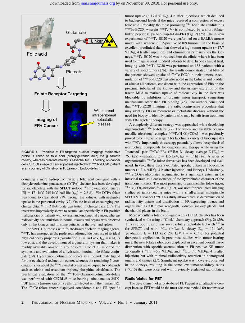
The concept of the FR-targeting strategy makes use of thevitamin folic acid as a molecular Trojan horse for selective deliveryof attached probes to FR-expressing cancer cells (Fig. 1). Com-pared with other targeting agents, such as monoclonal antibodies orpeptides, folic acid offers several advantages. It is small (441 Da),stable over a broad range of temperatures and pH values, and thusamenable for site-specific chemical modification. It is inexpensive,nonimmunogenic, and binds to the FR with high affinity even afterconjugation to a diagnostic or therapeutic cargo.
Because folic acid–targeted imaging agents can serve as non-invasive diagnostic tools to assess the location and severity of FR-positive cancer, a variety of folic acid–conjugated imaging agentshave been developed and evaluated in vitro and in vivo. Folic acidconjugates of probes for optical imaging, MRI, and nuclear imag-ing by SPECT and PET are reported in the literature (Fig. 1) (8,9).Because of the outstanding features of SPECT and PET (e.g., highsensitivity) the overall usage of nuclear medicine procedures isexpanding rapidly. Thus, recently, folate-based SPECT and PETtracers have attracted the greatest interest.
FOLIC ACID RADIOCONJUGATES
Radiofolates for SPECT and Potential TherapyOne of the first designs of a folic acid radioconjugate for SPECT
used deferoxamine for chelation of the g-emitting radioisotope 67Ga(10). Tumor targeting was successfully achieved with 67Ga-deferox-amine-folate in mice bearing FR-positive tumor xenografts (;8.5percentage injected dose per gram [%ID/g], 4 h after injection). How-ever, significant hepatobiliary excretion of the tracer led to unfavor-able abdominal accumulation of radioactivity (11). With the aim of
Received Jun. 23, 2010; revision accepted Sep. 8, 2010.For correspondence or reprints contact: Cristina Muller, Center for
Radiopharmaceutical Sciences ETH-PSI-USZ, Paul Scherrer Institute, CH-5232 Villigen-PSI, Switzerland.E-mail: [email protected] ª 2011 by the Society of Nuclear Medicine, Inc.
FOLIC ACID RADIOCONJUGATES • Muller and Schibli 1
by on November 30, 2018. For personal use only. jnm.snmjournals.org Downloaded from
designing a more hydrophilic tracer, a folic acid conjugate with adiethylenetriamine pentaacetate (DTPA) chelator has been developedfor radiolabeling with the SPECT isotope 111In (g-radiation: energy[E] 5 171 keV, 245 keV, half-life [t1/2] 5 2.8 d). 111In-DTPA-folatewas found to clear about 97% through the kidneys, with negligibleuptake in the peritoneal cavity (12). On the basis of encouraging pre-clinical data, 111In-DTPA-folate was tested in clinical trials (13). Thetracerwas impressively shown to accumulate specifically in FR-positivemalignancies of patients with ovarian and endometrial cancer, whereasradioactivity accumulation in normal tissues and organs was observedonly in the kidneys and, in some patients, in the liver and spleen.
For SPECT purposes with folate-based nuclear imaging agents,99mTc has emerged as the preferred radionuclide because of its idealphysical decay properties (g-radiation: E5 140 keV, t1/25 6 h), itslow cost, and the development of a generator system that makes itreadily available on-site in any hospital. Guo et al. reported thesynthesis and evaluation of a hydrazinonicotinamide-folate conju-gate (14). Hydrazinonicotinamide serves as a monodentate ligandfor the octahedral technetium center, whereas the remaining 5 coor-dination sites about the 99mTc-metal center are occupied by coligandssuch as tricine and trisodium triphenylphosphine trisulfonate. Thepreclinical evaluation of the 99mTc-hydrazinonicotinamide-folatewas performed with C57BL/6 mice bearing subcutaneous 24JK-FBP tumors (mouse sarcoma cells transfected with the human FR).The 99mTc-folate tracer displayed considerable and FR-specific
tumor uptake (;17.8 %ID/g, 4 h after injection), which declinedto background levels if the mice received a coinjection of excessfolic acid. Probably the most promising 99mTc-folate candidate is99mTc-EC20, wherein 99mTc(V) is complexed by a short folate-linked peptide (Cys-Asp-Dap-D-Glu-Pte) (Fig. 2) (15). The in vivoexperiments of 99mTc-EC20 were performed on a BALB/c mousemodel with syngeneic FR-positive M109 tumors. On the basis ofexcellent preclinical data that showed a high tumor uptake (;17.7%ID/g, 4 h after injection) and elimination primarily via the kid-neys, 99mTc-EC20 was introduced into the clinic, where it has beenused to image several hundred patients to date. In one clinical trial,imaging with 99mTc-EC20 was performed on 155 patients with avariety of solid tumors (16). The results demonstrated that 68% ofthe patients showed uptake of 99mTc-EC20 in their tumors. Accu-mulation of 99mTc-EC20 was also noted in the kidneys and bladderof almost all patients, consistent with the expression of FRs on theproximal tubules of the kidney and the urinary excretion of thetracer. Mild to marked uptake of radioactivity in the liver wasblockable by inhibitors of organic anion transport, suggestingmechanisms other than FR binding (16). The authors concludedthat 99mTc-EC20 imaging is a safe, noninvasive procedure thatmay identify FRs in recurrent or metastatic diseases without theneed for biopsy to identify patients who may benefit from treatmentwith FR-targeted therapy.
A completely different strategy was approached while developingorganometallic 99mTc-folates (17). The water- and air-stable organo-metallic tricarbonyl complex [99mTc(H2O)3(CO)3]1 was previouslyproved to be a versatile reagent for labeling a variety of bioconjugateswith 99mTc. Importantly, this strategypotentially allows the synthesis ofisostructural compounds for diagnosis and therapy while using the“matched” pair 99mTc/188Re (188Re: b2-decay, average E [Eav] 5763 keV, g-radiation, E 5 155 keV, t1/2 5 17 h) (18). A series oforganometallic 99mTc-folate derivatives has been developed and eval-uated. In vivo, these tracers exhibited specific uptake in FR-positivetumors (;2–4 %ID/g, 4 h after injection) and kidneys. Undesirably,99mTc(CO)3-radiofolates accumulated to a significant extent in theintestinal tract as a consequence of the hydrophobic character of thetricarbonyl-moiety. The most promising organometallic folate tracer,99mTc(CO)3-histidine-folate (Fig. 2), was used for preclinical imagingstudies of tumor-bearing mice with a small-animal multipinholeSPECT/CT scanner (19). This study allowed precise determination ofradioactivity uptake and distribution in FR-expressing tissues andorgans such as KB tumor xenografts, kidneys, salivary glands, andthe choroid plexus in the brain.
More recently, a folate conjugate with a DOTA chelator has beensynthesized while using a “Click”-chemistry approach (Fig. 2) (20).This radioconjugate was successfully radiolabeled with 111Infor SPECT and with 177Lu (177Lu: b2-decay, Eav 5 134 keV,g-radiation, E 5 113 keV, 208 keV, t1/2 5 6.7 d) for potentialtherapeutic application. In preclinical studies with tumor-bearingmice, the new folate radiotracer displayed an excellent overall tissuedistribution with specific accumulation in FR-positive KB tumorxenografts (111In, ;5.8 %ID/g, and 177Lu, 7.5 %ID/g, 4 h afterinjection) but with minimal radioactivity retention in nontargetedorgans and tissues (21). Significant uptake was, however, observedin the kidneys, resulting in the same low tumor-to-kidney ratios(,0.15) that were observed with previously evaluated radiofolates.
Radiofolates for PETThe development of a folate-based PETagent is an attractive con-
cept because PETwould be the most accurate method for noninvasive
FIGURE 1. Principle of FR-targeted nuclear imaging: radioactive
probe is linked to folic acid (pteroylglutamic acid) via glutamate
moiety, whereas pteroate moiety is essential for FR binding on cancer
cells. SPECT image of cancer patient injectedwith 99mTc-EC20 (patientscan courtesy of Christopher P. Leamon, Endocyte Inc.).
2 THE JOURNAL OF NUCLEAR MEDICINE • Vol. 52 • No. 1 • January 2011
by on November 30, 2018. For personal use only. jnm.snmjournals.org Downloaded from

diagnosis of cancer, particularly of small metastases. Mathias et al.reported the radiosynthesis of the first PET folate tracers, 66Ga- and68Ga-deferoxamine-folate (22). 68Ga is a generator isotope with ashort half-life (89% b1-decay, Eav 5 830 keV, t1/2 5 68 min),whereas 66Ga has a relatively long half-life but an unfavorably highpositron energy (56% b1-decay, Eav 5 1,740 keV, t1/2 5 9.5 h). Inthis study, FR-positive tumors and kidneys were clearly visualizedon small-animal PET images of a KB tumor–bearing mouse 25 hafter injection of 66Ga-deferoxamine-folate. However, the samedrawback of a high intestinal accumulation of radioactivity thatwas reported for 67Ga-deferoxamine-folate also hampered a furtherdevelopment of these PET folates.
The design of a 18F-radiolabeled folate tracer is a promisingapproach because, compared with other radionuclides, 18F (97%b1-decay, Eav5 250 keV, t1/2 5 110 min) displays excellent decaycharacteristics for PET. The first 18F-folate tracer reported in theliterature was a folic acid conjugate with 4-fluorbenzylamine as aprosthetic group, referred to as 18F-fluorobenzylamine-folate (Fig. 2)(23). 18F-fluorobenzylamine was coupled with ester-activatedfolic acid to obtain g- and a-18F-fluorobenzylamine-folate isomersin a ratio of 4:1. The last reaction step yielded 15%–44% 18F-fluorobenzylamine-folate tracer after purification via high-performance liquid chromatography. PET studies performed with18F-fluorobenzylamine-folate in tumor-bearing mice were success-ful in visualizing KB tumor xenografts (;6.5 %ID/g, 2 h after
injection). Beside uptake in FR-expressing kidneys, massive radio-activity uptake was observed in the gallbladder (.250 %ID/g, 2 hafter injection) and the intestinal tract. To address the drawback of alow radiochemical yield experienced with 18F-fluorobenzylamine-folate, a more versatile radiosynthetic strategy was approached thatused a Click-chemistry reaction (24). The folate precursor, folicacid-g-(4-azido)-butylamide, was prepared according to a previ-ously described method (25). The radiosynthesis of the 18F-Click-folate (Fig. 2) comprised 2 main reaction steps. First, theprosthetic group, 6-18F-fluoro-1-hexyne, was produced from thecorresponding p-tosylate precursor with an excellent radiochemicalyield (70%–85%) and purity (.95%). The second reaction stepcomprised the 1,4-triazole formation by Cu(I)-catalyzed cycloaddi-tion of the 6-18F-fluoro-1-hexyne and folic acid g-(4-azido)-butyl-amide. This Click reaction succeeded without the need for protectiongroups and directly provided the final 18F-Click-folate (20). In vivostudies performed with KB tumor–bearing mice revealed a relativelyhigh and FR-specific tumor uptake (;3 %ID/g, 45 min after injec-tion) and a reasonable tumor-to-kidney ratio. However, the stronglylipophilic character of the 18F-Click-folate resulted again in highaccumulation of radioactivity in the bile (.600 %ID/g, 45 min afterinjection) and in the intestinal tract. Because both of these 18F-PETtracers provided suboptimal results, further investment in the designof 18F-PET folates will be necessary for optimization of both theradiosynthesis and the in vivo properties of the tracer.
FIGURE 2. Chemical structures of folic acid radioconjugates: 99mTc-EC20 (M 5 99mTc) (1), 99mTc(CO)3-histidine-folate (M 5 99mTc) (2), 18F-fluorobenzylamine-a-folate (3a), 18F-fluorobenzylamine-g-folate (3b), 18F-Click-folate (4), and DOTA-Click-folate (M 5 111In, 177Lu) (5).
FOLIC ACID RADIOCONJUGATES • Muller and Schibli 3
by on November 30, 2018. For personal use only. jnm.snmjournals.org Downloaded from

PERSPECTIVE
A critical aspect of the FR-targeting strategy is the physiologicexpression of FRs in the kidneys. Reabsorption of folates fromprimary urine via FRs is a physiologic process that preventsconstant loss of these important vitamins (26). Not surprisingly,small-molecular-weight folic acid radioconjugates undergo thesame fate, which results in a significant renal uptake of radioactiv-ity. Thus, folate-based radionuclide therapy with particle-emittingisotopes has not been envisaged so far because low tumor-to-kid-ney ratios (,0.15) of radiofolates would present a high risk fordamage to the kidneys.
Recent data suggest, however, that predosing with the antifolatepemetrexed significantly reduces kidney uptake of radiofolateswhile retaining the desired radiotracer accumulation in the tumor(Fig. 3) (21,27,28). The exact underlying mechanism of this obser-vation is not yet completely understood. However, the accessibilityof reasonable tumor-to-kidney ratios allows a therapeutic applica-tion of radiofolates to be taken into consideration now. But the useof pemetrexed—a chemotherapeutic agent with potential sideeffects—only for the sake of kidney protection could be problem-atic. On the other hand, a potential synergistic anticancer effect oftherapeutic radiofolates and pemetrexed would be a strong argu-ment to justify the use of this combination for tumor treatment.
CONCLUSION
Using folic acid radioconjugates for SPECT and PET of cancerhas proven to be a versatile strategy in preclinical and clinicalstudies. Although the SPECT tracer 99mTc-EC20 is currently usedin the clinic, a suitable PET folate tracer is still lacking. Thequestion of whether FR-targeted radionuclide therapy will be usedin the future depends on an appropriate folate tracer design and oncombination with substances that potentiate the therapeutic anti-tumor effect or protect individuals from the risk of nephropathy.
REFERENCES
1. Low PS, Henne WA, Doorneweerd DD. Discovery and development of folic-
acid-based receptor targeting for imaging and therapy of cancer and inflamma-
tory diseases. Acc Chem Res. 2008;41:120–129.
2. Sirotnak FM, Tolner B. Carrier-mediated membrane transport of folates in mam-
malian cells. Annu Rev Nutr. 1999;19:91–122.
3. Qiu A, Jansen M, Sakaris A, et al. Identification of an intestinal folate transporter and
the molecular basis for hereditary folate malabsorption. Cell. 2006;127:917–928.
4. Antony AC. Folate receptors. Annu Rev Nutr. 1996;16:501–521.
5. Parker N, Turk MJ, Westrick E, Lewis JD, Low PS, Leamon CP. Folate receptor
expression in carcinomas and normal tissues determined by a quantitative radio-
ligand binding assay. Anal Biochem. 2005;338:284–293.
6. Garin-Chesa P, Campbell I, Saigo PE, Lewis JL, Old LJ, Rettig WJ. Trophoblast
and ovarian cancer antigen LK26: sensitivity and specificity in immunopathol-
ogy and molecular identification as a folate-binding protein. Am J Pathol.
1993;142:557–567.
7. Shen F, Ross JF, Wang X, Ratnam M. Identification of a novel folate receptor, a
truncated receptor, and receptor type b in hematopoietic cells: cDNA cloning,
expression, immunoreactivity, and tissue specificity. Biochemistry. 1994;33:
1209–1215.
8. Ke CY, Mathias CJ, Green MA. Folate-receptor-targeted radionuclide imaging
agents. Adv Drug Deliv Rev. 2004;56:1143–1160.
9. Sega EI, Low PS. Tumor detection using folate receptor-targeted imaging agents.
Cancer Metastasis Rev. 2008;27:655–664.
10. Wang S, Lee RJ, Mathias CJ, Green MA, Low PS. Synthesis, purification, and
tumor cell uptake of 67Ga-deferoxamine-folate, a potential radiopharmaceutical
for tumor imaging. Bioconjug Chem. 1996;7:56–62.
11. Mathias CJ, Wang S, Low PS, Waters DJ, Green MA. Receptor-mediated target-
ing of 67Ga-deferoxamine-folate to folate-receptor-positive human KB tumor
xenografts. Nucl Med Biol. 1999;26:23–25.
12. Mathias CJ, Wang S, Waters DJ, Turek JJ, Low PS, Green MA. Indium-111-
DTPA-folate as a potential folate-receptor-targeted radiopharmaceutical. J Nucl
Med. 1998;39:1579–1585.
13. Siegel BA, Dehdashti F, Mutch DG, et al. Evaluation of 111In-DTPA-folate as
a receptor-targeted diagnostic agent for ovarian cancer: initial clinical results.
J Nucl Med. 2003;44:700–707.
14. Guo W, Hinkle GH, Lee RJ. 99mTc-HYNIC-folate: a novel receptor-based targeted
radiopharmaceutical for tumor imaging. J Nucl Med. 1999;40:1563–1569.
15. Leamon CP, Parker MA, Vlahov IR, et al. Synthesis and biological evaluation of
EC20: a new folate-derived, 99mTc-based radiopharmaceutical. Bioconjug Chem.
2002;13:1200–1210.
16. Fisher RE, Siegel BA, Edell SL, et al. Exploratory study of 99mTc-EC20 imaging
for identifying patients with folate receptor-positive solid tumors. J Nucl Med.
2008;49:899–906.
17. Muller C, Schubiger PA, Schibli R. Synthesis and in vitro/in vivo evaluation of
novel 99mTc(CO)3-folates. Bioconjug Chem. 2006;17:797–806.
18. Alberto R, Schibli R, Waibel R, Abram U, Schubiger AP. Basic aqueous chem-
istry of [M(OH2)3(CO)3]1 (M 5 Re, Tc) directed towards radiopharmaceutical
application. Coord Chem Rev. 1999;192:901–919.
19. Muller C, Forrer F, Schibli R, Krenning EP, de Jong M. SPECT study of folate
receptor-positive malignant and normal tissues in mice using a novel 99mTc-
radiofolate. J Nucl Med. 2008;49:310–317.
20. Mindt TL, Muller C, Stuker F, et al. A “click chemistry” approach to the efficient
synthesis of multiple imaging probes derived from a single precursor. Bioconjug
Chem. 2009;20:1940–1949.
21. Muller C, Mindt TL, de Jong M, Schibli R. Evaluation of a novel radiofolate in
tumour-bearing mice: promising prospects for folate-based radionuclide therapy.
Eur J Nucl Med Mol Imaging. 2009;36:938–946.
22. Mathias CJ, Lewis MR, Reichert DE, et al. Preparation of 66Ga- and 68Ga-
labeled Ga(III)-deferoxamine-folate as potential folate-receptor-targeted PET
radiopharmaceuticals. Nucl Med Biol. 2003;30:725–731.
23. Bettio A, Honer M, Muller C, et al. Synthesis and preclinical evaluation of a folic
acid derivative labeled with 18F for PET imaging of folate receptor-positive
tumors. J Nucl Med. 2006;47:1153–1160.
24. Ross TL, Honer M, Lam PYH, et al. Fluorine-18 click radiosynthesis and pre-
clinical evaluation of a new 18F-labeled folic acid derivative. Bioconjug Chem.
2008;19:2462–2470.
25. Mindt TL, Muller C, Melis M, de Jong M, Schibli R. “Click-to-chelate”: in vitro
and in vivo comparison of a 99mTc(CO)3-labeled N(tau)-histidine folate deriva-
tive with its isostructural, clicked 1,2,3-triazole analogue. Bioconjug Chem.
2008;19:1689–1695.
26. Birn H, Spiegelstein O, Christensen EI, Finnell RH. Renal tubular reabsorption of
folate mediated by folate binding protein 1. J Am Soc Nephrol. 2005;16:608–615.
27. Muller C, Bruhlmeier M, Schubiger AP, Schibli R. Effects of antifolate drugs on the
cellular uptake of radiofolates in vitro and in vivo. J Nucl Med. 2006;47:2057–2064.
28. Muller C, Schibli R, Krenning EP, de Jong M. Pemetrexed improves tumor
selectivity of 111In-DTPA-folate in mice with folate receptor-positive ovarian
cancer. J Nucl Med. 2008;49:623–629.
FIGURE 3. SPECT/CT images of female mice bearing humanovarian IGROV-1 tumor xenografts (arrows), 4 h after injection of111In-DTPA-folate alone (A) and in combination with predosed
pemetrexed (B) (28).
4 THE JOURNAL OF NUCLEAR MEDICINE • Vol. 52 • No. 1 • January 2011
by on November 30, 2018. For personal use only. jnm.snmjournals.org Downloaded from

Doi: 10.2967/jnumed.110.076018Published online: December 13, 2010.
2011;52:1-4.J Nucl Med. Cristina Müller and Roger Schibli
Positive Cancer−Folic Acid Conjugates for Nuclear Imaging of Folate Receptor
http://jnm.snmjournals.org/content/52/1/1This article and updated information are available at:
http://jnm.snmjournals.org/site/subscriptions/online.xhtml
Information about subscriptions to JNM can be found at:
http://jnm.snmjournals.org/site/misc/permission.xhtmlInformation about reproducing figures, tables, or other portions of this article can be found online at:
(Print ISSN: 0161-5505, Online ISSN: 2159-662X)1850 Samuel Morse Drive, Reston, VA 20190.SNMMI | Society of Nuclear Medicine and Molecular Imaging
is published monthly.The Journal of Nuclear Medicine
© Copyright 2011 SNMMI; all rights reserved.
by on November 30, 2018. For personal use only. jnm.snmjournals.org Downloaded from