folding units govern the cytochrome c alkaline transition
TRANSCRIPT
Folding Units Govern the Cytochrome cAlkaline Transition
Linh Hoang*, Haripada Maity, Mallela M. G. Krishna, Yan Lin andS. Walter Englander
The Johnson ResearchFoundation, Department ofBiochemistry and BiophysicsUniversity of PennsylvaniaSchool of MedicinePhiladelphia, PA 19104-6059USA
The alkaline transition of cytochrome c is a model for protein structuralswitching in which the normal heme ligand is replaced by anothergroup. Stopped flow data following a jump to high pH detect two slowkinetic phases, suggesting two rate-limiting structure changes. Resultsdescribed here indicate that these events are controlled by the same struc-tural unfolding reactions that account for the first two steps in the revers-ible unfolding pathway of cytochrome c. These and other results showthat the cooperative folding-unfolding behavior of protein foldons canaccount for a variety of functional activities in addition to determiningfolding pathways.
q 2003 Elsevier Ltd. All rights reserved
Keywords: protein folding; cytochrome c, alkaline transition; hydrogenexchange; protein function*Corresponding author
Introduction
The present work applies a hydrogen exchangemethod to the study of a protein structural tran-sition, the alkaline transition experienced byequine cytochrome c (Cyt c) at high pH. Althoughthe alkaline transition of Cyt c was identifiedsome 60 years ago,1 the underlying mechanism isstill unclear.2 The high pH transition replaces theburied Met80-S to heme Fe ligand with an externallysine. A similarly reconfigured state may play arole in some physiologically important interactionsof Cyt c at neutral pH (cytochrome oxidase,3,4
cardiolipin,5,6 lipid membranes;7,8 reviewed byRosell et al.9).
Stopped-flow spectroscopic studies of Cyt c fol-lowing a jump to high pH reveal two kineticsteps.10 An initial step involves the deprotonationof some unidentified buried group, controlled by astructural event that allows some solvent speciesto enter the protein. The second step physicallyremoves the normal heme ligand and brings a pro-tein surface residue in to replace it. The present
study identifies two structural reactions thatappear to control these two steps.
Previous work using native state hydrogenexchange methods (NHX) has shown that thesmall Cyt c protein (104 residues) is composed offive structural units (foldons) that engage in revers-ible unfolding and refolding behavior,11 – 14 evenunder fully native conditions. NHX methods canidentify each folding unit at amino acid resolutionand can measure its equilibrium stability andreversible unfolding and refolding rate.11 – 22 Thesesame unit foldons appear to determine a stepwisepathway that carries unfolded Cyt c to its nativestate.11,14 – 19 The present study correlates the firsttwo reactions in the reversible unfolding pathwaywith the two structural steps that determine thealkaline transition.
Results
The equilibrium alkaline transition
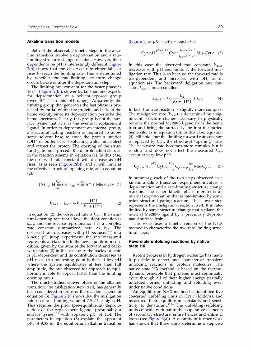
Figure 1 depicts the equilibrium alkaline tran-sition in several equine Cyt c variants, measuredby the loss of the 695 nm charge transfer bandwhich is specific for the native Met80-S to hemeFe ligation. The transition pKa for a recombinantpWT Cyt c (His26Asn/His33Asn) is 9.1 (Figure1(a)). The likely high pH replacement ligands arethe proximal lysine residues at positions 72, 73,and 79.23,24 When these three lysine residues are
0022-2836/$ - see front matter q 2003 Elsevier Ltd. All rights reserved
E-mail address of the corresponding author:[email protected]
Abbreviations used: Cyt c, equine cytochrome c; WT,wild-type; pWT, recombinant pseudo wild-typecytochrome c (His26Asn, His33Asn); foldon, cooperativefolding/unfolding unit; HX, hydrogen exchange;GdmCl, guanidinium chloride; HAR, homoarginine;N-yellow, nested yellow.
doi:10.1016/S0022-2836(03)00698-3 J. Mol. Biol. (2003) 331, 37–43

mutated to non-titratable residues the pKa for thetransition rises to 10.3. Similarly, Rosell et al.9
found a pKa of 10.5 (up from 8.7) when the samelysine residues were chemically blocked in yeastiso-1-Cyt c. These results favor the vicinal lysineresidues as the normal misligating groups.
Figure 1(b) shows another example. When all ofthe lysine residues in WT equine Cyt c are blockedby guanidination to form homoarginine (HAR),the pKa for the alkaline transition rises from 9.35to only 9.9. Similar results with all of the lysineresidues blocked in various ways have been takento suggest that non-lysine residues may be necess-ary to explain the alkaline transition (reviewed byRosell et al.9). However, these multiple modifi-cations are seriously perturbing. The misligatinggroup is different (high spin versus low spin).25
Also kinetic studies show that all of the 695 nmabsorbance is lost in a very fast phase that doesnot limit at increasing pH, and then some absor-bance slowly recovers (data not shown). Unmodi-fied Cyt c behaves very differently (Figure 2).
The present work focuses on the structurechanges that lead to the replacement of the Met80-
S ligand in unmodified WT Cyt c. Work in our lab-oratory and others show that various groups areable to misligate to the heme. The conclusionsreached here are most consistent with but do notdepend on the view that the normal replacementligand is drawn from the vicinal lysine residues.
Kinetics of the alkaline transition
Figure 2 shows stopped-flow results for the alka-line transition when WT equine Cyt c is mixed intohigh pH. The kinetics are biphasic.
A small fast phase reduces the amplitude of695 nm absorbance by 15% in the range above pH,10. It disappears at lower pH. The rate of thefast phase decreases as pH rises, limiting at16(^3) s21 at high pH. This phase registers theexposure and deprotonation of some buried groupnear the heme. The apparent pKa of this group isbelow pH 10, indicated by the fact that the full15% change is realized by pH 10.
The remaining A695 is then lost in a slowerphase indicating the replacement of the Met80-Sby another ligand. Again the amplitude (85%) isinsensitive to pH . 10. The rate of this phaseincreases as pH rises. It limits at 7.5(^0.4) s21.The pH dependence reflects the pre-equilibriumtitration of a surface group with pKa , 11.4, prob-ably the misligating lysine (see equation (3)below).
Similar results were found by Kihara et al.10 andHasumi.26 A small rate discrepancy is due to thedifferent temperatures used (20 8C versus 25 8C)and a small pH shift may be due to salt concen-tration (0.5 M versus 0.2 M). The data of Kiharaet al.10 suggest a subsequent increase in the fastphase rate beginning above pH 12. This is due tothe onset of a disruptive protein denaturation reac-tion described before.19 A more interesting increasein the fast phase rate occurs in some Cyt c homo-logs that have a stabilizing tyrosine residue at pos-ition 46.27 As discussed below, this observationhelps to identify the initial structural unfoldingreaction.
Figure 1. Equilibrium alkaline transition measured byCD or absorbance at 695 nm (20 8C). Data are fit by aone-proton titration curve. (a) Titration of pWT Cyt ccompared to a variant (HrsOpti-1) with the normalreplacement ligands removed by mutation (Lys72Gln/Lys73Ala/Lys79Arg). (b) Titration of WT equine Cyt ccompared to a variant with all of the lysine residuesblocked by guanidination to form homoarginine Cyt c(HAR-WT).
Figure 2. Kinetics of the alkalinetransition of WT equine Cyt c fol-lowed by absorbance at 695 and402 nm (20 8C). (a) A representativetrace at pH 10.5 and the corre-sponding residuals for one and twoexponential fits. (b) Amplitudesand rates of the fast and slowphases. Curves drawn for the fasterphase show the range of uncer-tainty (^20%).
38 Folding Units: Functional Role
Alkaline transition models
Both of the observable kinetic steps in the alka-line transition involve a deprotonation and a rate-limiting structure change reaction. However, theirdependence on pH is interestingly different. Figure2(b) shows that the observed rate either falls orrises to reach the limiting rate. This is determinedby whether the rate-limiting structure changeoccurs before or after the deprotonation step.
The limiting rate constant for the faster phase is16 s21 (Figure 2(b)), slower by far than one expectsfor deprotonation of a solvent-exposed group(over 106 s21 in this pH range). Apparently thetitrating group that generates the fast phase is pro-tected by burial within the protein, and it is in theheme vicinity since its deprotonation perturbs theheme spectrum. Clearly, this group is not the sur-face lysine that acts as the eventual replacementligand. In order to deprotonate an internal group,a structural gating reaction is required to allowsome solvent base to enter the protein interior(OH2 or buffer base þ solvating water molecules)and extract the proton. The opening of the struc-tural gate must precede the deprotonation step, asin the reaction scheme in equation (1). In this case,the observed rate constant will decrease as pHrises, as is seen (Figure 2(b)), and it will limit atthe effective structural opening rate, as in equation(2):
Cyt ccl-H Okop;1
kb;1
Cyt cop-HOK1
Hþ þ Mis-Cyt c ð1Þ
kobs;1 ¼ kop;1 þ kb;1½Hþ�
K1 þ ½Hþ�ð2Þ
In equation (2), the observed rate is kobs,1, the struc-tural opening rate that allows the deprotonation iskop,1, and the reverse reprotonation has a complexrate constant summarized here as kb,1. Theobserved rate decreases with pH because: (1) in akinetic pH jump experiment, the rate measuredrepresents a relaxation to the new equilibrium con-dition, given by the sum of the forward and back-ward rates; (2) in this case only the backward rateis pH-dependent and its contribution decreases aspH rises. (An interesting point is that, at low pHwhere the system equilibrates at less than fullamplitude, the rate observed for approach to equi-librium is able to appear faster than the limitingopening rate.)
The much-studied slower phase of the alkalinetransition, the misligation step itself, has generallybeen considered in terms of the reaction scheme inequation (3). Figure 2(b) shows that the misligationrate rises to a limiting value of 7.5 s21 at high pH.This requires the prior (pre-equilibrium) deproto-nation of the replacement ligand, presumably asurface lysine,2,28 with apparent pKa of 11.4. Theparameters in equation (3) explain the apparentpKa of 9.35 for the equilibrium alkaline transition
(Figure 1) as pKa ¼ pK2 2 logðkf=kbÞ:
Cyt c-H OpK2¼11:4
Cyt c Okf;2¼7:5 s21
kb;2¼0:02 s21Mis-Cyt c ð3Þ
In this case the observed rate constant, kobs,2,increases with pH and limits at the forward mis-ligation rate. This is so because the forward rate ispH-dependent and increases with pH, as inequation (4). The backward deligation rate con-stant, kb,2, is much smaller:
kobs;2 ¼ kf;2K2
K2 þ ½Hþ�þ kb;2 ð4Þ
In fact, the true reaction is slightly more complex.The misligation rate (kobs,2) is determined by a sig-nificant structure change necessary to physicallyremove the normal Met80-S ligand from the hemeiron and bring the surface lysine into the buriedheme site, as in equation (5). In this case, equation(4) still holds but the limiting forward rate constantis replaced by kop,2, the structural “opening” rate.The backward rate becomes more complex but itis slow and does not affect the measured kobs,except at very low pH:
Cyt ccl-H OpK2
Cyt ccl Okop;2
kcl;2
Cyt cop Ofast
kb;2
Mis-Cyt c ð5Þ
In summary, each of the two steps observed in akinetic alkaline transition experiment involves adeprotonation and a rate-limiting structure changereaction. The faster kinetic phase represents aninternal deprotonation that is rate-limited by someprior structural gating reaction. The slower steprepresents the misligation reaction itself. It is rate-limited by some structure change that replaces theinternal Met80-S ligand by a previously deproto-nated surface lysine.
This work uses a kinetic version of the NHXmethod to characterize the two rate-limiting struc-tural steps.
Reversible unfolding reactions by nativestate HX
Recent progress in hydrogen exchange has madeit possible to detect and characterize transientunfolding reactions in protein molecules. Thenative state HX method is based on the thermo-dynamic principle that proteins must continuallycycle through all of their higher energy partiallyunfolded forms, unfolding and refolding evenunder native conditions.
An equilibrium NHX method has identified fiveconcerted unfolding units in Cyt c (foldons), andmeasured their equilibrium constants and sensi-tivity to denaturant.11,14 The unfolding/refoldingunits coincide with naturally cooperative elementsof secondary structure, entire helices and entire Vloops (see Figure 3(a)). A quantity of further workhas shown that these units determine a stepwise
Folding Units: Functional Role 39

unfolding/refolding pathway,11,14,15,19 as dia-grammed in Figure 3(b).
A kinetic version of the NHX experiment, doneat relatively high pH (pH . 9) where HX is in theEX1 region, can measure the reversible unfoldingrates of these units.19 – 22 Fortunately for the presentinvestigation, the useful pH range of the kineticNHX experiment is the same as the pH range inwhich the alkaline transition occurs. Figure 4shows HX rate as a function of pH for a numberof measurable Cyt c hydrogen atoms in theNested-yellow (N-yellow) V loop and the Red Vloop (see Figure 3(a)).
At lower pH (EX2 region), HX rates are gov-erned by equilibrium structural unfolding reac-tions that intermittently expose the protectedamide hydrogen atoms to transient contact withsolvent. Here HX rate increases with pH becausethe chemical exchange reaction is catalyzed by OHion (equation (6)). The pH offset of HX rates forthe different amide protons in the EX2 region (seeFigure 4) is due to differences in their intrinsicchemical HX rates, determined by neighboringside-chain inductive and blocking effects.29,30 Athigh pH, HX rates are ultimately limited by therate of the unfolding reaction (EX1 condition) thatexposes the hydrogen atoms to exchange (equation(7)). The limiting rates at high pH exhibit a smallbut negligible spread.
Unfolding reactions and the alkaline transition
Figure 4 compares the unfolding rates for two Vloops with the two rate processes measured forthe alkaline transition. The averaged unfoldingrate for the N-yellow V loop protons at high pH is17(^2) s21 (15–20 s21 total range). This compareswith the rate measured for the fast phase of thealkaline transition in the same high pH region,16(^3) s21.
The averaged Red loop unfolding rate, 9(^2) s21
(7.5–10.8 s21 total range) matches the limiting rateof the misligation phase, 7.5(^0.4) s21. The bestagreement is found with residues Tyr74(7.5(^0.7) s21) and Ile75 (7.4(^0.5) s21). These
so-called marker protons exchange only when theRed loop unfolds.11,19 In the EX2 region the non-marker amides exchange more rapidly, by way oflocal fluctuations. Their extrapolation into the EX1region may receive a contribution from the localfluctuational pathways in addition to the dominantRed loop unfolding pathway. In any case, thesedifferences are small and do not mask the finalresult.
These results suggest that the two alkaline tran-sition reactions measured by stopped-flow arerate-limited by the transient unfolding of two unitloop structures (arrows in Figure 3(b)).
Discussion
Structure change in the alkaline transition andRed loop unfolding
The Red V loop (residues 71–85) protects theface of the heme that is misligated in the alkaline
Figure 3. Cyt c and its folding free energy profile. (a) Equilibrium native state HX results have identified five con-certed folding units, shown color-coded.11,14 (b) The Cyt c folding/unfolding pathway. Arrows show the unfoldingreactions that appear to rate-limit the two kinetic steps seen at high pH in alkaline transition studies.
Figure 4. Structural unfolding rates (color) comparedwith the two phases in the alkaline transition (black;20 8C in 0.5 M KCl). The limiting HX rates at high pHare the structural unfolding rates measured for protonsin the N-yellow and Red V loops. Within each unitdifferent protons initially reach the EX1 limit at some-what different pH due to their different intrinsic rateconstants.30 HX pulse labeling times and protonsmeasured are indicated.
40 Folding Units: Functional Role

transition (Figure 3(a)). It contains the native hemeligand (Met80-S) and also the lysine residues (72,73, 79) that appear to account for the high pH mis-ligation in unmodified Cyt c.9 In order to physi-cally remove Met80 from the heme and replace itwith one of the surface lysine residues, a signifi-cant distortion of this segment seems necessary.In fact a solution NMR study of alkaline Cyt c mis-ligated by Lys79 shows that the Red unit deviatesstrongly from the native structure whereas the N-yellow unit remains unchanged.31
Nelson & Bowler32 have previously suggestedthat the Red loop unfolding might mediate thealkaline transition. They found that these two equi-librium reactions have similar sensitivity to dena-turant, expressed in terms of an m valueðm ¼ DðRT ln KuÞ=D½GdmCl�Þ; which depends onthe surface newly exposed to solvent in the unfold-ing. The m value for the equilibrium alkaline tran-sition, 1.1–1.7 kcal/mol per M32 is close to the mvalue of 1.6 kcal/mol per M previously measuredfor Red unfolding by equilibrium native stateHX.11 In agreement, the present results show thatthe limiting rate for the misligation step matchesthe rate measured for the concerted unfolding ofthe Red loop at the same high pH condition.
The agreement of the kinetic rates, the agreementof the equilibrium m values, and the structuralrelationships required by the transition and seenby an NMR study consistently indicate that thenaturally occurring Red loop unfolding representsthe conformational rearrangement that limits theslower step in the alkaline transition.
Structure change in the alkaline transition andN-yellow loop unfolding
The faster phase of the alkaline transition pro-duces a transient intermediate form that has analtered heme spectral absorbance, apparentlycaused by the titration of some nearby internalgroup. The deprotonation of an internal grouprequires the prior opening of some gating structurethat would allow entry into the protein core ofsome solvent species able to act as a proton accep-tor (OH2, buffer base). That this occurs is sup-ported by the initial decrease in rate with pH(Figure 2(b); equation (2); see also Kihara et al.10).
The limiting rate for the fast phase of the alkalinetransition matches the rate for unfolding of theN-yellow loop at the same high pH condition(Figure 4(a)). The unfolding of the N-yellow loopwould allow solvent access to the heme crevice(see Figure 3(a)).
Saigo27 found an additional sensitivity to pH forthe fast phase rate in species that contain a tyrosineresidue at position 46 (tuna, bonito, rhesus) but notin other species with Phe46 (horse, sheep, dog,pigeon). Tyr46 is in the N-yellow loop. It forms astabilizing pH-sensitive H-bond. Its titrationðpKa . 11Þ increases the fast phase rate,27 appar-ently because the disruption of this bond poten-
tiates the determining structure change, consistentwith N-yellow unfolding.
In summary, the limiting gating rate for theinternal deprotonation reaction is equal to the N-yellow unfolding rate. Destabilization of the N-yellow loop accelerates the deprotonationreaction.27 N-yellow unfolding would open theheme crevice to invasion by solvent species. Theseobservations support the conclusion that the natu-rally occurring N-yellow loop unfolding representsthe conformational gating reaction that allows thedeprotonation of some buried heme-related group,seen as an initial heme spectral change in kineticalkaline transition experiments.
A general implication
It seems most noteworthy that the same tworeversible unfolding reactions that determine theCyt c alkaline transition represent the first twosteps in the Cyt c unfolding pathway (Figure 3(b)).The same steps may also limit the binding of exo-geneous ligands to the heme group.33 The accom-panying work14 notes the involvement of the N-yellow loop in other structural and evolutionaryactions. These results indicate that the foldon sub-structure of proteins, and particularly their natu-rally occurring reversible unfolding behavior, canact to determine not only folding/unfolding path-ways but also a number of other importantbehaviors.
Materials and Methods
Materials
Equine Cyt c was from Sigma Chemical Co. (WT Cytc). All experiments were done at 20 8C with 0.5 M KClto minimize charge effects.
Homoarginine modified WT Cyt c was prepared asdescribed by Hettinger et al.34 Complete modification ofall 19 lysine residues was confirmed by mass spec-trometry and purity was verified by SDS/polyacryl-amide gel electrophoresis.
The recombinant pWT Cyt c used has the potentiallymisligating histidine residues removed (His26Asn/His33Asn).35 In a selectively modified pWT protein(HrsOpti-1) the three lysine residues thought to accountfor the normal alkaline transition are replaced bymutation (Lys72Gln/Lys73Ala/Lys79Arg), a Gln residueblocks the N terminus by spontaneous cyclization, and anear terminal proline contributes to stability (Val3Pro).
Equilibrium titration
Equilibrium pH titrations measured by CD and absor-bance at 695 nm (5 nm bandwidth) used an automatedMicrolab 500 series titrator and continuous pH measure-ment in an AVIV CD Spectrometer model 202, regulatedat 20 8C. The titrator added incremental volumes ofNaOH, 0.5 M KCl (total added volume ,50 ml) to 2 mlof 0.4 mM protein in 0.5 M KCl, 10 mM KPO4, 10 mMBicine, 10 mM Caps. For each data point, equilibration
Folding Units: Functional Role 41

time was three minutes and the signal was averaged for30 seconds.
Stopped-flow kinetics
Stopped-flow kinetics used a Biologic SFM-4 instru-ment (20 8C). Syringes 1 and 2 contained buffers at pH 9and 12, with 50 mM Caps, 0.5 M KCl. Syringe 3 con-tained 0.7 mM protein (0.5 M KCl, 10 mM KPO4, pH 6).The protein was diluted tenfold with incremental mixesof syringes 1 and 2 to set pH, measured after collection.Data analysis used Igor Pro 4.0.
Kinetic native state hydrogen exchange
Hydrogen-bonded amides experience dynamic open-ing and closing reactions that separate the protectinghydrogen bond and allow hydrogen exchange with thebulk solvent. In steady state, the overall exchange rate(kex) is given by equation (6),36,37 where kop and kcl areopening and reclosing rates of the protecting H-bondand kch ¼ kint½OH2�: kint is an intrinsic second-order rateconstant which depends on temperature, nearest neigh-bor inductive and blocking effects, and isotopeeffects:29,30,38
kex ¼kopkch
kcl þ kchð6Þ
When kcl p kch (the EX1 monomolecular exchange limit),exchange rate is given by equation (7), allowing themeasurement of structural opening rates:37
kex ¼ kop ð7Þ
A native state strategy employs mild destabilants topromote larger structural unfoldings over localfluctuations11,19 so that they come to dominate theexchange measured. The high pH used in the presentwork to promote the alkaline transition and to makekch q kcl (EX1 condition) was adequate to serve this func-tion for the Red loop unfolding.
The very fast HX rate obtained at high pH wasmeasured by exposing the protein to the high pH con-dition for only a short time.20,21 A labeling pulse of either33 or 75 ms was used for the data set described here.19
The H to 2H exchange that occurs during the labelingpulse was measured by 2D NMR using the cross-peakassignments of Feng et al.39 for oxidized Cyt c.
Acknowledgements
This work was supported by research grantsfrom the NIH and the Mathers Foundation.
References
1. Theorell, H. & Akesson, A. (1941). Studies on cyto-chrome c. J. Am. Chem. Soc. 63, 1812–1818.
2. Wilson, M. T. & Greenwood, C. (1996). The alkalinetransition in ferricytochrome c. In Cytochrome c: AMultidisciplinary Approach (Scott, R. A. & Mauk,A. G., eds), pp. 611–634, University Science Books,Sausolito.
3. Dopner, S., Hudecek, J., Ludwig, B., Witt, H. &Hildebrandt, P. (2000). Structural changes in cyto-
chrome c oxidase induced by cytochrome c binding.A resonance Raman study. Biochim. Biophys. Acta,1480, 57–64.
4. Falk, K. E. & Angstrom, J. (1983). A 1H NMR longi-tudinal relaxation study of the interaction betweencytochrome c and cytochrome c oxidase. Biochim. Bio-phys. Acta, 722, 291–296.
5. Soussi, B., Bylund-Fellenius, A. C., Schersten, T. &Angstrom, J. (1990). 1H NMR evaluation of the ferri-cytochrome c-cardiolipin interaction. Effect of super-oxide radicals. Biochem. J. 265, 227–232.
6. Hildebrandt, P., Vanhecke, F., Buse, G., Soulimane, T.& Mauk, A. G. (1993). Resonance Raman study of theinteractions between cytochrome c variants and cyto-chrome c oxidase. Biochemistry, 32, 10912–10922.
7. Hildebrandt, P. & Stockburger, M. (1989). Cyto-chrome c at charged interfaces. 2. Complexes withnegatively charged macromolecular systems studiedby resonance Raman spectroscopy. Biochemistry, 28,6722–6728.
8. Hildebrandt, P., Heimburg, T. & Marsh, D. (1990).Quantitative conformational analysis of cytochromec bound to phospholipid vesicles studied by reson-ance Raman spectroscopy. Eur. J. Biophys. 18,193–201.
9. Rosell, F. I., Ferrer, J. C. & Mauk, A. G. (1998). Proton-linked protein conformational switching: definitionof the alkaline conformational transition of yeast iso-1-ferricytochrome c. J. Am. Chem. Soc. 120,11234–11245.
10. Kihara, H., Saigo, S., Nakatani, H., Hiromi, K., Ikeda-Saito, M. & Iizuka, T. (1976). Kinetic study of isomer-ization of ferricytochrome c at alkaline pH. Biochim.Biophys. Acta, 430, 225–243.
11. Bai, Y., Sosnick, T. R., Mayne, L. & Englander, S. W.(1995). Protein folding intermediates: native-statehydrogen exchange. Science, 269, 192–197.
12. Chamberlain, A. K. & Marqusee, S. (2000). Compari-son of equilibrium and kinetic approaches for deter-mining protein folding mechanisms. Advan. ProteinChem. 53, 283–328.
13. Bai, Y. & Englander, S. W. (1996). Future directions infolding: the multi-state nature of protein structure.Proteins: Struct. Funct. Genet. 24, 145–151.
14. Krishna, M. M. G., Lin, Y., Rumbley, J. N. &Englander, S. W. (2003). Cooperative omega loops incytochrome c: role in folding and function. J. Mol.Biol. 331, 29–36.
15. Xu, Y., Mayne, L. & Englander, S. W. (1998). Evidencefor an unfolding and refolding pathway in cyto-chrome c. Nature Struct. Biol. 5, 774–778.
16. Milne, J. S., Mayne, L., Roder, H., Wand, A. J. & Eng-lander, S. W. (1998). Determinants of protein hydro-gen exchange studied in equine cytochrome c.Protein Sci. 7, 739–745.
17. Bai, Y. (1999). Kinetic evidence for an on-pathwayintermediate in the folding of cytochrome c. Proc.Natl Acad. Sci. USA, 96, 477–480.
18. Rumbley, J., Hoang, L., Mayne, L. & Englander, S. W.(2001). An amino acid code for protein folding. Proc.Natl Acad. Sci. USA, 98, 105–112.
19. Hoang, L., Bedard, S., Krishna, M. M. G., Lin, Y. &Englander, S. W. (2002). Cytochrome c folding path-way: kinetic native-state hydrogen exchange. Proc.Natl Acad. Sci. USA, 99, 12173–12178.
20. Arrington, C. B. & Robertson, A. D. (2000). Micro-second to minute dynamics revealed by EX1-typehydrogen exchange at nearly every backbone
42 Folding Units: Functional Role

hydrogen bond in a native protein. J. Mol. Biol. 296,1307–1317.
21. Canet, D., Last, A. M., Tito, P., Sunde, M., Spencer,A., Archer, D. B. et al. (2002). Local cooperativity inthe unfolding of an amyloidogenic variant ofhuman lysozyme. Nature Struct. Biol. 9, 308–315.
22. Yan, S., Kennedy, S. & Koide, S. (2002). Thermodyn-amic and kinetic exploration of the energy landscapeof Borrelia burgdorferi OspA by native-state hydrogenexchange. J. Mol. Biol. 323, 363.
23. Smith, H. T. & Millett, F. (1980). Involvement oflysines-72 and -79 in the alkaline isomerization ofhorse heart ferricytochrome c. Biochemistry, 19,1117–1120.
24. Pollock, W. B., Rosell, F. I., Twitchett, M. B., Dumont,M. E. & Mauk, A. G. (1998). Bacterial expression of amitochondrial cytochrome c. Trimethylation of lys72in yeast iso-1-cytochrome c and the alkaline confor-mational transition. Biochemistry, 37, 6124–6131.
25. Stellwagen, E., Babul, J. & Wilgus, H. (1975). Thealkaline isomerization of lysine-modified ferricyto-chrome c. Biochim. Biophys. Acta, 405, 115–121.
26. Hasumi, H. (1980). Kinetic studies on isomerizationof ferricytochrome c in alkaline and acid pH rangesby the circular dichroism stopped-flow method. Bio-chim. Biophys. Acta, 626, 265–276.
27. Saigo, S. (1981). Kinetic and equilibrium studies ofalkaline isomerization of vertebrate cytochromes c.Biochim. Biophys. Acta, 669, 13–20.
28. Davis, L. A., Schejter, A. & Hess, G. P. (1974). Alka-line isomerization of oxidized cytochrome c. Equili-brium and kinetic measurements. J. Biol. Chem. 249,2624–2632.
29. Molday, R. S., Englander, S. W. & Kallen, R. G. (1972).Primary structure effects on peptide group hydrogenexchange. Biochemistry, 11, 150–158.
30. Bai, Y., Milne, J. S., Mayne, L. & Englander, S. W.
(1993). Primary structure effects on peptide grouphydrogen exchange. Proteins: Struct. Funct. Genet. 17,75–86.
31. Assfalg, M., Bertini, I., Dolfi, A., Turano, P., Mauk,A. G., Rosell, F. I. & Gray, H. B. (2003). Structuralmodel for an alkaline form of ferricytochrome c.J. Am. Chem. Soc. 125, 2913–2922.
32. Nelson, C. J. & Bowler, B. E. (2000). pH dependenceof formation of a partially unfolded state of a Lys73 ! His variant of iso-1-cytochrome c: implicationsfor the alkaline conformational transition of cyto-chrome c. Biochemistry, 39, 13584–13594.
33. Sutin, N. & Yandell, J. K. (1972). Mechanisms of thereactions of cytochrome c. Rate and equilibrium con-stants for ligand binding to horse heart ferricyto-chrome c. J. Biol. Chem. 247, 6932–6936.
34. Hettinger, T. P. & Harbury, H. A. (1964). Guanidatedcytochrome c. Proc. Natl Acad. Sci. USA, 52,1469–1476.
35. Rumbley, J. N., Hoang, L. & Englander, S. W. (2002).Recombinant equine cytochrome c in Escherichia coli:high-level expression characterization, and foldingand assembly mutants. Biochemistry, 41,13894–13901.
36. Linderstrøm-Lang, K. U. (1955). Deuterium exchangebetween peptides and water. Chem. Soc. Spec. Publ. 2,1–20.
37. Hvidt, A. & Nielsen, S. O. (1966). Hydrogenexchange in proteins. Advan. Protein Chem. 21,287–386.
38. Connelly, G. P., Bai, Y., Jeng, M. F. & Englander, S. W.(1993). Isotope effects in peptide group hydrogenexchange. Proteins: Struct. Funct. Genet. 17, 87–92.
39. Feng, Y., Roder, H., Englander, S. W., Wand, A. J. &Di Stefano, D. L. (1989). Proton resonance assign-ments of horse ferricytochrome c. Biochemistry, 28,195–203.
Edited by C. R. Matthews
(Received 6 February 2003; received in revised form 15 May 2003; accepted 27 May 2003)
Folding Units: Functional Role 43