fogsi matter page 29-31implantation+failure.pdf · 2017-09-19 · definition of repeated...
TRANSCRIPT
AIM (ADVANCED INFERTILITY MANAGEMENT) 160
FOGSI FOCUS
Introduction
The efficacy of assisted reproduction therapy in terms of live birth rates remained relatively constant. The rate limiting step appears to be implantation of the pre-embryo. Implantation requires the synchronization of multiple events including trophoblast development and timely expression of numerous molecules playing roles in apposition, nidation and invasion of the embryo into the endometrium. Failure of implantation in couples undergoing ART is a relatively common occurrence and may be a recurring phenomenon leading to despair in couples and frustration in their caregivers.
Definition of Repeated implantation failure (RIF)
Failure to achieve a pregnancy following 2–6 IVF cycles, in which more than 10 high-grade embryos were transferred to the uterus, was defined by various clinicians as RIF. After failure of three cycles in which reasonably good embryos were transferred, further investigation should be initiated.
Mechanism of Implantation
Initial apposition of trophoblast to uterus → subsequent adhesion and penetration of trophoblast into uterine epithelium. Disappearance of cadherins, and presence of b1 integrin are involved in implantation. Completeness of implantation occurs when the embryo prevents itself from rejection by the maternal immune system and when it induces its own blood supply.
Assumed aetiologies for repeated implantation failure (RIF)
RIF may be attributed to many factors. These can be grouped into three categories: decreased endometrial receptivity, embryonic defects and factors with combined effect. (Table 1)
Decreased endometrial receptivity
· Undiagnosed uterine pathology- Repeated hysteroscopic visualization revealed uterine abnormalities, mainly hyperplasia, polyps, endometritis, synechiae and leiomyomata. The effect of small fibroids or intramural fibroids without cavity distortion on implantation is uncertain.
· Thin endometrium- In a prospective large cohort studies it did not influence the cumulative pregnancy rates (PRs) particularly when high-quality embryos were transferred.
However, the concept that a minimum thickness (4–8 mm) is required to establish a clinical pregnancy is still arguable and should be considered in RIF.
· Local dysregulation of the normal expression or action of various cytokines.
Dr. Firuza Rajesh ParikhM.D., D.G.O., D.F.P., F.C.P.S., DIP.NBEDirector, Dept. of Assisted Reproduction and GeneticsJaslok Hospital and Research CentreEditor-in-Chief Fertility and Sterility Indian Edition
Dr. Mahendra Parikh MD, FICS, FICOG
Prof Emeritus Nowrosjee Wadia Maternity Hosp.Editor Emeritus Journal of Obst. & Gyn.President - Indian Society of Prenatal Diagnosis & TherapyFounder President ISARPast President FOGSI, ICOG, NARCHI
Dr. Madhavi PanpaliaM.B.B.S., M.S.(G & O)Clinical AssociateJaslok Hospital and Research Centre
RECURRENT IMPLANTATION FAILURE29

AIM (ADVANCED INFERTILITY MANAGEMENT)161
FOGSI FOCUS
Recurrent Implantation Failure contd.
1. Elevated endometrial NK cells, dysregulation of interleukin (IL) 12, 15 and 18. TH1 dominance.
2. High IL-1ß and low interferon-? and IL-10
3. Failure of appearance of a specific integrin –αVβ3 in the endometrium at the time of implantation
4. High levels of aromatase p450 mRNA
5. Changes in pinopode expression
6. High matrix metalloproteinases
· Immunological causes and Thrombophilia- Focus of many recent research efforts. The association of antiphospholipid or other autoantibodies with RIF has been shown in some early studies, but large prospective studies failed to reveal an association.
1. Some studies could not find any association between any of 18 specific antiphospholipid antibodies and RIF, whereas others found that ß2-glycoprotein-I antibodies were related to IVF failure. Antiphospholipids interfere with angiogenesis and inhibit invasion of embryonic cells.
2. Antibodies to annexin-V, which acts as an inhibitor of phospholipid-dependent coagulation and may be necessary for trophoblast differentiation, were found in greater incidence in women with RIF than in controls.
3. Association between peripheral natural killer (NK) cells and RIF in several small observational studies; however, in a recent review contended that there is no scientific evidence for such an association. Elevated levels associated with increased angiogenesis and peri-implantation blood flow leading to excessive oxidative stress.
4. Couples sharing HLA alleles are at high risk of RIF and recurrent biochemical pregnancies. However, preliminary report has never been confirmed. Frequent mutation is HLA G-725 C/G
5. Antinuclear antibodies-embryotoxic
6. Antithyroid antibodies-Marker of activated T cells which cause chemical pregnancy losses.
7. Increased rates of hereditary thrombophilia in women with RIF in some studies but not in others.
The prevalence of PAI-1 mutation and multiple thrombophilic gene mutations among patients with RIF was significantly higher than among fertile controls. Hence, screening of thrombophilia in RIF is still controversial.
Defective embryonic development
• Chromosomal abnormalities of the male or female partner, the gametes or the developing embryo- Increased frequency of female chromosomal abnormalities such as translocations, mosaics, inversion, deletion and chromosomal breakages, particularly at the centromere region. Increased incidence of sperm chromosomal abnormalities in patients with normal karyotype and RIF was also observed.
• The percentage of embryonic aneuploidy was higher in RIF compared with controls which fail to implant in a single cycle despite good morphology and developmental rate.
• The zona pellucida, which surrounds the mammalian oocyte, hardens naturally after fertilization to prevent polyspermia. Increased zona thickness was associated with lower implantation rates.

Zona hardening, can also affect hatching and be possible cause of RIF.
• Lack of specific culture conditions for embryonic development
Multifactorial causes
• Endometriosis as a cause for RIF has not been investigated directly, however all markers of reproductive process in endometriosis are decreased.
• Patients with hydrosalpinges have lower implantation and PRs. Hydrosalpinx fluid is either embryo-toxic or adversely affects the endometrium.
Investigations and Treatment
Suggested method for treatment of RIF (Table 2)
Improving endometrial receptivity
• Hysteroscopic correction of uterine cavity pathology
• Myomectomy- Favourable pregnancy and live birth rates seen following myomectomy. However, no prospective studies performed. Still hysteroscopic removal of submucous fibroids distorting the uterine cavity is recommended.
• Treatment of thin endometrial lining
o Low-dose aspirin
o Vaginal sildenafil- a type 5-specific PDE inhibitor that augments the vasodilatory effects of NO to improve uterine blood flow and endometrial proliferation.
o Estradiol creams
o Freezing embryos when endometrial lining less than 7mm and transferring them in a subsequent cycle where lining stimulated with high dose estrogen.
• Endometrial stimulation- Endometrial injury can cause a pseudo-decidual reaction that enhances implantation. However prospective controlled studies are required.
• Immunotherapy- There is evidence to suggest immunological factors cause RIF.
o Immunotherapy with intravenous immunoglobulin (IVIG) has been advised; however its effectiveness is still unresolved.
o Intralipid-20% intravenous fat emulsion-stimulates the immune system to remove danger signals that lead to pregnancy loss.
o Combined treatment with glucocorticoids and aspirin in autoantibodies seropositive patients
o Heparin-
• anticoagulant effect
• interacts with
• adhesion molecules: down regulates E, cadherdin expression( an intracellular adhesion molecule that has been shown to be involved in trophoblast differentiation and invasiveness),
AIM (ADVANCED INFERTILITY MANAGEMENT) 162
Recurrent Implantation Failure contd.
FOGSI FOCUS

AIM (ADVANCED INFERTILITY MANAGEMENT)163
FOGSI FOCUS
Recurrent Implantation Failure contd.
• growth factors: Heparin-binding Epidermal growth factor (HB-EGF) –blastocyst adhesion and invasion
• Cytokines and enzymes such as matrix metalloproteinases-modulate the potential for successful implantation.
o Aspirin
o Partners leukocytes- role controversial
Treatment of the embryos
• Preimplantation genetic screening for chromosomes 13, 16, 18, 21, 22.and selecting chromosomally normal embryos for transfer.
• Assisted hatching- To help release the embryos from their zona, different methods of assisted hatching have been developed. These involve the creations of an opening in the zona pellucida. . Three RCTs have shown that, in cases of RIF, AH significantly increases the pregnancy and implantation rate. However, in a systematic review of 23 RCTs, although clinical PR was significantly higher following AH, there was no effect on the 'take-home-baby rate'. No benefit of laser-AH in RIF could be found in a large European multicentre randomized trial
• Blastocyst transfer- more physiological approach because the human embryos enter the endometrial cavity only 5 days after fertilization, at the morula-blastocyst stage. Two large RCTs have shown that blastocyst transfer after RIF have significantly higher implantation and live birth rates. Improved embryo selection and uterine receptivity may explain the benefit of embryo transfer at the blastocyst stage for couples with RIF.
• Zygote intra-Fallopian Transfer (ZIFT)- Allows embryonic growth in the natural tubal environment. Initial retrospective studies were encouraging but later, results of a series of RCTs failed to demonstrate any advantage for ZIFT. The complexity and cost of ZIFT, led to the discontinuation of this method.
• Co-culture- Development of co-culture systems in which different cells having embryotrophic factors detoxifying agents. However, controversies exist regarding its benefit.
• Cytoplasmic transfer- An experimental procedure. Introduction of a small amount of ooplasm from a donor oocyte or zygote may alter the function of probable deficient oocytes. Cytoplasmic transfer is still considered an experimental procedure because it is not known whether the physiology of the early embryo is affected.
• Improving ET technique- Meta-analysis of randomized trials has shown that significantly higher PRs were obtained when an atraumatic ultrasound guidance technique was used and the embryos were deposited in the middle part of the uterine cavity. Many clinicians transfer large number of embryos after RIF; however, no comparative study has been published yet.
Multifactorial treatment options
• Endometriosis -Pretreatment for 3-6 months with GnRH agonists before ART significantly increases the ongoing PR. Most investigators agree that there is no benefit in the removal of endometriomas before IVF. The role of laparoscopic treatment of non-ovarian endometriosis in patients with failed IVF is controversial. Furthermore, surgery might be deleterious for ovarian reserve.

• Danazol- Immunosuppressive effects in vitro were shown long ago. In an RCT danazol treatment significantly increased PR. Danazol was found to increase receptivity of the endometrium and upgrade the endometrial aVß3 integrin.
·• Laparoscopic Salpingectomy of hydrosalpinges- is now recommended in all women with hydrosalpinx before IVF treatment. The pregnancy and live birth rates doubled following prophylactic salpingectomy.
• Stress can interfere with infertility treatments, and psychological interventions and various relaxation techniques, reduces anxiety and depression and possibly enhances conception success, but proof of their efficacy is lacking.
Conclusions
There are many known and unknown reasons for RIF, and we do not have the tools to diagnose the exact cause for the repeated failure in each case. However, after failure of three transfers of good-quality embryos in a unit with a PR of at least 30%, one should take some special measures. There are no hard data from RCTs that any of the treatments has a significant value, but on the contrary, everyone agrees that taking a different approach achieves a pregnancy in many cases that failed repeatedly.
After three failures, repeated hysteroscopy and a try of blastocyst transfer are highly recommended. A change in the stimulation protocol suggested. AH, PGS and co-culture are probably beneficial in experienced hands. Long-term use of danazol or GnRH agonists probably has a place in repeated failures with endometriosis. The use of IVIG is very controversial but may be justified after many failures in specific cases.
Steroids might have a place in patients with any sign of autoimmunity, and ZIFT has a place in cases of difficult embryo transfers.
Table 1. Assumed aetiologies for RIF
Decreased Endometrial Receptivity
Uterine cavity abnormalities
Thin Endometrium
Altered expression of adhesive molecules
Immunological factors
Thrombophilias
Defective embryonic development
Genetic abnormalities (male/ female/ gametes/ embryo)
Zona hardening
Suboptimal culture conditions
Multifactorial effectors
Endometriosis
Hydrosalpinges
Suboptimal ovarian stimulation
AIM (ADVANCED INFERTILITY MANAGEMENT) 164
Recurrent Implantation Failure contd.
FOGSI FOCUS

AIM (ADVANCED INFERTILITY MANAGEMENT)165
FOGSI FOCUS
Recurrent Implantation Failure
Table 2. Suggested methods for treatment of RIF
Improving endometrial receptivityHysteroscopic correction of cavity pathologyMyomectomyTreatment of thin endometriumEndometrial stimulation (biopsy)Immunotherapy ( intravenous immunoglobulin, steroids, aspirin, heparin
Treatment of embryos Preimplantation genetic screening Assisted hatching Zygote intra-Fallopian transfer Co-culture Blastocyst transfer Cytoplasmic transfer Improving ET technique
Multifactorial treatment options Treating endometriosis Danazol Salpingectomy for hydrosalpinges Tailoring stimulation protocols Psychological assistance
Bibliography
1. Acquired and inherited thrombophilia: implication in recurrent IVF and embryo transfer failure. Human Reproduction Vol.21, No.10 pp.2694-2698, 2006
2. Uterine natural killer cells and angiogenesis in recurrent reproductive failure. Human Reproduction Vol.24, No.1 pp.45-54, 2009
3. Mini Review –Developments in Reproductive Medicine; Investigation and treatment of repeated implantation failure following IVF-ET. Human Reproduction Vol.21, No.12 pp.3036-3043, 2006
4. Luteal phase empirical low molecular weight heparin administration in patients with failed ICSI embryo transfer cycles: a randomized open-labeled pilot trial. Human Reproduction Vol.24, No.7 pp.1640-1647, 2009
5. Treatment of repeated unexplained in vitro fertilization failure with intravenous immunoglobulin: a randomized, placebo-controlled Canadian trial. Fertility and Sterility Vol.74, No. 6, December 2000
6. Effect of vaginal sildenafil on the outcome of in vitro fertilization (IVF) after multiple IVF failures attributed to poor endometrial development. Fertility and Sterility Vol.78, No. 5, November 2002
7. http://www.illinoisivf.com/recurrent-pregnancy-loss/peri-implantation.html Site of Reproductive Medicine Institute, Chicago; Recurrent Pregnancy Loss Peri (Around )- Implantation
8. Molecular Mechanism of Implantation. A review in Keio Journal of Medicine, 45 (1) 37-43, 1996.
9. http://www.ivf1.com/index2.php?option=com_content&do_pdf=1&id=212 IVF Implantation Failure;Last updated Saturday 19 April 2008

Dr. Sulochana Gunasheela FRCS (EDIN), FRCOG (LOND.)Gunasheela Surgical & Maternity Hospital &
Gunasheela Assisted Reproduction Centre Pvt. Ltd.Bangalore
Dr. Devika GunasheelaMRCOG, Fellow In Reproductive Medicine (RGUHS) Gunasheela Surgical & Maternity Hospital &
Gunasheela Assisted Reproduction Centre Pvt. Ltd.Bangalore
AIM (ADVANCED INFERTILITY MANAGEMENT) 166
FOGSI FOCUS
INVITRO MATURATION (IVM)
What is IVM?
IVM stands for In Vitro Maturation of oocytes (maturation of the oocytes outside the body). The difference between IVM and conventional in vitro fertilisation (IVF) is that the eggs are immature when they are collected.
History of IVM
Pincus and Enzmann (1) in 1935 produced spontaneous maturation of immature rabbit oocytes removed from their natural ovarian environment and fertilization in vitro. Edwards in 1965 (2) was the first one to use this method on human beings as an effort to rescue immature oocytes seen in mixed cohorts contained in a harvest taken for standard IVF programme. Veeck et al. (3) went one step further and produced live offsprings in human beings from a mixed cohort. These are rescue oocytes with poor fertilization potential in view of the fact that they are probably dysmature, having failed to mature along with their sibling follicles in vivo despite exposure to the same levels of exogenous gonadotropins; they are also likely to show high rate of aneuploidy (4,5,6,). Cha et al (7) who reported a triplet birth occurring from unstimulated ovaries in a donor-oocyte programme in 1991, further reported 4 IVM babies in 1993.
Trounson et al reported his first IVM baby from PCOS in 1994. Barnes et al reported pregnancy in PCOS in 1995. He also reported his first blastocyst produced from IVM oocytes 110 hours after ICSI resulting in a live baby. From 1996, there was increasing number of groups worldwide reporting IVM pregnancies (see table 1,2,3 below).
Table 1
Deliveries and ongoing pregnancies in Europe
(facts and educated guesses)
Total No. – 320
Countries Deliveries and ongoing pregnancies
Scandinavia 150
Italy 77
France 40
Germany 20
Rest of Europe 33
H.I. Neilsen (8)
30

AIM (ADVANCED INFERTILITY MANAGEMENT)167
FOGSI FOCUS
Invitro Maturation (IVM) contd.
Table 2 (8)
Deliveries and ongoing pregnancies in Asia
(facts and educated guesses)
Total No. – 679
Countries Deliveries and ongoing pregnancies
Middle East 21
Japan 100
Vietnam 26
China 60
Korea 457
Rest of Asia 15
H.I. Neilsen (8)
What does IVM treatment involve?
Immature eggs are collected from unstimulated or minimally stimulated ovaries under ultrasound scan guidance. The immature eggs are then matured in the laboratory for 24-48 hours using a special culture medium with added small quantities of hormones. Intracytoplasmic Sperm Injection (ICSI) is used for fertilization of the matured eggs. Laser Assisted Hatching (LAH) is performed on these embryos before transferring them into the woman's womb.
Human primordial germ cells travel from yolk sac to the gonad from day 26 of pregnancy, and are then termed oogonia (9). Meiotic division usually commences gradually in the third month of gestation. The oogonia enlarge and acquire more intracellular organelles; these are then termed as oocytes.
Follicular formation in humans begins in the fourth month of gestation. For the assembly of follicles, rapid proliferation of the nearby stroma cells should occur, and the oocytes are surrounded by a single layer of flattened somatic cells, termed as granulosa cells, enclosed by a basement membrane (9,10). The cellular complexes enclosing the oocytes are called as primordial follicles which are 30-50 µm in diameter and can be identified in the humans from around 22 gestational weeks.
Most of the primordial follicles are activated when the granulosa cells become cuboidal, and these are then termed as primary follicles which are 50 µm-100 µm (0.1 mm) in diameter.
Immature Oocytes IVF 4 cell Embryo
Physiology of development of oogenesis in foetus to primary & secondary follicle

AIM (ADVANCED INFERTILITY MANAGEMENT) 168
FOGSI FOCUS
Stimulation for IVM
Mikkelsen et al. (11) designed a prospective randomized trial that compared early follicular phase FSH priming (150 IU daily for 3 days) versus no FSH priming. There was a difference in pregnancy rate of 29% with FSH, and zero pregnancy in non-primed cycles. It looks as though women with PCO and PCOS may benefit from a short course of FSH priming than those with regular menstrual cycle with normal looking ovaries.
Chian et al. (12) used single injections of 10,000 IU of HCG to prime follicles 36 hours before the immature oocytes were retrieved and reported on two clinical pregnancies. The same group further went on to study the usefulness of HCG priming in 24 patients with PCO and PCOS. He compared 13 patients who received HCG with 11 patients who did not have any injections (13). The observation was that there was a significant increase in the rate of in-vitro nuclear maturation (percentage of germinal vesicle breakdown and percentage of MII) in HCG group. Pregnancies were 38.5% in the HCG group and 27.5% in the non-HCG group and this did not show statistical significance.
Oocyte Retrieval
At the time of oocyte retrieval, a lead-follicle diameter of 8 and 12 mm and/or an endometrial thickness of >7 mm are regarded as adequate for injection of HCG. Some people insist on having the lead follicle reaching 10 mm (14), whereas other investigators believe this to be too late and propose cancelling the cycle (15,16). So, ultrasound criteria for oocyte retrieval show variable significance in between studies.
The guiding principle was to postpone the day of immature oocyte retrieval to a time when atresia of follicle must have just started but not yet established and before prolonged exposure to the putative detrimental endocrine and paracrine factors produced by the dominant follicle. In view of this, there are much controversies regarding optimal day of immature oocyte retrieval. But as a matter of fact, it has been shown by several people that oocytes can be retrieved from early follicular phase to late follicular phase or luteal phase or even in a pregnant woman at the time of caesarean section.
Regarding collection of oocytes in IVM programme, special needles are recommended. The needles for aspiration of immature follicles are slightly shorter than standard egg collection needle with a short bevel (made by COOK). They should be sturdy enough to get through vagina and ovarian cortex. The bevel is shortened so that there is no leakage of fluid. On the other hand, 17-guage 25mm long Swemed needle which is normally used for standard IVF can also be used for operating immature follicles (personal experience).
Technique of aspirating antral follicles
The vacuum setting for aspiration of immature oocytes is set between 40 – 70mm Hg as compared with 80 – 100mm Hg for standard IVF programme. This helps protection of immature oocyte with its thin covering of granulose cell without which they cannot survive. The actual technique of aspirating antral follicles differs from that for standard mature follicles. One may not see the evidence of visual collapse during aspiration of follicles while retrieving IVM oocytes. So, one must apply multiple passes of the needle throughout the ovary in a
trombone motion at the same time rotating the bevel of the needle.
Identification and Handling of Retrieved Immature Oocytes
The follicular fluid retrieved in IVM procedures is smaller in volume and contains lot of whole blood & blood products as ovarian tissue is repeatedly traumatized during aspiration. The recognition of a single oocyte of 100µ with hardly any evidence of surrounding granulose cells requires additional time. The aspirates can be filtered through a cell strainer device made of a nylon mesh of 70-µm pores (BD Falcon, Franklin Lakes, NJ) to facilitate identification of the oocytes (17).
Invitro Maturation ( )IVM contd.
AIM (ADVANCED INFERTILITY MANAGEMENT)169
FOGSI FOCUS
Invitro Maturation ( )IVM contd.
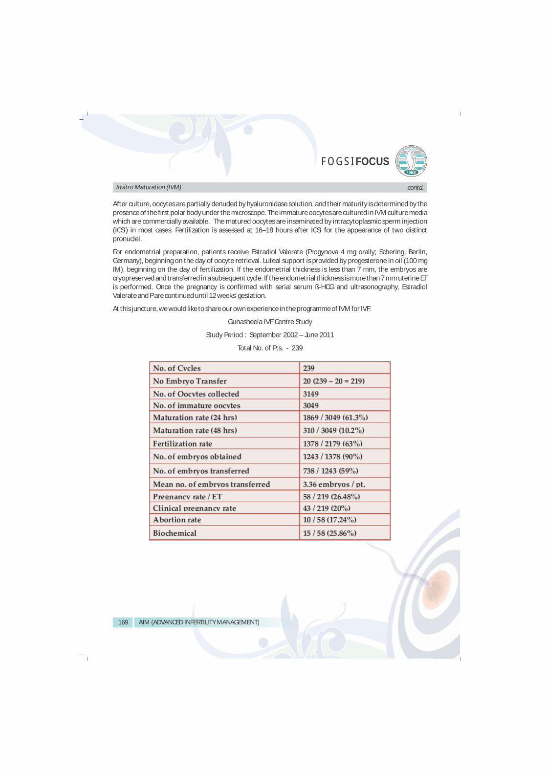
After culture, oocytes are partially denuded by hyaluronidase solution, and their maturity is determined by the presence of the first polar body under the microscope. The immature oocytes are cultured in IVM culture media which are commercially available. The matured oocytes are inseminated by intracytoplasmic sperm injection (ICSI) in most cases. Fertilization is assessed at 16–18 hours after ICSI for the appearance of two distinct pronuclei.
For endometrial preparation, patients receive Estradiol Valerate (Progynova 4 mg orally; Schering, Berlin, Germany), beginning on the day of oocyte retrieval. Luteal support is provided by progesterone in oil (100 mg IM), beginning on the day of fertilization. If the endometrial thickness is less than 7 mm, the embryos are cryopreserved and transferred in a subsequent cycle. If the endometrial thickness is more than 7 mm uterine ET is performed. Once the pregnancy is confirmed with serial serum ß-HCG and ultrasonography, Estradiol Valerate and P are continued until 12 weeks' gestation.
At this juncture, we would like to share our own experience in the programme of IVM for IVF.
Gunasheela IVF Centre Study
Study Period : September 2002 – June 2011
Total No. of Pts. - 239

AIM (ADVANCED INFERTILITY MANAGEMENT) 170
FOGSI FOCUS
Gunasheela IVF Centre Study
Study Period : September 2002 – June 2011
Total No. of Pts. - 239; Immature Oocytes – 3049
MINIMAL STIMULATION IN PCOS
Gunasheela IVF Centre Study
Study Period : September 2002 – June 2011
Total No. of Pts. - 49
Invitro Maturation ( )IVM contd.

AIM (ADVANCED INFERTILITY MANAGEMENT)171
FOGSI FOCUS
Invitro Maturation ( )IVM
In conclusion, IVM for IVF programme has come to stay. It is less expensive than traditional IVF, less dangerous to patients, because of total avoidance of the complications of life-threatening OHSS. It can also produce occasional pregnancies on women who are bordering on menopause. IVM can attract many more women to take up egg donation voluntarily.
After all said and done, the controversy of the carcinogenic potentiality of ovulation inducing drugs is still going on even as we continue to use them. Lessening of the quantum of the usage of these drugs, or even avoiding them, will come as a boon to treatment of assisted conception.
Reference :
1. Pincus G, Enzmann EV. Comparative behavior of mammalian eggs in vivo and in vitro. J Exp Med. 1935;62:665–678.
2. Edwards RG. Maturation in vitro of human ovarian oocytes. Lancet. 1965;2:926–929.
3. Veeck LL, Wortham JW, Witmyer J, Sandow BA, Acosta AA, Garcia JE, et al. Maturation and fertilization of morphologically immature human oocytes in a program of in vitro fertilization. Fertil Steril. 1983;39:594–602.
4. Nogueira D, Staessen C, Van de Velde H, Van Steirteghem A. Nuclear status and cytogenetics of embryos derived from in vitro-matured oocytes. Fertil Steril. 2000;74:295–298.
5. C, Wright DL, Mayer JF, Gibbons W, Muasher SJ, Lanzendorf SE. Human embryos derived from in vitro and in vivo matured oocytes: analysis for chromosomal abnormalities and nuclear morphology. J Assist Reprod Genet. 2000;17:284–292.
6. Emery BR, Wilcox AL, Aoki VW, Peterson CM, Carrell DT. In vitro oocyte maturation and subsequent delayed fertilization is associated with increased embryo aneuploidy. Fertil Steril. 2005;84:1027–1029.
7. Cha K, Koo JJ, Choi DH, Han SY and Yoon TK. Pregnancy after in vitro fertilization of human follicular oocytes collected from non stimulated cycles, their culture in vitro and their transfer in a donor oocyte program. Fertil Steril 1991; 55,109–113.
nd8. H. I. Nielsen. History and Fundamentals of Oocyte Maturation in Vitro. 2 International symposium on In Vitro Maturation of Oocytes. Lyon. 2007
9. Van den Hurk R., Abir R., Telfer E.E. and Bevers M.M. Preantral and antral follicles as possible source for fertilizable oocytes in human and bovine. Hum. Reprod. Update 2000; 6, 457-474.
10. Gougeon A. Regulations of ovarian follicular development in primates: facts and hypotheses. Endocrinol. Rev. 1996; 17, 121-154.
11. Mikkelsen AL, Lindenberg S. Benefit of FSH priming of women with PCOS to the in vitro maturation procedure and the outcome: a randomized prospective study. Reproduction. 2001;122:587–592.
12. Chian RC, Buckett WM, Too LL, Tan SL. Pregnancies resulting from in vitro matured oocytes retrieved from patients with polycystic ovary syndrome after priming with human chorionic gonadotropin. Fertil Steril. 1999;72:639–642.
13. Chian RC, Buckett WM, Tulandi T, Tan SL. Prospective randomized study of human chorionic gonadotrophin priming before immature oocyte retrieval from unstimulated women with polycystic ovarian syndrome. Hum Reprod. 2000;15:165–170.
14. Mikkelsen AL, Smith SD, Lindenberg S. In-vitro maturation of human oocytes from regularly menstruating women may be successful without follicle stimulating hormone priming. Hum Reprod. 1999;14:1847–1851.
15. Le Du A, Kadoch IJ, Bourcigaux N, Doumerc S, Bourrier MC, Chevalier N, et al. In vitro oocyte maturation for the treatment of infertility associated with polycystic ovarian syndrome: the French experience. Hum Reprod. 2005;20:420–424.
16. Cobo AC, Requena A, Neuspiller F, Aragones M, Mercader A, Navarro J, et al. Maturation in vitro of human oocytes from unstimulated cycles: selection of the optimal day for ovum retrieval based on follicular size. Hum Reprod. 1999;14:1864–1868.
17. Hreinsson J, Rosenlund B, Friden B, Levkov L, Ek I, Suikkari AM, et al. Recombinant LH is equally effective as recombinant HCG in promoting oocyte maturation in a clinical in-vitro maturation programme: a randomized study. Hum Reprod. 2003;18:2131–2136.

AIM (ADVANCED INFERTILITY MANAGEMENT) 172
FOGSI FOCUS
OOCYTE DONATION & EMBRYO DONATION
Oocyte Donation
The advent of IVF-ET has given birth to not only thousands of babies but also countless number of newer techniques which aid in infertility. Egg donation is part of the process of third party reproduction as part of ART (Assisted Reproductive Technology).
The concept of oocyte donation is about 100 years old, first attempted in rabbit. The first transfer of a fertilized egg from one human to another resulting in pregnancy was reported in July 1983 and subsequently led to the
.1announcement of the first egg-donation-produced human birth on February 3, 1984
Donor oocytes and Embryo donation has given women a chance to get pregnant and give birth to a child which otherwise would not have been possible. Oocyte and embryo donation as practiced today now accounts for approximately 5% of in vitro fertilization recorded births.
A need for egg donation may arise for a number of reasons. The indications of oocyte donation could be divided into two main groups:
1. Without ovarian function
a) Ovarian genesis
b) Premature Ovarian Failure
• Idiopathic
• Genetic
• Immunological
• Iatrogenic
c) Menopausal
d) Bilateral oopherectomy
2. With Ovarian Function
a) Poor responders
b) Failed IVFs due to poor quality oocytes
Dr. Kanthi Bansal MD, DGO, FICOGDirector, Safal Fertility Foundation, AhmedabadEndometriosis Committee Chairperson, FOGSIHonorary Librarian, ISARInternational Affairs Chairperson, ISARSecretary Gujarat Chapter, ISAR
31

AIM (ADVANCED INFERTILITY MANAGEMENT)173
FOGSI FOCUS
Oocyte Donation & Embryo Donation contd.
c) Presence of genetic disease
d) Inaccessible ovaries
Donors include the following types:
Professional Donors responding to advertisement: These donors do it for monetary reasons. They are often anonymous donors typically recruited by egg donor agencies or, sometimes, IVF programs. This group yield good quality of oocytes as the selection could be carried out with strict criteria of age & fertility.
Known Donors or Designated donors: This form of oocyte donation is nearly on altruistic grounds e.g. a friend or relative brought by the patients to serve as a donor specifically to help them. In Sweden, couples who bring such a donor still get another person as a donor, gets advanced on the waiting list for their own procedure, and that donor becomes a "cross donor".
Patients undergoing IVF/ICSI treatment willing to share the extra eggs retrieved: Women who go through in vitro fertilization may be willing to donate unused eggs to such a program, where the egg recipients' together help paying the cost of the IVF procedure.
Egg donation carries risks for both donor and recipient sometimes; it must be made clear that the procedure for the donor, and the medication given, is the same as the medication given for IVF procedure.
The long-term impact of egg donation on donors has not been well studied, but apparently some evidence 2,3 suggests an increased risk of ovarian cancer, and effects on fertility . One in five women report psychological
effects from donating their eggs, both good and bad.
Factors Determining Selection of Egg Donors
1. AGE: The most important characteristic of the egg donor is age. The genetic material is transferred from the donor to the recipient. Chromosomal anomalies increase with increasing age, so the donor should preferably be less than 35 years.

2. PHYSICAL FEATURES: most of the recipients are keen to have the physical characteristics matched to their own. The cast, community, hair, eyes, height, weight, color and built of the donor should closely match to the recipient.
3. BLOOD GROUP: The blood group should be matched as far as possible. Special care should be taken where the recipient couples are Rh –ve to prevent complication of Rh immunization.
4. PARITY: Pregnancy rates are definitely higher when the oocyte donation program is carried out from parous donors. Care should be taken to see that the donor does not conceive. Further still, parous donor who is already sterilized is preferred.
Age 21-35, proved to be fertile, sterilized, physical characteristics and blood group matched to the recipient and motto should be altruistic.
Protocols for preparation of Endometrium
Estrogen and progesterone are administered to create a suitable milieu for successful embryo transfer.
This can be divided into two groups:
1. Preparation in recipients without ovarian function
2. Preparation of recipients with ovarian function.
Preparation in recipients without ovarian function
Exogenous estrogen and progesterone have to be administered to develop the endometrium for successful implantation. There are two methods:
A. Incremental Method: This is more physiological pattern of hormone replacement. It is not very popular as it is difficult to follow. There is also very limited window available for embryo transfer, as there is less chance of synchronization. In this method estrogen is given in an incremental dose and patient has to
thfollow a schedule fixed on a daily bases. Progesterone is started from 15 day.
B. Constant dose: The follicular phase in this protocol is variable. A high dose of estradiol valerate, 2 mg t.d.s or q.d.s is administered daily which is supra-physiological dose. And progesterone is given either in the form of injection 100 mg/day or through vaginal pessary at a dose 400 mg b.d. Progesterone is started at Safal Fertility Foundation, one day before oocyte retrieval from the donor. At our center recipients are given a daily dose of estradiol valerate 2 mg and if donor is available in cycle estrogen is increased. If donor is not available than progesterone is given and menstrual cycle is completed. This helps the recipients who are on a waiting list to be taken into programme anytime.
Preparation of Recipients with Ovarian follicle
This again can be divided into two groups:
A. Recipient with regular cycles: In this group recipient can have donation programme in the natural cycle. thThe cycle has to be monitored for LH surge so that the embryo transfer can be performed on the 5 after
LH surge.
B. Recipients with irregular cycles: GnRH analogues have to be administered in the form of daily
injections/spray/depot. Type of GnRH analogue could be decided by interaction with patient. Different GnRH agonists available in the market are buserelin, leulerolide, triptorelin, goserelin, naferelin. Estrogen has to start once the down regulation is ascertained. The dose and time of administration of
AIM (ADVANCED INFERTILITY MANAGEMENT) 174
Oocyte Donation & Embryo Donation contd.
FOGSI FOCUS

AIM (ADVANCED INFERTILITY MANAGEMENT)175
FOGSI FOCUS
Oocyte Donation & Embryo Donation contd.
estrogen and progesterone remains the same as explained in the earlier group. Estrogen can be given in other forms like conjugated estrogen. 1.25 mg thrice daily, estradiol transdermal application0.1-0.4 mg/day, injection estradiol valerate 2-6 mg/twice a day.
Monitoring Endometrial Development
The endometrium and endometrial thickness can be used for success of oocyte donation programme. The endometrial thickness is 8-12mm is considered ideal for embryo transfer.
Preparation of Endometrium
Women with Menstruation
1. Normal size of uterus
2. Hysteroscopy
3. GnRH-a + estrogen
Women without menstruation
1. Give 3 to 6 months of estrogens + progesterone till
-minimum 3 months of bleeding
-UCL is 5 to 6 cm
-endometrial thickness 8 to 9 mm
2. Hysteroscopy
3. Egg donation programme
AIM (ADVANCED INFERTILITY MANAGEMENT) 176
Oocyte Donation & Embryo Donation contd.
FOGSI FOCUS
Key process involved in egg donation
Recipient evaluation (USG to monitor development of eggs, blood tests to check E2 levels)↓
Donor Recruitment↓
Donor Screening↓
Obtaining informed consent from recipient and donor↓
Synchronization of donor and recipient cycles↓
Prescription of hormones for the recipient↓
Ovarian stimulation of donor↓
Egg retrieval from donors↓
Fertilization of eggs↓
Embryo Transfer↓
Maintenance of pregnancy in recipient
Factors affecting pregnancy rates:
1. Recipient's age: There may be decline in the uterine receptivity with aging but very few women above the age of 50 have been studied.
2. Donor age: Donor age must ideally be less than 35 years
3. Embryo Quality: Pregnancy improves with quality but grade 4 embryos gives 20 % pregnancy rate.
4. Sequencial attempts: Probability of pregnancy using donor is the same in the first three attempts but declines afterwards.

AIM (ADVANCED INFERTILITY MANAGEMENT)177
FOGSI FOCUS
Oocyte Donation & Embryo Donation contd.
Embryo Donation
Embryo donation is an alternative approach to assisted reproduction that meets the needs of single women as well as older and less affluent couples. It is a cost-effective and efficacious means of achieving pregnancy. Embryo donation is more often contemplated than performed. Embryo donation is where extra embryos from a successful IVF of a couple are given to other couples or women for transfer with the goal of producing a successful pregnancy. Embryos for embryo donation may also be created specifically for embryo transfer using donor eggs and sperm, or in some cases donor eggs and donor sperm. It may thus be seen as a combination of sperm donation and egg donation, since what is donated is a combination of these.
Source of Embryo Donation
There are many young patients who need IVF / ICSI procedure, but who cannot afford it. These patients produce lot of eggs and hence many embryos.
After taking proper informed consent of these patients, some of the extra embryos are used for the recipients. In return, the costs of the drugs of the younger patient are borne by the recipient. This embryo sharing is beneficial for both the patients.
Alternatively, during the IVF treatment best quality embryos are transferred into the womb of the patient. The extra embryos can be frozen. If the patient gets pregnant and do not want more children, they often agree to donate their embryos to other infertile couples, to help them to start a family.
There are selected groups of patients to whom embryo donation is recommended:
· When both the partners are infertile
· Couples who are at a high risk of passing on genetic disorders to their offspring.
· Women with recurrent IVF failures
.4· Families who cannot afford the huge fees needed for egg donation may prefer embryo adoption
Embryo donors

AIM (ADVANCED INFERTILITY MANAGEMENT) 178
Oocyte Donation & Embryo Donation contd.
FOGSI FOCUS
The primary motivation for donation is altruism. The decision to donate the embryos is complicated by personal 5
factors, including the possibility that a genetic sibling of their children may result.
Moral or religious views that dictate embryo use only for pregnancy make donation the only plausible option. Patients who have achieved their own family-building goals and are comfortable with disclosing personal information are more likely to donate, as are patients who perceive nurture to be more influential than genetic
6lineage in parental bonding.
Donor embryo recipients
Candidates include those who require donor oocytes (typically less available than donor embryos) and/or 7donor sperm, but have inadequate financial resources for these procedures. Patients who are unsuccessful
with conventional treatments are also suitable for this program.
Matching donor embryos with recipient(s)
There are no published guidelines for matching donor embryos with recipients. A survey of reproductive health 8
professionals showed that matching was equally done by practices and recipients. Programs that routinely offer embryo donation may elect to provide a list from which patients may choose their donor embryos. This list may include non-identifying physical characteristics and whether or not screening was performed. Recipients should be provided ample non-identifying personal, psychological, and health information about the embryo
9donors to maximize their informed decision to accept the embryos for transfer.
Screening and informed consent: Recipients
Potential recipients must sign informed consent for receiving donated embryos, undergo infectious disease 10screening and testing, and comply with program policies for the actual embryo transfer. Psychological
counseling should be strongly considered. Informed consent must include willingness to take full responsibility for offspring that may result, as well as release the donors and the program from liability from potential complications with the pregnancy or offspring.
Endometrium Preparation Procedure:
The preparation of endometrium is similar in both Oocyte donation and Embryo donation programs.
Embryo Donation versus Adoption
Embryo donation is the first choice prior to adoption Unlike traditional adoption, the couple undergoes a medical rather than a legal procedure to have a baby. For infertile couples, embryo donation offers a great opportunity to be pregnant, to bond with their child prior to birth, and to give birth. In addition, embryo donation may be much more affordable than traditional adoption.
Embryo donation also offers couples privacy and secrecy, so that they do not need to worry about societal 11acceptance of their adopted child.
Success Rate:
The pregnancy rate in our clinic is 40-50% per cycle. The reason for the high pregnancy rate is twofold. Firstly, these are often excellent quality embryos. Also, since we prepare the endometrium using exogenous hormones, the uterine receptivity to these embryos is usually very good. When a "fresh cycle" is followed by a "frozen cycle", the success rate with donor eggs goes up to approximately 80%.

AIM (ADVANCED INFERTILITY MANAGEMENT)179
FOGSI FOCUS
Oocyte Donation & Embryo Donation
References:
1. Blakeslee, Sandra (1984-02-04). "Infertile Woman Has Baby Through Embryo Transfer". The New York Times. Retrieved 2009-
11-05.
2. Textbook of Assisted Reproductive Techniques, Laboratory and Clinical Perspectives, edited by David K. Gardner, 2001
3. Galindo, A.; Bodri, D.; Guillen, J.J.; Colodron, M.; Vernaeve, V.; Coll, O. (2009). "Triggering with HCG or GnRH agonist in
GnRH antagonist treated oocyte donation cycles: a randomized clinical trial". Gynecol Endocrinol 25 (1): 60–6
4. Fuscaldo G, Russell S, Gillam L. How to facilitate decisions about surplus embryos: Patients' views. Hum Reprod. 2007; 22:3129–3138.
5. Nachtigall RD, Becker G, Friese C, et al. Parents' conceptualization of their frozen embryos complicates the disposition decision. Fertil Steril. 2007;84:431–434.
6. Hammarberg K, Tinney L. Deciding the fate of supernumerary frozen embryos: A survey of couples' decisions and the factors influencing their choice. Fertil Steril. 2006;86:86–91.
7. Hum. Reprod. (2001) 16 (6): 1120-1128.-for embro donor & recipient
8. Hoffman DI, Zellman GL, Fair CC, et al. Cryopreserved embryos in the united States and their availability for research. Fertil Steril. 2003;79:1063–1069.
9. Grumankin AD, Sisti D, Caplan AL. Embryo disposal practices in IVF clinics in the united States. Politics Life Sci. 2004;22:4–8.
10. Practice Committee of the American Society for Reproductive Medicine and the Practice Committee of Society for Assisted Reproductive Technology. 2008 Guidelines for Gamete and embryo Donation. Fertil Steril. 2008;90:s30–s44
11. deLacey S. Decisions for the fate of frozen embryos: Fresh insights into patients' thinking and their rationales for donating or discarding embryos. Hum Reprod. 2007;22:1751–1758