fnip1 regulates skeletal muscle fiber type … regulates skeletal muscle fiber type specification,...
TRANSCRIPT

Fnip1 regulates skeletal muscle fiber typespecification, fatigue resistance, and susceptibilityto muscular dystrophyNicholas L. Reyesa, Glen B. Banksb, Mark Tsanga, Daciana Margineantuc, Haiwei Gud, Danijel Djukovicd, Jacky Chana,Michelle Torresa, H. Denny Liggitta, Dinesh K. Hirenallur-Sa, David M. Hockenberyc, Daniel Rafteryd,e,and Brian M. Iritania,1
aThe Department of Comparative Medicine, University of Washington, Seattle, WA 98195-7190; bDepartment of Neurology, University of Washington,Seattle, WA 98195; cClinical Division, Fred Hutchinson Cancer Research Center, Seattle, WA 98109-1024; dDepartment of Anesthesiology and Pain Medicine,Mitochondria and Metabolism Center, Northwest Metabolomics Research Center, University of Washington, Seattle, WA 98109-8057; and ePublic HealthSciences Division, Fred Hutchinson Cancer Research Center, Seattle, WA 98109-1024
Edited* by Robert N. Eisenman, Fred Hutchinson Cancer Research Center, Seattle, WA, and approved December 8, 2014 (received for review July 14, 2014)
Mammalian skeletal muscle is broadly characterized by the presenceof two distinct categories of muscle fibers called type I “red” slowtwitch and type II “white” fast twitch, which display marked differ-ences in contraction strength, metabolic strategies, and susceptibil-ity to fatigue. The relative representation of each fiber type canhave major influences on susceptibility to obesity, diabetes, andmuscular dystrophies. However, the molecular factors controllingfiber type specification remain incompletely defined. In this study,we describe the control of fiber type specification and susceptibilityto metabolic disease by folliculin interacting protein-1 (Fnip1). UsingFnip1 null mice, we found that loss of Fnip1 increased the represen-tation of type I fibers characterized by increased myoglobin, slowtwitch markers [myosin heavy chain 7 (MyH7), succinate dehydro-genase, troponin I 1, troponin C1, troponin T1], capillary density,and mitochondria number. Cultured Fnip1-null muscle fibers hadhigher oxidative capacity, and isolated Fnip1-null skeletal muscleswere more resistant to postcontraction fatigue relative to WT skel-etal muscles. Biochemical analyses revealed increased activation ofthe metabolic sensor AMP kinase (AMPK), and increased expressionof the AMPK-target and transcriptional coactivator PGC1α in Fnip1null skeletal muscle. Genetic disruption of PGC1α rescued normallevels of type I fiber markers MyH7 and myoglobin in Fnip1-nullmice. Remarkably, loss of Fnip1 profoundly mitigated muscle dam-age in a murine model of Duchenne muscular dystrophy. Theseresults indicate that Fnip1 controls skeletal muscle fiber type spec-ification and warrant further study to determine whether inhibitionof Fnip1 has therapeutic potential in muscular dystrophy diseases.
folliculin | BHD | AMPK | mTOR | PGC1α
Mammalian skeletal muscle is composed of a mosaic ofmuscle fiber types (type I, type IIa, type IIb, and type IIx),
which are categorized based on differences in the abundance ofmyosin heavy chain (MHC) proteins, mitochondria, and capillarydensity, strength, fatigue resistance, and metabolic strategies (seeref. 1 for review). Type I “slow twitch” fibers are deep red incolor because of high concentrations of myoglobin and highdensities of blood capillaries, which support sustained aerobicactivity. Type I fibers are also rich in mitochondria, have in-creased contraction endurance with lesser strength potential, anduse predominantly oxidative phosphorylation for energy pro-duction. In contrast, type IIb “fast twitch” fibers are pale in colordue to low concentrations of myoglobin, contain comparativelylow numbers of mitochondria, and rely more heavily on anaer-obic glycolysis for energy production. These characteristics allowtype II fibers to have considerable strength and contractionspeed, but only for short anaerobic bursts of activity before themuscles fatigue. Type IIa/x fibers have hybrid characteristicsbetween type I and type IIb fibers in that they have intermediatenumbers of mitochondria and oxidative potential resulting in
moderate strength and improved resistance to fatigue. Becauseslow twitch fibers use predominantly fatty acid oxidation forenergy production, increasing the representation of type I fibersprovides increased protection against obesity and related meta-bolic disorders including diabetes (2–5). Hence, identifying mol-ecules that regulate fiber type conversion can profoundly impactsusceptibility to metabolic diseases and can influence the patho-physiology of muscular dystrophies.Over the last decade, studies using transgenic and knockout
mice, and chemical agonists and antagonists, have resulted in theidentification of several factors that regulate skeletal muscle fibertype differentiation. In particular, the master metabolic sensor AMPkinase (AMPK) has emerged as a key regulator of mitochondrialbiogenesis, type I fiber type specification, and endurance adapta-tions during chronic exercise (6, 7). AMPK is activated in responseto metabolic cues such as low energy (high AMP/low ATP), changesin intracellular Ca2+, and exercise (see ref. 8 for review). Uponactivation, AMPK helps maintain energy homeostasis by stimu-lating mitochondrial biogenesis, ATP production, and autoph-agy, while concurrently inhibiting ATP consumption mediated bymammalian target of rapamycin (mTOR), a major regulator ofcell growth and protein synthesis. AMPK regulates muscle metab-olism and differentiation by synergizing with Ca2+ signaling tomodulate expression and stability of the transcriptional regu-lators peroxisome proliferator-activated receptor-γ coactivator-1α(PGC1α) and PGC1β, which further activate entire genetic
Significance
Folliculin interacting protein-1 (Fnip1) is an intracellular proteinknown to interact with folliculin (a protein mutated in Birt HoggDube’ Syndrome) and the master metabolic sensor AMP kinase.However, the roles of Fnip1 in mammalian development andfunction are unclear. In this study, we used mice deficient inFnip1 to show that Fnip1 regulates skeletal muscle fiber typespecification. Mice deficient in Fnip1 were significantly enrichedfor highly oxidative skeletal muscle that is more resistant tofatigue than wild-type muscle. Loss of Fnip1 also decreasedmuscle damage in a mouse model of Duchenne muscular dys-trophy. These results reveal a previously unidentified functionfor Fnip1 and suggest that pharmacological inhibition of Fnip1may reduce muscle damage in patients with muscular dystrophy.
Author contributions: N.L.R., G.B.B., D.M., H.D.L., D.M.H., D.R., and B.M.I. designed re-search; N.L.R., G.B.B., M. Tsang, D.M., H.G., D.D., J.C., M. Torres, and D.K.H.-S. performedresearch; and N.L.R., G.B.B., and B.M.I. wrote the paper.
The authors declare no conflict of interest.
*This Direct Submission article had a prearranged editor.1To whom correspondence should be addressed. Email: [email protected].
This article contains supporting information online at www.pnas.org/lookup/suppl/doi:10.1073/pnas.1413021112/-/DCSupplemental.
424–429 | PNAS | January 13, 2015 | vol. 112 | no. 2 www.pnas.org/cgi/doi/10.1073/pnas.1413021112

programs involved in mitochondrial biogenesis, oxidative metab-olism, and slow twitch fiber specification (9–12). However, themolecules that link AMPK and PGC1α to fiber type specificationare poorly understood.Through the use of a random ENU mutagenesis strategy in
mice to identify novel immune regulatory genes (13), we pre-viously identified a novel mouse pedigree that lacks expression offolliculin interacting protein-1 (Fnip1) due to a 32-bp deletion inthe Fnip1 gene (14). Fnip1−/− mice were identified by an absenceof B lymphocytes, which was attributed to a block in B-cell de-velopment and survival at the pre–B-cell stage. Although thefunctions of Fnip1 are poorly understood, Fnip1 protein inter-acts with folliculin, Fnip2 (a related Fnip family member), andall three subunits (α, β, γ) of AMPK (15–17). Mutations in theBhd gene encoding folliculin results in Birt–Hogg–Dube (BHD)Syndrome, a rare human condition characterized by hamartomaformation, pulmonary cysts, and development of renal tumors(18). In the current study, we found that the loss of Fnip1 resultsin a pronounced shift of skeletal muscle fiber type toward type Islow twitch and type IIa mixed fibers, due in part to increasedactivation of PGC1α. These results identify a previously un-identified pathway involving Fnip1 in the differentiation of skel-etal muscle fiber types and suggest that Fnip1 may be part ofa complex linking AMPK with PGC1α.
ResultsLoss of Fnip1 Increases the Representation of Type I Skeletal MuscleFibers. Upon gross observation, skeletal muscle from Fnip1−/−
mice appeared dark red in coloration compared with wild-type(WT) skeletal muscle (Fig. 1A and Fig. S1). Because slow-twitchfibers appear deeper red relative to fast-twitch fibers, we in-vestigated whether loss of Fnip1 alters the composition of skel-etal muscle fiber types. We stained sections of gastrocnemiusmuscles from Fnip1−/− and WT mice for enzymes and proteinsthat are differentially expressed in type I relative to type IIbfibers, including succinate dehydrogenase (SDH; a mitochon-drial enzyme involved in oxidative respiration), metachromaticATPase (pH 4.7) (which allows for differential myofibrillar stainuptake), and slow twitch-specific myosin heavy chain 7 (MYH7).As shown in Fig. 1B, WT gastrocnemius (lateral head) musclesconsisted of a mixture of SDH+ type I and SDH− type IIb fibers.However, Fnip1−/− muscle contained almost exclusively SDH+
fibers. Fnip1−/− muscle also contained an excess of ATPase+(Fig. 1C), MyH7+ (Fig. 1D and Fig. S1), and MyH2+ myofibers(Fig. S1) relative to WT muscle. Transmission electron micros-copy also revealed increased numbers of subsarcolemmal mito-chondria in Fnip1−/− muscle relative to WT muscle (Fig. 1E).Morphometric analyses with anti-wheat germ agglutinin (WGA)and anti-CD31 (to identify capillaries) showed that Fnip1-nullmuscle fibers are smaller in diameter (Fig. 1F) and contain in-creased numbers of capillaries relative to WT muscle fibers (Fig.1G), which is consistent with increased representation of slow-twitch myofibers.To further define the apparent shift in muscle fiber types
following the loss of Fnip1, we performed quantitative real-timePCR (qPCR) and immunoblotting to determine the abundanceof mRNA and proteins characteristic of type I versus type IIbmyofibers. qPCR revealed an ∼threefold increase in MyoglobinmRNA expression in Fnip1−/− skeletal muscle compared withcontrols, and pronounced increases in mRNA expression of cy-toskeletal markers of slow twitch muscle including Troponin Islow (Tnni1), Tnnc1, Tnnt1, and MyH7 (Fig. 2A), with corre-sponding decreases in the transcript levels of MyH4, which isa marker of type IIb fast-twitch fibers (Fig. 2B). Immunoblotanalyses confirmed significant increases in Myoglobin, MyH7,and cytochrome C proteins in Fnip1 null gastrocnemius musclecompared with WT muscle (Fig. 2C).Because increased activity could theoretically result in in-
creased oxidative fibers in Fnip1−/− mice, we measured ambu-latory activity levels in Fnip1−/− versus Fnip1+/− mice. We foundno significant difference in activity levels during either the light
or dark cycles (Fig. S2). These results collectively suggest thatloss of Fnip1 results in a significant shift in the representation ofskeletal muscle fibers from fast twitch type IIb to slow twitch typeI and mixed type IIa fibers.
Fnip1 Null Skeletal Muscle Contains Increased Numbers of HighlyFunctional Mitochondria. To further define the role of Fnip1 infiber type specification, we assessed the normal levels of Fnip1protein in muscles with type IIb characteristics [extensor digitalislongus (EDL) and gastrocnemius] versus type I characteristics(Soleus). We found that in WT skeletal muscles, Fnip1 protein isexpressed in muscles rich in type IIb fibers (EDL, gastrocne-mius), but is low or absent in soleus muscles, which are rich intype I fibers (Fig. S3). These results are consistent with the no-tion that Fnip1 has a role in fiber type specification. To char-acterize the consequences of Fnip1 loss on the metabolic capacityof muscle fibers, we assessed levels of key mitochondrial genetranscripts and measured the metabolic capacity of myofibrilsfrom Fnip1−/− and WT gastrocnemius muscle. qPCR analysis in-dicated that relative to WT muscle, disruption of Fnip1 resultedin increased levels of mitochondrial gene transcripts including
BSDH
C ATPase4.7
DMYH7
AWildtype Fnip1-/- Wildtype Fnip1-/-
E
Rel
ativ
eF
iber
Siz
eD
istr
ibut
ion
( Pix
elu n
i tsx
100 /
fiber
)
0 5 10 15 200 5 10 15Percent of Fibers
F
EM(1000x)
G
Fnip1-/- Wildtype
p=5 x 10⁻⁸
CD31WGA
Wildtype Fnip1-/-
0
1
2
3
#Capillaries/fiber
4-6
12-1416-18
0-2
8-10
20-2224-2628-3032-3436-3840-4244-4648-5052-5456-58 p=4 x 10⁻⁸
Fig. 1. Fnip1 null skeletal muscle is characterized by deep red coloration;increased SDH, ATPase pH 4.7, and MyH7 staining; increased mitochondria,decreased myofibril size, and increased capillary density indicative of slowtwitch skeletal muscle. (A) Representative photographs showing WT andFnip1−/− skeletal muscle after euthanasia. (B–D) Immunohistochemistrystaining of mitochondrial and slow twitch myofiber markers was performedon cross-sections of the lateral head, gastrocnemius muscle. Shown arerepresentative SDH (B); ATPase (pH 4.7) (C); and myosin heavy chain 7(MyH7) (D) stained sections from n = 3–5 8-wk-old mice per group. (E) Increasedmitochondria in Fnip1−/− muscle. Electron micrographs of the gastrocnemiusmuscle taken from 8-wk-old male mice. Shown are representative 1,000×images. (F) Representative wheat germ agglutinin (WGA) stained cross-sectionsof WT and Fnip1 null gastrocnemius muscles. Images were analyzed by fluo-rescence microscopy, and cross-sectional area was measured. Bar graphs depictrelative fiber size distribution of a total of 100 individual fibers per genotype(n = 3 mice per group). Wt mean = 3,929 (pixels per fiber), Fnip1−/− mean =2,199 (pixels per fiber); P values shown. (G) Increased capillary density inFnip1−/− relative to WT gastrocnemius muscle. Sections from 8- to 12-wk-oldmice were stained with WGA and anti-CD31. Shown are representative im-munofluorescence photos. Graphs represent the mean and SEM of fourFnip1−/− and 4 Fnip1+/− gastrocnemius muscles. P values are shown. (Scalebars: B, D, and G, 100 μm; E, 2 μm.)
Reyes et al. PNAS | January 13, 2015 | vol. 112 | no. 2 | 425
DEV
ELOPM
ENTA
LBIOLO
GY

cytochrome B (Cytb), ATP synthase lipid binding protein(Atp5γ1), and uncoupling protein 3 (Ucp3) (Fig. S3).To assess the functional consequences of the increased mito-
chondria mass in Fnip1−/− skeletal muscle, we isolated adultmyofibers from Fnip1−/− and WT mice and assessed basal bio-energetic metabolism by using the Seahorse XF analyzer, whichmeasures oxygen consumption rate (OCR; a measure of oxidativephosphorylation) on live cultured cells. We found that Fnip1−/−
myofibrils exhibit significantly increased basal OCR relative toWTmyofibrils (Fig. S3), suggesting that disruption of Fnip1 resultsin a pronounced expansion of functional mitochondria.To better define this observed metabolic shift, we performed
targeted metabolomics using liquid chromatography tandemmass spectrometry (LC-MS/MS) to measure alterations in levelsof 158 metabolites associated with major metabolic pathways.We found that Fnip1−/− gastrocnemius muscle contained in-creased metabolites associated with amino acid signaling (glu-tamine, asparagine, and glutamic acid), and progression of theTCA cycle (α-ketoglutaric acid, aconitate), consistent with in-creased mTOR signaling (further discussed below) and increasedoxidative metabolism (Table S1). Collectively, our results areconsistent with a normal role for Fnip1 in the generation and/ormaintenance of type IIb fibers, and a shift to oxidative type I andIIa fibers following disruption of Fnip1.
Increased Activation of AMPK and PGC1α in Fnip1 Null Skeletal Muscle.Because Fnip1 directly interacts with AMPK, we compared RNAand protein levels of components of the AMPK signaling pathwayin gastrocnemius muscle from Fnip1−/− and WT mice. Levels ofphosphorylated AMPK and phosphorylated acetyl CoA carbox-ylase 1 (ACC1) were increased in Fnip1−/− skeletal muscle relativeto WT muscle, which is indicative of activated AMPK (Fig. 3A).Expression of the AMPK target and transcriptional coactivatorPGC1α was significantly elevated at both the protein (Fig. 3A) andmRNA (Fig. 3B) level in skeletal muscle from Fnip1−/− relative toWT mice (19, 20). Transcript levels of Pparα, Pparδ, and PGC1β
(coactivation targets of PGC1α) (6) were also increased in Fnip1−/−
mice. These results collectively suggest that disruption of Fnip1results in increased basal activation of AMPK, which targetsmultiple transcriptional programs controlled by its substratesincluding PGC1α (6).
Increased mTORC1 Activity in Fnip1 Null Skeletal Muscle.Mammaliantarget of rapamycin consists of the rapamycin-sensitive mTORC1complex (mTOR, Raptor, Pras-40, Deptor, mLST8) and therapamycin-insensitive mTORC2 complex (mTOR, Rictor, mSin1,Protor, Deptor, and mLST8) (21). mTORC1 promotes cell growthby inducing protein synthesis and cell cycle progression in re-sponse to amino acids and growth factors. In contrast, mTORC2regulates cell survival and cytoskeletal organization in responseto growth factors. Under energy-deficient conditions, activatedAMPK inhibits energy consuming cellular growth driven by themTORC1 pathway in part by phosphorylating and activating themTORC1 inhibitor TSC2, and by phosphorylating and inhibitingthe mTORC1-activating adaptor Raptor (8). Because mTORC1has been shown to enhance mitochondrial biogenesis in skeletalmuscle, we sought to determine whether mTORC1 is regulated byFnip1. Immunoblot analysis revealed increased phosphorylationof ribosomal S6 protein (S6R) and EIF4E-binding protein 1(P-4EBP1) in Fnip1−/− gastrocnemius muscle relative to WTmuscle (Fig. S4). P-S6R and P-4EBP are downstream productsof mTORC1 activation and are commonly used as markers ofmTORC1 activity. mTORC1 is also regulated by a negative-feedback loop resulting in reduced expression of growth factorreceptors and decreased PI3K signaling (21). Indeed, we founda reduction of biphosphorylated glycogen synthase kinase-3(GSK-3) which is phosphorylated by protein kinase B (AKT), adownstream effecter of PI3 kinase (Fig. S4). Inhibition of GSKactivation is likely a consequence of mTORC1 self-regulationthrough inhibition of PI3-kinase and subsequently AKT.We next examined whether hyperactive mTOR might be re-
sponsible for the shift in skeletal muscle fiber type following dis-ruption of Fnip1. We treated Fnip1−/− mice with the mTORC1inhibitor rapamycin beginning at conception (i.e., breeding pairswere treated with rapamycin diet) through weaning until 6 wk ofage when gastrocnemius muscles were harvested and analyzed.We found that long-term rapamycin treatment failed to reducelevels of the slow twitch markers MyH7, myoglobin, and PGC1α inFnip1−/− mice relative to WT mice despite inhibition mTORC1activity (pS6R) to below basal WT levels (Fig. S4). These resultscollectively support a role of Fnip1 in skeletal muscle fiber typerepresentation relatively independent of mTORC1.
Fnip1 Null Skeletal Muscle Is More Resistant to Fatigue. To investigatethe functional significance of the shift in fiber type following
A
B C
Fig. 2. Fnip1 null skeletal muscle expresses increased levels of slow twitch-specific genes and proteins. (A and B) Gastrocnemius muscle RNA wasextracted from 8-wk-old male mice. Gene expression was measured by qPCR.Shown are the means ± SEM from 4 to 6 mice per group. mRNA expressionlevels are shown as Fnip1−/− relative to WT mice. (C) Immunoblotting ofproteins characteristic of slow twitch muscle fibers. Proteins were isolatedfrom gastrocnemius muscle. Shown are representative immunoblots show-ing three independent mice per group. Gapdh is shown as loading controls.P values are shown (Student’s t test).
A B
Fig. 3. Fnip1−/− skeletal muscles exhibit hyperactivation of the AMPK/PGC1αpathway. (A) Immunoblots were performed on protein lysate extracted fromgastrocnemius muscles of age- and sex-matched mice. Shown are representa-tive immunoblots from five mice of each genotype. Numbers 1, 2, and 3 rep-resent individual mice of each genotype. (B) Expression of ERRα, Pparα, Pparδ,PGC1α, and PGC1β were determined via qPCR on RNA extracted from thegastrocnemius of age- and sex-matched mice. Shown are bar graphs depictingthe mean ± SEM from six mice per group.
426 | www.pnas.org/cgi/doi/10.1073/pnas.1413021112 Reyes et al.
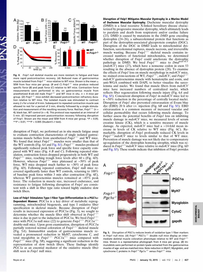
disruption of Fnip1, we performed an in situ muscle fatigue assayto evaluate contraction characteristics of single isolated gastroc-nemius muscle bellies from anesthetized Fnip1−/− and WT mice.We found that intact Fnip1−/− muscles were smaller than that ofthe WT controls (Fig. 4A and Fig. S1). Fnip1−/− muscles producedsignificantly reduced peak force and specific force capacity com-pared with WT mice (Fig. 4 B and C). Following repeated stim-ulation, contraction forces dropped progressively in both WT andFnip1−/− mice, reaching trough force levels after 60 s (Fig. 4D).However, whereas Fnip1−/− mice plateaued at ∼50% of peakforce, WT mice dropped much further to ∼30% of peak force(Fig. 4D). Following repeated contraction, Fnip1 null mice re-covered significantly faster than WT controls, returning to 100%of baseline peak force within 3 min after contraction (Fig. 4E),whereas WT gastrocnemius muscles remained at ∼85% peakforce. The reduction in muscle size, increased endurance, andresistance to fatigue following disruption of Fnip1 are consis-tent with a shift in fiber type ratio toward highly oxidative slowtwitch fibers.
Loss of Fnip1 Stimulates Type I Fiber Type Differentiation in a PGC1α-Dependent Manner. PGC1α is a key driver of metabolic reprog-ramming, mitochondrial biogenesis, and type I oxidative fiberspecification in skeletal muscle. Because disruption of Fnip1results in increased expression of PGC1α (Fig. 3), we sought todetermine whether the muscle fiber shift observed in Fnip1−/−
mice is due in part to the induction of PGC1α. We bred Fnip1−/−
mice with PGC1α null mice (22) to generate Fnip1−/− PGC1α−/−double-null mice. Upon gross examination, disruption of PGC1αpartially restored normal coloration of Fnip1−/− skeletal muscle(Fig. 5A). Immunoblot analysis of gastrocnemius muscle re-vealed a pronounced reduction in MyH7, cytochrome C, andtotal myoglobin in Fnip1−/− PGC1α−/− mice compared withFnip1−/− mice (Fig. 5B), suggesting a significant reduction in therepresentation of slow twitch fibers. These findings identifyPGC1a as an essential mediator of the oxidative muscle fibershift seen in Fnip1 null mice.
Disruption of Fnip1 Mitigates Muscular Dystrophy in a Murine Modelof Duchenne Muscular Dystrophy. Duchenne muscular dystrophy(DMD) is a fatal recessive X-linked hereditary disease charac-terized by progressive muscular dystrophy, which ultimately leadsto paralysis and death from respiratory and/or cardiac failure(23). DMD is caused by mutations in the DMD gene encodingdystrophin (24–26), a subsarcolemmal protein that functions aspart of the dystrophin-associated glycoprotein complex (DGC).Disruption of the DGC in DMD leads to mitochondrial dys-function, sarcolemmal ruptures, muscle necrosis, and irreversiblemuscle wasting. Because Fnip1−/− skeletal muscle contains in-creased numbers of functional mitochondria, we determinedwhether disruption of Fnip1 could ameliorate the dystrophicpathology in DMD. We bred Fnip1−/− mice to Dmdmdx-4CV
(mdx4CV) mice (27), which have a nonsense codon in exon 53resulting in the absence of dystrophin protein (28). To examinethe effects of Fnip1 loss on muscle morphology in mdx4CV mice,we stained cross-sections of WT, Fnip1−/−, mdx4CV, and Fnip1−/−
mdx4CV gastrocnemius muscle with hematoxylin and eosin, andanti-WGA combined with DAPI, to better visualize the sarco-lemma and nuclei. We found that muscle fibers from mdx4CVmice have increased numbers of centralized nuclei, whichreflects fiber regeneration following muscle injury (Fig. 6A andFig. S5). Concurrent disruption of Fnip1 in mdx4CV mice led toa ∼50% reduction in the percentage of centrally located nuclei.Disruption of Fnip1 also prevented extravasation of Evans bluedye (EBD) 20 h after i.v. injection (Fig. 6B and Fig. S5). EBDextravasation is a common measure of increased vascular andcellular permeability that occurs following muscle damage. Tofurther assess the potential benefits of Fnip1 loss on inhibitingmuscle damage in mdx4CV mice, we measured levels of serumcreatine kinase (CK), which is a sensitive measure of muscledamage. As expected, mdx4CV mice had a ∼14-fold mean in-crease in levels of CK relative to WT mice (Fig. 6C). Re-markably, disruption of Fnip1 profoundly reduced CK levels inFnip1−/−mdx4CV mice to levels similar to that of WT mice.Prevention of muscle damage was not likely due to compensatoryup-regulation of the dystrophin homolog utrophin, which was re-duced in Fnip1−/−mdx4CV mice relative to mdx4CV mice (Fig. 6Dand Fig. S5). These results indicate that inhibition of Fnip1 provides
051020253035404550
PeakForce(mNx100)
SpecificForce(KN/m2 )
0
50
100
200
250
300
0255075100125150175
WT Fnip1-/-
0
20
40
60
80
100
2400 60 120 180time (s)
%PeakForce
D
Gastroc.Mass(Mg)A B C
%Recovery
3 min 5 min
EWT Fnip1-/- WT Fnip1-/-
WTFnip1-/-
02040
60
80100
Fig. 4. Fnip1 null skeletal muscles are more resistant to fatigue and havemore rapid postcontraction recovery. (A) Reduced mass of gastrocnemiusmuscle isolated from Fnip1−/− mice relative to WT mice. Shown is the mean ±SEM from four mice per group. (B and C) Fnip1−/− mice produce reducedspecific force (B) and peak force (C) relative to WT mice. Contraction forcemeasurements were performed in situ on gastrocnemius muscle fromanesthetized 8-wk-old male Fnip1−/− and WT mice in situ. n = 4 mice pergroup. (D) Fnip1−/− mice exhibit decreased postcontraction refractory dura-tion relative to WT mice. The isolated muscle was stimulated to contractevery 2 s for a total of 4 min. Subsequent to repeated contraction muscle wasallowed to rest for a period of 3 min, directly followed by a single stimula-tion and measurement of the resulting recovery force. Red bar, Fnip1−/− (n =4); black bar, WT control (n = 4). This protocol was repeated at an interval of5 min. (E) Improved percent postcontraction recovery following disruptionof Fnip1. Shown are the mean and SEM from 4 mice per group. *P < 0.05;**P < 0.01; ***P < 0.005 (Student’s t test).
Wildtype
Fnip1-/-
Myoglobin
MyH7
gapdh
PGC1
Fnip1-/-
PGC1-/-
CytoC
A B
WT
Fnip1-/-
Fnip1-/-
Pgc1
Fig. 5. Disruption of PGC1α reduces levels of oxidative type 1 fiber markersin Fnip1 null mice. (A) Fnip1−/−PGC1α−/− double null mice display an inter-mediate skeletal muscle coloration phenotype relative to WT and Fnip1−/−
mice. Shown is a representative photograph from 4 mice per group. (B) Im-munoblots were performed on protein lysate extracted from the gastrocnemiusmuscles of age- and sex-matchedmice. Note reduction inMyH7, Myoglobin, andcytochrome C proteins in Fnip1−/−Pgc1α−/− versus Fnip1−/− mice.
Reyes et al. PNAS | January 13, 2015 | vol. 112 | no. 2 | 427
DEV
ELOPM
ENTA
LBIOLO
GY

significant protection against muscle damage in the mdx4CV modelof Duchenne muscular dystrophy.
DiscussionThe benefits of endurance exercise on skeletal muscle functionand resistance to obesity occur in part through the metabolicreprogramming of myofibers, which results in increased utiliza-tion of fatty acids, improved resistance to fatigue, and increasedprotection against obesity and metabolic diseases including di-abetes and cardiovascular disease (2–5). In this study, we foundthat disruption of Fnip1 results in a shift in skeletal muscle fibertype from predominantly type IIb glycolytic fibers to type I andtype IIa oxidative fibers that are rich in mitochondria andcapillaries and preferentially use oxidative phosphorylation overglycolysis. Our results identify Fnip1 as an integral member of asignaling pathway involved in programming skeletal muscle fiberspecification, and suggest that inhibition of Fnip1 has “exercisemimetic” properties, which has potential to profoundly impactoverall metabolic health.How might Fnip1 regulate fiber type specification? AMPK is
hyperactivated in Fnip1 null muscle based on increased phos-phorylation of AMPK at Thr172, and increased phosphorylationof ACC1, an AMPK target involved in lipid metabolism. AMPKwas shown to be a major signaling molecule involved in speci-fying skeletal muscle fiber type differentiation and mitochondrialbiogenesis (29) in response to endurance exercise and chronicenergy deprivation. For example, gain-of-function mutations in
the AMPK γ3 subunit in mice increased mitochondrial bio-genesis and oxidative potential in glycolytic skeletal muscle, andprovided protection from dietary-induced insulin resistance (30).Similarly, exercise training and the AMPK agonist AICAR in-creased oxidative fibers, running endurance, and glucose uptakein adult mice (6, 31). Conversely, reduced AMPK activity inAMPKα2 (32, 33) or AMPK β1β2 null mice (34) lead to de-creased skeletal muscle mitochondrial function and increasedinsulin resistance (31, 35). Thus, our results are consistent withFnip1 regulating mitochondrial biogenesis and fiber type de-termination in part by directly or indirectly regulating AMPK.Because our study was performed by whole body deletion ofFnip1, we cannot discern whether the shift in skeletal musclefiber type is autonomous to muscle cells. However, activity levelswere not increased in Fnip1−/− mice, suggesting that increasedactivity is not causing the shift in fiber representation.To further define the mechanism whereby Fnip1 regulates
mitochondrial biogenesis and fiber type determination, weassessed levels of PGC1α, a master transcriptional coactivatorknown to regulate these processes downstream of AMPK.PGC1α, and the related PCG1β, have been shown to initiatea program of mitochondrial biogenesis and oxidative phosphor-ylation by increasing transcription of components of the electrontransport chain, TCA cycle, and fatty acid oxidation (36). PGC1αis preferentially expressed in “red” oxidative fibers, and trans-genic expression of PGC1α (4) or PCG1β (5) in skeletal musclefibers increases mitochondrial content, cellular respiration, andfatigue resistance. In our study, we found that disruption ofFnip1 results in increased levels of PGC1α transcripts and pro-tein. Genetic disruption of PGC1α significantly reduced ex-pression of the slow-twitch specific MyH7, myoglobin, andcytochrome C in PGC1α−/−Fnip1−/− relative to Fnip1−/− gas-trocnemius muscles. These results demonstrate that disruptionof Fnip1 results in increased levels of PGC1α protein, which,in turn, contributes to increased mitochondrial biogenesis andmuscle fiber type switch.It is not clear how loss of Fnip1 results in increased PGC1α.
Fnip1 physically interacts with folliculin, Fnip2, HSP90, and allthree subunits of AMPK. AMPK stimulates mitochondria bio-genesis and increases oxidative phosphorylation resulting in ATPproduction, in part by increasing expression of PGC1α and byphosphorylating PGC1α (20), resulting in increased proteinstabilization. Other studies have shown that AMPK phosphor-ylates both Fnip1 and folliculin (15), and inhibition of folliculinin mice also induces mitochondrial biogenesis and skeletalmuscle fiber type switch in a PGC1α-dependent manner (37).Hence, the complex of Fnip1 and folliculin may function ina negative feedback loop to inhibit or “turn off” AMPK, PGC1α,and oxidative metabolism following AMPK activation (Fig. S6).In the absence of Fnip1 or folliculin, AMPK is hyperactivated,resulting in increased PGC1α expression and oxidative metabo-lism. Consistent with this notion, a recent study concluded thatfolliculin represses AMPK activation and PGC1α up-regulationin primary mouse embryonic fibroblasts (38), and a separate studyconcluded that folliculin represses AMPK in Caenorhabditiselegans (39), although mechanism(s) for repression were not de-termined in either study. However, whereas loss of Fnip1 mimicsmany aspects of AMPK activation in skeletal muscle, there arealso important differences. For example, others have shown thatactivated AMPK inhibits cell growth by inhibiting mTOR. Wefound that both AMPK and mTOR are hyperactivated in Fnip1−/−
skeletal muscle, pre-B cells, and iNKT cells (40), suggesting thatFnip1 could have several functions including “turning off” AMPKfollowing activation and coupling AMPK to mTOR inhibition.Muscular dystrophy diseases are often typified by defective
mitochondria (41), and slow oxidative fibers have been shown tobe more resistant to dystrophic pathology than fast, glycolyticfibers (42). In this study, we found that inhibition of Fnip1attenuates severe dystrophic pathology in the mdx4CV murinemodel of DMD (43). Overexpression of PGC1α specifically inskeletal muscle also protects against muscle dystrophy (44),
Cre
atin
eK
inas
e(F
old
rela
ti ve
toW
T)C D
A B
Eva
nsD
yeE
xtra
vasa
tion WT
mdxFnip1-/-
Fnip1-/-
mdx4CV
051015202530354045
Fnip1-/-
mdx4
CV
Fnip1-/-
mdx4
CVPer
cent
cent
raln
ucle
i .04
Rel
ativ
eU
trop
hin
A(U
tro/
Gap
dh)
0
.5
1
1.5
2
2.5
mdx4
CV
Fnip1-/-
mdx4
CV
.05
0
5
10
15
20
25 .004.0006
mdx4
CV
Fnip1-/-
Fnip1-/-
mdx4
CV
WT
Fig. 6. Disruption of Fnip1 significantly reduces muscle fiber damage andrestores muscle fiber integrity in muscular dystrophy mice. Fnip1−/− micewere bred to mdx4CV mice to generate mice of the indicated genotypes. (A)Quadriceps muscles were stained with hematoxylin and eosin (H and E), andwheat germ agglutinin plus DAPI, which assists in visualizing plasma mem-branes and nuclei. Nuclei were counted in 10 random fields per genotype(n = 3 per genotype) in a blinded fashion. (B) Loss of Fnip1 expression sig-nificantly reduces extravasation of EBD in mdx4CV mice. Shown are repre-sentative images of cross-sections of gastrocnemius muscle harvested frommice of the listed genotypes. Dye leakage was analyzed by fluorescent mi-croscopy. (C) Inhibition of Fnip1 significantly reduces serum creatine kinaselevels in mdx4CV mice. Creatine kinase levels were determined by colori-metric assay. n = 5–6 mice per group. (D) Utrophin protein expression in de-creased in Fnip1−/− mice. Protein was extracted from quadricepts muscle, andutrophin was measured by immunoblotting. Bar graphs represent the mean ±SEM of three mice per genotype. P values are shown (Student’s t test).
428 | www.pnas.org/cgi/doi/10.1073/pnas.1413021112 Reyes et al.

suggesting that inhibition of Fnip1 may act in part through in-duction of PGC1α. Given the role of Fnip1 in skeletal musclefiber type differentiation, capillary density, mitochondrial bio-genesis, and resistance to fatigue, these results warrant furtherinvestigation as to whether pharmacological inhibition of Fnip1may provide an innovative strategy to improve muscle functionon patients with muscular dystrophy diseases and/or improve theresponses of patients with obesity-associated disease.
Materials and MethodsTransgenic Animals. Fnip1−/− mice were developed as described (14). Themajority of the studies were performed on 8- to 12-wk-old male mice. PGC1α(22) and DmdMdx4CV (27) mice were obtained from Jackson Laboratories andthe Chamberlain laboratory, respectively. Mice were housed under specificpathogen-free conditions. Animal studies were reviewed and approved bythe Institutional Care and Use Committee of the University of Washington.
Immunohistochemistry and Metachromatic ATPase Staining. Gastrocnemiusmuscles were collected and were flash frozen in OCT before sectioning. Allspecial staining was performed at the Histology and Imaging Core. High powerimmunohistochemistry images were performed on the lateral head after re-moval of the soleus muscle. MyH7 antibody was obtained from Sigma-Aldrich.
Serum Creatine Kinase Assay. Creatine kinase was measured in serum accordingto manufacturer’s instructions [Creatinine Kinase-SL Reagent Kit (Sekisui Diag-nostics P.E.I. Inc.)].
EBD Assay. A 1% solution of EBD was injected i.p. at a volume dose of 1% ofbody weight. Approximately 20 h following injection, mice were killed andthe gastrocnemius muscles were snap frozen in OCT media. Leakage of EBDwas analyzed by fluorescence microscopy (44).
ACKNOWLEDGMENTS. We thank the Ruddell laboratory for assistancewith the fluorescent microscopy. This study was supported by NIH GrantsK26RR024462, R56AI092093, P30-DK035816, and R25RR032027 (to B.M.I.).
1. Schiaffino S, Reggiani C (2011) Fiber types in mammalian skeletal muscles. Physiol Rev91(4):1447–1531.
2. Stuart CA, et al. (2013) Slow-twitch fiber proportion in skeletal muscle correlates withinsulin responsiveness. J Clin Endocrinol Metab 98(5):2027–2036.
3. Hambrecht R, et al. (1997) Effects of endurance training on mitochondrial ultra-structure and fiber type distribution in skeletal muscle of patients with stable chronicheart failure. J Am Coll Cardiol 29(5):1067–1073.
4. Lin J, et al. (2002) Transcriptional co-activator PGC-1 alpha drives the formation ofslow-twitch muscle fibres. Nature 418(6899):797–801.
5. Arany Z, et al. (2007) The transcriptional coactivator PGC-1beta drives the formationof oxidative type IIX fibers in skeletal muscle. Cell Metab 5(1):35–46.
6. Narkar VA, et al. (2008) AMPK and PPARdelta agonists are exercise mimetics. Cell134(3):405–415.
7. Hardie DG (2011) Energy sensing by the AMP-activated protein kinase and its effectson muscle metabolism. Proc Nutr Soc 70(1):92–99.
8. Gowans GJ, Hardie DG (2014) AMPK: A cellular energy sensor primarily regulated byAMP. Biochem Soc Trans 42(1):71–75.
9. Wu H, et al. (2002) Regulation of mitochondrial biogenesis in skeletal muscle by CaMK.Science 296(5566):349–352.
10. Naya FJ, et al. (2000) Stimulation of slow skeletal muscle fiber gene expression bycalcineurin in vivo. J Biol Chem 275(7):4545–4548.
11. Kramerova I, et al. (2012) Impaired calcium calmodulin kinase signaling and muscleadaptation response in the absence of calpain 3. Hum Mol Genet 21(14):3193–3204.
12. Parsons SA, Wilkins BJ, Bueno OF, Molkentin JD (2003) Altered skeletal muscle phe-notypes in calcineurin Aalpha and Abeta gene-targeted mice. Mol Cell Biol 23(12):4331–4343.
13. Appleby MW, Ramsdell F (2003) A forward-genetic approach for analysis of the im-mune system. Nat Rev Immunol 3(6):463–471.
14. Park H, et al. (2012) Disruption of Fnip1 reveals a metabolic checkpoint controlling Blymphocyte development. Immunity 36(5):769–781.
15. Baba M, et al. (2006) Folliculin encoded by the BHD gene interacts with a bindingprotein, FNIP1, and AMPK, and is involved in AMPK and mTOR signaling. Proc NatlAcad Sci USA 103(42):15552–15557.
16. Hasumi H, et al. (2008) Identification and characterization of a novel folliculin-interacting protein FNIP2. Gene 415(1-2):60–67.
17. Takagi Y, et al. (2008) Interaction of folliculin (Birt-Hogg-Dubé gene product) witha novel Fnip1-like (FnipL/Fnip2) protein. Oncogene 27(40):5339–5347.
18. Linehan WM, et al. (2009) Hereditary kidney cancer: Unique opportunity for disease-based therapy. Cancer 115(10, Suppl):2252–2261.
19. Zong H, et al. (2002) AMP kinase is required for mitochondrial biogenesis in skeletalmuscle in response to chronic energy deprivation. Proc Natl Acad Sci USA 99(25):15983–15987.
20. Jäger S, Handschin C, St-Pierre J, Spiegelman BM (2007) AMP-activated protein kinase(AMPK) action in skeletal muscle via direct phosphorylation of PGC-1alpha. Proc NatlAcad Sci USA 104(29):12017–12022.
21. Laplante M, Sabatini DM (2012) mTOR signaling in growth control and disease. Cell149(2):274–293.
22. Lin J, et al. (2004) Defects in adaptive energy metabolism with CNS-linked hyperac-tivity in PGC-1alpha null mice. Cell 119(1):121–135.
23. Ruegg UT (2013) Pharmacological prospects in the treatment of Duchenne musculardystrophy. Curr Opin Neurol 26(5):577–584.
24. Koenig M, et al. (1987) Complete cloning of the Duchenne muscular dystrophy (DMD)cDNA and preliminary genomic organization of the DMD gene in normal and af-fected individuals. Cell 50(3):509–517.
25. Hoffman EP, Brown RH, Jr, Kunkel LM (1987) Dystrophin: The protein product of theDuchenne muscular dystrophy locus. Cell 51(6):919–928.
26. Monaco AP, et al. (1986) Isolation of candidate cDNAs for portions of the Duchennemuscular dystrophy gene. Nature 323(6089):646–650.
27. Bulfield G, Siller WG, Wight PA, Moore KJ (1984) X chromosome-linked musculardystrophy (mdx) in the mouse. Proc Natl Acad Sci USA 81(4):1189–1192.
28. Im WB, et al. (1996) Differential expression of dystrophin isoforms in strains of mdxmice with different mutations. Hum Mol Genet 5(8):1149–1153.
29. Röckl KS, et al. (2007) Skeletal muscle adaptation to exercise training: AMP-activatedprotein kinase mediates muscle fiber type shift. Diabetes 56(8):2062–2069.
30. Garcia-Roves PM, Osler ME, Holmström MH, Zierath JR (2008) Gain-of-function R225Qmutation in AMP-activated protein kinase gamma3 subunit increases mitochondrialbiogenesis in glycolytic skeletal muscle. J Biol Chem 283(51):35724–35734.
31. Mu J, Brozinick JT, Jr, Valladares O, Bucan M, Birnbaum MJ (2001) A role for AMP-activated protein kinase in contraction- and hypoxia-regulated glucose transport inskeletal muscle. Mol Cell 7(5):1085–1094.
32. Reznick RM, et al. (2007) Aging-associated reductions in AMP-activated protein kinaseactivity and mitochondrial biogenesis. Cell Metab 5(2):151–156.
33. Jørgensen SB, et al. (2007) Role of AMPKalpha2 in basal, training-, and AICAR-inducedGLUT4, hexokinase II, and mitochondrial protein expression in mouse muscle. Am JPhysiol Endocrinol Metab 292(1):E331–E339.
34. O’Neill HM, et al. (2011) AMP-activated protein kinase (AMPK) beta1beta2 musclenull mice reveal an essential role for AMPK in maintaining mitochondrial content andglucose uptake during exercise. Proc Natl Acad Sci USA 108(38):16092–16097.
35. Thomson DM, et al. (2007) Skeletal muscle and heart LKB1 deficiency causes de-creased voluntary running and reduced muscle mitochondrial marker enzyme ex-pression in mice. Am J Physiol Endocrinol Metab 292(1):E196–E202.
36. Rowe GC, Safdar A, Arany Z (2014) Running forward: New frontiers in enduranceexercise biology. Circulation 129(7):798–810.
37. Hasumi H, et al. (2012) Regulation of mitochondrial oxidative metabolism by tumorsuppressor FLCN. J Natl Cancer Inst 104(22):1750–1764.
38. Yan M, et al. (2014) The tumor suppressor folliculin regulates AMPK-dependentmetabolic transformation. J Clin Invest 124(6):2640–2650.
39. Possik E, et al. (2014) Folliculin regulates ampk-dependent autophagy and metabolicstress survival. PLoS Genet 10(4):e1004273.
40. Park H, Tsang M, Iritani BM, Bevan MJ (2014) Metabolic regulator Fnip1 is crucial foriNKT lymphocyte development. Proc Natl Acad Sci USA 111(19):7066–7071.
41. Katsetos CD, Koutzaki S, Melvin JJ (2013) Mitochondrial dysfunction in neuromusculardisorders. Semin Pediatr Neurol 20(3):202–215.
42. Webster C, Silberstein L, Hays AP, Blau HM (1988) Fast muscle fibers are preferentiallyaffected in Duchenne muscular dystrophy. Cell 52(4):503–513.
43. Handschin C, et al. (2007) PGC-1alpha regulates the neuromuscular junction programand ameliorates Duchenne muscular dystrophy. Genes Dev 21(7):770–783.
44. Chan MC, et al. (2014) Post-natal induction of PGC-1α protects against severe muscledystrophy independently of utrophin. Skelet Muscle 4(1):2.
Reyes et al. PNAS | January 13, 2015 | vol. 112 | no. 2 | 429
DEV
ELOPM
ENTA
LBIOLO
GY