fluorinated 4-quinolones induce hyperproduction interleukin 2 · proc. nati. acad. sci. usa vol....
TRANSCRIPT
Proc. Nati. Acad. Sci. USAVol. 86, pp. 2809-2813, April 1989Immunology
Fluorinated 4-quinolones induce hyperproduction of interleukin 2KRISTIAN RIESBECK*, JAN ANDERSSONt, MARTIN GULLBERGt, AND ARNE FORSGREN*§*Department of Medical Microbiology, University of Lund, Malmo General Hospital, S-214 01 Malmo, Sweden; tDepartment of Immunology,Biomedical Center, University of Uppsala, S-75123 Uppsala, Sweden; and tUnit for Applied Cellular and Molecular Biology,University of Umef, S-90187 Ume&, Sweden
Communicated by Jan G. Waldenstrom, December 15, 1988 (received for review September 16, 1988)
ABSTRACT The fluorinated 4-quinolones are a "new"group of antibiotics with a broad antibacterial spectrum. Theyare already widely used in clinical practice. Previous studieshave shown that these drugs increase the uptake of [3H]-thymidine into DNA of mitogen-stimulated lymphocytes butinhibit cell growth and immunoglobulin secretion. This studyshows that the 4-quinolones strongly (up to 100 times) increasethe recovery of interleukin 2 (IL-2) in culture supernatants ofphytohemagglutinin (PHA)-stimulated normal human lympho-cytes and also prolong the kinetics of IL-2 production. Theeffect was significant at clinically achievable concentrations (5jmg/ml). In addition to hyperproduction of IL-2, the level ofRNA hybridizing with a human IL-2 cDNA probe was alsointensely elevated (16-32 times) in PHA-stimulated lympho-cytes cultured with ciprofloxacin (80 jag/ml). The mechanismresponsible for 4-quinolone-mediated effects on T cells is atpresent unclear, but evidence is presented that suggests theeffect is not exerted at the level of protein kinase C activation.Ciprofloxacin at 80 jig/ml also decreased the expression ofIL-2receptors measured by immunofluorescence with CD 25 anti-bodies and a radiolabeled IL-2 binding assay. At the sameconcentration of ciprofloxacin, there was a very low expressionof the transferrin receptor and the cell size increased very littlein human lymphocytes after PHA stimulation. The enhanced1L-2 production by 4-quinolones may contribute to side effectsreported when these drugs are used for treatment of patients.
The "new" 4-quinolones are strongly bactericidal and havea very broad antibacterial spectrum. They are fluorinatedpredecessors to nalidixic acid and the antibacterial activity isdue to inhibition of DNA synthesis resulting from inhibitionof DNA gyrase activity (1). Ciprofloxacin, enoxacin, nor-floxacin, ofloxacin, and pefloxacin have been most exten-sively studied but many other 4-quinolones have been syn-thesized and are under development. Because the 4-quinolones have proven to be highly effective in most clinicaltrials, they are already widely used in clinical practice. Ingeneral, the drugs appear to be safe and well tolerated (2).
In previous studies, we have shown that the new 4-quinolones significantly increase the uptake of [3H]thymidineinto DNA of mitogen-stimulated lymphocytes. In addition,ciprofloxacin, one of the most powerful 4-quinolones, whichwas investigated in more detail, inhibited cell growth, cell-cycle progression, and immunoglobulin secretion (3, 4).T lymphocytes can be activated by a variety of stimuli,
including specific monoclonal antibodies and mitogenic lec-tins. The appropriate stimulation of T lymphocytes has beenshown to induce the production of the growth factor inter-leukin 2 (IL-2), as well as specific receptors for IL-2 (5, 6),and it is the subsequent interaction between IL-2 and itsreceptor that leads to initiation of DNA synthesis andproliferation (7-10). The functional receptor for IL-2 iscomposed of two noncovalently linked 55-kDa (Tac antigen;
CD 25) and 75-kDa polypeptide chains (11-13). IL-2 is alymphokine that has attracted much attention during recentyears and, considering our previous studies on the effects ofnew 4-quinolones on T lymphocytes (3, 4), it was of interestto evaluate the effects of these antibiotics on the IL-2 andIL-2 receptor system. Quite unexpectedly, 4-quinoloneshave a dramatic synergistic effect on phytohemagglutinin(PHA)-induced IL-2 production resulting in hyperproductionof IL-2, which was demonstrated both at the level of IL-2activity in culture supernatants as well as induction andaccumulation of specific mRNA for IL-2. In contrast, how-ever, the effect of 4-quinolones on the IL-2 receptor expres-sion was less pronounced.
MATERIALS AND METHODSAntimicrobial Agents. Fresh solutions of the following
preservative-free drugs were used: amifloxacin and cipro-floxacin (Bayer, Wuppertal, F.R.G.), enoxacin (Warner-Lambert, Ann Arbor, MI), norfloxacin (Astra, Sodertalje,Sweden), ofloxacin (Hoechst, Frankfurt), pefloxacin (RhonePoulenc, Antom, France), nalidixic acid (Sterling, NewYork), bensylpenicillin (Astra), and gentamicin (Schering,Kenilworth, NJ).Lymphocytes and Culture Conditions. Human peripheral
lymphocytes (PBLs) were obtained from heparinized bloodfrom healthy donors by centrifugation on a step gradient ofmixed Isopaque (1 part) and Macrodex (2 parts), followed byseparation on Ficoll-Isopaque (Lymphoprep; Pharmacia).Lymphocytes were cultured at a density of 1 x 10⁶ cells perml in RPMI 1640 medium supplemented with 10% fetal calfserum and 12 ,ug of gentamicin per ml in a humidifiedatmosphere of 5% C02/95% air. PHA (Wellcome) was usedat a final concentration of 1 ,ug/ml.
IL-2 Biological Activity. IL-2 activity in lymphocyte culturesupernatants was determined by the IL-2 concentration-dependent stimulation of proliferation of a cloned cytolyticT-lymphocyte line CTLL-2 (14). CTLL proliferation wasmonitored by [3H]thymidine incorporation. [3H]Thymidine(1 ,uCi/ml; 85.6 Ci/mmol; 1 Ci = 37 GBq; New EnglandNuclear) was added during the last 4 hr of a 24-hr cultureperiod in the presence of serial 1:2 dilutions of the experi-mental sample and a standard IL-2 preparation calibrated toan international IL-2 standard preparation (Biological Re-sponse Modifiers Program). The IL-2 concentration(units/ml) in the experimental samples was obtained bycomparison of regression lines for experimental samples andstandard preparation.Immunofluorescence Staining and Flow Cytometer Analysis.
Fluorescein-conjugated anti-IL-2 receptor monoclonal anti-bodies (CD 25) (Becton Dickinson), antibodies to the trans-ferrin receptor (OKT 9) (Ortho Diagnostics), and fluorescein-
Abbreviations: IL-2, interleukin 2; PHA, phytohemagglutinin; PBL,peripheral lymphocyte; CTLL, cytolytic T lymphocyte; PDB, phor-bol dibutyrate; PMA, phorbol 12-myristate 13-acetate.§To whom reprint requests should be addressed.
2809
The publication costs of this article were defrayed in part by page chargepayment. This article must therefore be hereby marked "advertisement"in accordance with 18 U.S.C. §1734 solely to indicate this fact.
Dow
nloa
ded
by g
uest
on
May
20,
202
0

Proc. Natl. Acad. Sci. USA 86 (1989)
conjugated rabbit anti-mouse antibodies (Dakopatts, Glos-trup, Denmark) were used according to the manufacturers'instructions.A FacScan flow cytometer (Becton Dickinson) in which
10,000 events from every sample were registered and storedin the computer was used. The autofluorescence was deter-mined with nonconjugated lymphocytes. By setting a gate,PBLs with a lower grade of viability as revealed by propidiumiodine were not registered.RNA Isolation, Blotting, and Autoradiography. Total cel-
lular RNA was prepared from 200 x 106 cells by theguanidinium isothiocyanate/cesium chloride method (15). AMinifold I apparatus (Schleicher & Schull, Dassel, F.R.G.)was used to blot the RNA onto nitrocellulose paper (Schlei-cher & Schull membrane filter; 0.45 ,m) as described by themanufacturer. The samples were diluted serially 1:2 and thefirst dilution represented 10 jig of total RNA. For electro-phoretic blot hybridization, 5 ,ug of total RNA was loadedonto formaldehyde-agarose gels after denaturation at +65°Cin formamide (16) and subsequently blotted to nitrocellulose.All filters were hybridized at +65°C in a mixture containing5 x SSC (1x SSC = 0.15M NaCl/0.015M sodium citrate), 5 xDenhardt's solution ( x Denhardt's solution = 0.02% bovineserum albumin/0.02% Ficoll/0.02% polyvinylpyrrolidone),0.1% SDS, and 0.05 M EDTA for 14-16 hr. Final stringencyin the washings was 0.1 x SSC/0.1% SDS, 60°C, after whichthe filters were exposed to x-ray film for 24 hr at -70°C withan intensifying screen.DNA Probes. A probe containing the entire coding region
for human IL-2 was constructed from a partial cDNAobtained from P. Peterson (University of Uppsala) to whichsynthetic oligonucleotides were added. Upon screening, thesynthetic cDNA displayed the correct IL-2 sequence as
published by Taniguchi et al. (17). The cDNA probe specificfor 8-actin (PAL 41) was a kind gift from M. Buchingham(Pasteur Institute, Paris) to T. Leanderson in our laboratory.Probes were labeled with [32P]dCTP (specific activity, 3000Ci/mmol) by nick-translation (16) and the free nucleotideswere separated by passage over a Sephadex G-50 finecolumn.
Radiolabeled IL-2 Binding Assay. Preparation of biosyn-thetically radiolabeled [3H]IL-2 and radiolabeled IL-2 bind-ing to intact cells was performed as described (18). All cellswere prepared for the assay by centrifugation, followed byincubation at 37°C in IL-2-free RPMI 1640 medium for 4 hr tofacilitate dissociation and/or degradation of endogenouslybound IL-2. The calculated values of the number of bindingsites per cell were obtained by Scatchard analysis of equi-librium binding data after subtraction of the nonspecificbinding determined in the presence of 150-fold molar excess
10380pg
12s102E
101- 0 / < 2 0 p2
5g910~~~~~~
0 24 48 72hours
FIG. 1. Kinetics of IL-2 production by PHA-activated normalhuman lymphocytes (106 cells per ml) cultured with ciprofloxacin inconcentrations of 80 (A), 20 (e), and 5 (*) ,g/ml, and without drug(*). IL-2 activity in culture supernatants was measured by a CTLLassay. Mean values for experiments with lymphocytes from fourdifferent donors are indicated.
Table 1. Peak IL-2 activity in culture supernatants of PHA-stimulated human lymphocytes cultured with 4-quinolones
Drug concentration, ,ug/ml
Drug 80 20 5
Amifloxacin 35 ± 10 (1) 39 ± 11 (1) 16 + 4 (1)Ciprofloxacin 617 ± 204 (2) 53 ± 16 (2) 17 + 3 (1)Enoxacin 712 ± 218 (2) 148 ± 70 (2) 15 ± 2 (1)Norfloxacin 58 ± 22 (2) 89 ± 40 (1) 21 ± 5 (1)Ofloxacin 101 ± 42 (2) 27 ± 7 (1) 14 ± 2 (1)Pefloxacin 235 ± 41 (2) 48 ± 11 (1) 16 ± 2 (1)Nalidixic acid 11 ± 2 (1) 12 ± 2 (1) 10 ± 1 (1)
No drug 9 ± 2 (1)
Normal human lymphocytes (106 cells per ml) were cultured withPHA (1 ug/ml) in RPMI 1640 medium with the addition of 4-quinolones at different concentrations. The values demonstrate peakIL-2 activity during 24-72 hr of incubation and are the mean values± SEM ofexperiments with lymphocytes from six healthy donors foreach concentration and drug. The values for all drugs and concen-trations except nalidixic acid were significantly different from thecontrols (P < 0.05). Numbers in parentheses indicate day of harvest.
of unlabeled IL-2. The lower detection limit of receptor sitesper cell was 50.
32p Labeling of Cells and Analysis of the 80-kDa Phospho-protein. Cells were prelabeled with carrier-free 32P, for 6 hr asdescribed (19). Cells were thereafter treated with ciproflox-acin in the presence or absence of mitogenic stimulation withanti-CD3 or the phorbol ester phorbol dibutyrate (PDB) andsubsequently lysed in lysis buffer (100 mM NaH2PO4/5 mMphenylmethylsulfonyl fluoride/10 mM EDTA/5 mMEGTA/10 mM NaF/20 mM sodium pyrophosphate/1% Tri-ton X-100/0.15 M NaCI, pH 7.5). Enrichment for the 80-kDaphosphoprotein was performed on the cleared lysate asdescribed (20). The resulting phosphoproteins were analyzedby SDS/PAGE on an 11% gel under reducing conditions (2).To improve the resolution of the 80-kDa protein, gels weresubjected to alkali treatment (20, 21). Thereafter, gels weredried down onto paper for autoradiography.
Statistics. Student's t test for paired data was used tocalculate the statistical significance of differences.
RESULTS4-Quinolones Induce Hyperproduction of IL-2. The charac-
teristic kinetics of IL-2 accumulation in culture supernatantsofnormal human lymphocytes stimulated with PHA is shownin Fig. 1. In cultures not exposed to 4-quinolones, there wasa transient peak at 24 hr of culture of maximally 10 units ofIL-2 per ml of supernatant. After 48 and 72 hr of culture, verylow levels of IL-2 could be detected in the culture medium.Identical kinetics and level of IL-2 production were alsoobtained in cultures containing 5-80 ,ug of the "old" non-
Table 2. IL-2 activity in Jurkat cells incubated with substimu-latory concentrations of PMA and UCHT-1 antibodies(anti-T3) with and without ciprofloxacin
Ciprofloxacin, PMA,,ug/mlAg/ml 0 1.25 5 20
0 <0.25 0.5 0.5 0.520 <0.25 5 8.5 1180 <0.25 6.5 11 17
The T-cell line Jurkat was cultured at a concentration of 1 x 10⁶cells per ml in RPMI 1640 medium in the presence of UCTH-1antibodies at 4 ,ug/ml and the indicated concentration of PMA;ciprofloxacin IL-2 activity in the supernatants was determined after18 hr of culture. Results are expressed as IL-2 units per ml of culturesupernatant as determined by CTLL assay. Lower limit of the assaywas 0.25 unit/ml.
2810 Immunology: Riesbeck et al.
Dow
nloa
ded
by g
uest
on
May
20,
202
0
Proc. Natl. Acad. Sci. USA 86 (1989) 2811
Ciprofloxacin Time Ch3cpg/ml)0 18
80
0
80 48
IL-2 ACTIN
* * * *
18 0**** *a **
48 * * * 0 * * * ' 4
*b * * @ A&IL 0
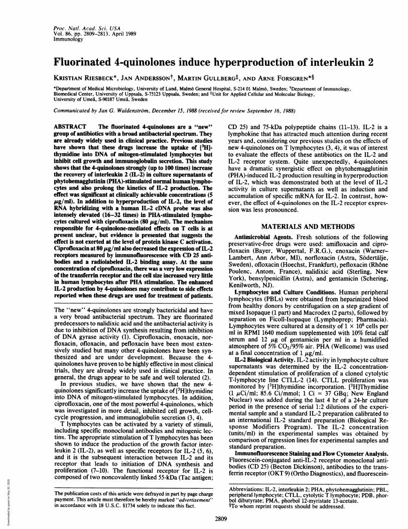
FIG. 2. Ciprofloxacin induces hyperinduction of IL-2 mRNA. Autoradiogram of dot blotting of cDNA (IL-2 and actin) with 1:2 dilutions ofmRNA for IL-2 and actin from PHA-stimulated human lymphocytes grown at a density of 1 x 10⁶ cells per ml for 18 and 48 hr in the presenceor absence of ciprofloxacin added to cultures at the onset.
fluorinated 4-quinolone nalidixic acid, gentamicin, or ben-zylpenicillin per ml (data not shown).However, ciprofloxacin, one of the new fluorinated 4-
quinolones, caused a dose-dependent increase in the recov-
ery of IL-2 in the culture supernatants. The levels were
strongly increased after only 24 hr, and accumulated IL-2activity continued to increase until 72 hr of culture at thehighest antibiotic concentration (80 ,ug/ml). The peak levelsof IL-2 in the presence of 80 ,ug of ciprofloxacin per ml were
up to 100 times higher than the IL-2 levels in the antibiotic-free controls. The effect of lower concentrations (5-20,ug/ml) of ciprofloxacin was more modest with declining IL-2levels after 48 hr.
Similar kinetics of increased IL-2 concentration in culturesupernatants was also demonstrated when PHA-stimulatedlymphocytes were incubated with new 4-quinolones otherthan ciprofloxacin. The highest levels were obtained withenoxacin and pefloxacin. Table 1 summarizes the peakconcentrations of IL-2 activity obtained during 72 hr ofincubation with 5, 20, and 80 ,g of the different 4-quinolonesper ml. As shown, there were also increased IL-2 levels withall new 4-quinolones at 5 ,ug/ml, a concentration that isachievable in serum in patients after oral administration ofthe new 4-quinolones. In cultures containing the new 4-quinolones in the absence of PHA, IL-2 could not berecovered in the supernatants (data not shown). Controlexperiments showed that the 4-quinolones did not by them-selves influence the CTLL assay of concentrations used forIL-2 determinations.To study IL-2 production in a clonal T-cell line in the
absence of monocytes, the T-cell leukemia cell line Jurkatwas used. This cell line produces IL-2 when stimulated withmonoclonal antibodies to the T3 antigen (anti-CD3) in thepresence of a phorbol ester-i.e., PDB or phorbol 12-myristate 13-acetate (PMA) (22). When Jurkat cells were
exposed to suboptimal concentrations of these stimuli, an
increased (0.5 unit/ml) IL-2 production occurred, but addi-tion of ciprofloxacin caused a strong enhancement of IL-2production (5-17 units/ml) (Table 2). However, ciprofloxa-cin could not in the absence of the other stimuli cause IL-2production in Jurkat cells (data not shown). Ciprofloxacinthus seems to have a direct effect on the responding T-cellpopulation and cannot replace the action of phorbol esters.
Ciprofloxacin Increases Specifically the Level of IL-2 mRNAin PHA-Stimulated Lymphocytes. The data presented aboveshow increased levels of IL-2 in supernatants of activatedlymphocytes incubated with the new 4-quinolones. To pro-vide further evidence of an increased IL-2 production, thelevel of IL-2 mRNA was measured. In human lymphocytes,the concentration ofRNA hybridizing to a human IL-2 cDNAwas intensely elevated (16-32 times) in cells stimulated withPHA and cultured with ciprofloxacin (80 ,ug/ml) as revealedby dot blot hybridization (Fig. 2). As also shown, the level ofactin mRNA was not affected by ciprofloxacin. Electropho-retic blot hybridization analysis revealed that the increase inhybridization to the IL-2 probe was due to the selective
increase in one band corresponding to the native 1.2-kilobaseIL-2 mRNA (data not shown).
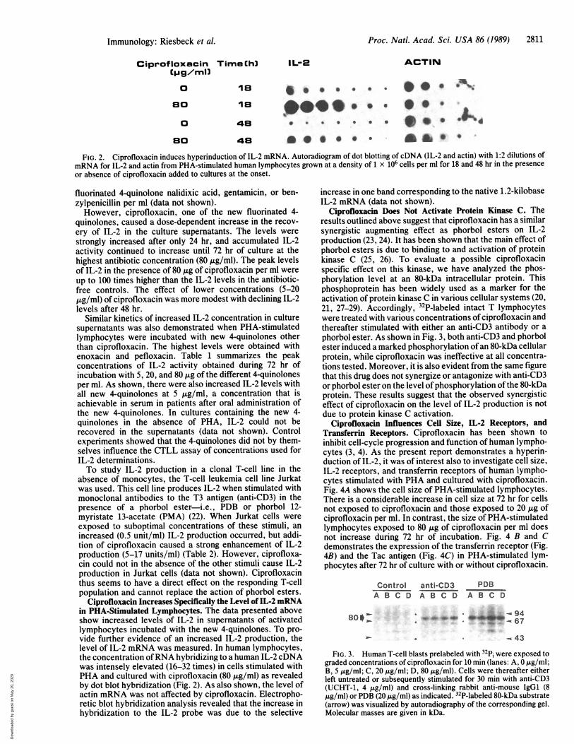
Ciprofloxacin Does Not Activate Protein Kinase C. Theresults outlined above suggest that ciprofloxacin has a similarsynergistic augmenting effect as phorbol esters on IL-2production (23, 24). It has been shown that the main effect ofphorbol esters is due to binding to and activation of proteinkinase C (25, 26). To evaluate a possible ciprofloxacinspecific effect on this kinase, we have analyzed the phos-phorylation level at an 80-kDa intracellular protein. Thisphosphoprotein has been widely used as a marker for theactivation of protein kinase C in various cellular systems (20,21, 27-29). Accordingly, 32P-labeled intact T lymphocyteswere treated with various concentrations of ciprofloxacin andthereafter stimulated with either an anti-CD3 antibody or a
phorbol ester. As shown in Fig. 3, both anti-CD3 and phorbolester induced a marked phosphorylation ofan 80-kDa cellularprotein, while ciprofloxacin was ineffective at all concentra-tions tested. Moreover, it is also evident from the same figurethat this drug does not synergize or antagonize with anti-CD3or phorbol ester on the level of phosphorylation of the 80-kDaprotein. These results suggest that the observed synergisticeffect of ciprofloxacin on the level of IL-2 production is notdue to protein kinase C activation.
Ciprofloxacin Influences Cell Size, IL-2 Receptors, andTransferrin Receptors. Ciprofloxacin has been shown toinhibit cell-cycle progression and function of human lympho-cytes (3, 4). As the present report demonstrates a hyperin-duction of IL-2, it was of interest also to investigate cell size,IL-2 receptors, and transferrin receptors of human lympho-cytes stimulated with PHA and cultured with ciprofloxacin.Fig. 4A shows the cell size of PHA-stimulated lymphocytes.There is a considerable increase in cell size at 72 hr for cellsnot exposed to ciprofloxacin and those exposed to 20 ,ug ofciprofloxacin per ml. In contrast, the size of PHA-stimulatedlymphocytes exposed to 80 ,ug of ciprofloxacin per ml doesnot increase during 72 hr of incubation. Fig. 4 B and Cdemonstrates the expression of the transferrin receptor (Fig.4B) and the Tac antigen (Fig. 4C) in PHA-stimulated lym-phocytes after 72 hr of culture with or without ciprofloxacin.
Control anti-CD3 PDB
A B C D A B C D A B C D
- 67
W* K43
FIG. 3. Human T-cell blasts prelabeled with 32p; were exposed tograded concentrations of ciprofloxacin for 10 min (lanes: A, 0 ,tg/ml;B, 5 ,ug/ml; C, 20 ug/ml; D, 80 ,ug/ml). Cells were thereafter eitherleft untreated or subsequently stimulated for 30 min with anti-CD3(UCHT-1, 4 ,tg/ml) and cross-linking rabbit anti-mouse IgG1 (8,tg/ml) or PDB (20 ,ug/ml) as indicated. 32P-labeled 80-kDa substrate
(arrow) was visualized by autoradiography of the corresponding gel.Molecular masses are given in kDa.
S %
Immunology: Riesbeck et al.
800J-
Dow
nloa
ded
by g
uest
on
May
20,
202
0

2812 Immunology: Riesbeck et al.
0
275
100 200FSC
102OKT -9
102CD 25
FIG. 4. Flow cytometer analysis of cell size (A), transferrin receptors (B), and IL-2 receptors (C) of human PHA-stimulated lymphocytesincubated for 72 hr with ciprofloxacin at 80 Ag/ml (------) or 20 ,Ag/ml (........) or no antibiotic (. ). Cell size of fresh cells (A) (-) andautofluorescense (B and C) (-) of cells not conjugated was also included. The PBLs were stained with CD 25 and OKT-9 antibodies. FSC(forward scattering) expresses the relative cell size. The y axes represent the relative number of cells.
The transferrin receptor showed the same intensity ofexpres-sion on lymphocytes exposed to 20 ug of ciprofloxacin per mlas on those lymphocytes not exposed to antibiotics. How-ever, with ciprofloxacin at 80 ,ug/ml, there was a very lowexpression of transferrin receptor. A less pronounced de-crease in the expression of the 55-kDa chain of the IL-2receptor (Tac antigen) was also demonstrated by CD 25antibodies after 72 hr ofincubation ofPHA-stimulated humanlymphocytes with ciprofloxacin (80 tug/ml) (Fig. 4C). On theother hand, with ciprofloxacin at 20 pg/ml, the expression ofCD 25 was enhanced as compared to lymphocytes stimulatedwith PHA but not exposed to ciprofloxacin.To provide further evidence ofan altered expression of the
IL-2 receptor by ciprofloxacin, high-affinity receptors forIL-2 were measured by an assay with radiolabeled IL-2.PHA-stimulated lymphocytes incubated for 72 hr with ci-profloxacin at 5 Ag/ml demonstrated 1950 receptor sites perlymphocyte, which was about the same as lymphocytes notexposed to ciprofloxacin (Table 3). In contrast, lymphocytesexposed to ciprofloxacin at 20 ,ug/ml showed 5000 sites perml and those incubated with 80 ,ug/ml showed 1200 sites percell. Thus, our data demonstrate that ciprofloxacin causestwo different effects on the IL-2 receptor system. At highconcentrations (80 ,ug/ml), there is a decrease and, atmoderate concentrations (20 jig/ml), there is an increase inIL-2 receptors.
DISCUSSION
Our results show that a new group of antibacterial drugs, the4-quinolones, in concert with a lectin PHA are powerfulstimulators of IL-2 production. This was demonstrated bothby an increased IL-2 activity in culture supernatants and byincreased IL-2 mRNA in PHA-stimulated lymphocytes whenexposed to ciprofloxacin, which is one of the most powerfulnew 4-quinolones. In addition, at moderate concentrations ofciprofloxacin (20 ug/ml), there was an increase in high-affinity receptors for IL-2 as well as the 55-kDa antigendetected by the monoclonal antibody CD 25. However, a
Table 3. High-affinity receptors on PHA-stimulated humanlymphocytes incubated with and without ciprofloxacin
Ciprofloxacin, IL-2 receptor Affinity,ttg/ml sites per cell pM
0 2000 195 1950 24
20 5000 3180 1200 21
Normal human lymphocytes (106 cells per ml) were incubated withciprofloxacin for 72 hr and after washing and additional incubation tofacilitate dissociation ofendogenously bound IL-2, IL-2 high-affinityreceptors were determined with radiolabeled IL-2.
very high concentration of ciprofloxacin (80 Ag/ml) caused areduction of the same receptor. At the same concentration ofciprofloxacin, the transferrin receptor was almost completelyabolished and cell size did not increase. None ofthese effectson IL-2 or its receptor was produced by the 4-quinolones inthe absence of PHA.Our results on IL-2 were quite unexpected considering the
fact that we previously have reported that ciprofloxacininhibits cell growth by =50% at 20 pg/ml and completely at80 Ag/ml. In addition, immunoglobulin secretion by humanlymphocytes stimulated by pokeweed mitogen or Epstein-Barr virus was inhibited 50% with ciprofloxacin at 5 ,ug/mland almost completely at 20 ttg/ml. In addition, all new4-quinolones tested at 1.5-25 ug/ml caused an increaseduptake ofradiolabeled thymidine after 3-5 days of incubationwith PHA-stimulated human lymphocytes (3, 4).The molecular target for 4-quinolones in human lympho-
cytes causing increased IL-2 production was not revealed byour studies. A selective IL-2 gene expression resulting froma toxic effect by the 4-quinolones seems to be an unlikelyexplanation for the apparent discrepancy between the in-creased IL-2 production and the inhibited lymphocyte cellfunctions. The similarities with the action of the tumor-promoting phorbol ester, PMA or PDB, which stimulatesprotein kinase C, has to be pointed out. Optimal IL-2 mRNAinduction requires the concerted action of PMA or PDB anda lectin like PHA or monoclonal antibodies directed againstthe T3 antigen complex (20, 21, 27-30). In the present study,ciprofloxacin induced increased IL-2 production in the Jurkatcell line when substimulatory concentrations of PMA andantibodies directed toward the T3 antigen complex wereused. However, by analysis of phosphorylation of endoge-nous protein kinase C substrates, we were unable to detectany stimulatory effect by ciprofloxacin on this kinase.The lymphokine IL-2 has a short half-life (ti/2, 3-5 min) in
humans, which is consistent with its role as a transient signalwithin the immune system, a signal that must be cleared toavoid chronic and possible deleterious stimulation (31). Inunstimulated normal human blood lymphocytes, IL-2mRNAis undetectable but stimulation with mitogens causes its rapidaccumulation (32). In activated human PBL, IL-2 mRNAlevels decline rapidly on removal of the inducing agents,indicating that transcription continues only as long as theactivating signal is present. In addition, it has been suggestedthat an A+U-rich untranslated region in IL-2 mRNA and inthe mRNA of other mediators of inflammation, and in a fewother mRNAs, is responsible for an instability (33, 34). IL-2mRNA degradation presumably by a labile RNase, has beenreported to be selectively inhibited by cycloheximide andactinomycin D, inhibitors of protein and RNA synthesis,respectively (33). This leads to superinduction. Quinolonesmay inhibit individual genes at concentrations lower than
375
0
-<--trA
Proc. Natl. Acad. Sci. USA 86 (1989)
Dow
nloa
ded
by g
uest
on
May
20,
202
0

Proc. Natl. Acad. Sci. USA 86 (1989) 2813
those inhibiting gyrase activity (35, 36). It is possible thatIL-2-regulating mechanisms are particularly sensitive tociprofloxacin in eukaryotic cells.
In general, clinical side effects of the new 4-quinolones aremild to moderately severe and discontinuation of therapybecause of drug toxicity is necessitated in only 1-3% ofpatients (2). Central nervous system side effects are reportedby 1-5% of patients. Stimulatory effects may occur becausequinolones inhibit receptor binding of y-aminobutyric acid,an inhibitory transmitter (37). However, this effect wasmainly apparent at high concentrations of 4-quinolones. Sideeffects of high dose IL-2 administration also involve thecentral nervous system in a rather high number of patients(38, 39). Considering the findings in this report that 4-quinolones, even at the clinically achievable concentration of5 Ag/ml, increase the IL-2 level and at high concentrationsdramatically influence IL-2 production in PHA-stimulatedhuman lymphocytes in vitro, it is possible that effects of new4-quinolones on the central nervous system are secondary totheir ability to mediate increased IL-2 production in stimu-lated T cells.
This investigation was supported in part by grants from theSwedish Medical Research Council (Project B88-16X-07489-03A andProject B88-16X-03163-16B).
1. Smith, J. T. (1984) Pharm. J. 233, 299-305.2. Smith, C. R. (1987) J. Antimicrob. Chemother. 19, 709-712.3. Forsgren, A., Schlossman, S. F. & Tedder, T. F. (1987) Anti-
microb. Agents Chemother. 31, 768-773.4. Forsgren, A., Bredberg, A., Pardee, A. B., Schlossman, S. F.
& Tedder, T. F. (1987) Antimicrob. Agents Chemother. 31,774-779.
5. Moller, G., ed. (1984) Immunol. Rev. 81.6. Meuer, S. C., Hussey, R. E., Cantrell, D. A., Hodgdon, J. C.,
Schlossman, S. F., Smith, K. A. & Reinherz, E. L. (1984)Proc. Natl. Acad. Sci. USA 81, 1509-1513.
7. Moller, G., ed. (1980) Immunol. Rev. 51.8. Robb, R. J., Munck, A. & Smith, K. A. (1981) J. Exp. Med.
154, 1455-1474.9. Cantrell, D. A. & Smith, K. A. (1983) J. Exp. Med. 158, 1895-
1911.10. Cantrell, D. A. & Smith, K. A. (1984) Science 224, 1312-1316.11. Tsudo, M., Kozak, R. W., Goldman, C. K. & Waldmann,
T. A. (1986) Proc. Natl. Acad. Sci. USA 83, 9694-9698.12. Sharon, M., Klausner, R. D., Cullen, B. R., Chizzonite, R. &
Leonard, W. J. (1986) Science 234, 859-863.13. Wang, H. M. & Smith, K. A. (1987) J. Exp. Med. 166, 1055-
1069.
14. Gillis, S., Ferm, M. H., Ou, W. & Smith, K. A. (1978) J.Immunol. 120, 2027-2032.
15. Chirgwin, J. M., Przybyla, A. E., MacDonald, R. J. & Rutter,W. J. (1978) Biochemistry 18, 5294-5299.
16. Maniatis, T., Fritsch, E. F. & Sambrook, J. (1982) MolecularCloning:A Laboratory Manual (Cold Spring Harbor Lab., ColdSpring Harbor, NY), pp. 109-112, 202-203.
17. Taniguchi, T., Matsui, H., Fujita, T., Takaoka, C., Kashima,N., Yoshimoto, R. & Hamuro, J. (1983) Nature (London) 301,305-310.
18. Gullberg, M. (1986) EMBO J. 5, 2171-2178.19. Friedrich, B., Noreus, K., Cantrell, D. & Gullberg, M. (1988)
Immunobiology (Stuttgart) 176, 465-478.20. Friedrich, B. & Gullberg, M. (1988) Eur. J. Immunol. 18, 489-
492.21. Isacke, C. M., Meisenhelder, J., Brown, K. D., Gould, K. L.,
Gould, S. J. & Hunter, T. (1986) EMBO J. 5, 2889-2898.22. Weiss, A., Wiskocil, R. L. & Stobo, J. D. (1984) J. Immunol.
133, 123-128.23. Truneh, A., Albert, F., Goldstein, P. & Schmitt-Verhulst,
A. M. (1985) Nature (London) 313, 318-320.24. Weiss, A., Imboden, J., Wiscocil, R. & Stobo, J. (1984) J. Clin.
Immunol. 4, 165-171.25. Castagna, M., Takai, Y., Kaibuchi, K., Sano, K., Kikkawa, U.
& Nishizuka, Y. (1982) J. Biol. Chem. 257, 7847-7851.26. Nishizuka, Y. (1986) Science 233, 305-312.27. Rozengurt, E., Rodriguez-Pena, M. & Smith, K. A. (1983)
Proc. Natl. Acad. Sci. USA 80, 7244-7248.28. Albert, K. A., Walaas, S. I., Wang, J. K. T. & Greengard, P.
(1986) Proc. Natl. Acad. Sci. USA 83, 2822-2826.29. Blackshear, P. J., Wen, L., Glynn, B. P. & Witters, L. A.
(1986) J. Biol. Chem. 261, 1459-1469.30. Granelli-Piperno, A., Andrus, L. & Steinman, R. M. (1986) J.
Exp. Med. 163, 922-937.31. Hooton, J. W. L., Gibbs, C. & Paetkau, V. (1985) J. Immunol.
135, 2464-2473.32. Kronke, M., Leonard, W. J., Depper, J. M. & Greene, W. C.
(1985) J. Exp. Med. 161, 1593-1598.33. Shaw, J., Meerovitch, K., Bleackley, R. C. & Paetkov, V.
(1988) J. Immunol. 140, 2243-2248.34. Caput, D., Beutler, B., Hartog, K., Thayer, R., Brown-Shimer,
S. & Cerami, A. (1986) Proc. Natl. Acad. Sci. USA 83, 1670-1674.
35. Hussy, P., Maas, G., Tummler, B., Grosse, F. & Shomburg, U.(1986) Antimicrob. Agents Chemother. 29, 1073-1078.
36. McClure, W. (1985) Annu. Rev. Biochem. 54, 171-204.37. Tsuji, A., Sato, H., Kume, Y., Tamai, I., Okezaki, E., Nagata,
0. & Kato, H. (1988) Antimicrob. Agents Chemother. 32, 190-194.
38. Rosenstein, M., Ettinghausen, S. E. & Rosenberg, S. A. (1986)J. Immunol. 137, 735-742.
39. Rosenberg, S. A., Lotze, M. T., Muul, L. M., Chang, A. E.,Avis, F. P., Leitman, S., Linehan, W. M., Robertson, C. N. &White, D. E. (1987) N. Engl. J. Med. 316, 889-897.
Immunology: Riesbeck et al.
Dow
nloa
ded
by g
uest
on
May
20,
202
0