fluorescent carbon dots from mono- and polysaccharides ... · fluorescent carbon dots from mono-...
TRANSCRIPT

675
Fluorescent carbon dots from mono- and polysaccharides:synthesis, properties and applicationsStephen Hill and M. Carmen Galan*
Review Open Access
Address:School of Chemistry, University of Bristol, Cantock’s Close, BristolBS8 1TS, UK
Email:M. Carmen Galan* - [email protected]
* Corresponding author
Keywords:fluorescent carbon dots; monosaccharides; nanomaterials;nanotechnology applications; polysaccharides
Beilstein J. Org. Chem. 2017, 13, 675–693.doi:10.3762/bjoc.13.67
Received: 23 January 2017Accepted: 30 March 2017Published: 10 April 2017
This article is part of the Thematic Series "The glycosciences".
Guest Editor: A. Hoffmann-Röder
© 2017 Hill and Galan; licensee Beilstein-Institut.License and terms: see end of document.
AbstractFluorescent carbon dots (FCDs) are an emerging class of nanomaterials made from carbon sources that have been hailed as poten-
tial non-toxic replacements to traditional semiconductor quantum dots (QDs). Particularly in the areas of live imaging and drug
delivery, due to their water solubility, low toxicity and photo- and chemical stability. Carbohydrates are readily available chiral bio-
molecules in nature which offer an attractive and cheap starting material from which to synthesise FCDs with distinct features and
interesting applications. This mini-review article will cover the progress in the development of FCDs prepared from carbohydrate
sources with an emphasis on their synthesis, functionalization and technical applications, including discussions on current chal-
lenges.
675
IntroductionNanotechnology applied to biological and biomedical problems
has seen an explosion of research in recent years [1]. Func-
tional nanomaterials that can carry biologically relevant mole-
cules have become very useful for drug delivery, sensing and
catalysis to name just a few applications. As a result, nanomate-
rials exhibiting novel electronic and optical properties, having
controlled size, geometry, surface distribution and functionality
have been developed as materials for probing biological interac-
tions and in biomedical applications [2-6]. Among these novel
type of probes, luminescent semiconductors, quantum dots
(QDs), which possess a narrow emission spectra and common
excitation, superior photostability and electron density when
compared to organic fluorophores, in addition to bright visible
emission, have become particularly popular for their versatility
as non-isotopic detection labels which are amenable to live cell
imaging and immunoassay applications [7]. In particular,
cadmium-based QDs (e.g., CdS, CdSe, CdSe/ZnS) are com-
monly used for in vitro biological studies due to their well-
established synthesis and functionalisation strategies, tuneable
emission profiles and high quantum yields of fluorescence

Beilstein J. Org. Chem. 2017, 13, 675–693.
676
(QYs) [8-11]. However, the presence of heavy metals like Cd2+,
and the associated concerns surrounding heavy metal toxicity
has meant that their in vivo applications are restricted [12].
Therefore, the development of fluorescent nanoparticles that are
able to replicate QD fluorescence properties without exhibiting
long term toxicity profiles, has become very relevant.
The term carbon dots (CDs) has been coined to describe a new
class of carbon-based nanomaterials which are typically
discrete, quasi-spherical nanoparticles, with sizes usually less
than 10 nm in diameter (although bigger sizes have recently
been reported). These relatively new nanomaterials have found
many applications in the fields of photo- and electrocatalysis,
chemical sensing, biosensing, bioimaging and nanomedicine,
due to their unique tuneable photoluminescence (PL) properties,
chemical inertness, high water solubility, ease and low cost fab-
rication and more importantly, low toxicity profiles. The latter
makes these fluorescent nanomaterials attractive for a wide
range of in vivo applications, which has been the topic of
several recent reviews [13-15]. Following the serendipitous
discovery by Xu et al. during the separation and purification of
single-walled carbon nanotubes (SWCNTs) [16], the develop-
ment of synthetic methodologies to access these fluorescent
nanomaterials combined with their myriad of applications, has
led to CDs being hailed as the potential non-toxic successors to
traditional semiconductor QDs, particularly in the areas of live
imaging and drug delivery.
Synthetic approaches to access CDs can be classified into two
broad categories: top-down or bottom-up syntheses. Top-down
methods are characterised by using a bulk carbon substrate as
the starting material; using conditions that remove nanoparti-
cles from the bulk substrate such as electrochemistry, chemical
oxidation, arc discharge or laser ablation, carbon-based nano-
particles can be obtained. Typical substrates used are single/
multi-walled carbon nanotubes, graphite, graphene or candle
soot, amongst many others [15,17]. The crystalline make-up of
top-down derived CDs is usually highly sp2 in character, which
is transferred from the sp2-enriched starting materials, e.g.,
graphite or graphene. Conversely bottom-up methodologies rely
on the use of a molecular precursor which can be treated in such
a way as to seed the formation of a CD. Typical starting materi-
als include amino acids, citric acid, biomass and carbohydrates
to name but a few, which can be reacted using thermal decom-
position, chemical or hydrothermal oxidation, microwave, acid-
mediated reflux, ultrasonic irradiation or silica nanoparticle-
templated synthesis [18-23]. Unlike their top-down equivalents,
the CDs derived from these methods are usually less sp2 crys-
talline and tend to have more amorphous morphologies. It
should be stated that no two CD preparations lead to the same
type of nanoparticle, as any changes to the ratio and composi-
tion of starting materials, additives, solvent, temperature, type
of vessel, etc., does have an effect on the final molecular com-
position and architecture of the CD. Resultantly, differential
properties are easily acquired through minor manipulations of
the CD synthesis. To date, the de novo rational design of
bottom-up syntheses of CDs for advanced application is limited
in the literature.
Carbohydrates are one of the most diverse and important class
of biomolecules in nature and offer well-defined chiral scaf-
folds primed for modification at the anomeric position and
alcohol functionalities. Therefore, the use of carbohydrates as a
starting material for synthesizing CDs is extremely attractive
not only due to their abundance, availability and heterogeneity,
but also due to their high water solubility, low-carbonisation
temperatures, low cost and typically inherently lack toxicity.
With all these options available to tune the synthesis of CDs, it
is no surprise that researchers have already began to see the
benefits of carbohydrates when considering the synthesis of
novel FCDs with improved properties. For example, simple
monosaccharides such as glucose, glucosamine, mannose, fruc-
tose and their derivatives and common disaccharides, e.g.,
sucrose, lactose, and maltose have been employed to prepare
fluorescent carbon dots (FCDs) using different methodologies
[13,24]. Similarly, important carbohydrate-based biopolymers
such as cellulose, chitin, chitosan, dextran, cyclodextrin, and
hyaluronic acid, which differ not only in elemental composition,
but also in chemo-physical properties, have also been success-
fully utilised in the preparation of CDs, where their differences
allow tailoring of the CD structure and properties [25].
In this review, we focus on the most recent approaches de-
veloped to prepare fluorescent CDs using mono-, oligo- and
polysaccharides as the main carbon source.
ReviewFluorescent carbon dots synthesised frommonosaccharidesGlucose-based fluorescent carbon dotsSustainable syntheses of CDs have driven researchers to find
readily available, cheap and renewable carbon sources of which
the monosaccharide glucose is an ideal candidate. Not only is
glucose cheap and commercially available, but also has a low
carbonisation temperature, ring-opens readily to afford a reac-
tive aldehyde moiety which can be further exploited for conju-
gations, polymerisations and (hetero)aromatic formation, which
are all ideal for generating CDs [26,27]. For these reasons, in
addition to the inherent low toxicity and high water solubility of
glucose, this particular monosaccharide has been extensively
used as an ideal carbon source for CD formation, under a range
of experimental conditions.

Beilstein J. Org. Chem. 2017, 13, 675–693.
677
Scheme 1: Microwave-driven reaction of glucose in the presence of PEG-200 to afford blue-emissive CDs.
Scheme 2: Two-step synthesis of TTDDA-coated CDs generated from acid-refluxed glucose.
The microwave-assisted synthesis of FCDs from a glucose solu-
tion in the presence of poly(ethylene glycol)-200 (PEG-200) by
Yang et al. is, to the best of our knowledge, the first reported
example involving a carbohydrate moiety (Scheme 1) [18]. The
water-soluble nanoparticles exhibited an amorphous core, as
deduced by X-ray diffraction (XRD), while Fourier-trans-
formed infrared (FTIR) spectroscopy analysis indicated the
presence of a range of oxygen-containing functionalities, e.g.,
alcohols, ethers and carboxylic acids on the CD surface, which
are likely the reason for the high water solubility exhibited by
the nanomaterial. This type of chemical profile is typical of
standard bottom-up synthesised CDs [15,28]. Interestingly, the
team was also able to show that the use of PEG-200, as a sur-
face passivation agent (SPA), was crucial for favourable photo-
luminescence (PL) properties and QYs of up to 6.3% were
achieved. The use of SPAs is among one of two main tech-
niques that are widely employed to improve the PL properties
of FCDs. SPAs are argued to provide uniform PL trapping sites
on the CD surface, alongside promoting new functionality that
can work, in tandem with the core, to turn-on fluorescence.
Another example, which highlights the importance of surface
passivation and how SPAs can be used to modify and tune CD
chemical and physical properties, was reported by Travas-
Sejdic et al. [23]. They also employed glucose as the carbon
precursor which, after refluxing in aqueous H2SO4, yielded car-
bonaceous nanoparticles with observable PL (Scheme 2).
Further treatment with aqueous HNO3 under reflux, yielded
nanoparticles of weak PL (QY = 1%). The PL properties could
be improved upon introducing surface passivation, which was
achieved by heating the weakly fluorescent CDs in a solution of
4,7,10-trioxa-1,13-tridecanediamine (TTDDA) for 72 hours at
120 °C to give a nanomaterial with QY values of up to 13%.
FTIR studies suggested that TTDDA incorporation onto CDs
occurred via amide formation, from the reaction between sur-
face carboxylic acids and the corresponding amine SPA. This
was further supported by the change in zeta-potential (ZP)
values which shifted from −37.3 mV (non-passivated CD) to
3.46 mV (TTDDA passivated CD). Similar carbonaceous mate-
rials were obtained when the team used sucrose or starch as
starting carbohydrate materials.
In addition to microwave and acid reflux-mediated glucose
dehydration reactions, the group of Wang developed an alterna-
tive protocol that combined glucose with monopotassium phos-
phate (KH2PO4) in a Teflon-lined autoclave chamber with
heating to 200 °C for 12 h (Scheme 3) [29]. The fluorescence
emission could be tuned by changing the ratio of sugar and
KH2PO4. For instance a molar ratio of 1:26 (glucose/KH2PO4)
afforded blue-fluorescent CDs (QY = 0.02), whereas a 1:36
ratio yielded green-fluorescent CDs (QY = 0.01). In the absence
of KH2PO4, irregular black carbon aggregates were obtained.
Raman and TEM analysis showed both types of FCDs had
graphitic crystallinity. This example highlights that an inorgan-
ic-based dehydrating agent could be used instead of a tradi-
tional diamine SPA to induce CD dehydration and affect their
PL properties. Most carbohydrate-derived CDs emit in the blue
area of the visible section of the electromagnetic spectrum
under UV/high energy blue excitation. However, most
mammalian cells are also autofluorescent in this particular
Beilstein J. Org. Chem. 2017, 13, 675–693.
678
region [30]. As a result, the majority of CDs produced with blue
emission have QYs that are not suitable for bioimaging applica-
tions. CDs with multicolour/excitation-dependent emission that
can be red shifted and avoid the cellular autofluorescence
window, are a good alternative. Unfortunately, the CD fluores-
cence tends to lose intensity upon red-shifting the excitation. An
ideal CD probe for bioimaging applications will have either a
high QY in the blue, or adequate green to red emission. Thus,
the green-emissive glucose-based CDs produced by Wang et al.
are ideal for this type of application and the team showed their
applicability in cell internalisation studies with HepG2 cells
[29]. The green CDs were non-toxic to cells at concentrations of
up to 625 μg/mL and exposures of 72 h. Laser scanning
confocal microscopy (LSCM) demonstrated cell internalization,
making these materials a good candidate as a bioimaging agent.
Scheme 3: Glucose-derived CDs using KH2PO4 as a dehydratingagent to both form and tune CD’s properties.
In 2011 Qu et al. developed a tuneable synthesis of FCDs by
selecting a different inorganic ion and carbohydrate combina-
tions using microwave irradiation as the heating source, demon-
strating that both the starting material and dehydrating agent of
choice can allow tuning/manipulation of the fluorescence prop-
erties of the system [31]. It was found that irradiation times of
14 min could be employed to afford CDs from glycerol, glycol,
glucose or sucrose. The source of the inorganic ion was impor-
tant too, as increasing the valency of either the anion or cation
would lead to a greater ability to dehydrate the carbon precur-
sor. An ideal balance of cation and anion valence was found
when using CuSO4 which afforded CDs with QY of up to 9.5%,
in-line with the state-of-the-art at the time.
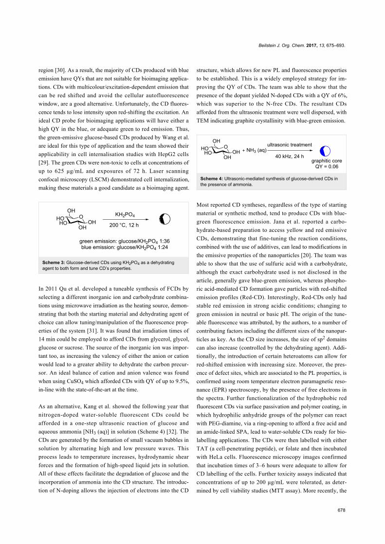
As an alternative, Kang et al. showed the following year that
nitrogen-doped water-soluble fluorescent CDs could be
afforded in a one-step ultrasonic reaction of glucose and
aqueous ammonia [NH3 (aq)] in solution (Scheme 4) [32]. The
CDs are generated by the formation of small vacuum bubbles in
solution by alternating high and low pressure waves. This
process leads to temperature increases, hydrodynamic shear
forces and the formation of high-speed liquid jets in solution.
All of these effects facilitate the degradation of glucose and the
incorporation of ammonia into the CD structure. The introduc-
tion of N-doping allows the injection of electrons into the CD
structure, which allows for new PL and fluorescence properties
to be established. This is a widely employed strategy for im-
proving the QY of CDs. The team was able to show that the
presence of the dopant yielded N-doped CDs with a QY of 6%,
which was superior to the N-free CDs. The resultant CDs
afforded from the ultrasonic treatment were well dispersed, with
TEM indicating graphite crystallinity with blue-green emission.
Scheme 4: Ultrasonic-mediated synthesis of glucose-derived CDs inthe presence of ammonia.
Most reported CD syntheses, regardless of the type of starting
material or synthetic method, tend to produce CDs with blue-
green fluorescence emission. Jana et al. reported a carbo-
hydrate-based preparation to access yellow and red emissive
CDs, demonstrating that fine-tuning the reaction conditions,
combined with the use of additives, can lead to modifications in
the emissive properties of the nanoparticles [20]. The team was
able to show that the use of sulfuric acid with a carbohydrate,
although the exact carbohydrate used is not disclosed in the
article, generally gave blue-green emission, whereas phospho-
ric acid-mediated CD formation gave particles with red-shifted
emission profiles (Red-CD). Interestingly, Red-CDs only had
stable red emission in strong acidic conditions; changing to
green emission in neutral or basic pH. The origin of the tune-
able fluorescence was attributed, by the authors, to a number of
contributing factors including the different sizes of the nanopar-
ticles as key. As the CD size increases, the size of sp2 domains
can also increase (controlled by the dehydrating agent). Addi-
tionally, the introduction of certain heteroatoms can allow for
red-shifted emission with increasing size. Moreover, the pres-
ence of defect sites, which are associated to the PL properties, is
confirmed using room temperature electron paramagnetic reso-
nance (EPR) spectroscopy, by the presence of free electrons in
the spectra. Further functionalization of the hydrophobic red
fluorescent CDs via surface passivation and polymer coating, in
which hydrophilic anhydride groups of the polymer can react
with PEG-diamine, via a ring-opening to afford a free acid and
an amide-linked SPA, lead to water-soluble CDs ready for bio-
labelling applications. The CDs were then labelled with either
TAT (a cell-penetrating peptide), or folate and then incubated
with HeLa cells. Fluorescence microscopy images confirmed
that incubation times of 3–6 hours were adequate to allow for
CD labelling of the cells. Further toxicity assays indicated that
concentrations of up to 200 μg/mL were tolerated, as deter-
mined by cell viability studies (MTT assay). More recently, the

Beilstein J. Org. Chem. 2017, 13, 675–693.
679
Scheme 5: Tryptophan-derived CDs used for the sensing of peroxynitrite in serum-fortified cell media.
Scheme 6: Glucose-derived CDs conjugated with methotrexate for the treatment of H157 lung cancer cells.
same team developed hydrophobic yellow and red emissive
CDs via the degradation of ascorbic acid in the presence of
oleophilic oleylamine [13]. The CDs were similarly polymer
coated with poly(maleic anhydride-alt-1-octadecene) that could
subsequently be functionalised with hydrophilic PEG-diamine,
providing an amine functionality for conjugation with glucos-
amine, histidine, arginine and folate. The yellow/red emissive
loaded-CDs were shown to be viable bioimaging probes in live
cells, since their emission did not overlap with the cell autofluo-
rescence.
Recent years have seen an increase in synthetic reports of large
scale N-doped CDs with good QYs from carbohydrate starting
materials. For example in 2014, Leitão et al. described the
microwave synthesis of CDs using 2.5 g of glucose and 0.3 g of
tryptophan as the N-dopant/surface passivation agent
(Scheme 5) [33]. The resultant CDs had a QY of up to 12%
(34-times higher than that of the undoped CDs). Interestingly,
the N-doped CDs in this report had a 20 nm diameter, as deter-
mined by TEM, which is contrary to the generally held belief
that CDs’ particular properties are only observed below a diam-
eter of 10 nm, which is not the case here and has since been ob-
served in one other carbohydrate-derived CD synthesis [34].
The team demonstrated the utility of the glucose/tryptophan-
derived CDs as a sensor of peroxynitrite anions (NO3−) in solu-
tion. The peroxynitrite anion is one of the key reactive species
which is implicated in various metabolic and physiological pro-
cesses [35]. Thus, it is important to provide analytical methods
to detect and quantify its presence, however, due to its high re-
activity, low concentration levels and quick diffusion, it has
been traditionally difficult to detect. The team was able to show
significant quenching of the CDs via tryptophan oxidation of
the exposed residues on the surface of CDs (Scheme 5). Post-
oxidation fluorescence is compromised and therefore can be
used as a signal for selectively sensing peroxynitrite up to con-
centrations of 1.5 μM (with a linear regression between
2.5–50 μM). The sensing ability of the nanoparticles was exhib-
ited in serum-fortified samples, which can be regarded as a
biomimetic for complex biological media.
A number of glucose-based CDs has been reported in recent
years as drug-delivery vehicles. In 2015, Yunus et al. synthe-
sized CDs by the ultrasonication of glucose or sucrose in the
presence of oxidising conditions afforded by H3PO4/H2SO4
(Scheme 6) [36]. The resultant CDs were blue emissive and the
use of strong oxidising conditions during their synthesis
afforded CDs with surface carboxylic acids that could be func-
tionalised. Surface conjugation with PEG-diamine afforded a
steric blocking, enhanced permeation and retention (EPR) shell,
whilst providing an amine functionality for further surface
conjugation. The anticancer drug methotrexate (MTX), which is
a well-studied drug used to treat various types of cancer includ-

Beilstein J. Org. Chem. 2017, 13, 675–693.
680
Scheme 7: Boron-doped blue-emissive CDs used for sensing of Fe3+ ion in solution.
Scheme 8: N/S-doped CDs with aggregation-induced fluorescence turn-off to temperature and pH stimuli.
ing lung cancer, was then conjugated via EDC-mediated amide
coupling chemistry (Scheme 6) [37]. The MTX-CDs were inter-
nalised into H157 lung cancer cells and compared with cells
exposed to unfunctionalised amine-bearing CDs. While the
amine-CDs showed no cellular toxicity, MTX-CDs were highly
toxic to H157 cell cultures, highlighting the potential applicabil-
ity of carbohydrate-derived CDs as vehicles for the delivery of
conventional cancer therapeutics.
More recently, in addition to the introduction of electron-donat-
ing heteroatoms such as N or S as dopant agents to improve the
PL properties of CDs, the use of boron as an additive, which is
an electron-accepting element, has also been explored by Hao et
al. [38]. The CDs were produced by the addition of boric acid
(B(OH)3) into the hydrothermal carbonisation of glucose, using
a Teflon autoclave at 180 °C for 12 h (Scheme 7). The resultant
fluorescent nanoparticles had an average diameter of 4 nm and
were negatively charged with ZP values of −40.7 mV. XPS and
FTIR analysis confirmed the presence of B in the CD structure.
Although, the addition of boron did not change the typical blue
fluorescence profile significantly, when compared to other re-
ported heteroatom-doping syntheses, the fluorescence of the
B-CDs was dynamically quenched by Fe3+ ions. Mechanistic
studies suggested that a dynamic quenching model was preva-
lent at low concentrations due to interactions between Fe3+ and
the CD surface, possibly indicating the interception of an
excited CD state by the Fe3+ ion that leads to fluorescence
quenching. The group exemplified the applicability of the mate-
rial by demonstrating the ability of the B-CD to sense Fe3+ in
tap water samples with a limit of detection of 242 nM, which
complies with U.S. Environmental Protection Agency stan-
dards.
Having shown that chemical doping with heteroatoms within
the CD synthesis can lead to materials with improved PL and
physicochemical properties, this has encouraged research
groups to focus their efforts to study the effect of using differ-
ent heteroatoms simultaneously. For example, in 2015 Zhang et
al. demonstrated that the energy intensive, hydrothermal treat-
ment of glucose in the presence of glutathione (acting as both
SPA and N/S heteroatom dopant) conducted at 180 °C for 22 h,
resulted in CDs with QYs of up to 7%. The obtained CDs had
blue-emissive fluorescence under UV excitation, a standard fea-
ture of bottom-up produced carbohydrate-derived CDs
(Scheme 8) [39]. Whereas most synthetic CDs from glucose
sources generally show a fluorescence-decay response to one or
several transition metals. Surprisingly, the CDs produced in the
presence of glutathione had a very stable fluorescence output,
which was unaffected by a wide-range of transition metal
cations. The new CD’s fluorescence intensity was, however,
sensitive to changes in both pH and temperature. The CDs were
shown to aggregate and change emission from pH 3 to 9, which
the authors attribute to the ionisation of the surface function-
ality (Scheme 8). The feature is reversible as demonstrated by
monitoring the PL intensity at a given excitation at different
pHs and during several iterations. Similarly the CDs were
shown to have an emission-intensity dependence on the temper-
ature. Upon increasing the temperature from 15 to 90 °C, 52%
of the fluorescence was lost, without any red or blue shift in the
emission maximum. The mechanism for this change was also
attributed to nanoparticle aggregation, with CD agglomeration
occurring at higher temperatures.
Two different groups reported, nearly concurrently their results,
in the use of both N and P as dopants in their CD syntheses. For

Beilstein J. Org. Chem. 2017, 13, 675–693.
681
Scheme 9: N/P-doped hollow CDs for efficient drug delivery of doxorubicin.
Scheme 10: N/P-doped CDs applied to the sensing of Fe3+ ions in mammalian T24 cells.
instance, Dong et al. described the development of dual-doped
hollow CDs using a wet-chemical method from glucose in the
presence of 1,2-ethylenediamine (EDA) and conc. H3PO4
(Scheme 9) [40]. During the exothermic reaction, a foam was
produced which resulted in green-emissive N,P-doped CDs that
were hollow and had a diameter of 10 nm as determined by
HRTEM and AFM. Despite the hollowness of the CDs, the
nanoparticles exhibit an excitation-dependent emission profile
akin to other glucose-derived, bottom-up synthesised CDs. The
material’s unique properties deemed them ideal candidates for
drug-delivery purposes. The team chose doxorubicin (DOX) as
the model drug for this purpose and loading of DOX onto the
CD was demonstrated via a change in the ZP from −9.3 mV to
−0.13 mV, suggesting that an electrostatic interaction between
the positively charged amino group in DOX and the negatively
charged groups on the CD surface can take place. Also van der
Waals and π–π stacking interactions were attributed as contrib-
uting factors to the DOX loading and DOX incorporation into
the hollow CD cavity. Drug release at acidic pHs, further sup-
ported the proposed electrostatic interactions between DOX and
the CDs. Further studies showed initial efficacy of the
DOX–CD adduct as a beneficial drug-delivery system, even in
animal models.
On the other hand, Zhao et al. described an alternative synthe-
sis for N/P-doped CDs. Hydrothermal oxidation of glucose,
phosphoric acid and aqueous ammonia, as the nitrogen source,
in a Teflon-lined autoclave followed by heating at 160 °C for
5 h afforded blue-emissive CDs under 365 nm excitation
(Scheme 10) [41]. A high QY of 30% was obtained, which is
one of the highest reported for a carbohydrate-derived CD to
date. Interestingly, it was observed that the fluorescence of
these N/P-CDs was strongly dependent on the local concentra-
tion of Fe3+. With increasing concentrations of the metal
leading to fluorescence decay of the CDs, which was attributed
to the interception of an excited state on the CD by the Fe3+ ion.
The selectivity towards Fe3+ was demonstrated against a panel
of other transition and alkali metals and a detection limit for
Fe3+ of 1.8 nM was established. The glucose-derived blue-
emissive CD could be readily internalised into T24 cells, with-
out significant cell death, and used to detect the presence of
exogenously added Fe3+ (Scheme 10).
It is important to highlight that small changes in the nitrogen
source (EDA vs ammonia), ratio of reagents and reaction condi-
tions can lead to marked differences in fluorescence, physical
and chemical properties of the nanomaterials, as demonstrated
with these two parallel reports for hollow-green and solid blue-
emitting CDs.
A systematic study has been performed by the Travas-Sejdic
group on the synthesis of CDs from either citric acid or glucose
starting materials in the presence of either TTDDA or dopa-
mine, in order to evaluate how the choice of carbon and
nitrogen sources plays a key role in the final properties of these
nanomaterials [42]. The authors found that the average size of

Beilstein J. Org. Chem. 2017, 13, 675–693.
682
Scheme 11: Comparative study of CDs formed from glucose and N-doped with TTDDA and dopamine.
CDs prepared was dependent on both, the carbon source, e.g.,
CDs from citric acid were larger than the ones derived from
glucose, and the nitrogen source, e.g., CDs derived from dopa-
mine were larger than those using TTDDA. The authors attri-
bute this observation to the fact that citric acid possesses readily
available carbonyl groups (as opposed to the masked aldehyde
in the carbohydrate) that can readily react with basic TTDDA or
dopamine to form stable intermediates; while glucose mostly
interacts with the amine dopants through intramolecular forces
such as van der Waals’ forces and hydrogen bonds. The latter
weaker interactions cause the intermediates to break down into
small fragments during the heating process resulting in smaller
CDs. In the case of N-dopant agents, the presence of a bulky
phenyl ring in dopamine was reasoned to be the possible cause
for the somewhat larger sizes observed. In addition, it was
found that PL properties were mostly dependent on the
N-source, with optimum QY of up to 29.5% (for glucose) or
33.9% (for citric acid) when using TTDDA, as opposed to
dopamine (Scheme 11). These results show that by experimen-
tally probing the reaction conditions and fully characterising the
obtained materials, a better understanding of the underpinning
mechanisms of CD formation and PL mechanisms will be
gained, which in turn will lead to improved materials with high
QYs.
Non-glucose monosaccharide-based fluorescentcarbon dotsIn addition to glucose, different monosaccharides and polyols
have also been utilised as carbon sources for the synthesis of
FCD, although this approach is less common.
The ability of glycerol to undergo dehydration and polymerisa-
tion in the presence of amino groups makes it a cheap and suit-
able candidate as a molecular precursor for CD synthesis. To
that end, Liu et al. demonstrated in 2011 that the microwave-
assisted pyrolysis of glycerol in the presence of TTDDA
afforded blue-emissive CDs with a QY of 12% (Scheme 12)
[43]. The particles had preeminent multicolour emission, which
was excitation-dependent. Importantly, the team demonstrated
that TTDDA was crucial as a passivating agent for optimal
levels of fluorescence. The method was also applicable to other
carbon sources such as glucose, sucrose, glucan and starch. The
Scheme 12: Formation of blue-emissive CDs from the microwave irra-diation of glycerol, TTDDA and phosphate.
novel nanomaterials were found to be useful in live cell bio-
imaging applications. The team carried out cell viability studies
(MTT assay) and after treatment of HepG2 cells with these
multicolour emissive CDs, 100% cell viability was recorded
with concentrations of up to 240 μg/mL of the CDs, while sig-
nificant toxicity was seen at concentrations at and above
400 μg/mL. CDs (100 μg/mL) were also incubated with HepG2
for 24 h and laser scanner confocal microscopy (LCSM) was
used to image the internalization of the CDs within the cells
using the green, yellow and red channels, demonstrating their
utility.
In a similar fashion, xylitol was used as a CD molecular precur-
sor, in the presence of HCl and ethylene diamine (EDA), in a
2 min microwave-mediated synthesis of CDs developed by Kim
et al. [44]. The team successfully demonstrated that to improve
the blue emission of the nanoparticles, HCl was crucial. In the
absence of HCl as an additive, the QY was only 0.38%, where-
as in the presence of HCl, a significant increase of the QY to
7% was observed (Scheme 13). Interestingly, Cl atoms are in-
corporated as part of the CD structure in as much as 9.14%,
based on the elemental analysis, demonstrating that in addition
to N-dopant agents, Cl sources such as HCl, are key as SPAs
that can improve the PL properties of FCDs. The Cl/N-doped
CDs were incubated with WI38 and HeLa cell lines and the cell
viability was studied by an MTT assay. It was found that no
cytotoxicity was observed up to CD concentrations of
100 μg/mL, while slight levels of toxicity were detected at con-
centrations of up to 1000 μg/mL. Fluorescence microscopy
analysis of HeLa cells treated with CDs at 100 μg/mL for 24 h
showed cell internalisation as monitored by their multicolour
emissive properties, in addition LCSM confirmed their remark-
able photostability too, as long exposure times lead to no
obvious photobleaching.

Beilstein J. Org. Chem. 2017, 13, 675–693.
683
Scheme 13: Xylitol-derived N-doped CDs with excellent photostability demonstrating the importance of Cl incorporation to the fluorescence proper-ties.
Scheme 14: Base-mediated synthesis of CDs with nanocrystalline cores, from fructose and maltose, without forcing reaction conditions.
Fructose and maltose combinations have also been used as an
alternative to glucose as the carbon source. The Ostrikov team
developed a room temperature preparation of weakly emissive
CDs (QY 2%) by mixing a 500 mM aqueous solution of fruc-
tose and maltose (a glucose 1,4-linked disaccharide) with a
500 mM solution of NaOH and NaHCO3 also dissolved in
water (Scheme 14) [45]. The resulting clear mixture was moni-
tored until a colour change towards a yellow colouration was
observed after approximately 60 minutes of mixing. Upon exci-
tation by 405 nm lasers, a green fluorescence was recorded. It
was found that the concentration of the solution was essential
for the formation of CDs. Although this method does not
produce highly fluorescent CDs, the example shows that green-
emitting CDs can be made in a reaction without either strong
heating, N-doping or surface passivation occurring. Also
remarkably, the CDs produced by this method were found by
HRTEM to have graphite crystallinity. This feature is interest-
ing as, until now, it was thought that this type of crystallinity in
a bottom-up constructed nanomaterial was only possible under
energy intensive/forcing conditions.
We have already established that an effective method for modu-
lating the properties of CDs is to introduce heteroatoms, with
the use of N-dopant agents being the most common. The
majority of methods discussed thus far, utilise cheap, readily
available neutral carbohydrate such as glucose as the carbon
source in combination with a nitrogen-containing molecule.
Glucosamine hydrochloride, which is a byproduct from the
hydrolysis of chitosan and chitin polysaccharides found on crus-
tacean shells, bears an amine functionality at C-2 and offers all
the advantages of glucose, while already containing an N atom.
A few examples in the literature have already utilised this sugar
as the starting material in the synthesis of CDs with interesting
results. One of the earliest examples of the hydrothermal prepa-
ration of FCDs using glucosamine hydrochloride was shown by
Wang et al. [34]. A one-step process whereby an aqueous
(deionised) solution of the amine-containing glucoside was
heated in an autoclave to 140 °C for 12 h, which after several
days of dialysis, led to strongly green-emitting CDs with a
35 nm average diameter. Interestingly, the authors observed that
under the same reaction conditions, glucose did not generate
CDs. The authors proposed that polymerisation of glucosamine
molecules followed by aromatisation via intramolecular dehy-
dration, leads to a burst of nucleation when the aromatic cluster
supersaturation is reached. This burst of nucleation takes place
and the carbon nuclei grow to partially nanocrystalline CDs
with certain hydrophilic functional groups in the surface.
Raman, FTIR and XPS data confirmed the presence of aromat-
ic amines, hydroxy and carboxy groups on the CD surface.
Subsequently, Liu et al. reported the hydrothermal synthesis of
amino-functionalised green fluorescent CDs using glucosamine
hydrochloride in the presence of excess sodium pyrophosphate
(Na4P2O7) (Scheme 15) [19]. The team showed that heating an
aqueous mixture of the sugar and Na4P2O7 for 10 h to 180 °C in
a Teflon-lined autoclave resulted in green fluorescent N/P-
doped CDs with QYs of up to 17% with an excitation indepen-
dent emission which could be modulated by varying the con-
centration of Na4P2O7 in the starting mixture. Also, the higher
the concentration of pyrophosphate, the less aggregation prod-
uct was observed. The resultant CDs were then effectively
coupled to hyaluronate (a long-chain polymer containing
repeating disaccharide units of glucuronate-β1->3-N-acetyl-
glucosamine) stabilised gold nanoparticles (AuNPs) and used as
a sensitive and selective probe to monitor hyaluronidase enzy-
matic activity, which is an enzyme that breaks down

Beilstein J. Org. Chem. 2017, 13, 675–693.
684
Scheme 15: N/P-doped green-emissive CDs working in tandem with hyaluronic acid-coated AuNPs to monitor hyaluronidase activity.
hyaluronate (Scheme 15). As hyaluronate is used to stabilise the
AuNPs, any enzymatic activity that degrades the polymer
would result in AuNP aggregation, which in turn modulates the
absorption properties of the AuNPs. The latter has a favourable
overlap with the emission spectra of CDs, when stabilised.
Hence a turn-on of the CD fluorescence is indicative of enzyme
activity.
More recently, Galan et al. reported the 3 min one-step synthe-
sis of blue-emitting CDs from glucosamine hydrochloride in the
presence of TTDDA using microwave irradiation with QYs of
up to 17% (Scheme 16) [46]. While most reported syntheses
afford CDs with sp2 crystalline or amorphous cores, the team
showed that the resultant nanoparticles had an sp3 nanocrys-
talline core, as determined by HRTEM and Raman spectrosco-
py. The authors attributed this observation to the relatively mild
conditions used. They also showed that the presence of HCl was
critical for the PL properties of the CD and that the formation of
C–Cl bonds, as determined by Raman and FTIR spectroscopy,
yielded the chlorine as a crucial auxochrome, which is in agree-
ment to results previously reported by Kim et al. [44].
Scheme 16: Three-minute microwave synthesis of Cl/N-doped CDsfrom glucosamine hydrochloride and TTDDA to afford bottom-up syn-thesised CDs with an sp3 nanocrystalline core.
Mechanism studies of the reaction by 1H, 13C, FTIR and React-
IR helped to identify the key reaction intermediates
(Scheme 17). The loss of the anomeric proton/carbon with for-
mation of an aldehyde was observed within the first 90 seconds
of the reaction, after which time, amide formation and sp2-
centre formation/aromatisation were also observed. React-IR
studies under hydrothermal conditions, but at a lower tempera-
ture of 70 °C, helped the team to identify a reactive iminium
species, which is formed from the reaction between the sugar
aldehyde and an amine present in the reaction mixture, and is a
key intermediate in the initial stages of nanoparticle formation.
Trapping of the iminium electrophile could allow oligomer for-
mation and dehydration, leading to the formation of the sp3-
enriched nanocrystalline core. In the second phase of the reac-
tion, following the loss of bulk water, further carbonisation
occurs and aromaticity is then generated on the outer layers of
the core. Surface passivation by TTDDA can now take place via
either incorporation of TTDDA into the surface heteroaro-
matics or amide bond formation. Amide formation can occur
either through surface-bound carboxylic acids reacting directly
with an amine (e.g., TTDDA or sugar-derived amine) or
through the nucleophilic attack of an alcohol to the iminium
electrophile, followed by rearrangement of the resulting
imidate.
The work by Mandal et al. has also recently sought to provide
some insights into nanoparticle formation and PL mechanism
for sugar-derived CDs [47]. The team studied the reaction be-
tween sucrose and H3PO4 to afford excitation-independent
orange-red emissive CDs (Ex = 365 nm), which were readily
soluble in organic solvents such as DCM and MeCN
(Scheme 18). Mechanistic investigations showed evidence of

Beilstein J. Org. Chem. 2017, 13, 675–693.
685
Scheme 17: Mechanism for the formation of N/Cl-doped CDs via key aldehyde and iminium intermediates, monitored by 1H and 13C NMR, FTIR andReact-IR studies.
Scheme 18: Phosphoric acid-mediated synthesis of orange-red emissive CDs from sucrose.
Scheme 19: Proposed HMF dimer, and its formation mechanism, that upon aggregations bestows orange-red emissive on sucrose-derived CDs.
hydroxymethylfurfural (HMF) derivatives as the major compo-
nent in the preparation of this type of CD, as evidenced by 1H,13C NMR, FTIR and MALDI–MS (Scheme 19).
The authors proposed that initial acid catalysed degradation of
the sucrose disaccharide to its monosaccharide constituents
fructose and glucose, followed by glucose isomerisation to fruc-
tose, leads to HMF formation following three dehydration steps.
Indeed, HMF formation has been identified as a dehydration
product in reactions with glucose, fructose and sucrose under
acidic conditions [48]. Furthermore, the team was able to show
that instead of polymeric furfural structures, HMF dimers are

Beilstein J. Org. Chem. 2017, 13, 675–693.
686
Scheme 20: Different polysaccharide-derived CDs in the presence of PEG-200 and how the starting material composition is conferred to the CDproducts.
produced via the acid-catalysed ether formation between HMF
and fructose followed by subsequent dehydration, and undergo
aggregation to form fluorescent CDs. Although, it is clear that
small changes in the reaction conditions and reagents do have a
significant effect in the final nanoparticle properties. These
results provide evidence that aggregation of furfural intermedi-
ates or other heteroaromatic species could be responsible for the
PL and physicochemical properties observed.
Fluorescent carbon dots synthesised frompolysaccharidesPolysaccharides are essentially polymeric sugar molecules
composed of monosaccharide units coupled together via glyco-
sidic linkages to form long linear or branched chains. Some of
the most common polysaccharides found in nature include
cellulose, starch, glycogen or chitin [49]. Upon hydrolysis,
these structures break down into smaller fragments such as
oligosaccharides or monosaccharide units. Thus, it is unsur-
prising that these naturally occurring materials have also been
used as CD precursors. Many CD syntheses report the use of
biomass, particularly sourced from plant matter, which is essen-
tially a huge source of naturally derived polysaccharides
combined with smaller amounts of other organic molecules,
e.g., amino acids, which can act as dopant agents. Some exam-
ples include the use of garlic [50], orange juice [51], onion
waste [52] and general kitchen waste [53]. For the purpose of
this review, we will concentrate on describing examples where
defined and commercially available polysaccharides are used
for the synthesis of FCDs and how these materials compare to
CDs made using their monomeric counterparts.
Many different polysaccharides with different elemental com-
position and structural morphologies are available and as seen
for monosaccharide-derived CDs, the different features and
functional groups present in those distinct carbohydrate chains,
will have an effect in the final properties of the CDs synthe-
sised from them. Pramanik et al. exploited this hypothesis in the
synthesis of CDs from three different polysaccharides: chitosan
(Chi-CDs), alginic acid (Alg-CDs) and starch (S-CDs) in the
presence of PEG-200 under identical microwave conditions
(Scheme 20) [54]. TEM analysis of the samples highlighted that
a range of morphologies and sizes were obtained depending on
the polymer used. For example, S-CDs afforded the smallest
particle size distribution (1–2 nm) but little morphological
uniformity. On the other hand, Chi-CDs appear to have a
distinctly spherical morphology with a size range of 2–10 nm
and Alg-CDs also exhibit a distinct spherical morphology with
a size range of 2–4 nm. Interestingly, an inverse correlation be-
tween the size of the CD and the fluorescence output was estab-
lished, the smallest S-CDs gave the best fluorescence intensity
of the three samples, while the largest Chi-CDs had the lowest.
FTIR analysis provided evidence that the starting polysaccha-
ride functional group composition is conferred onto the CDs.
For instance, alginic acid has one carboxylic acid group per
monomer unit, whereas chitosan is an amine-containing poly-
saccharide; analysis of the different CDs showed higher intensi-
ties for peaks attributed to carboxylic acid C=O bonds in both
Alg-CDs and Chi-CDs. Similarly Chi-CDs showed an abun-
dance of amine functionality, while S-CDs spectra had many
more signals that could be assigned to alcohol groups and some
carboxylic acid functionality, with the latter probably generated
during the reaction. The authors further demonstrated the appli-
cability of the different materials in heavy metal sensing. To
that end, each CD sample was exposed to the same concentra-
tion (0.001 M) of divalent metal cations Cu2+, Cd2+, Sn2+ or
Zn2+ in solution and the fluorescence response monitored. The
starch-based S-CDs showed an interesting PL response, where-
as the fluorescence output increased when in solution with all

Beilstein J. Org. Chem. 2017, 13, 675–693.
687
Scheme 21: Tetracycline release profiles for differentially-decorated CDs.
metal ions tested, in the case of Cu2+, a significant reduction in
fluorescence was recorded. The authors proposed that Cu2+, due
to its paramagnetic nature, could quench the S-CD fluorescence
via a photoinduced electron transfer mechanism, in which Cu2+
is reduced to Cu+. The presence of Cu+ was confirmed by
selected area electron diffraction (SAED) in the TEM analysis
of the CD surface.
CDs from chitosan hydrogels have also been reported by
Chowdhury et al. [55]. The hydrogels were synthesised from a
mixture of acetic acid, glycerol and chitosan, as a more stable
starting material for CDs. Microwave irradiation of the
hydrogel yielded UV-blue emissive CDs with a range of sizes
from 0.6–8.7 nm (as determined by DLS). Zeta-potential analy-
sis yielded a value of +27 mV, indicative of an abundance of
amino groups and as expected from an amino group containing
chitosan starting material. In addition, the group also investigat-
ed CDs prepared from chitosan/Ag and chitosan/Au nanocom-
posites, which were incorporated while preparing the chitosan
hydrogels. It was observed that although the emission of the
new CDs was broad and less well defined, there was an en-
hancement in the PL emission for the Ag or Au-doped CDs.
Subsequently in 2014, the same group was able to show that
coating of calcium alginate (CA) beads with chitosan hydrogel-
based CDs yielded a new nanomaterial that could be employed
as a pH-responsive drug-delivery vehicle (Scheme 21) [56]. The
CDs were used as a protective layer onto the CA beads and
tetracycline (TC) was loaded onto the CD–CA beads. It was
shown that a two-fold increase on drug loading was seen when
compared to uncoated beads. Subsequently, TC release at a
range of pH values was studied over a 96 h period and it was
found that 70% of TC release takes place at low pH (pH 1)
when compared to 36% release at pH 7 and 27% at pH 12. In
order to improve the drug delivery profile of the complex, the
authors developed a β-cyclodextrin/tetracycline (β-TC)
host–guest inclusion complex, which allows a second “barrier”
of release. The pre-formulated β-TC complex was loaded onto
the CD–CA beads and not only were higher loading levels
measured (90%), but also a slower rate of TC release at each pH
value was recorded, as expected from a more stable drug/
nanocomplex. The results reported here are good examples of
the potential applications of amine-coated CDs as important
components in drug-delivery applications.
Chitin, which is a cheap and readily available linear polysaccha-
ride comprised of β-1,4-linked N-acetyl-D-glucosamine units, is
the second most abundant biopolymer in nature and forms the
backbone of crustaceans and insects exoskeleton and is also
found in the cell wall of yeast and fungi [57]. Chitin is also the
precursor of chitosan, which is formed by N-deacetylation to
partially free amino groups, and is notoriously insoluble in
water. Despite this fact, Shchipunov et al. demonstrated in
2015, the first hydrothermal synthesis of CDs derived from
chitin in a Teflon-lined autoclave at 180 °C for 3 h in the pres-
ence of HNO3, all in deionised water [58]. The CDs produced
in this manner were purified from unreacted/unsolubilised

Beilstein J. Org. Chem. 2017, 13, 675–693.
688
Scheme 22: Hyaluronic acid (HA) and glycine-derived CDs, suspected to be decorated in unreacted HA, allowing receptor-mediated cell uptake.
chitin via several filtration, centrifugation and dialysis steps.
The N-doped CDs were blue-emitting under UV excitation with
apparent long-term, bench stable fluorescence. These results
might suggest further opportunities in the field for these type of
less water soluble N-containing polysaccharides.
Hyaluronic acid is another N-containing polysaccharide
composed of repeating dimeric units of glucoronic acid and
N-acetyl-D-glucosamine units and which forms the core of
complex proteoglycan aggregates found in the extracellular
matrix [57]. The team of Du and Shao et al. reported the synthe-
sis of N-doped hyaluronic acid-derived CDs and their applica-
tion as drug delivery vectors [59]. Following standard hydro-
thermal synthetic procedures as previously described for other
CD preparations, hyaluronate was heated in a Teflon-lined auto-
clave in the presence of glycine, which was found to be
key, to 200 °C for 4 h to yield CDs of under 10 nm in size
(Scheme 22). Structural analysis of the resultant CDs indicated
the presence of carbonyl-containing functional groups such as
carboxylic acids and amides, which coated a graphitic-type
core. The nanoparticles exhibit excitation-dependent emission
and were blue-emissive under UV excitation, but green when
excited at 496 nm. Although no NMR characterisation was
carried out on the samples, the authors proposed that due to the
polymeric nature of the starting material, the resulting N-doped
CD cores might be decorated by unreacted/fragmented
hyaluronic acid (HA–CD). Subsequent cell feeding experi-
ments with HA–CD with HeLa and U251 cells, revealed that
upon internalisation the CDs where found to localise in the
cytoplasm and particularly around the nucleus. Due to the large
amounts of internalisation a receptor-mediated endocytosis was
proposed. The particles were used as fluorescent probes to
target CD44 high expression in tumour cells, opening the door
for these types of polysaccharide-based nanomaterials in other
targeted live cell labelling, imaging and drug-delivery applica-
tions.
In addition to amine containing polysaccharide, other neutral
carbohydrate-based polymers have also been reported in the
synthesis of CDs. Cyclodextrin is a cyclic glucose polymer that
is commonly available in its α, β and γ forms, each correspond-
ing to the number of glucose units (6, 7 and 8, respectively). In
2014, Wang et al. reported the synthesis of CDs from each of
the different cyclodextrins via an acidic, hydrothermal treat-
ment at 70 °C for 4 h (Scheme 23) [60]. Reaction of each type
of cyclodextrin afforded quasi-spherical nanoparticles with a
size range of 2.5 ± 0.8 nm and with an amorphous carbon core.
The materials obtained had a range of alcohol and carbonyl-
containing functionalities present on their surface. QYs were
measured to range from 9% to 13%, which were dependant on
the type of cyclodextrin utilised, with each CD showing a green
emission under UV irradiation and excitation-independent emis-
sion from 360 to 460 nm excitation, which is typical of a
uniform morphology. The authors proposed that the uniform
emission could be attributed to either the uniform size distribu-
tion or the uniform surface state giving a single quantum dot-
like emission profile. This interesting report highlights the fact
that a reducing sugar is not essential to produce CDs and that
under acidic and forcing conditions this type of starting materi-
als can still undergo acetal hydrolysis, dehydration, aromatisa-
tion and carbonisation to yield CDs. The resulting cyclodextrin-
derived CDs were then used for the detection of Ag+ ions in
solution. It was found initially that mixing AgNO3 in an
aqueous solution of CDs in sunlight resulted in the formal
reduction of Ag+ to elemental Ag0, which was thought to
proceed via the adhesion of Ag+ to the CD surface, followed by
reduction in the presence of sunlight, which promotes the exci-
tation of the reducing electron to a higher energetic state
(Scheme 23). Through UV–vis absorbance and TEM measure-
ments it was evident that a surface layer of plasmonic Ag
existed on their surface. The PL intensity of the CDs was modi-
fied in a linear manner with Ag+ concentrations, and as such the
nanoparticles could be utilised as a fluorescence probe to detect
Ag+ in solution up to concentrations of 0–25 μM.
β-Cyclodextrin has also been utilised in the synthesis of CDs
using a surface passivation and inorganic dehydration method.
The groups of Yang and Teo et al. demonstrated that the synthe-
sis of excitation-independent green emissive CDs could be
achieved through the reaction of β-cyclodextrin in the presence
of oligoethyleneimine (OEI) and phosphoric acid under thermo-
lysis conditions (90 °C) for 2 h (Scheme 24) [61]. It was

Beilstein J. Org. Chem. 2017, 13, 675–693.
689
Scheme 23: Cyclodextrin-derived CDs used for detection of Ag+ ions in solution, based on the formal reduction of Ag+ to afford plasmonic Ag.
Scheme 24: Cyclodextrin and OEI-derived CDs, coated with hyaluronic acid and DOX, to produce an effective lung cancer cell drug-delivery vehicle.

Beilstein J. Org. Chem. 2017, 13, 675–693.
690
Scheme 25: Cellulose and urea-derived N-doped CDs with green-emissive fluorescence.
demonstrated that the presence of phosphoric acid was crucial
for the formation of fluorescent CDs, as the control reaction, in
the absence of acid, did not produce emissive CDs. AFM and
TEM indicated quasi-spherical CDs of 2–4 nm, while FTIR and
XPS indicated that nitro groups were present within the CD
structure. The green-emissive CDs were photostable at a wide
range of pH values (1–13) and over long-exposure to excitation
sources. These results indicated the advantages of inorganic-ion
mediated dehydration and the use of N-doping via surface
passivation to achieve QYs up to 30%. Due to the use of OEI,
the CDs were positively charged as measured by ZP and as a
result the novel nanomaterial could form nanocomplexes with
negatively-charged polymers. To demonstrate their applicabili-
ty, CDs were successfully decorated with hyaluronic acid (HA),
a negatively-charged polysaccharide, and shown by AFM, DLS
and TEM, to have formed nano-aggregates of up to 250 nm.
Interestingly, the emissive properties of the CDs were un-
changed upon complexation to HA. The resultant nano-aggre-
gates were then loaded with doxorubicin (DOX) and a strong
correlation between dose and cell death was demonstrated in
lung cancer H1299 cells (Scheme 24).
Cellulose is the most abundant organic molecule on Earth and is
a linear polysaccharide comprised of repeating β-1,4-linked
glucose units. Similarly to cyclodextrin, cellulose does not
contain N-functionalities, which has been shown to be crucial
for superior PL properties in CDs. In order to exploit the abun-
dance, renewable and cheap advantages offered by using cellu-
lose as the starting material, an N-doping strategy is needed.
The group of Yao described recently the formation of CDs from
cellulose (the specific type is not defined in the original report)
via hydrothermal treatment in the presence of urea (Scheme 25)
[62]. The resultant CDs were blue-green emissive with excita-
tion-dependent emission and a QY of up to 21%. The high QY
and favourable fluorescence properties were, in-part, attributed
to the presence of auxochromic N within the architecture of the
CDs. Subsequent CD internalisation experiments in MC3T3
osteoblast cells indicated that after exposure times of up to 24 h,
the cell viability was unchanged when using concentrations of
CDs of up to 250 µg/mL. LCSM experiments showed that the
CDs were readily internalised into the cells and could poten-
tially find uses in drug-delivery applications.
ConclusionFCDs have only been around for a little over a decade and yet,
it has become clear that these novel fluorescent nanomaterials
have tremendous potential in many applications such as metal
sensing, photocatalysis and as probes for bioimaging and bio-
medical applications and they offer a cheaper and non-toxic al-
ternative to other metal-based fluorescent nanomaterials, e.g.,
semiconductor QDs. In this review, we have described a num-
ber of synthetic approaches to access FCDs using mono-, oligo-
and polysaccharides, as cheap and readily available starting ma-
terials and the data has been collated in Table 1. Methods de-
scribed include thermal decomposition, chemical or hydrother-
mal oxidation under autoclave, ultrasonic or microwave-
assisted conditions. The presence of defects in the CD structure
has been proposed to be important with regards to their PL
properties. Additionally, the use of surface passivating agents to
provide uniform PL trapping sites on the CD surface and the
introduction of electron-donating heteroatoms as dopant agents,
have been shown to improve and help tune the PL properties of
these interesting nanomaterials. Not one synthesis is the same, it
has been made evident that small changes in the synthetic
scheme employed to access CDs, have an impact on the final
chemical and physical properties of the nanoparticles obtained
(See Table 1). Thus, careful consideration needs to be given to
the type of carbon source used (carbohydrates being inherently
heterogeneous provide an abundant and cheap source to be
explored among other materials), reagent ratios/concentration,
presence or absence of dopant agent/s (N, P, S or B) and their
sources, and type of chemical process employed. Although a
full mechanism of CD formation has not been elucidated to
date, initial mechanistic studies on the formation of CDs from
carbohydrates, have suggested that carbohydrate ring-opening
to the aldehyde, which can then react with available nucleo-
philes in the reaction mixture is key. Subsequent dehydration/
aromatization events can take place, which lead to the produc-
tion of N-heteroaromatic structures on the CD surface. The
ability to tune the CD synthesis to produce different nanodots,
offers unique opportunities and renders these materials
amenable to a wide range of applications, as we have briefly de-
scribed in this review. On the other hand, CD quantum yields
are still lower in comparison to their direct competitors (semi-
conductor QDs) and efforts are currently being devoted to

Beilstein J. Org. Chem. 2017, 13, 675–693.
691
Table 1: Summary of carbohydrate-derived CDs synthetic protocols and properties.
Carbohydrate Heteroatomdopant/SPAa
Syntheticconditions
Fluorescenceprofileb
Size [nm] Crystallinity Principlefunctionality
Ref.
glucose PEG-200 microwave blue to green(Dep)
2–4 amorphous C=C, C=O (acid),OH, C-O
[18]
glucosamineHCl
Na4P2O7 Teflon-autoclave,reflux
green (Ind) 4 – C=C, C=O, C-O,C-N, -OH, -NH2
[19]
not specified various hydrothermal blue to Red various various various [20]glucose TTDDA H2SO4, HNO3
refluxblue (Dep) 5 graphitic C=C, C=O
(acid/amide), OH,C-O
[23]
glucose KH2PO4 Teflon-autoclave,reflux
blue or green(Dep)
2–5 graphitic C=C, C=O (acid),OH, C-O
[29]
glucose phosphate microwave blue to green(Dep)
2 – C=C, C=O (acid),OH, C-O
[31]
glucose NH3 ultrasonic blue (Dep) 10 graphitic C=C, C=O (acid),OH, N-aromatics,
C-O
[32]
glucose tryptophan microwave blue (Ind) 20 – C=C, C=O (acid),OH, N-aromatics,
C-O
[33]
glucosamineHCl
– Teflon-autoclave,reflux
green (Ind) 30 crystalline C=C, C=O, C-N,C-O, O-H
[34]
glucose PEG-diamine H+/ultrasonic blue (-) 10 – C=C, C=O(acid/amide), OH,
C-O, N-H
[36]
glucose boric acid Teflon-autoclave,reflux
blue (Dep) 3–5 – C=C, C=O (acid),-OH, B-OH
[38]
glucose glutathione hydrothermal blue to green(Dep)
2.5 amorphous C=C, C=O, C-O,N-H, Oxidised S
[39]
glucose EDA, conc.H3PO4
hydrothermal blue to green(Dep)
10 hollow C=C, C=O, C=N,-OH, P=O, P-C
[40]
glucose NH3, H3PO4 Teflon-autoclave,reflux
blue (Dep) 3 graphitic C=C, C=O, P-C,P-N, P-O
[41]
glucose TTDDA ordopamine
hydrothermal blue or green(Dep)
2–7 crystalline C=C, C=O, -OH,-NH2
[42]
glycerol TTDDA microwave blue to green(Dep)
3.5 amorphous C=C, C=O(amide), -OH,
-NH2
[43]
xylitol EDA, HCl microwave blue (Dep) 4–5 graphitic C=C, C=O(amide), -OH,
C-N, -Cl
[44]
fructose/maltose – NaOH/NaHCO3,rt
green (Dep) 3–5 graphitic C=C, C=O, C-O,-OH
[45]
glucosamineHCl
TTDDA microwave blue (Dep) 2–5 sp3 crystalline C=C, C=O(amide), C-O,
C-N, C-Cl
[46]
sucrose – H3PO4,hydrothermal
orange-red(Ind)
associated toHMF dimeraggregation
4 graphitic(molecularcrystallinity)
C=C, C=O (acid),C-O, -OH
[47]
severalpolysaccharides
PEG-200 microwave blue (Dep) 1–10(substrate
dependent)
– substratedependent
[54]
chitosan glycerol, AcOHhydrogel
microwave UV to blue(Dep)
1–8 – C=C, -NH2, C-O,-OH
[55,56]
chitin – HNO3,Teflon-autoclave,
reflux
blue (Dep) 4-8 graphitic C=C, C=O(amide), -NH2,
-OH
[58]

Beilstein J. Org. Chem. 2017, 13, 675–693.
692
Table 1: Summary of carbohydrate-derived CDs synthetic protocols and properties. (continued)
hyaluronic acid glycine Teflon-autoclave,reflux
blue (Dep) 2–4 graphitic C=C, C=O(amide), -NH2,
C-O
[59]
cyclodextrin – HCl,hydrothermal
green (Ind) 2.5 amorphous C=C, C=O (acid),C-O, -OH
[60]
cyclodextrin OEI hydrothermal green (Ind) 2–4 – C=C, C=O(anhydride,
amide), C-O, -OH,-NH2
[61]
cellulose urea hydrothermal blue (Dep) 4 graphitic C=C, C=O(amide), C-N,
C-O, -NH2, -OH
[62]
aSurface passivating agent, bmajor fluorescence emission range highlighted; Ind = excitation-independent emission, Dep = excitation-dependentemission.
improve their PL properties. As we gain a better understanding
at the molecular level of the mechanism of photoluminescence
and chemical formation of these exciting nanomaterials, we will
be able to devise procedures to access designer materials for
specific applications. It is clear that the future of this field is
“CD” bright.
References1. El-Boubbou, K.; Huang, X. Curr. Med. Chem. 2011, 18, 2060.
doi:10.2174/0929867117956561442. Katz, E.; Willner, I. Angew. Chem., Int. Ed. 2004, 43, 6042.
doi:10.1002/anie.2004006513. Marradi, M.; Martín-Lomas, M.; Penadés, S.
Adv. Carbohydr. Chem. Biochem. 2010, 64, 211.doi:10.1016/S0065-2318(10)64005-X
4. Wu, C.; Chiu, D. T. Angew. Chem., Int. Ed. 2013, 52, 3086.doi:10.1002/anie.201205133
5. Vácha, R.; Martinez-Veracoechea, F. J.; Frenkel, D. ACS Nano 2012,6, 10598. doi:10.1021/nn303508c
6. Canton, I.; Battaglia, G. Chem. Soc. Rev. 2012, 41, 2718.doi:10.1039/c2cs15309b
7. Reichardt, N. C.; Martin-Lomas, M.; Penadés, S. Chem. Soc. Rev.2013, 42, 4358. doi:10.1039/c2cs35427f
8. Gao, X.; Cui, Y.; Levenson, R. M.; Chung, L. W. K.; Nie, S.Nat. Biotechnol. 2004, 22, 969. doi:10.1038/nbt994
9. Hou, B.; Benito-Alifonso, D.; Kattan, N.; Cherns, D.; Galan, M. C.;Fermin, D. J. Chem. – Eur. J. 2013, 19, 15847.doi:10.1002/chem.201302722
10. Benito-Alifonso, D.; Tremel, S.; Hou, B.; Lockyear, H.; Mantell, J.;Fermin, D. J.; Verkade, P.; Berry, M.; Galan, M. C.Angew. Chem., Int. Ed. 2014, 53, 810. doi:10.1002/anie.201307232
11. Hou, B.; Benito-Alifonso, D.; Webster, R.; Cherns, D.; Galan, M. C.;Fermin, D. J. J. Mater. Chem. A 2014, 2, 6879.doi:10.1039/C4TA00285G
12. Derfus, A. M.; Chan, W. C. W.; Bhatia, S. N. Nano Lett. 2004, 4, 11.doi:10.1021/nl0347334
13. Lim, S. Y.; Shen, W.; Gao, Z. Chem. Soc. Rev. 2015, 44, 362.doi:10.1039/C4CS00269E
14. Miao, P.; Han, K.; Tang, Y.; Wang, B.; Lin, T.; Cheng, W. Nanoscale2015, 7, 1586. doi:10.1039/C4NR05712K
15. Baker, S. N.; Baker, G. A. Angew. Chem., Int. Ed. 2010, 49, 6726.doi:10.1002/anie.200906623
16. Xu, X.; Ray, R.; Gu, Y.; Ploehn, H. J.; Gearheart, L.; Raker, K.;Scrivens, W. A. J. Am. Chem. Soc. 2004, 126, 12736.doi:10.1021/ja040082h
17. Dong, Y.; Pang, H.; Yang, H. B.; Guo, C.; Shao, J.; Chi, Y.; Li, C. M.;Yu, T. Angew. Chem., Int. Ed. 2013, 52, 7800.doi:10.1002/anie.201301114
18. Zhu, H.; Wang, X.; Li, Y.; Wang, Z.; Yang, F.; Yang, X.Chem. Commun. 2009, 5118. doi:10.1039/b907612c
19. Liu, S.; Zhao, N.; Cheng, Z.; Liu, H. Nanoscale 2015, 7, 6836.doi:10.1039/C5NR00070J
20. Bhunia, S. K.; Saha, A.; Maity, A. R.; Ray, S. C.; Jana, N. R. Sci. Rep.2013, 3, 1473. doi:10.1038/srep01473
21. Hou, J.; Yan, J.; Zhao, Q.; Li, Y.; Ding, H.; Ding, L. Nanoscale 2013, 5,9558. doi:10.1039/c3nr03444e
22. Pan, D.; Zhang, J.; Li, Z.; Zhang, Z.; Guo, L.; Wu, M. J. Mater. Chem.2011, 21, 3565. doi:10.1039/c0jm03763j
23. Peng, H.; Travas-Sejdic, J. Chem. Mater. 2009, 21, 5563.doi:10.1021/cm901593y
24. Li, H.; Kang, Z.; Liu, Y.; Lee, S.-T. J. Mater. Chem. 2012, 22, 24230.doi:10.1039/c2jm34690g
25. Yang, Y.; Cui, J.; Zheng, M.; Hu, C.; Tan, S.; Xiao, Y.; Yang, Q.; Liu, Y.Chem. Commun. 2012, 48, 380. doi:10.1039/C1CC15678K
26. Klinger, K. M.; Liebner, F.; Fritz, I.; Potthast, A.; Rosenau, T.J. Agric. Food Chem. 2013, 61, 9004. doi:10.1021/jf4019596
27. Hwang, H.-I.; Hartman, T. G.; Rosen, R. T.; Lech, J.; Ho, C.-T.J. Agric. Food Chem. 1994, 42, 1000. doi:10.1021/jf00040a031
28. Cayuela, A.; Soriano, M. L.; Carrillo-Carrión, C.; Valcárcel, M.Chem. Commun. 2016, 52, 1311. doi:10.1039/C5CC07754K
29. Yang, Z.-C.; Wang, M.; Yong, A. M.; Wong, S. Y.; Zhang, X.-H.;Tan, H.; Chang, A. Y.; Li, X.; Wang, J. Chem. Commun. 2011, 47,11615. doi:10.1039/c1cc14860e
30. Berezin, M. Y.; Achilefu, S. Chem. Rev. 2010, 110, 2641.doi:10.1021/cr900343z
31. Wang, X.; Qu, K.; Xu, B.; Ren, J.; Qu, X. J. Mater. Chem. 2011, 21,2445. doi:10.1039/c0jm02963g
32. Ma, Z.; Ming, H.; Huang, H.; Liu, Y.; Kang, Z. New J. Chem. 2012, 36,861. doi:10.1039/c2nj20942j

Beilstein J. Org. Chem. 2017, 13, 675–693.
693
33. Simões, E. F. C.; da Silva, J. C. G. E.; Leitão, J. M. M.Anal. Chim. Acta 2014, 852, 174. doi:10.1016/j.aca.2014.08.050
34. Yang, Z.-C.; Li, X.; Wang, J. Carbon 2011, 49, 5207.doi:10.1016/j.carbon.2011.07.038
35. Pacher, P.; Beckman, J. S.; Liaudet, L. Physiol. Rev. 2007, 87, 315.doi:10.1152/physrev.00029.2006
36. Ajmal, M.; Yunus, U.; Matin, A.; Ul Haq, N. J. Photochem. Photobiol., B2015, 153, 111. doi:10.1016/j.jphotobiol.2015.09.006
37. Gottesman, M. M.; Fojo, T.; Bates, S. E. Nat. Rev. Cancer 2002, 2, 48.doi:10.1038/nrc706
38. Wang, F.; Hao, Q.; Zhang, Y.; Xu, Y.; Lei, W. Microchim. Acta 2016,183, 273. doi:10.1007/s00604-015-1650-1
39. Wang, C.; Xu, Z.; Cheng, H.; Lin, H.; Humphrey, M. G.; Zhang, C.Carbon 2015, 82, 87. doi:10.1016/j.carbon.2014.10.035
40. Gong, X.; Zhang, Q.; Gao, Y.; Shuang, S.; Choi, M. M. F.; Dong, C.ACS Appl. Mater. Interfaces 2016, 8, 11288.doi:10.1021/acsami.6b01577
41. Shi, B.; Su, Y.; Zhang, L.; Huang, M.; Liu, R.; Zhao, S.ACS Appl. Mater. Interfaces 2016, 8, 10717.doi:10.1021/acsami.6b01325
42. Peng, H.; Li, Y.; Jiang, C. L.; Luo, C. H.; Qi, R. J.; Huang, R.;Duan, C. G.; Travas-Sejdic, J. Carbon 2016, 100, 386.doi:10.1016/j.carbon.2016.01.029
43. Liu, C.; Zhang, P.; Tian, F.; Li, W.; Li, F.; Liu, W. J. Mater. Chem. 2011,21, 13163. doi:10.1039/c1jm12744f
44. Kim, D.; Choi, Y.; Shin, E.; Jung, Y. K.; Kim, B.-S. RSC Adv. 2014, 4,23210. doi:10.1039/c4ra01723d
45. Li, Y.; Zhong, X.; Rider, A. E.; Furman, S. A.; Ostrikov, K. Green Chem.2014, 16, 2566. doi:10.1039/c3gc42562b
46. Hill, S. A.; Benito-Alifonso, D.; Morgan, D. J.; Davis, S. A.; Berry, M.;Galan, M. C. Nanoscale 2016, 8, 18630. doi:10.1039/C6NR07336K
47. Gude, V.; Das, A.; Chatterjee, T.; Mandal, P. K.Phys. Chem. Chem. Phys. 2016, 18, 28274. doi:10.1039/C6CP05321A
48. Vigier, K. D. O.; Benguerba, A.; Barrault, J.; Jérôme, F. Green Chem.2012, 14, 285. doi:10.1039/C1GC16236E
49. Shi, L. Int. J. Biol. Macromol. 2016, 92, 37.doi:10.1016/j.ijbiomac.2016.06.100
50. Zhao, S.; Lan, M.; Zhu, X.; Xue, H.; Ng, T.-W.; Meng, X.; Lee, C.-S.;Wang, P.; Zhang, W. ACS Appl. Mater. Interfaces 2015, 7, 17054.doi:10.1021/acsami.5b03228
51. Sahu, S.; Behera, B.; Maiti, T. K.; Mohapatra, S. Chem. Commun.2012, 48, 8835. doi:10.1039/c2cc33796g
52. Bandi, R.; Gangapuram, B. R.; Dadigala, R.; Eslavath, R.; Singh, S. S.;Guttena, V. RSC Adv. 2016, 6, 28633. doi:10.1039/C6RA01669C
53. Himaja, A. L.; Karthik, P. S.; Sreedhar, B.; Singh, S. P. J. Fluoresc.2014, 24, 1767. doi:10.1007/s10895-014-1465-1
54. Chandra, S.; Pathan, S. H.; Mitra, S.; Modha, B. H.; Goswami, A.;Pramanik, P. RSC Adv. 2012, 2, 3602. doi:10.1039/c2ra00030j
55. Chowdhury, D.; Gogoi, N.; Majumdar, G. RSC Adv. 2012, 2, 12156.doi:10.1039/c2ra21705h
56. Gogoi, N.; Chowdhury, D. J. Mater. Chem. B 2014, 2, 4089.doi:10.1039/c3tb21835j
57. Rinaudo, M. Prog. Polym. Sci. 2006, 31, 603.doi:10.1016/j.progpolymsci.2006.06.001
58. Shchipunov, Y. A.; Khlebnikov, O. N.; Silant'ev, V. E.Polym. Sci., Ser. B 2015, 57, 16. doi:10.1134/S1560090415010121
59. Zhang, M.; Fang, Z.; Zhao, X.; Niu, Y.; Lou, J.; Zhao, L.; Wu, Y.;Zou, S.; Du, F.; Shao, Q. RSC Adv. 2016, 6, 104979.doi:10.1039/C6RA22210B
60. Hu, M.; Yang, Y.; Gu, X.; Hu, Y.; Huang, J.; Wang, C. RSC Adv. 2014,4, 62446. doi:10.1039/C4RA11491D
61. Yang, C.; Thomsen, R. P.; Ogaki, R.; Kjems, J.; Teo, B. M.J. Mater. Chem. B 2015, 3, 4577. doi:10.1039/C5TB00467E
62. Shen, P.; Gao, J.; Cong, J.; Liu, Z.; Li, C.; Yao, J. ChemistrySelect2016, 1, 1314. doi:10.1002/slct.201600216
License and TermsThis is an Open Access article under the terms of the
Creative Commons Attribution License
(http://creativecommons.org/licenses/by/4.0), which
permits unrestricted use, distribution, and reproduction in
any medium, provided the original work is properly cited.
The license is subject to the Beilstein Journal of Organic
Chemistry terms and conditions:
(http://www.beilstein-journals.org/bjoc)
The definitive version of this article is the electronic one
which can be found at:
doi:10.3762/bjoc.13.67