fluid management of the critically ill: when, how … r prowle ma msc md mrcp fficm consultant in...
TRANSCRIPT

John R Prowle MA MSc MD MRCP
FFICM
Consultant in Intensive Care
& Renal Medicine
Fluid Management of the Critically Ill:
When, How Much and What?

Too much fluid is bad…


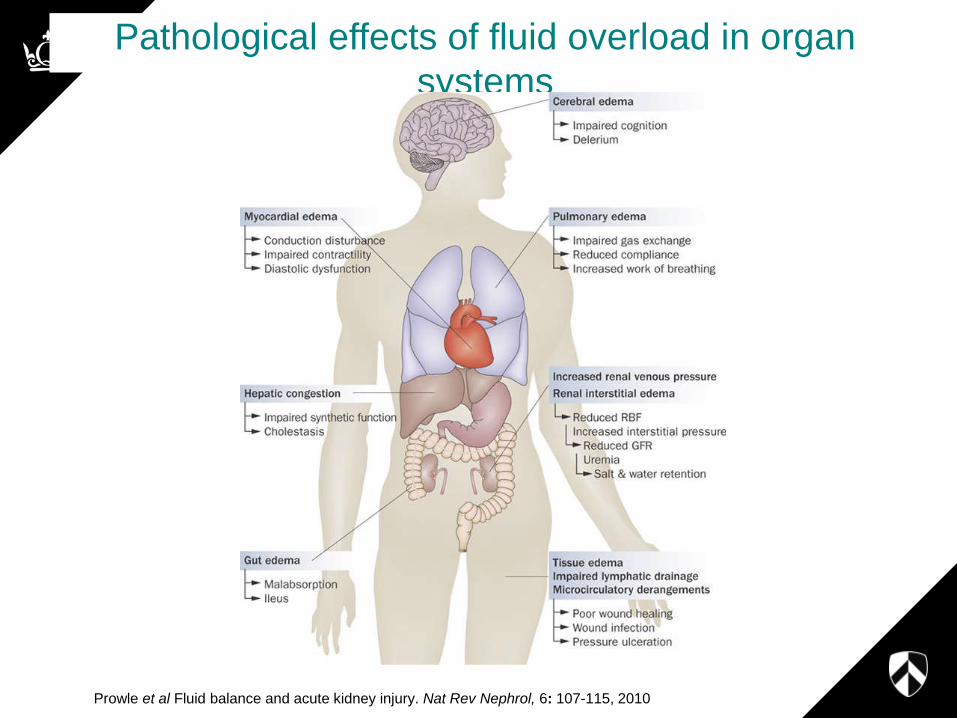
Pathological effects of fluid overload in organ
systems
Prowle et al Fluid balance and acute kidney injury. Nat Rev Nephrol, 6: 107-115, 2010

Resolution of Fluid Overload
with RRT and Survival

All d
emo
nstrate h
arm asso
ciated w
ith flu
id o
verload
o
r be
nefit fro
m its reso
lutio
n

Questions
• To what extent is fluid overload a marker of illness severity (measure of unknown confounders) and to what extent an avoidable cause of iatrogenic morbidity and mortality?
• Does fluid overload itself contribute to the initiation or persistence of AKI?
– Can we treat this?
• Does the avoidance or treatment of fluid overload with RRT (or diuretics) improve outcomes?
– If so when?

Fluid overload and interstitial oedema
may contribute to the maintenance of
AKI
Prowle, Kirwan, Bellomo Nat Rev Nephrol 2013


Systematic approach to fluid
management in critical illness
• Resuscitate appropriately early
• Avoid need for removal as much as
possible by then appropriately limiting
intake
• Ultrafiltration as a component in an active
fluid management strategy

Prowle, Kirwan, Bellomo Nat Rev Nephrol 2013

• Indication of potentially inadequate oxygen
delivery
• New or worsening organ dysfunction
• Lactic acidosis
• Clinical examination
• Measures of tissue oxygenation (Near Infra-red
Spectrocopy, Gastric tonometry)
• Measures of Cardiac Output / Tissue Perfusion
• Cardiac index
• Stroke volume
• Ejection volume
• Venous saturation of oxygen
• Microcirculatory imaging

Measures of Potential
Volume Responsiveness
• Stroke volume or pulse pressure variation
• Echocardiography
• Passive straight leg raising
• Haemodynamic response to fluid challenge
• Central venous or pulmonary artery pressure
• Real-time haematocrit changes to fluid removal

Prowle, J. R. et al. (2013) Nat. Rev. Nephrol. doi:10.1038/nrneph.2013.232

Quantification of fluid overload
• Clinical Examination
• Serial Weights
• Cumulative Fluid Balance
• Chest X-ray
• Oxygenation indices
• Lung Ultrasound
• Intra-abdominal pressure
• Echocardiography
• Bioimpedance body composition analysis

Goals in mechanical fluid
removal
• Resolve fluid overload and its adverse
effects on organ function
• Allow necessary interventions
– Nutrition
– Drugs
• Prevent overt hypovolaemia
– Secondary ischaemic injury
– Adverse neuroendocrine responses
• Avoid complications of RRT

It is possible to be both fluid
overloaded and volume
responsive
• Response needs to be tailored to
– Stage of and severity of illness
– Acute and chronic organ dysfunction
– Response to fluid administration or removal

Decisions in prescribing ultrafiltration
• Goal (Long term)
– Extent of fluid overload
– “Euvolaemia”
• Tolerance of fluid removal (Short term)
– Rate of removal
– Rate of vascular refilling
– Ability of circulation to tolerate transient reduction in
intravascular volume
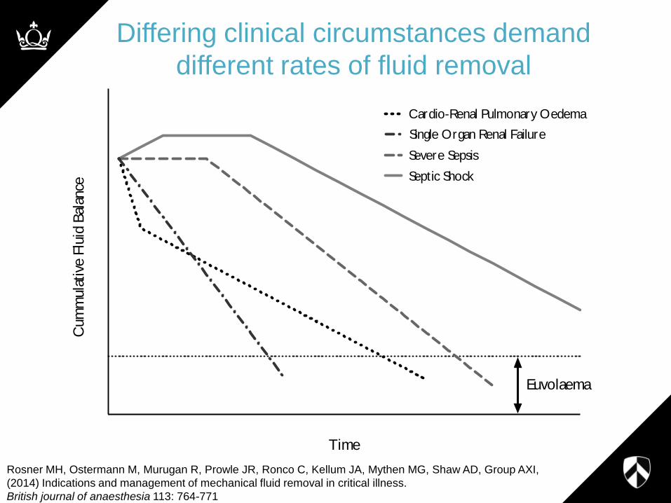
Time
Cum
mula
tive
Flu
id B
alan
ce
Single Organ Renal Failure
Cardio-Renal Pulmonary Oedema
Severe Sepsis
Septic Shock
Euvolaema
Differing clinical circumstances demand
different rates of fluid removal
Rosner MH, Ostermann M, Murugan R, Prowle JR, Ronco C, Kellum JA, Mythen MG, Shaw AD, Group AXI,
(2014) Indications and management of mechanical fluid removal in critical illness.
British journal of anaesthesia 113: 764-771

Conclusions
• Fluid overload is a common occurrence in the critically ill and is link to adverse outcomes in particular in association with AKI
• Limiting or resolving fluid overload may require earlier recourse to RRT
• Prescribing fluid removal requires attention to
– Total fluid excess
– Rate of vascular refilling
– Haemodynamic stability
• Treatment will need to be individualized and responses frequently reassessed
Methods to guide UF in CRRT
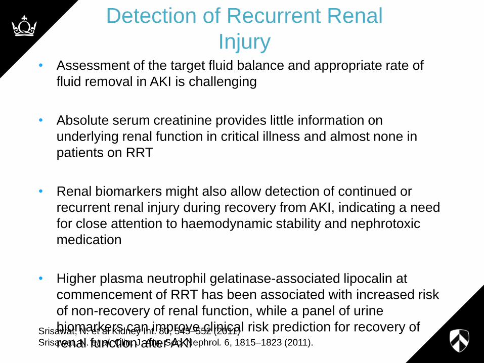
Detection of Recurrent Renal
Injury • Assessment of the target fluid balance and appropriate rate of
fluid removal in AKI is challenging
• Absolute serum creatinine provides little information on
underlying renal function in critical illness and almost none in
patients on RRT
• Renal biomarkers might also allow detection of continued or
recurrent renal injury during recovery from AKI, indicating a need
for close attention to haemodynamic stability and nephrotoxic
medication
• Higher plasma neutrophil gelatinase-associated lipocalin at
commencement of RRT has been associated with increased risk
of non-recovery of renal function, while a panel of urine
biomarkers can improve clinical risk prediction for recovery of
renal function after AKI
Srisawat, N. et al Kidney Int. 80, 545–552 (2011)
Srisawat, N. et al. Clin. J. Am. Soc. Nephrol. 6, 1815–1823 (2011).

Monitoring for Fluid removal:
• Bioimpedance analysis techniques
– quantify the expansion of extracellular and
intracellular fluid volume
– Quantification of Fluid Overload
• Relative blood volume monitoring
– Real-time monitoring of plasma refilling during
ultrafiltration is possible using real-time
monitoring of blood haematocrit
– Tolerance of Fluid Removal

Bioimpedance

Body Weight
Fat mass
Fat Free Mass
(100%)
Body Cell Mass
Visceral protein
Intracellular Water
(~30%)
Extracellular Water
(~45%)
Bone Mineral
(~7%)
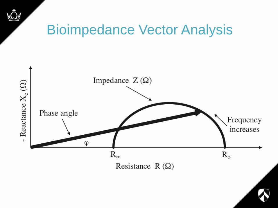
Bioimpedance Vector Analysis


Relative Blood Volume


E-Mail [email protected]
Original Paper
Blood Purif 2013;35:202–208
DOI: 10.1159/000346630
Sensitivity of Blood Volume Monitoring for Fluid Status Assessment in Hemodialysis Patients
Francisco Maduell Marta Arias Elisabet Massó Néstor Fontseré
Montserrat Carrera Manel Vera Aleix Cases Josep M. Campistol
Department of Nephrology, Hospital Clinic Barcelona, Barcelona , Spain
Introduction
Despite many technological advances in hemodialysis therapy, the management and optimization of fluid status remains a major challenge in the field of renal replace-ment therapy [1] . Fluid overload (FO)-related hospital-izations are high [2] and it has been shown that patients who present long-term FO suffer from increased mortal-ity [3] . A multicenter study in eight European dialysis centers has revealed that 25% of all patients are volume overloaded [4] .
Fluid management comprises two main aspects: (i) the short-term (intradialytic) preservation of blood volume to avoid hypotensive episodes, and (ii) the long-term maintenance of fluid status below a critical level beyond which cardiovascular damage may occur.
Usually, patients with high FO tend to be more stable during the treatment because of increased vascular refill-ing [5, 6] . Some patients, however, may present high FO but still suffer from intradialytic episodes, possibly due to cardiac impairment, antihypertensive medication or oth-er factors degrading vascular refilling [7] . It has been pro-posed that careful long-term fluid reduction in these pa-tients may improve their cardiac status and subsequently lead to more intradialytic stability [4] .
Key Words
Bioimpedance · Blood volume monitoring · Fluid overload ·
Fluid status assessment · Hemodialysis
Abstract
Background/Aims: This study investigates the use of blood
volume monitoring (BVM) markers for the assessment of
fluid status. Methods: Predialysis fluid overload (FO) and
BVM data were collected in 55 chronic hemodialysis pa-
tients in 317 treatments. Predialysis FO was measured us-
ing bioimpedance spectroscopy. The slope of the intravas-
cular volume decrease over time normalized by ultrafiltra-
tion rate (Slope4h) was used as the primary BVM marker
and compared against FO. Results: Average relative blood
volume curves were well separated in different FO groups
between 0 and 5 liters. Receiver-operating characteristics
analysis revealed that the sensitivity of BVM was moderate
in median FO ranges between 1 and 3 liters (AUC 0.60–
0.65), slightly higher for volume depletion of FO <1 liter
(AUC 0.7) and highest for excess fluid of FO >3 liters (AUC
0.85). Conclusion : Devices that monitor blood volume are
well suited to detect high FO, but are not as sensitive at
moderate or low levels of fluid status.
Copyright © 2013 S. Karger AG, Basel
Received: August 28, 2012
Accepted: December 11, 2012
Published online: March 13, 2013
Francisco Maduell Canals, MD Servicio de Nefrología, Hospital Clinic Barcelona C/Villarroel, 170 ES–08036 Barcelona (Spain) E-Mail fmaduell @ clinic.ub.es
© 2013 S. Karger AG, Basel0253–5068/13/0353–0202$38.00/0
www.karger.com/bpu
Dow
nlo
ade
d b
y:
Cla
ude
Mo
ore
Health
Scie
nce
s L
ib -
Univ
of V
irg
inia
137
.54.8
0.1
08 -
8/2
3/2
013
12:5
7:1
8 P
M
BIA & RBV In Combination (maintenance HD - high UF rate)
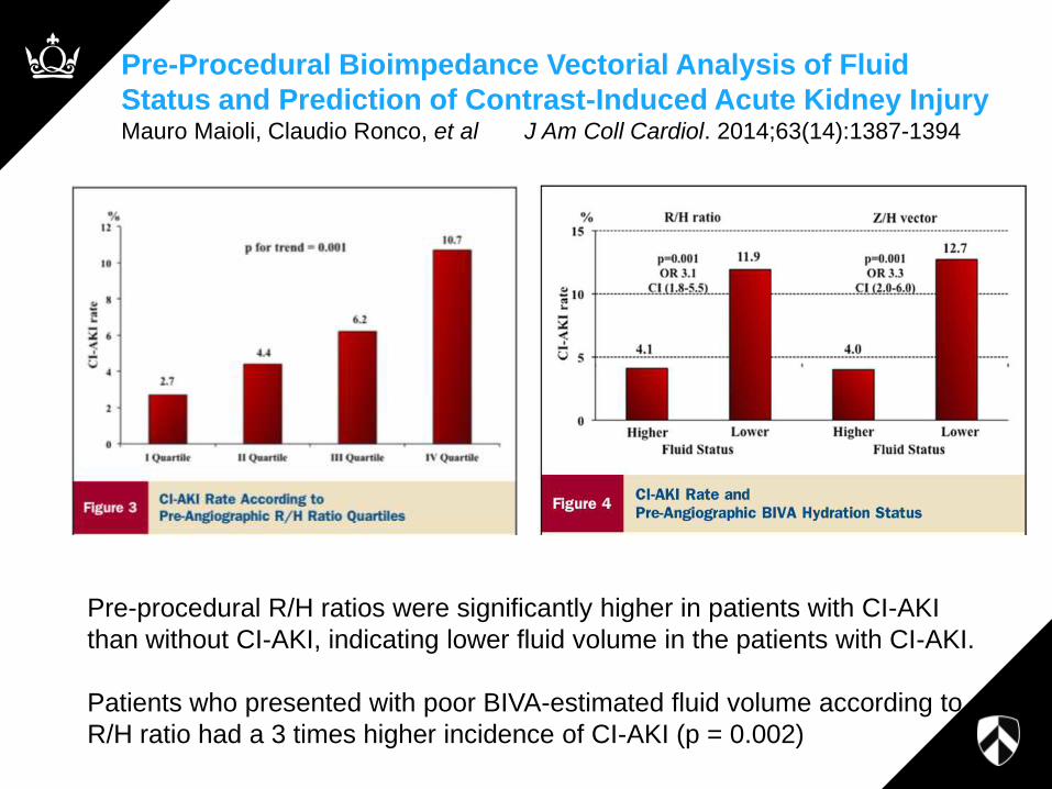
Pre-Procedural Bioimpedance Vectorial Analysis of Fluid
Status and Prediction of Contrast-Induced Acute Kidney Injury Mauro Maioli, Claudio Ronco, et al J Am Coll Cardiol. 2014;63(14):1387-1394
Pre-procedural R/H ratios were significantly higher in patients with CI-AKI
than without CI-AKI, indicating lower fluid volume in the patients with CI-AKI.
Patients who presented with poor BIVA-estimated fluid volume according to
R/H ratio had a 3 times higher incidence of CI-AKI (p = 0.002)

Case #1: Managing Fluid Overload in
Critical Illness with CRRT
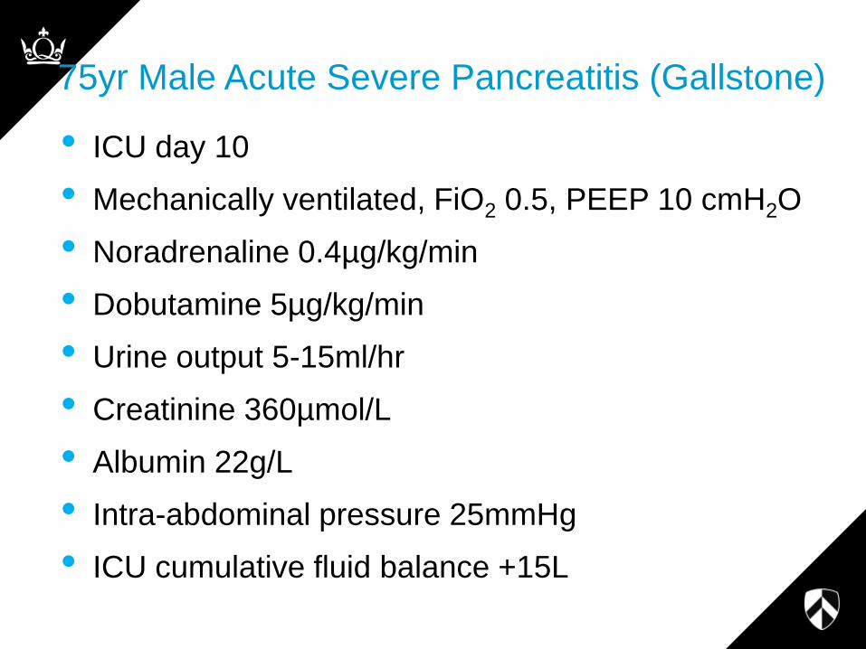
75yr Male Acute Severe Pancreatitis (Gallstone)
• ICU day 10
• Mechanically ventilated, FiO2 0.5, PEEP 10 cmH2O
• Noradrenaline 0.4µg/kg/min
• Dobutamine 5µg/kg/min
• Urine output 5-15ml/hr
• Creatinine 360µmol/L
• Albumin 22g/L
• Intra-abdominal pressure 25mmHg
• ICU cumulative fluid balance +15L

Multi-frequency BIA (5, 50, 100, 200kHz)
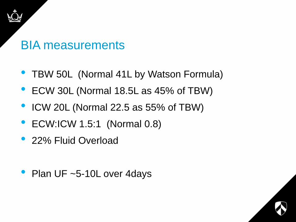
BIA measurements
• TBW 50L (Normal 41L by Watson Formula)
• ECW 30L (Normal 18.5L as 45% of TBW)
• ICW 20L (Normal 22.5 as 55% of TBW)
• ECW:ICW 1.5:1 (Normal 0.8)
• 22% Fluid Overload
• Plan UF ~5-10L over 4days

Day 13
• 7.5L UF over 3 days
• Noradrenaline and Dobutamine off in 36h
• Pressure support ventilation with falling oxygen requirement
• IAP fell to 15mmHg
• UO ~15ml/hr
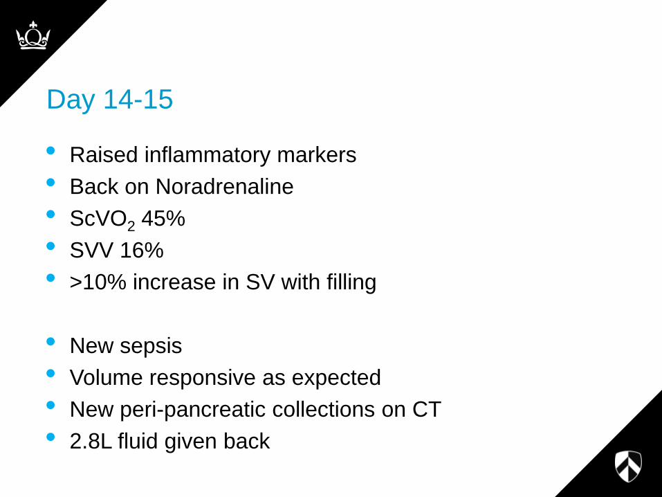
Day 14-15
• Raised inflammatory markers
• Back on Noradrenaline
• ScVO2 45%
• SVV 16%
• >10% increase in SV with filling
• New sepsis
• Volume responsive as expected
• New peri-pancreatic collections on CT
• 2.8L fluid given back

-10000 -5000 0
35
40
45
50
55
Fluid Balance (ml)
To
tal B
od
y W
ate
r (B
IA)

-10000 -5000 0
35
40
45
50
55
Fluid Balance (ml)
Inclusive of 500ml/day insensible losses
TB
W (
L)

-10000 -5000 0
15
20
25
30
35
Fluid Balance (ml) Inclusive of 500ml/day insensible losses
EC
W (
L)


Bioimpedance - Conclusions
• Performance appears variable – occasionally very good
• There is a lack of easily applicable gold standard for
body fluid assessment
• Plasma volume is a undetermined
• Plasma electrolytes and fluid collections are a
challenge
• Can we set a cut off maybe something equivalent to
10% FO as a target for BIA guided therapy??
• We would need to assess this prospectively

Case #2: RRT Prescription in Severe Uraemia
• 22 yr old man 182cm
• Previous fit university rugby player
• Unwell and progressively weak over 1 year
• Saw family practitioner: BP 200/105
• Bloods
– Creatinine 2100µmol/L (24mg/dl)
– Urea 75mmol/L (BUN 210mg/dl)
– HCO3 8, K 7.4, Na 142, PO4 4, Ca 1.85, Hb 7.8

RRT
• Haemodialysis in Renal Ward
• CRRT
• Issues
– Potassium
– Disequilibrium
– Hypocalcaemia

0 5 10 15 200
20
40
60
80
Time h
Ure
a m
mo
l/L
Interstitium
Blood
0 5 10 15 200
20
40
60
80
Time h
Ure
a m
mo
l/L
Blood
Brain
H2O

Haemeodialysis - Decisions
• Time / Frequency
• Blood flow
• Dialyzer size
• Acid concentrate composition (K and Ca)
• Sodium concentration (130-155mmol/L)
• Bicarbonate concentration (20-40mmol/L plus
acetate)
• Ultrafiltration rate

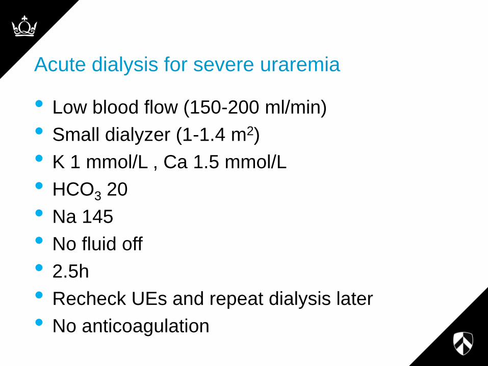
Acute dialysis for severe uraremia
• Low blood flow (150-200 ml/min)
• Small dialyzer (1-1.4 m2)
• K 1 mmol/L , Ca 1.5 mmol/L
• HCO3 20
• Na 145
• No fluid off
• 2.5h
• Recheck UEs and repeat dialysis later
• No anticoagulation

A safer way?

CRRT for treatment of Severe Uraemia
• Only choices are Dose and Potassium replacement
• Start K 0 and 15-20 ml/kg/h Effluent Flow Rate
• Run continuously
• Monitor Urea, K
• Add in UF, Potassium replacement and increase dose
to 25ml/kg/h when appropriate
• This is simply and easily done within existing CRRT
protocols
• Only issue is use of heparin (avoid)
– Pre-dilute - dose is not an issue
– Citrate – watch pH and Calcium

CRRT replacement/dialysis solutions

0 5 10 15 200
20
40
60
80
Time h
Ure
a m
mo
l/L
Brain
Blood
CRRT Urea changes with 25ml/kg/h CRRT