flower-shaped gold nanoparticles: synthesis, characterization and their application as sers-active...
TRANSCRIPT

This content has been downloaded from IOPscience. Please scroll down to see the full text.
Download details:
IP Address: 134.151.40.2
This content was downloaded on 22/08/2014 at 10:41
Please note that terms and conditions apply.
Flower-shaped gold nanoparticles: synthesis, characterization and their application as SERS-
active tags inside living cells
View the table of contents for this issue, or go to the journal homepage for more
2011 Nanotechnology 22 055702
(http://iopscience.iop.org/0957-4484/22/5/055702)
Home Search Collections Journals About Contact us My IOPscience

IOP PUBLISHING NANOTECHNOLOGY
Nanotechnology 22 (2011) 055702 (7pp) doi:10.1088/0957-4484/22/5/055702
Flower-shaped gold nanoparticles:synthesis, characterization and theirapplication as SERS-active tags insideliving cellsSanda Boca1, Dumitrita Rugina2, Adela Pintea2,Lucian Barbu-Tudoran3 and Simion Astilean1
1 Nanobiophotonics Center, Institute for Interdisciplinary Research in Nanobioscience,Babes-Bolyai University, Treboniu Laurian 42, 400271 Cluj-Napoca, Romania2 Department of Biochemistry, University of Agricultural Sciences and Veterinary Medicine,Manastur 3-5, 400372, Cluj-Napoca, Romania3 Electron Microscopy Center, Faculty of Biology and Geology, Babes-Bolyai University,Clinicilor 5-7, 400006, Cluj-Napoca, Romania
E-mail: [email protected] and [email protected]
Received 1 October 2010, in final form 16 November 2010Published 22 December 2010Online at stacks.iop.org/Nano/22/055702
AbstractThe detection of Raman signals inside living cells is a topic of great interest in the study of cellbiology mechanisms and for diagnostic and therapeutic applications. This work presents thesynthesis and characterization of flower-shaped gold nanoparticles and demonstrates theirapplicability as SERS-active tags for cellular spectral detection. The particles were synthesizedby a facile, rapid new route that uses ascorbic acid as a reducing agent of gold salt. Twotriarylmethane dyes which are widely used as biological stains, namely malachite green oxalateand basic fuchsin, were used as Raman-active molecules and the polymer mPEG-SH as cappingmaterial. The as-prepared SERS-active nanoparticles were tested on a human retinal pigmentepithelial cell line and found to present a low level of cytotoxicity and high chemical stabilitytogether with SERS sensitivity down to picomolar particle concentrations.
(Some figures in this article are in colour only in the electronic version)
1. Introduction
The surface-enhanced Raman (SERS) activity of noble metalnanoparticles, given by the strong amplification of thescattering cross-sections of the molecules adsorbed thereonor situated in their close vicinity, has led to a wide rangeof bioanalytical applications, from DNA sequence detectionto intracellular studies [1–3]. Beyond analytical detection,an increasing interest is given nowadays to the fabrication ofSERS-based spectral tags composed of a metallic nanoparticle,a spectroscopic encoding chromophore (called a Raman tag)and a polymeric shell for protection and bioconjugation [4].Traditionally, fluorescent tags based on organic fluorophoresor semiconductor quantum dots (QDs) have been used forin vitro/in vivo labeling assays. However, spectroscopic
labels based on gold nanoparticles and Raman tags canopen a valid alternative to the above assays due to severaladvantages they present. For instance, SERS tags can exhibitsuperior brightness to semiconductor quantum dots, especiallyin the near-infrared (NIR) region, and better photochemicalstability than ordinary fluorescent tags [5, 6]. On the otherhand, the narrower fingerprint of SERS spectra and theability to efficiently excite simultaneously many dye encodednanoparticles at a single wavelength makes this class ofoptical tags potential candidates for combinatorial codingand multiplexed cellular spectral imaging [7]. Moreover,SERS-coded gold nanoparticles were recently demonstrated tooperate as multifunctional tools for imaging and treatment oftumors in vivo by photothermal therapy in the NIR spectralwindow [8]. Finally, noble metal nanoparticles, especially
0957-4484/11/055702+07$33.00 © 2011 IOP Publishing Ltd Printed in the UK & the USA1

Nanotechnology 22 (2011) 055702 S Boca et al
gold, are considered to be biocompatible in contrast with thecytotoxicity of semiconductor quantum dots, which is decisivefor applications in nanomedicine [9].
Therefore a variety of SERS reporter complexesconsisting of nanostructures such as gold nanoshells,Au–Ag core–shell particles, composite organic–inorganicnanoparticles, organosulfur molecules and polymeric coatingshas been developed and tested until now for cell and tissuelabeling [10–12]. Nevertheless, it is still highly desirablefor ultrasensitive assays to design brighter SERS labels thatcombine facile and versatile synthesis with high chemicalstability. As the Raman scattering enhancement is mainly dueto the surface plasmon resonance of the metal nanostructure(electromagnetic enhancement), there is a direct interest innanoparticles with sharp morphologies which are able tosustain the local field enhancement and generate so called hotspots [13–15].
Of particular interest are multi-branched nanoparticles,also called flower-shaped, urchinlike or bumpy gold nanopar-ticles which exhibit strong plasmon resonances close to theNIR window of biological transparency and, particularly, highelectromagnetic field localized at their protrusions [16–18].Currently flower-shaped gold nanoparticle (gold nanoflower)synthesis implies multiple-step, relatively expensive proce-dures and the use of hazardous chemical reactants, whichin turn requires supplementary detoxification procedures forbiological application [19]. On the other hand, producing bio-compatible labels consisting of polymer or protein protectedflower-shaped nanoparticles remains a challenging task andonly a few examples have proved their efficiency in practicalapplications inside living organisms [20].
Unlike previous studies, herein we report a fast andlow cost method to prepare gold nanoparticles of flowerlikeshape using the ascorbate as reduction agent, withoutinvolving any seeds or surfactants. Widely used asbiological stains, malachite green (MG) oxalate and basicfuchsin (BF) molecules were selected as Raman-activereporters and their SERS readouts were successfully testedin the cytosol of a line of epithelial cells from humanretina. The stability of nanoparticle–reporter conjugates isa crucial parameter for successful assays based on SERSlabels. We specifically addressed this point by protectingthe nanoparticle–reporter conjugates with thiol-modifiedpoly(ethylene) glycol (PEG), a nontoxic, hydrophilic polymerwhich, in addition, improves particles’ biocompatibility andtheir systemic retention [21]. Furthermore, we considered theproblem of cellular nanoparticle uptake and assessed the levelof toxicity of the designed SERS tags, which is of paramountimportance for any practical implementation in nanomedicine.
2. Experimental details
2.1. Chemicals and materials
Hydrogen tetrachloroaurate (III) trihydrate (HAuCl4·3H2O)
was purchased from Sigma-Aldrich (Germany). An-alytical grade L-ascorbic acid (C6H8O6) was obtainedfrom Reactivul (Bucuresti). Malachite green oxalate
([(C23H25N2)·(C2HO4)]2·C2H2O4) and basic fuchsin(C20H19N3HCl) were purchased from Merck (Germany). α-methoxy-ω-mercapto poly(ethylene glycol) (mPEG-SH) ofmolecular weight 5 kDa was obtained from Iris biotechGmbH (Germany). GIBCO Dulbecco’s Modified Eagle’sMedium (DMEM) was purchased from Invitrogen (Carlsbad,California, USA). Fetal bovine serum, penicillin, strepto-mycin, amphotericin B, and 3-(4,5-dimethylthiazol-2-yl)-2,5-diphenyltetrazolium bromide (MTT) were purchased fromSigma (St. Louis, USA). The dimethylsulfoxide, sodiumpyruvate and all the other chemicals used were of analyticalgrade and supplied by Merck (Germany). The other reagentsused during the experiments were analytical grade and wereused without further purification. Distilled–deionized water(ddH2O) was used in all aqueous solutions and rinsingprocedures.
2.2. Equipment and characterization methods
Optical extinction spectra were measured with a Jasco V-670 spectrophotometer over a spectral range between 400 and1100 nm with a spectral resolution of 2 nm. The mean diameterof the gold nanoparticles was determined by transmissionelectron microscopy (TEM) imaging using a JEOL modelJEM1010 microscope. The SERS spectra of the conjugatedgold nanoparticles in aqueous solution were recorded usingfor excitation an NIR (785 nm) laser line from a diode laserusing a portable Raman spectrophotometer (R-3000CN fromRaman Systems) with 1 cm−1 spectral resolution and anintegration time of 10 s. The SERS spectra of the conjugatednanoparticles inside living cells were recorded using the He–Ne laser line (633 nm) with 3 mW power through a 100×oil immersion (NA = 1.4) of a confocal Raman microscope(Witec alpha300R).
2.3. Sample preparation
Gold nanoflower synthesis involves only two chemicalreactants: L-ascorbic acid (L-AA), having the role of goldsalt reducer and tetrachloroauric acid (HAuCl4), used asnanoparticle initiator. The nanoparticles were prepared bythe rapid mixture of 20 ml of a solution of 19.8 × 10⁻³ ML-AA with 200 μl of 10−2 M HAuCl4 at ice temperature. Themixtures rapidly turned colorless and finally faint blue.
For nanoparticle conjugation, malachite green oxalate(MG) and basic fuchsin (BF) solutions were added dropwiseto a few milliliters of nanoparticle solutions and shaken forseveral minutes. The Raman reporter molecule concentrationand nanoparticle binding time were carefully established toachieve enough reporter molecules/particle without causing asevere nanoparticle aggregation. Then, appropriate volumes of10−6 mM mPEG-SH were added to the nanoparticle solutionsand allowed to sit for 1 h so that the polymer would be able tofully stabilize the particles. At the end, the nanoparticles werecentrifuged at 6000 rpm for 20 min and washed in ddH2O toremove any unreacted products.
2
Nanotechnology 22 (2011) 055702 S Boca et al
2.4. Cell line culture and gold nanoparticle incubation
Human adult retinal pigment epithelial cells D407 weremaintained in Dulbecco’s Modified Eagle’s Medium sup-plemented with 10% fetal bovine serum, 1 mM sodiumpyruvate, 100 U ml−1 penicillin, 100 μg ml−1 streptomycin,and 2.5 μg ml−1 amphotericin B, at 37 ◦C, 5% CO2, and95% relative humidity. For MTT 3-(4,5-dimethylthiazol-2-yl)-2,5-diphenyltetrazolium bromide viability and SERSmeasurements the cells were seeded in 96-well plates at aconcentration of 1 × 10⁴ and in 60 mm diameter Petri dishesrespectively. After reaching 90% confluence, the growthmedium was removed and the cells were treated with basicfuchsin gold nanoflowers (BF-GNFs).
2.5. MTT viability assay
D407 cells were plated (10 000 cells per well) in 96-wellplates and after the cells attached, they were incubated for24 h with basic fuchsin gold nanoflowers (BF-GNFs) byvarying the colloidal concentration between 0.47 × 10⁻¹³ and5.64 × 10−13 M. The tetrazolium salt was used for assayingthe quantification of living metabolically active cells, basedupon the principle that MTT works by being metabolizedby mitochondrial dehydrogenase to form a formazan dye.Thereby the water-soluble end-product was measured at awavelength of 550 nm with reference wavelength at 690 nm,with a microplate reader HT BioTek Synergy (BioTekInstruments, USA). Briefly, the cells were washed with PBSand 200 μl MTT solution in HBSS buffer was added toeach well. After 2 h of incubation the MTT reagent wasremoved and the formazan particles were solubilized with200 μl DMSO. Viability was expressed as the percentageof actively proliferative cells and subsequently a relationshipbetween viability and treatment concentration was plotted infigure 6. Statistical analyses were performed using an analysisof variance with Dunnett’s multiple comparison test (P <
0.05). Data are expressed as mean±SEM (n = 4) and ∗ meanssignificant, ∗∗ very significant, ∗ ∗ ∗ extremely significant.
3. Results and discussion
3.1. Flowerlike gold nanoparticle characterization
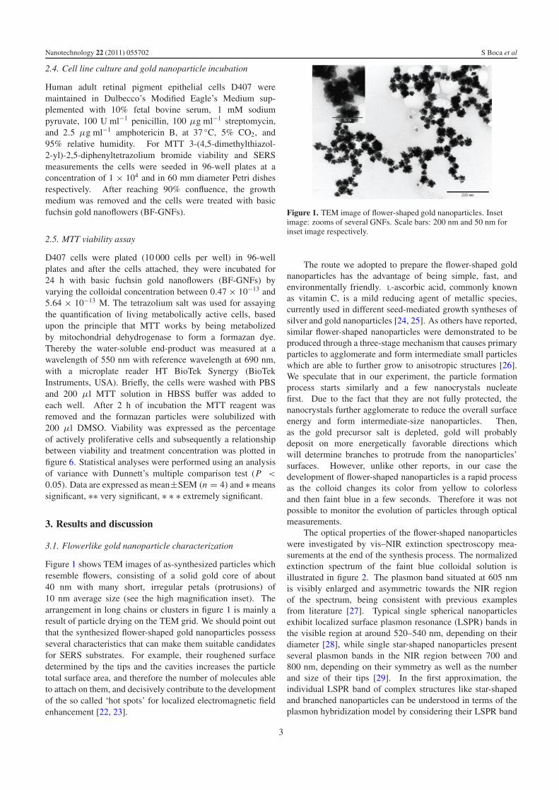
Figure 1 shows TEM images of as-synthesized particles whichresemble flowers, consisting of a solid gold core of about40 nm with many short, irregular petals (protrusions) of10 nm average size (see the high magnification inset). Thearrangement in long chains or clusters in figure 1 is mainly aresult of particle drying on the TEM grid. We should point outthat the synthesized flower-shaped gold nanoparticles possessseveral characteristics that can make them suitable candidatesfor SERS substrates. For example, their roughened surfacedetermined by the tips and the cavities increases the particletotal surface area, and therefore the number of molecules ableto attach on them, and decisively contribute to the developmentof the so called ‘hot spots’ for localized electromagnetic fieldenhancement [22, 23].
Figure 1. TEM image of flower-shaped gold nanoparticles. Insetimage: zooms of several GNFs. Scale bars: 200 nm and 50 nm forinset image respectively.
The route we adopted to prepare the flower-shaped goldnanoparticles has the advantage of being simple, fast, andenvironmentally friendly. L-ascorbic acid, commonly knownas vitamin C, is a mild reducing agent of metallic species,currently used in different seed-mediated growth syntheses ofsilver and gold nanoparticles [24, 25]. As others have reported,similar flower-shaped nanoparticles were demonstrated to beproduced through a three-stage mechanism that causes primaryparticles to agglomerate and form intermediate small particleswhich are able to further grow to anisotropic structures [26].We speculate that in our experiment, the particle formationprocess starts similarly and a few nanocrystals nucleatefirst. Due to the fact that they are not fully protected, thenanocrystals further agglomerate to reduce the overall surfaceenergy and form intermediate-size nanoparticles. Then,as the gold precursor salt is depleted, gold will probablydeposit on more energetically favorable directions whichwill determine branches to protrude from the nanoparticles’surfaces. However, unlike other reports, in our case thedevelopment of flower-shaped nanoparticles is a rapid processas the colloid changes its color from yellow to colorlessand then faint blue in a few seconds. Therefore it was notpossible to monitor the evolution of particles through opticalmeasurements.
The optical properties of the flower-shaped nanoparticleswere investigated by vis–NIR extinction spectroscopy mea-surements at the end of the synthesis process. The normalizedextinction spectrum of the faint blue colloidal solution isillustrated in figure 2. The plasmon band situated at 605 nmis visibly enlarged and asymmetric towards the NIR regionof the spectrum, being consistent with previous examplesfrom literature [27]. Typical single spherical nanoparticlesexhibit localized surface plasmon resonance (LSPR) bands inthe visible region at around 520–540 nm, depending on theirdiameter [28], while single star-shaped nanoparticles presentseveral plasmon bands in the NIR region between 700 and800 nm, depending on their symmetry as well as the numberand size of their tips [29]. In the first approximation, theindividual LSPR band of complex structures like star-shapedand branched nanoparticles can be understood in terms of theplasmon hybridization model by considering their LSPR band
3

Nanotechnology 22 (2011) 055702 S Boca et al
Figure 2. Normalized extinction spectrum of gold nanoflowers(GNFs) in aqueous solution. The inset image shows the colloidalsolution.
to be the result of the interaction of plasmon resonances ofsimpler structures [30, 31]. As the LSPR band measured in thisstudy extends from the visible to the NIR, the gold nanoflowersreported here can be regarded as complex hybrids of spheresand stars that are not highly monodisperse and their individualLSPR bands are lost in ensemble measurements.
3.2. Particle stability measurements
We first compared the optical extinction spectra of uncoatednanoparticles (black solid curves) and Raman reporter taggednanoparticles (color dashed curves). In figure 3(A) thetwo spectra look almost identical. A few nanometer red-shift of the plasmonic resonance is observed for MG taggednanoparticles which is consistent with a local change in theoptical refractive index due to the electrostatic interactionbetween the cationic reporters and the negatively chargednanoparticles. A more pronounced effect can be observedfor BF tagged gold nanoflowers (figure 3(B)) whose plasmonband is visibly enlarged and 15 nm red-shifted after addingthe reporter molecules. Although both MG and BF possess
the electronic transition in this spectral region, their molecularabsorption bands are not detectable at this low concentration(less than 10−7 M), being practically masked by the Auplasmon extinction band.
Since the nanoparticles are to be exploited as SERS tagsinside cells, they should retain their optical properties and thestability of the attached Raman reporter in the physiologicalmedium. Therefore, the stability of encoded gold nanoparticlescoated with a protective polymeric shell was checked viaa reliable aggregation test. This was done by mixing theconjugates with a high molarity aqueous solution of NaCl salt(1 M), known to induce particle aggregation in the case ofnonprotected gold colloids [32]. Figure 3 depicts the recordednormalized extinction spectra of PEGylated malachite green(figure 3(A)) and basic fuchsin (figure 3(B)) encoded goldnanoflowers as resulted from the aggregation test. Nosignificant changes in the LSPR spectral position and shapewere noticed, which means that the PEG provided effectivestabilization against aggregation. We further tested the stabilityof the Raman signal under conditions which simulate a realapplication and the SERS measurements are discussed later.
3.3. Assessment of Raman encoded gold nanoparticles asSERS tags
The SERS applicability of flower-shaped nanoparticles wasinvestigated for two molecules with affinities for the negativecharged nanoparticle gold surface, malachite green oxalateand basic fuchsin. Figures 4(A) and (B) show the SERSspectra of PEGylated malachite green (MG-GNFs) and basicfuchsin (BF-GNFs) tagged nanoflowers respectively, recordedin solution using 785 nm NIR excitation. The SERSspectra exhibit characteristic vibrational bands of the twomolecules, which are clearly distinguishable, irrespectiveof the closely similar molecular structure of the twotriarylmethane chromophores [33, 34]. Therefore, strongvibrational bands at 224, 417, 1167, 1368 cm−1 for MG-GNFs and at 271, 420, 1171, 1373 cm−1 for BF-GNFsrespectively are assigned to the totally symmetric breathingmode bonds to the central carbon, out-of-plane benzene ringdeformation, C–H bending and N-phenyl stretching according
Figure 3. Normalized extinction spectra of MG-gold nanoflowers (A) and of BF-gold nanoflowers (B) recorded in the absence (solid anddashed lines) and in the presence (dotted lines) of 1 M NaCl solution. Solid lines represent uncoated particles while broken lines representtagged particles.
4

Nanotechnology 22 (2011) 055702 S Boca et al
Figure 4. SERS spectra of malachite green (A) and basic fuchsin gold nanoflowers (B) in solution recorded under 785 nm excitation.
Figure 5. SERS spectra of MG-GNFs (A), and BF-GNFs (B) in solution recorded under NIR excitation. Spectra ii are recorded immediatelyafter adding 1 M salted solution, spectra iii are recorded after several days of particle solution storage at room temperature, while spectra ivare recorded after several days of particle solution storage at room temperature in the presence of NaCl solution. Spectrum i is characteristicto as-prepared tags.
to the literature [35]. Two strong additional bands are presentfor MG-GNFs at 796 cm−1 (out-of-plane C–H (benzene e1g))and at 1612 cm−1 (N-phenyl and C–C stretching) while BF-GNFs present a supplementary strong vibrational band at1584 cm−1, characteristic of the stretching and bending of thearomatic ring.
We further tested the stability of the SERS signal aftertransferring the MG- and BF-tagged nanoflowers into asimulated physiological medium made of highly concentratedsalted solution (spectra ii) and for periods of several days ofstorage at room temperature (spectra iii and iv in figure 5).In the case of malachite green oxalate we observed a signalincrease just after adding salt in the colloidal solution followedby a slight decrease after several days. A slight aggregation ofthe particles can explain the increase observed at the beginningof the experiments and it is likely that the ionic strength ofthe environment can induce the loss of a part of the cationicdye molecules bound to the nanoparticles. On the contrary,basic fuchsin encoded nanoflowers demonstrated an increasedstability and the SERS signal is conserved after salt additionand after several days, due probably to stronger binding ontothe nanoparticle surfaces via amine chemical groups.
It is important to note that the reporter molecules are notdisplaced by PEG-SH polymer anchored on the nanoparticlesurfaces, although the molecules are absorbed throughelectrostatic interactions and delocalized pi-electrons [36]. Itis plausible that due to its high bulkiness, the thiol-PEG layerprotects and maintains the adsorbed reporter dyes trapped nearthe gold nanoparticle surface by steric shielding. This isalso sustained by other studies on dye encoded nanoparticleswhere the Raman tagging was found to be a one way process,meaning that if particles were first coated by the polymer, theSERS signal from the reporter molecules was blocked andonly ions or smaller molecules having a thiol functional groupwere demonstrated to be able to pass the polymeric cappinglayer [37, 38]. These results confirmed that the Raman encodedgold nanoparticles developed here are reliable for applicationsin physiological media.
3.4. Effect of gold nanoparticles on D407 cell viability
As basic fuchsin tagged nanoparticles showed increased signalstability, we have chosen them for further experiments insideliving cells. Therefore, we first tested the BF-GNF particles’
5

Nanotechnology 22 (2011) 055702 S Boca et al
Figure 6. MTT test of proliferation (viability %) for epithelial cellsfrom human retina (D407) treated with various concentrations ofBF-GNFs. Zero concentration is the control sample of cellsincubated under identical experimental conditions but withoutnanoparticles.
cytotoxicity using MTT assay in order to establish the properparticle concentration that can be safely used and yet toobtain sufficient SERS response. We found that the viabilityand proliferation of D407 cells is highly dependent on theparticle concentration and that in our tagged nanoparticledesign the Raman tags should be below any potentially harmfulconcentration, which is lower than 2.82 × 10⁻¹³ M as canbe seen in the plotted graph in figure 6. A marked decreaseof D407 cell viability for higher nanoparticle concentrationsdemands that their use be restrained above the thresholdconcentration. However, this is not a particular response ofBF tagged nanoparticles since similar viability tests dependingon particle concentration were observed for untagged goldnanoparticles [39]. In fact, the PEG polymer on thenanoparticle surface acts as a protective layer, impeding thetrapped Raman molecules from being released in the cellular
medium, and possibly deteriorating the cells. Moreover,the methoxy distal ends of PEG-SH capped nanoparticlesare highly resistant to chemical or enzymatic oxidation andtherefore less prone to nonspecific protein binding, as provedin the literature [40].
3.5. SERS inside living cells
Proved to be one of the most sensitive approaches for bioassayapplications, particularly in living cells, nanoparticle-basedSERS techniques have been successfully used for intracellularstudies [41]. However, the huge number of molecules foundin cell cytoplasm is often an impediment to the extractionof relevant biological information. Therefore, more sensitivenanosensors were developed by the use of molecules that canimage specific phenomena inside cells or can be bound todesired cellular organelles [42, 43].
To demonstrate the reliability of our SERS tags, weincubated D407 cells in the presence of PEGylated BF encodedgold nanoparticles for 24 h. Figure 7(A) shows two SERSspectra which highly resemble: the Raman encoded particles’signature collected as reference from a colloidal solution(spectrum ii) and the spectrum collected from inside the cellsfrom where the nanoparticles were internalized (spectrum iii).No Raman signal could be obtained from nonloaded cellsor from the free of particles nuclear region for loaded cells(spectrum i). We believe that SERS nanoparticle tags weredelivered to cells by a natural endocytotic pathway andaccumulated inside late endosomes and in the endoplasmaticreticulum region near the nucleus as can be seen in the opticalimage in figure 7(B). We cannot exclude the formation ofdimers or small clusters during cell incubation. Yet, theirsize should be adequate to penetrate most of the eukaryoticcell membranes, especially cells with increased permeabilityand retention such as macrophages or cancer cells, but theyare unlikely to access the cell nucleus. Notably, such particleclusters are welcome for trapping Raman-active molecules.However, few nanoparticle clusters could be bound also ontothe cell membrane. Even so, no interfering signal from the
Figure 7. (A) SERS spectra of as-prepared BF-GNFs in aqueous solution (ii), inside one selected region (marked by arrows) of a cell (iii) andfrom cells free of particles (i) recorded under 633 nm excitation. The spectra are baseline corrected for better visualization. (B) Transmittedlight optical image of three D407 cells loaded with Raman encoded gold nanoflowers.
6
Nanotechnology 22 (2011) 055702 S Boca et al
polymeric nanoparticle coating or from cellular constituentssuch as membrane proteins was observed, meaning that theexternal molecules are ‘locked out’. Moreover, we observedthat even after long time exposure the dyes do not photo-bleachand the reporter–nanoparticle complex spectral signature isconserved, without any carbonaceous contamination. Theseresults additionally prove that our encoded nanoparticles arehighly stable and protected from protein adsorption, beingable to conserve their Raman identity in vitro, and thereforeamenable to future spectral imaging applications inside livingorganisms.
4. Conclusions
Our work reports the synthesis and characterization of a classof biocompatible, spectroscopic encoded flower-shaped goldnanoparticles, which are very effective for surface-enhancedRaman detection inside living cells. We demonstrated thatas-prepared PEGylated gold nanoparticles are highly stable insalted solution such as the biological medium and that theiroptical and SERS spectroscopic signals are also conservedin time. When tested on epithelial cells from humanretina, the particles demonstrated a low level of toxicity forthe concentrations required for SERS investigation. Themaintenance of a particle SERS signal together with the abilityof fluorescent emission makes this class of tags be potentialcandidates for combinatorial coding and multiplexed spectralimaging inside living cells. Moreover, this nanoparticleplatform, allowing future conjugation of specific ligands toPEGylated coating, can demonstrate the applicability of ourSERS encoded nanoparticles for in vivo tumor detection andimaging.
Acknowledgment
This work was supported by CNCSIS-UEFISCSU, projectnumber PNII-ID PCCE 129/2008.
References
[1] Cao Y C, Jin R and Mirkin C A 2002 Science 297 1536[2] Tanabe K 2008 J. Phys. Chem. C 112 15721[3] Sun S, Thompson D, Schmidt U, Graham D and
Leggett G J 2010 Chem. Commun. 46 5292[4] Qian X, Peng X H, Ansari D O, Yin-Goen Q, Chen G,
Shin D M, Yang L, Young A N, Wang M D and Nie S 2008Nat. Biotechnol. 26 83
[5] Njoki P N, Lim I I S, Mott D, Park H Y, Khan B, Mishra S,Sujakumar R, Luo J and Zhong C J 2007 J. Phys. Chem. C111 14664
[6] Yake A M, Snyder C E and Velegol D 2007 Langmuir 23 9069[7] Matschulat A, Drescher D and Kneipp J 2010 ACS Nano
4 3259[8] von Maltzahn G, Centrone A, Park J H, Ramanathan R,
Sailor M J, Hatton A T and Bhatia S N 2009 Adv. Mater.21 3175
[9] Bhattacharya R and Mukherjee P 2008 Adv. Drug Deliv. Rev.60 1289
[10] Ochsenkuhn M A, Jess P R T, Stoquert H, Dholakia K andCampbell C J 2009 ACS Nano 3 3613
[11] Kumar G V P, Shruti S, Vibha B, Reddy B A A, Kundu T Kand Narayana C 2007 J. Phys. Chem. C 111 4388
[12] Kim J H et al 2006 Anal. Chem. 78 6967[13] Hu X, Wang T, Wang L and Dong S 2007 J. Phys. Chem. C
111 6962[14] Esenturk E N and Hight W A R 2009 J. Raman Spectrosc.
40 86[15] Iosin M, Toderas F, Baldeck P and Astilean S 2008 J. Opt. Adv.
Mater. 10 2285[16] Nehl C L, Liao H and Hafner J H 2006 Nano Lett. 6 683[17] Hrelescu C, Sau T K, Rogach A L, Jackel F and
Feldmann J 2009 Appl. Phys. Lett. 94 153113[18] Kim J H, Kang T, Yoo S M, Lee S Y, Kim B and Choi Y K
2009 Nanotechnology 20 235302[19] Wang L, Wei G, Guo C, Sun L, Sun Y, Song Y, Yang T and
Li Z 2008 Colloids Surf. A 312 148[20] Xie J, Zhang Q, Lee J Y and Wang D I C 2008 ACS Nano
2 2473[21] Paciotti G F, Kingston D G I and Tamarkin L 2006 Drug Dev.
Res. 6 47[22] Chen T, Wang H, Chen G, Wang Y, Feng Y, Teo W S,
Wu T and Chen H 2010 ACS Nano 4 3087[23] Farcau C and Astilean S 2010 J. Phys.Chem. C 114 11717[24] Murphy C J, Sau T K, Gole A M, Orendorff C J, Gao J, Gou L,
Hunyadi S E and Li T 2005 J. Phys. Chem. B 109 13857[25] Andreescu D, Sau T K and Goia D V 2006 J. Colloid Interface
Sci. 298 742[26] Wang W, Yang X and Cui H 2008 J. Phys. Chem C 112 16348[27] Yu K, Kelly K L, Sakai N and Tatsuma T 2008 Langmuir
24 5849[28] Boca S C, Farcau C and Astilean S 2009 Nucl. Instrum.
Methods Phys. Res. B 267 406[29] Dondapati S K, Sau T K, Hrelescu C, Klar T A, Stefani F D and
Feldmann J 2010 ACS Nano 4 6318[30] Prodan E, Radloff C, Halas N J and Nordlander P 2003 Science
302 419[31] Nehl C L and Hafner J H 2008 J. Mater. Chem. 18 2415[32] Mangeney C, Ferrage F, Aujard I, Marchi-Artzner V, Jullien L,
Ouari O, El Djouhar R, Laschewsky A, Vikholm I andSadowski J W 2002 J. Am. Chem. Soc. 124 5811
[33] Christiansen S H, Becker M, Fahlbusch S, Michler J,Sivakov V, Andra G and Geiger R 2007 Nanotechnology18 035503
[34] Efremov E V, Ariese F, Brinkman U A T and Gooijer C 2004Anal. Chim. Acta 508 127
[35] Lueck B H, Daniel D C and McHale J L 1993 J. RamanSpectrosc. 24 363
[36] Frazen S, Folmer J C W, Glomm W R and O’Neal R 2002J. Phys. Chem. A 106 6533
[37] Boca S C and Astilean S 2010 Nanotechnology 21 235601[38] Merican Z, Schiller T L, Hawker C J, Fredericks P M and
Blakey I 2007 Langmuir 23 10539[39] Mahmood M et al 2010 J. Appl. Toxicol. 30 74[40] Prime K L and Whitesides G M 1993 J. Am. Chem. Soc.
115 10714[41] Kneipp K, Haka A S, Kneipp H, Badizadegan K, Yoshizawa N,
Boone C, Shafer-Peltier K E, Motz J T, Dasari R R andFeld M S 2002 Appl. Spectrosc. 56 150
[42] Kneipp J, Kneipp H, McLaughlin M, Brown D andKneipp K 2006 Nano Lett. 6 2225
[43] Ximei Q, Li J and Nie S 2009 J. Am. Chem. Soc. 131 7540
7