flexibly deployed pax genes in eye development at studies ...flexibly deployed pax genes in eye...
TRANSCRIPT

Flexibly deployed Pax genes in eye development atthe early evolution of animals demonstrated bystudies on a hydrozoan jellyfishHiroshi Sugaa,1, Patrick Tschoppa,2, Daria F. Graziussia,3, Michael Stierwaldb,4, Volker Schmidb,5, and Walter J. Gehringa,6
aDepartment of Cell Biology, Biozentrum, and bInstitute of Zoology, Pharmazentrum, University of Basel, CH-4056 Basel, Switzerland
Contributed by Walter J. Gehring, June 14, 2010 (sent for review April 8, 2010)
Pax transcription factors are involved in a variety of develop-mental processes in bilaterians, including eye development, a roletypically assigned to Pax-6. Although no true Pax-6 gene has beenfound in nonbilateral animals, some jellyfish have eyes with com-plex structures. In the cubozoan jellyfish Tripedalia, Pax-B, anortholog of vertebrate Pax-2/5/8, had been proposed as a regulatorof eye development. Here we have isolated three Pax genes (Pax-A, Pax-B, and Pax-E) from Cladonema radiatum, a hydrozoan jel-lyfish with elaborate eyes. Cladonema Pax-A is strongly expressedin the retina, whereas Pax-B and Pax-E are highly expressed in themanubrium, the feeding and reproductive organ. Misexpression ofCladonema Pax-A induces ectopic eyes in Drosophila imaginaldiscs, whereas Pax-B and Pax-E do not. Furthermore, CladonemaPax-A paired domain protein directly binds to the 5′ upstream re-gion of eye-specific Cladonema opsin genes, whereas Pax-B doesnot. Our data suggest that Pax-A, but not Pax-B or Pax-E, is in-volved in eye development and/or maintenance in Cladonema.Phylogenetic analysis indicates that Pax-6, Pax-B, and Pax-A be-long to different Pax subfamilies, which diverged at the latestbefore the Cnidaria–Bilateria separation. We argue that our data,showing the involvement of Pax genes in hydrozoan eye develop-ment as in bilaterians, supports the monophyletic evolutionaryorigin of all animal eyes. We then propose that during the earlyevolution of animals, distinct classes of Pax genes, which may haveplayed redundant roles at that time, were flexibly deployed foreye development in different animal lineages.
biodiversity | Cladonema radiatum | Cnidaria | evo-devo | gene duplication
The Pax gene family encodes transcription factors involved ina variety of developmental processes in metazoans (1–3). It
can be subdivided into five subfamilies—Pax-4/6, Pax-2/5/8, Pax-1/9, Pax-3/7, and pox neuro (poxn)—on the basis of structuralcomparison and phylogenetic relationship (4). These subfamiliesdiverged from each other at the latest before the separation ofcnidarians and bilaterians (4, 5).It is widely accepted that Pax-6, a member of Pax-4/6 subfam-
ily, is one of the most significant components of the gene networkthat controls eye development in many bilaterians (e.g., refs. 6, 7);mutations in Pax-6 genes cause severe eye defects both in mam-mals and inDrosophila (8, 9), and Pax-6 genes cloned from diversebilaterians can ectopically initiate eye development both in Dro-sophila and in Xenopus when they are misexpressed (6, 10, 11).Cnidaria are the earliest branching animal phylum containing
species with multicellular eyes, which sometimes show complexstructures such as lens, iris, pigmented layer, andphotosensitive layer(12). Among the five classes of the phylum Cnidaria (Anthozoa,Hydrozoa, Cubozoa, Scyphozoa, and recently recognized Staur-ozoa), four of them (Hydrozoa, Cubozoa, Scyphozoa, and Staur-ozoa) include eye-bearing species (12). However, no bona fide Pax-6has been identified in cnidarians to date.Kozmik et al. (13) have proposed that Pax-B, a member of the
Pax-2/5/8 subfamily, is responsible for eye development in Tripe-dalia cystophora, a cubozoan jellyfish. Tripedalia Pax-B is expressedin the rhopalia, which are batteries of sensory organs including
eyes. This gene is also able to transactivate the promoters of a Tri-pedalia crystallin gene and a Drosophila rhodopsin gene in cellculture, and it induces ectopic eyes in Drosophila when misex-pressed in imaginal discs. These data suggest that Pax-B is re-sponsible for eye development in Tripedalia. The authors havefurther proposed that Pax-6 diverged from Pax-B (Pax-2/5/8) bygene duplication in the bilaterian lineage after the separationfrom cnidarians and was independently recruited for controllingeye development in the bilaterian lineage (13, 14). Accordingly,they have hypothesized that cnidarian and bilaterian eyes aroseindependently.Besides the class Cubozoa, the class Hydrozoa includes several
species with complex eyes (12). Sun et al. (15) have investigatedCladonema californicum, a hydrozoan jellyfish bearing eyes. How-ever, they have found only a Pax-B gene, whose function has notbeen well studied in Cladonema.In this study, we have isolated and characterized three Pax genes
from Cladonema radiatum, which possesses eyes with elaboratestructures including lens, pigmented cell layer, and photosensitivecell layer (16, 17) (Fig. 1A). Our data suggest that Pax-A, a memberof the poxn subfamily, rather than Pax-B, plays a major role in theCladonema eye.We argue that our results support a hypothesis thatall of the animal eyes have a single evolutionary origin (6). Wepropose that, for development and/or maintenance of eyes, distinctlineages of animals flexibly selected different classes (correspond-ing to subfamilies) of Pax genes, the ancestors of which may havehad redundant roles in the common ancestor of cnidarians andbilaterians.
ResultsIdentification of Pax Genes from Hydrozoan Jellyfish and Marine Sponge.By performing degenerate PCR with multiple primer combinations,we obtained three cDNAs encoding Pax proteins from C. radiatum.Two of the cloned Pax genes show high sequence similarities andidentical domain structures (Fig. 1B) to previously identified cni-darian Pax genes, Pax-A and Pax-B; C. radiatum Pax-A and Pax-B(designated CrPax-A andCrPax-B) show 100% and 83%amino acid
Author contributions: H.S., P.T., D.F.G., V.S., and W.J.G. designed research; H.S., P.T., D.F.G.,andM.S. performed research; H.S., P.T., D.F.G., M.S., V.S., and W.J.G. analyzed data; and H.S.wrote the paper.
The authors declare no conflict of interest.
Data deposition: Sequence data from this report have been deposited in the GenBank/European Molecular Biology Laboratory/DNA Data Base in Japan database under acces-sion nos. AB379656–AB379659, AB332437, and AB439133.1Present address: Parc Cientifíc de Barcelona, Universitat de Barcelona, 08028 Barcelona, Spain.2Present address: Department of Zoology and Animal Biology, Sciences III, University ofGeneva, CH-1211 Geneva, Switzerland.
3Present address: Institut für Entwiklungsbiologie, Universität zu Köln, 50923 Köln, Germany.4Present address: Patent Department, Novartis Pharma AG, 4056 Basel, Switzerland.5Deceased April 1, 2008.6To whom correspondence should be addressed. E-mail: [email protected].
This article contains supporting information online at www.pnas.org/lookup/suppl/doi:10.1073/pnas.1008389107/-/DCSupplemental.
www.pnas.org/cgi/doi/10.1073/pnas.1008389107 PNAS | August 10, 2010 | vol. 107 | no. 32 | 14263–14268
EVOLU
TION
Dow
nloa
ded
by g
uest
on
Mar
ch 1
, 202
0

identity in the paired domain (PD) to Hydra magnipapillata Pax-Aand Pax-B, respectively. The third gene (CrPax-E) does not showa particularly close relationship to any of the cnidarian Pax genesidentified thus far. Interestingly, an identical domain structure isshared by CrPax-E and Drosophila Eyegone (Eyg), both of themhaving a highly divergent N-terminal subdomain (called PAI) of PD,and a complete homeodomain (HD), but lacking an octapeptidesequence (Fig. 1B).To gain further insights into the diversity of Pax genes in basal
metazoans, we investigated several additional organisms. Amongmetazoans, poriferans (sponges) show the simplest body plans,lacking nervous system and true organs. Although some spongelarvae show photosensitive responses (18, 19), they seem to usedifferent cell- and molecular-level mechanisms of photorecep-tion from those of eumetazoans (18–21). All of the Pax genesidentified so far from various species of demosponges are of thePax-2/5/8 type. Only one Pax cDNA that we could obtain fromthe marine sponge Microciona prolifera by degenerate PCR isclosely related to the Pax-2/5/8 of the freshwater sponge Ephy-datia fluviatilis (22) (92% amino acid identity in the PD). Theonly Pax gene found in the complete genome sequence of themarine sponge Amphimedon queenslandica (named AmqPaxB)(23), and another Pax gene recently found in another marinesponge, Chalinula loosanoffi (24), also belong to the Pax-2/5/8subfamily. From the genome sequence of the choanoflagellateMonosiga brevicollis, a protist thought to be the closest relative tometazoans (25), no Pax gene was found.
Phylogenetic Tree Analyses of the Pax Gene Family. We inferreda phylogenetic tree based on a comparison of the whole PDsequences by the maximum likelihood (ML) method (Fig. 1C).The phylogenetic tree provides supports to the previous classifi-cation of the Pax genes into five subfamilies: Pax-4/6, Pax-2/5/8,Pax-1/9, Pax-3/7, and pox neuro (poxn) (4, 5). The ctenophore Paxgenes form an independent cluster, being away from the othermetazoan Pax subfamilies. It remains unclear whether they rep-resent a novel subfamily or it is simply an artifact caused by thehigh evolutionary rate (4).In Fig. 1C, CrPax-A and CrPax-B are classified into the poxn and
Pax-2/5/8 subfamilies, respectively. The classification of CrPax-Ewas carried out separately because its PD is incomplete. Theidentical domain structure shared by CrPax-E and Drosophila Eygsuggests their close evolutionary relationship. Searches of the ge-nome sequences of Anopheles, two species of nematodes, and Hy-dra revealed in each organism the presence of a Pax gene thatlacks a PAI subdomain or has a highly divergent one, as happensin CrPax-E and Drosophila eyg. Phylogenetic analyses using thesequences of the second half of PD (RED subdomain) providesa strong support to the independent clustering of all those Paxgenes that have the truncated PDs (Fig. S2; Bayesian posteriorprobability of 1.0 and extended local bootstrap probability of 90%).We therefore propose that CrPax-E and eyg compose a novelsubfamily (eyg subfamily) together with the other Pax genesbearing the degenerated PDs. To determine the branching po-sition of the eyg subfamily, we exhaustively evaluated all of the
0.1
90
95
97
100
97
33
84
57
29
75
ctenophore Pax
Pax-4/6cnidarians
Pax-2/5/8(Pax-B)
poxn(Pax-A/C)
eyg(Pax-E)
Pax-1/9Pax-3/7(Pax-D)
0.1
A
B
poxn
Pax-2/5/8
eyg
Drosophila Poxn
Drosophila poxn
Drosophila eyg
Drosophila ey
Drosophila D-Pax2
Drosophila Spa
Cladonema Pax-A
Cladonema Pax-A
human Pax2
human Pax6
human Pax2
Tripedalia Pax-B
Tripedalia Pax-B
Ephydatia Pax-2/5/8
Ephydatia Pax-2/5/8
Cladonema Pax-B
Cladonema Pax-B
Microciona Pax-2/5/8
Microciona Pax-2/5/8
Drosophila EygCladonema Pax-E
Cladonema Pax-E
PD OP HD
Acropora Pax-Cmb
ut
A
C
B
1
2
Acropora Pax-A
*
*
*
*
*
*
*
*
poliferans
Fig. 1. Pax genes cloned from Cladonema and Microciona, and molecular phylogenetic tree of the Pax family. (A) Medusa of C. radiatum. Arrowheadindicates an eye. b, tentacle bulb; m, manubrium; t, tentacle; u, umbrella. Photo courtesy of Claudia List. (B) Structures of Pax proteins. Those cloned in thisstudy are marked by asterisks. Red box, green ellipse, and blue box represent the PD, octapeptide (OP), and HD, respectively. Octapeptide-like motifs of thepoxn subfamily are found at amino acid positions 360–367 for Drosophila Poxn, 435–442 for Cladonema Pax-A, 324–331 for Acropora Pax-A, and 153–160 forAcropora Pax-C. Symbols with dashed line and pale color indicate highly divergent sequences. (C) The ML tree inferred from a comparison of the whole PDamino acid sequences. See Fig. S1 for details. Two possible root positions are indicated according to previous publications (4, 5, 22) (white arrowheads). Theeyg subtree derived from an additional ML analysis (Fig. S2B), which is performed on the basis of comparison of the latter halves (RED subdomains) of the PDsequences, is shown in a box. Black arrowheads indicate two possible ML positions of the eyg subtree suggested by further ML analyses based either ona comparison of the RED subdomain sequences (arrowhead A; Fig. S3A) or on that of the RED subdomain plus the whole HD sequences (arrowhead B; Fig.S3B). Because complete HDs are present only in Pax-C among the members of the poxn subfamily, Pax-A and poxn were excluded from the latter analysis. Theposition of the eyg subtree is therefore ambiguously shown at the root of poxn subtree (gray ellipse). Subtrees corresponding to distinct subfamilies areshaded gray and the subfamily names are shown on the right, with the names of cnidarian members in parentheses. Cnidarian sequences and poriferansequences are shown in red and blue, respectively. Red and blue circles indicate the cnidarians–bilaterians and the poriferans–eumetazoans splits, re-spectively. Filled rhombi indicate the gene duplications that gave rise to the subfamilies. The extended local bootstrap probability is shown at each branchthat separates subfamilies.
14264 | www.pnas.org/cgi/doi/10.1073/pnas.1008389107 Suga et al.
Dow
nloa
ded
by g
uest
on
Mar
ch 1
, 202
0
possible branching positions of the eyg subtree on the ML tree inFig. 1C by log-likelihood values. These analyses using (i) onlythe RED subdomains and (ii) the RED subdomains plus thewhole HDs indicated two different branching positions (Fig. 1C;arrowheads A and B, respectively) as most likely, although none ofthem excluded the other possibilities statistically significantly (Fig. S3).Consistent with previous reports (4, 26), our phylogenetic tree
analysis strongly suggests that the majority of gene duplications(subfamily-generating duplications; indicated by black rhombi inFig. 1C) that gave rise to different Pax subfamilies occurredbefore the separation of cnidarians and bilaterians (Fig. 1C, redcircles), no matter where the tree root is situated (white arrow-heads) (4, 5, 22). The affiliation of the sponge Pax genes to thePax-2/5/8 subfamily (4, 22) suggests an even earlier occurrence(before the separation of poriferans and eumetazoans) of theseduplications (4, 22). It is thus likely that at the latest the commonancestor of cnidarians and bilaterians once possessed a gene be-longing to the Pax-4/6 subfamily, which was subsequently lost orhas not yet been found in the cnidarian lineage.
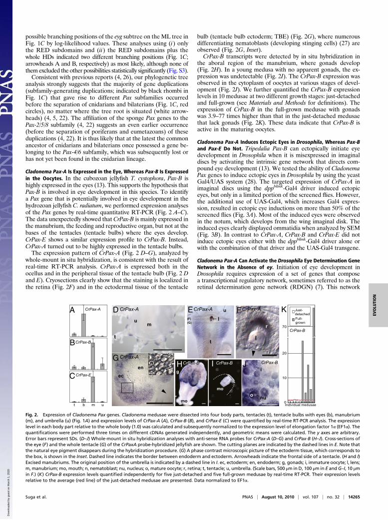
Cladonema Pax-A Is Expressed in the Eye, Whereas Pax-B Is Expressedin the Oocytes. In the cubozoan jellyfish T. cystophora, Pax-B ishighly expressed in the eyes (13). This supports the hypothesis thatPax-B is involved in eye development in this species. To identifya Pax gene that is potentially involved in eye development in thehydrozoan jellyfish C. radiatum, we performed expression analysesof the Pax genes by real-time quantitative RT-PCR (Fig. 2 A–C).The data unexpectedly showed that CrPax-B is mainly expressed inthe manubrium, the feeding and reproductive organ, but not at thebases of the tentacles (tentacle bulbs) where the eyes develop.CrPax-E shows a similar expression profile to CrPax-B. Instead,CrPax-A turned out to be highly expressed in the tentacle bulbs.The expression pattern of CrPax-A (Fig. 2 D–G), analyzed by
whole-mount in situ hybridization, is consistent with the result ofreal-time RT-PCR analysis. CrPax-A is expressed both in theocellus and in the peripheral tissue of the tentacle bulb (Fig. 2 Dand E). Cryosections clearly show that the staining is localized inthe retina (Fig. 2F) and in the ectodermal tissue of the tentacle
bulb (tentacle bulb ectoderm; TBE) (Fig. 2G), where numerousdifferentiating nematoblasts (developing stinging cells) (27) areobserved (Fig. 2G, Inset).CrPax-B transcripts were detected by in situ hybridization in
the aboral region of the manubrium, where gonads develop(Fig. 2H). In a young medusa with no apparent gonads, the ex-pression was undetectable (Fig. 2I). The CrPax-B expression wasobserved in the cytoplasm of oocytes at various stages of devel-opment (Fig. 2J). We further quantified the CrPax-B expressionlevels in 10 medusae at two different growth stages: just-detachedand full-grown (see Materials and Methods for definitions). Theexpression of CrPax-B in the full-grown medusae with gonadswas 3.9–77 times higher than that in the just-detached medusaethat lack gonads (Fig. 2K). These data indicate that CrPax-B isactive in the maturing oocytes.
Cladonema Pax-A Induces Ectopic Eyes in Drosophila, Whereas Pax-Band Pax-E Do Not. Tripedalia Pax-B can ectopically initiate eyedevelopment in Drosophila when it is misexpressed in imaginaldiscs by activating the intrinsic gene network that directs com-pound eye development (13). We tested the ability of CladonemaPax genes to induce ectopic eyes in Drosophila by using the yeastGal4/UAS system (28). The targeted expression of CrPax-A inimaginal discs using the dppblink-Gal4 driver induced ectopiceyes, but only in a limited portion of the screened flies. However,the additional use of UAS-Gal4, which increases Gal4 expres-sion, resulted in ectopic eye inductions on more than 50% of thescreened flies (Fig. 3A). Most of the induced eyes were observedin the notum, which develops from the wing imaginal disk. Theinduced eyes clearly displayed ommatidia when analyzed by SEM(Fig. 3B). In contrast to CrPax-A, CrPax-B and CrPax-E did notinduce ectopic eyes either with the dppblink-Gal4 driver alone orwith the combination of that driver and the UAS-Gal4 transgene.
Cladonema Pax-A Can Activate the Drosophila Eye Determination GeneNetwork in the Absence of ey. Initiation of eye development inDrosophila requires expression of a set of genes that composea transcriptional regulatory network, sometimes referred to as theretinal determination gene network (RDGN) (7). This network
A
B
C
t b m u
CrPax-E
CrPax-B
CrPax-B
CrPax-A
l
mo
mo
gen
ec
CrPax-B CrPax-B CrPax-B
0
10
20
70
t
l(F)(G)
r
u Just-detachedFull-grown
E FCrPax-A CrPax-A CrPax-A
CrPax-A
D
H IG
J
K
i
nu
um
t
o
n
Individual medusae
Fig. 2. Expression of Cladonema Pax genes. Cladonema medusae were dissected into four body parts, tentacles (t), tentacle bulbs with eyes (b), manubrium(m), and umbrella (u) (Fig. 1A) and expression levels of CrPax-A (A), CrPax-B (B), and CrPax-E (C) were quantified by real-time RT-PCR analysis. The expressionlevel in each body part relative to the whole body (1.0) was calculated and subsequently normalized to the expression level of elongation factor 1α (EF1α). Thequantifications were performed three times on different cDNAs generated independently, and geometric means were calculated. The y axes are arbitrary.Error bars represent SDs. (D–J) Whole-mount in situ hybridization analyses with anti-sense RNA probes for CrPax-A (D–G) and CrPax-B (H–J). Cross-sections ofthe eye (F) and the whole tentacle (G) of the CrPaxA probe-hybridized jellyfish are shown. The cutting planes are indicated by the dashed lines in E. Note thatthe natural eye pigment disappears during the hybridization procedure. (G) A phase contrast microscopic picture of the ectoderm tissue, which corresponds tothe box, is shown in the Inset. Dashed line indicates the border between endoderm and ectoderm. Arrowheads indicate the frontal side of a tentacle. (H and I)Excised manubriums. The original position of the umbrella is indicated by a dashed line in I. ec, ectoderm; en, endoderm; g, gonads; i, immature oocyte; l, lens;m, manubrium; mo, mouth; n, nematoblast; nu, nucleus; o, mature oocyte; r, retina; t, tentacle; u, umbrella. (Scale bars, 500 μm in D, 100 μm in E and G–I, 10 μmin F.) (K) CrPax-B expression levels quantified independently for five just-detached and five full-grown medusae by real-time RT-PCR. Their expression levelsrelative to the average (red line) of the just-detached medusae are presented. Data normalized to EF1α.
Suga et al. PNAS | August 10, 2010 | vol. 107 | no. 32 | 14265
EVOLU
TION
Dow
nloa
ded
by g
uest
on
Mar
ch 1
, 202
0

contains a positive feedback transcriptional loop comprising eye-less (ey; Drosophila Pax-6), sine oculis, eyes absent, and dachshund(7, 29). Initially, this transcriptional loop was thought to be ignitedonly by the activation of ey by twin of eyeless (toy; another Pax-6gene) (29). However, it has been shown that toy alone is able toinitiate the transcriptional loop in the absence of ey by directly ac-tivating another loopmember, sine oculis (30). To examine how thejellyfish Pax gene activates the Drosophila RDGN, we testedwhether CrPax-A can still induce ectopic eyes in the absence of ey.Indeed, the expression of CrPax-A, driven by the dppblink enhancer,induced ectopic eyes in an ey null mutant (eyJ5.71) background(Fig. 3 C and D). Our data indicate that the CrPax-A initiates theDrosophila RDGN by activating the components of the transcrip-tional loop, even in the absence of ey, as toy does.
Both Cladonema Pax-A and Pax-B Can Substitute for Pax-2 in theDrosophila Eye. In the Drosophila spapol mutant (Fig. 3E), the ex-pression of D-Pax2, a member of the Pax-2/5/8 subfamily, in coneand primary pigment cells is abolished, resulting in a severelydisturbed development of ommatidial cells (31). We used thismutant to test whether the Cladonema Pax genes can substitutefor the D-Pax2 functions in ommatidial cells, by performing res-cue experiments. Interestingly, not only CrPax-B, an ortholog ofD-Pax2, but also CrPax-A significantly rescued the spapol eyephenotype when they were expressed under the control of thecone- and pigment cell–specific enhancer (spa enhancer) (31, 32)of D-Pax2. CrPax-E, however, did not rescue the phenotype. SEMpictures clearly show that the hexagonal shape of each ommatid-ium and the regular arrangement of interommatidial bristles werelargely recovered both by the expression of CrPax-A and by that ofCrPax-B in the developing eye (Fig. 3 F–I).
CrPax-A PD Directly Binds to the Upstream Regions of Eye-SpecificOpsin Genes. Previous publications had proposed that DrosophilaEy directly regulates the expression of rhodopsin genes throughits HD (33, 34). Likewise, Tripedalia Pax-B can transactivate a lacZreporter gene under the control of a Drosophila rhodopsin pro-moter, presumably through its HD (13). Cladonema Pax-A, how-ever, lacks anHD.We therefore tested the possibility that CrPax-Acan still directly bind the promoter regions of Cladonema eye-
specific opsin genes, which had been characterized in our previousstudy (17), in the absence of an HD. We screened ≈1 kb of 5′promoter fragments by performing EMSA with PD proteins usingsets of probes that cover the fragments. In two examined opsingenes (CropG1 and CropN1), one and two CrPax-A–specific PDbinding sites (red vertical lines in Fig. 4A) were identified, re-spectively (Fig. 4B). No or very faint binding was detected when theputative binding sites were mutated (Fig. 4B). The sequences ofthe identified CrPax-A PD binding sites moderately match thechordate Pax-6 PD consensus binding sequence (35) (Fig. S4). Incontrast, CrPax-B PD showed little, if any, binding to any of thetested probes (Fig. 4B; only the three probes that showed theCrPax-A binding are shown). These results are consistent withthe notion that Pax-A, but not Pax-B, is involved in eye develop-ment and/or maintenance in Cladonema.
DiscussionFunctional Diversity of Cladonema Pax Genes. In this study, we havecharacterized three Pax genes (CrPax-A, CrPax-B, and CrPax-E)from C. radiatum, a hydrozoan jellyfish. From the expressionanalyses, we assume that CrPax-A is involved in eye developmentand/or maintenance, whereas CrPax-B is required for the oocytematuration process. Although CrPax-E is predominantly expressedin the manubrium, attempts to detect its transcripts by in situ hy-bridization were not successful, probably owing to its low expres-sion level. Interestingly, the dramatic gene up-regulation observedfor CrPax-B upon gonad formation (Fig. 2K) was not observedfor CrPax-E, suggesting that they exert different functions in themanubrium.The expression ofCrPax-A in the TBE, in addition to the retina,
suggests its involvement in nematogenesis because the TBE is thespecialized site for the tentacular nematocyte differentiation in
A
E F G H I
B C D
spapol D-Pax2 CrPax-A
CrPax-A CrPax-A; ey
CrPax-B CrPax-E
ad v
p
Fig. 3. Ectopic eye induction in Drosophila and rescue of the spapol mutantphenotype by Cladonema Pax genes. (A) CrPax-A was expressed under thecontrol of dppblink-Gal4 driver with UAS-Gal4. Arrowhead indicates the in-duced eye. (B) SEM picture of the induced eye. (C) Misexpression of CrPax-Aunder the control of dppblink-Gal4 driver in a homozygous ey null mutant(eyJ5.71) background induced ectopic eyes (arrowhead). The anteroposterior(a-p) and dorsoventral (d-v) axes are shown. Note that the natural com-pound eye is absent. The genotype of ey mutant was confirmed by theabsence of the second exon of ey (30) by PCR. (D) SEM picture of the inducedeye. (E) Eye phenotype of the spapol homozygous fly. (F–I) Rescue experi-ments of the spapol mutant phenotype. UAS-D-Pax2 (F; positive control),UAS-CrPax-A (G), UAS-CrPax-B (H), and UAS-CrPax-E (I) transgenic lines werecrossed with the spa-Gal4 driver line in a spapol homozygous background.(Scale bars, 30 μm.)
200 bp
CropG1A
B
CropN1
65
65
44.1 4.2
4.3 33.13.2
21
4
3
12
5.1
70
+ m + m + m + m + m + m
80100
CropG1 - 5.1 CropN1 - 4.3 CropN1 - 3.2
Pax-A Pax-B Pax-A Pax-B Pax-A Pax-B
Fig. 4. CrPax-A PD binding sites in the upstream region of eye-specificCladonema opsins. (A) Schematic drawing showing the positions of theprobes generated for EMSA. Probes to which CrPax-A PD bound are shownin red. Red vertical bars indicate the positions of the CrPax-A binding sites,which were precisely identified by the use of mutated probes. Black tri-angles, arrows, and gray boxes represent TATA boxes, transcription startingsites, and protein coding regions, respectively. (B) EMSA for the three posi-tive probes: probe 5.1 of CropG1, and probe 4.3 and 3.2 of CropN1. +, wild-type probes; m, mutated probe. The size (bp) of the probe is shown at left.Arrowheads indicate the band shifts caused by the binding of proteins. Notethat the bindings of CrPax-B PD are very faint or undetectable (white ar-rowhead), even though the same amount of the proteins as CrPax-A PDwere used. CrPax-B PD was separately proven to be active by the use ofa control probe carrying D-Pax2 binding sites (Materials and Methods).
14266 | www.pnas.org/cgi/doi/10.1073/pnas.1008389107 Suga et al.
Dow
nloa
ded
by g
uest
on
Mar
ch 1
, 202
0

hydrozoan jellyfish (27, 36, 37). Like bilaterian Pax genes (2, 3,38), CrPax-A seems to be involved in multiple aspects of de-velopmental processes.
Involvement of Pax-A in Cladonema Eye Development and/or Mainte-nance. By targeted expression experiments, we have shown thatCrPax-A is able to ectopically initiate eye development in Dro-sophila, whereas CrPax-B and CrPax-E are not. This agrees withthe results of our expression analyses, which show that onlyCrPax-A is highly expressed in the eye (Fig. 2).The Drosophila ey gene includes an eye-specific enhancer that
contains Pax-6 protein binding sites (29). It is possible that anintroduced Pax gene simply augments the endogenous Ey pro-tein level, which then induces the ectopic eyes. By showing theability of CrPax-A to induce ectopic eyes in Drosophila in an eynull mutant, however, we have demonstrated that the inductionof ectopic eyes by CrPax-A is not solely the effect of this jellyfishPax gene-mediated induction of the Drosophila endogenous ey. Itseems that, even in the absence of Drosophila Ey protein, theRDGN components expression is directly or indirectly inducedby the CrPax-A protein.The ability of the Cladonema Pax-A PD to bind to the 5′ up-
stream regions of two eye-specific opsin genes raises the possi-bility that CrPax-A directly regulates the expression of opsingenes during eye development and/or maintenance. In contrast,CrPax-B PD is incapable of binding to the same sequences. Thesedata are consistent with the expression analyses and the ectopiceye induction experiments. The direct interaction of Pax proteinswith opsin promoters might have contributed to the evolution ofancestral animal eyes (13). Intercalation of other transcriptionfactors into this simple regulatory cascade may explain how thecomplex gene network regulating eye development evolved (39).It should be noted, however, that DNA–protein interactionsdemonstrated by EMSA do not always reflect the same inter-actions in vivo. Studies on other species of jellyfish bearing eyesshould allow testing of our hypothesis.
Flexible Choice of Distinct Pax Genes for Eye Development in DifferentAnimal Lineages. Pax-6 has been characterized as one of the centralcomponents of the gene network that controls eye development inmost of the bilaterians studied to date (6). In Tripedalia, a cubo-zoan jellyfish that seems to lack a bona fide Pax-6, Pax-B has beenimplicated in eye development (13). We have now shown that Pax-A, rather than Pax-B, seems to be involved in eye developmentand/or maintenance in Cladonema, a hydrozoan jellyfish. Pax-6,Pax-B, and Pax-A belong to different Pax subfamilies, which di-verged from each other by gene duplication at the early stage ofanimal evolution, at the latest before the separation of cnidariansand bilaterians. It is thus very likely that for eye development and/or maintenance, three distinct animal lineages use three distantlyrelated Pax genes, which were generated by gene duplication be-fore the three animal lineages separated from each other.It can be argued that these data provide evidence in favor of
three independent origins of animal eyes during evolution (13). Ifit is the case, however, completely different sets of transcriptionfactors could have been recruited for eye development in differentanimal lineages. The observation that the three animal lineagesuse genes that belong to the same gene family (i.e., Pax family) intheir eyes rather supports the hypothesis of the monophyleticorigin of all animal eyes (6). This hypothesis is further supportedby the functional conservation of the Six family genes that areinvolved in eye development and/or regeneration both in Clado-nema and in several bilaterians studied to date (40–44). In addi-tion, we recently cloned the Cladonema homolog of eyes absent,one of the main components of Drosophila RDGN (45), anddetected its expression in the eye. Recent publications revealedthat not only the transcription factor network controlling eyedevelopment but also its potential downstream targets that in-
deed constitute the eye, such as genes involved in photoreception,phototransduction, and pigmentation, are also well conservedbetween cnidarians and bilaterians (17, 46). The common an-cestry of cnidarian eyes and bilaterian ones seems to be the mostreasonable interpretation of these data.In our present model, gene duplications that gave rise to distinct
subfamilies occurred most likely before the separation of poriferansand eumetazoans (1 in Fig. 5), as suggested by the statistical tests inrefs. 4 and 22. We cannot, however, completely eliminate the pos-sibility that some (or all) of these duplications postdate the porifer-ans–eumetazoans split (dashed line in Fig. 5) (4) until more spongePax genes that do not belong to the Pax-2/5/8 subfamily are found.When the ancestral animal eye evolved in the common ancestor ofcnidarians and bilaterians, Pax genes may have been recruited ascomponents of the genenetwork responsible for eye development (2in Fig. 5). We assume that, at this stage, several classes (corre-sponding to subfamilies) of Pax genes were redundantly involved inthis network. After the divergence of bilaterians and cnidarians onone hand, and hydrozoans and cubozoans on the other hand, thethree distinct animal lineages selected different classes of Pax genesfor the roles in eye development and/or maintenance (3 in Fig. 5).Such molecular-level opportunism is often observed in evolution(e.g., lens crystallins) (47). Interestingly, formation of some bilat-erian eyes seems to be Pax-6 independent (ref. 14 for review). Thissuggests that the gene network directing eye development can beanomalously modified, making Pax-6 dispensable for eye de-velopment in some bilaterian lineages (4 in Fig. 5).Our model predicts that genes from different Pax subfamilies
may still retain to some extent the ability to perform each other’sfunction redundantly. The ability of Cladonema Pax-A (poxnsubfamily), Pax-B (Pax-2/5/8 subfamily), andDrosophila ey and toy(Pax-4/6 subfamily) to rescue the D-Pax2 (Pax-2/5/8 subfamily)mutant spapol in the Drosophila eye is in agreement with thisprediction (Fig. 3 G and H and ref. 13). Similarly, misexpressedD-Pax2 induces eyes in the imaginal discs, as ey and toy do (13).Also at the molecular level, chordate Pax-6 and Pax-2 PDs rec-ognize almost identical sequences, even though they show cleardifferences in their affinities to certain DNA sequences (35).In summary, our study uncovers the diversity of the Pax genes used
for development and/or maintenance of animal eyes. We proposethat in the ancestral animal eye, which is likely to have evolved inthe common ancestor of cnidarians and bilaterians, different classes(corresponding to the present subfamilies) of Pax genes were re-dundantly recruited and may then have been flexibly selected indistinct animal lineages for their roles in eye development.
Materials and MethodsDetailed descriptions of animal culture, fly strains, and all of the technical in-formationregarding thegenecloningand sequencing, real-timePCR,molecularphylogenetic tree analysis, in silico search for Pax genes, in situ hybridization,protein expression, and EMSA are found in the SI Materials and Methods.
ACKNOWLEDGMENTS. We thank M. Noll (University of Zurich, Zurich) for flystrains; P. Callaerts (Katholieke Universiteit Leuven, Louvain, Belgium) andJ. Blanco (Insitute of Medical Biology, Singapore, Singapore) for the PD control
Porifera Hydrozoa
Vertebrates
DrosophilaCubozoa
Cnidaria
41 2
3
Bilateria
Pax-A
Pax-B
Pax-6
Fig. 5. Evolution of Pax genes deployed for animal eye development. 1:Gene duplications that gave rise to distinct Pax classes (corresponding tosubfamilies) occurred. 2: The ancestral animal eye evolved and differentclasses of Pax genes were redundantly recruited for eye development. 3: Ineach of three different animal lineages, a specific Pax gene was selected forthe eye development. 4: Pax genes responsible for eye development werealtered in some bilaterians. See text for detailed description.
Suga et al. PNAS | August 10, 2010 | vol. 107 | no. 32 | 14267
EVOLU
TION
Dow
nloa
ded
by g
uest
on
Mar
ch 1
, 202
0

probe; and M. M. Burger (Friedrich Miescher Institute, Basel, Switzerland) forMicrociona cDNA; J. Blanco and D. Papadopoulos for critically reading theman-uscript; and D. Duboule for allowing P.T. to continue the project. H.S. was sup-
ported by Yamada Science Foundation for a long-term visit. This work wassupported by the Swiss National Foundation and the Kantons of Basel-Stadtand Basel-Landschaft.
1. Mansouri A, Goudreau G, Gruss P (1999) Pax genes and their role in organogenesis.Cancer Res 59(7, Suppl):1707s–1709s, discussion 1709s–1710s.
2. Chi N, Epstein JA (2002) Getting your Pax straight: Pax proteins in development anddisease. Trends Genet 18:41–47.
3. Lang D, Powell SK, Plummer RS, Young KP, Ruggeri BA (2007) PAX genes: Roles indevelopment, pathophysiology, and cancer. Biochem Pharmacol 73:1–14.
4. Hoshiyama D, Iwabe N, Miyata T (2007) Evolution of the gene families forming thePax/Six regulatory network: Isolation of genes from primitive animals and molecularphylogenetic analyses. FEBS Lett 581:1639–1643.
5. Matus DQ, Pang K, Daly M, Martindale MQ (2007) Expression of Pax gene familymembers in the anthozoan cnidarian, Nematostella vectensis. Evol Dev 9:25–38.
6. Gehring WJ (2004) Historical perspective on the development and evolution of eyesand photoreceptors. Int J Dev Biol 48:707–717.
7. Silver SJ, Rebay I (2005) Signaling circuitries in development: Insights from the retinaldetermination gene network. Development 132:3–13.
8. Hill RE, et al. (1991) Mouse small eye results from mutations in a paired-like homeobox-containing gene. Nature 354:522–525.
9. Quiring R, Walldorf U, Kloter U, Gehring WJ (1994) Homology of the eyeless gene ofDrosophila to the Small eye gene in mice and Aniridia in humans. Science 265:785–789.
10. Halder G, Callaerts P, Gehring WJ (1995) Induction of ectopic eyes by targetedexpression of the eyeless gene in Drosophila. Science 267:1788–1792.
11. Onuma Y, Takahashi S, Asashima M, Kurata S, Gehring WJ (2002) Conservation of Pax6 function and upstream activation by Notch signaling in eye development of frogsand flies. Proc Natl Acad Sci USA 99:2020–2025.
12. Martin VJ (2002) Photoreceptors of cnidarians. Can J Zool 90:1703–1722.13. Kozmik Z, et al. (2003) Role of Pax genes in eye evolution: A cnidarian PaxB gene
uniting Pax2 and Pax6 functions. Dev Cell 5:773–785.14. Kozmik Z (2008) The role of Pax genes in eye evolution. Brain Res Bull 75:335–339.15. Sun H, Dickinson DP, Costello J, Li WH (2001) Isolation of Cladonema Pax-B genes and
studies of the DNA-binding properties of cnidarian Pax paired domains. Mol Biol Evol18:1905–1918.
16. Weber C (1981) Structure, histochemistry, ontogenetic development, and regenerationof the ocellus of Cladonema radiatum Dujardin (Cnidaria, Hydrozoa, Anthomedusae).J Morphol 167:313–331.
17. Suga H, Schmid V, Gehring WJ (2008) Evolution and functional diversity of jellyfishopsins. Curr Biol 18:51–55.
18. Leys SP, Degnan BM (2001) Cytological basis of photoresponsive behavior in a spongelarva. Biol Bull 201:323–338.
19. Maldonado M, Durfort M, McCarthy DA, Young CM (2003) The cellular basis ofphotobehavior in the tufted parenchymella larva of demosponges. Mar Biol 143:427–441.
20. Björn LO, Rasmusson AG (2009) Photosensitivity in sponge due to cytochrome coxidase? Photochem Photobiol Sci 8:755–757.
21. Leys SP, Cronin TW, Degnan BM, Marshall JN (2002) Spectral sensitivity in a spongelarva. J Comp Physiol A Neuroethol Sens Neural Behav Physiol 188:199–202.
22. Hoshiyama D, et al. (1998) Sponge Pax cDNA related to Pax-2/5/8 and ancient geneduplications in the Pax family. J Mol Evol 47:640–648.
23. Larroux C, et al. (2008) Genesis and expansion of metazoan transcription factor geneclasses. Mol Biol Evol 25:980–996.
24. Hill A, et al. (2010) Origin of Pax and Six gene families in sponges: Single PaxB andSix1/2 orthologs in Chalinula loosanoffi. Dev Biol 343:106–123.
25. King N, et al. (2008) The genome of the choanoflagellate Monosiga brevicollis andthe origin of metazoans. Nature 451:783–788.
26. Miller DJ, et al. (2000) Pax gene diversity in the basal cnidarian Acropora millepora(Cnidaria, Anthozoa): Implications for the evolution of the Pax gene family. Proc NatlAcad Sci USA 97:4475–4480.
27. Denker E, Manuël M, Leclère L, Le Guyader H, Rabet N (2008) Ordered progression ofnematogenesis from stem cells through differentiation stages in the tentacle bulb of
Clytia hemisphaerica (Hydrozoa, Cnidaria). Dev Biol 315:99–113.28. Brand AH, Perrimon N (1993) Targeted gene expression as a means of altering cell
fates and generating dominant phenotypes. Development 118:401–415.29. Czerny T, et al. (1999) twin of eyeless, a second Pax-6 gene of Drosophila, acts
upstream of eyeless in the control of eye development. Mol Cell 3:297–307.30. Punzo C, et al. (2004) Functional divergence between eyeless and twin of eyeless in
Drosophila melanogaster. Development 131:3943–3953.31. Fu W, Noll M (1997) The Pax2 homolog sparkling is required for development of cone
and pigment cells in the Drosophila eye. Genes Dev 11:2066–2078.32. Jiao R, et al. (2001) Headless flies generated by developmental pathway interference.
Development 128:3307–3319.33. Sheng G, Thouvenot E, Schmucker D, Wilson DS, Desplan C (1997) Direct regulation of
rhodopsin 1 by Pax-6/eyeless in Drosophila: Evidence for a conserved function inphotoreceptors. Genes Dev 11:1122–1131.
34. Papatsenko D, Nazina A, Desplan C (2001) A conserved regulatory element present inall Drosophila rhodopsin genes mediates Pax6 functions and participates in the fine-tuning of cell-specific expression. Mech Dev 101:143–153.
35. Czerny T, Busslinger M (1995) DNA-binding and transactivation properties of Pax-6:Three amino acids in the paired domain are responsible for the different sequencerecognition of Pax-6 and BSAP (Pax-5). Mol Cell Biol 15:2858–2871.
36. Bouillon J (1994) Compendium of Zoology, Cnidaria, Ctenophora, Part 2, ed Grassé PP(Masson, Paris), Vol III, pp 165–209 (in French).
37. Campbell RD (1988) The Biology of Nematocysts, eds Hessinger DA, Lenhoff HM(Academic Press, San Diego), pp 123–142.
38. Mansouri A, Hallonet M, Gruss P (1996) Pax genes and their roles in cell differentiationand development. Curr Opin Cell Biol 8:851–857.
39. Gehring WJ, Ikeo K (1999) Pax 6: Mastering eye morphogenesis and eye evolution.Trends Genet 15:371–377.
40. Cheyette BN, et al. (1994) The Drosophila sine oculis locus encodes a homeodomain-
containing protein required for the development of the entire visual system. Neuron12:977–996.
41. Kawakami K, Sato S, Ozaki H, Ikeda K (2000) Six family genes—structure and functionas transcription factors and their roles in development. Bioessays 22:616–626.
42. Pineda D, et al. (2000) Searching for the prototypic eye genetic network: Sine oculis is
essential for eye regeneration in planarians. Proc Natl Acad Sci USA 97:4525–4529.43. Seo HC, Drivenes Ø, Ellingsen S, Fjose A (1998) Expression of two zebrafish homologues
of the murine Six3 gene demarcates the initial eye primordia. Mech Dev 73:45–57.44. Stierwald M, Yanze N, Bamert RP, Kammermeier L, Schmid V (2004) The Sine oculis/Six
class family of homeobox genes in jellyfish with and without eyes: Development and
eye regeneration. Dev Biol 274:70–81.45. Pignoni F, et al. (1997) The eye-specification proteins So and Eya form a complex and
regulate multiple steps in Drosophila eye development. Cell 91:881–891.46. Kozmik Z, et al. (2008) Assembly of the cnidarian camera-type eye from vertebrate-
like components. Proc Natl Acad Sci USA 105:8989–8993.47. Wistow G (1993) Lens crystallins: Gene recruitment and evolutionary dynamism.
Trends Biochem Sci 18:301–306.
14268 | www.pnas.org/cgi/doi/10.1073/pnas.1008389107 Suga et al.
Dow
nloa
ded
by g
uest
on
Mar
ch 1
, 202
0