flexible nanopillar-based immunoelectrochemical biosensor
TRANSCRIPT

Park et al. Nano Convergence (2020) 7:29 https://doi.org/10.1186/s40580-020-00239-2
FULL PAPER
Flexible nanopillar-based immunoelectrochemical biosensor for noninvasive detection of Amyloid betaYoo Min Park1†, Junhyoung Ahn2†, Young Sun Choi1, Jae‑Min Jeong3, Seok Jae Lee1, Jae Jong Lee2*, Bong Gill Choi4* and Kyoung G. Lee1*
Abstract
The noninvasive early detection of biomarkers for Alzheimer’s disease (AD) is essential for the development of specific treatment strategies. This paper proposes an advanced method for fabricating highly ordered and flexible nanopillar‑based electrochemical biosensors by the combination of soft/photolithography and metal evaporation. The nanopil‑lar array (NPA) exhibits high surface area containing 1500 nm height and 500 nm diameter with 3:1 ratio. In regard with physical properties of polyurethane (PU) substrate, the developed NPA is sustainable and durable to external pressure such as bending and twisting. To manipulate the NPA surface to biocompatible, the gold was uniformly deposited on the PU substrate. The thiol chemistry which is stably modified on the gold surface as a form of self‑assembled monolayer was employed for fabricating the NPA as a biocompatible chip by covalently immobilize the antibodies. The proposed nanopillar‑based immunoelectrochemical biosensor exhibited good and stable electro‑chemical performance in β‑amyloid (Aβ) detection. Moreover, we successfully confirmed the performance of the as‑developed sensor using the artificial injection of Aβ in human tear, with sensitivity of 0.14 ng/mL and high repro‑ducibility (as a standard deviation below 10%). Our findings show that the developed nanopillar‑based sensor exhibits reliable electrochemical characteristics and prove its potential for application as a biosensor platform for testing at the point of care.
Keywords: Noninvasive, Flexible, Nanopillar array, Immunoelectrochemical sensor, Alzheimer’s disease
© The Author(s) 2020. This article is licensed under a Creative Commons Attribution 4.0 International License, which permits use, sharing, adaptation, distribution and reproduction in any medium or format, as long as you give appropriate credit to the original author(s) and the source, provide a link to the Creative Commons licence, and indicate if changes were made. The images or other third party material in this article are included in the article’s Creative Commons licence, unless indicated otherwise in a credit line to the material. If material is not included in the article’s Creative Commons licence and your intended use is not permitted by statutory regulation or exceeds the permitted use, you will need to obtain permission directly from the copyright holder. To view a copy of this licence, visit http://creat iveco mmons .org/licen ses/by/4.0/.
1 IntroductionAlzheimer’s disease (AD) is an irreversible progres-sive brain disorder that is common among the elderly. At present, the main techniques for diagnosing AD are brain imaging from positron emission tomography and magnetic resonance imaging [1–5]. Despite the great
social and economic costs of this disease, there has been slow progress in effectively treating AD, which has been exacerbated by the lack of powerful diagnostic methods. Although brain imaging is a powerful tool for diagnosing AD, intensive efforts have been made to achieve early AD diagnosis by analyzing biomarkers from biological fluid, especially from noninvasively acquired fluids such as tear and saliva. Moreover, given the relatively high protein content in tear fluid, it has become an excellent and via-ble candidate for the early diagnosis of AD [6–8].
Among different kinds of biomarker for AD, it was first proposed that the accumulation of β-amyloid (Aβ) pep-tide in brain tissue may cause neurodegeneration in AD. More recently, various research groups have reported that Aβ in ocular fluids could potentially be used as a
Open Access
*Correspondence: [email protected]; [email protected]; [email protected]†Yoo Min Park, Junhyoung Ahn contributed equally1 Division of Nano‑Bio Sensor/Chip Development, National NanoFab Center (NNFC), Daejeon 34141, Republic of Korea2 Department of Nano Manufacturing Technology, Nano‑Convergence Mechanical Systems Research Division, Korea Institute of Machinery & Materials (KIMM), Daejeon 34103, Republic of Korea4 Department of Chemical Engineering, Kangwon National University, Samcheok 25913, Republic of KoreaFull list of author information is available at the end of the article
Page 2 of 9Park et al. Nano Convergence (2020) 7:29
biomarker for the diagnosis of AD [9]. In particular, low concentration of Aβ aggregates under sub-nanomolar level were discovered in parts of the eye such as the lens and retina in AD patients [10–16]. Additionally, the pre-vious studies reveals the possibility of AD evaluation by verifying the concentration change of Aβ in the tear between healthy people with patients. Therefore, the Aβ aggregates in the tear sample could be used as a prom-ising biomarker. The human eye is anatomically closer to the brain than other human organs; Aβ can be rela-tively easily obtained there without any surgical proce-dure. To date, few methods including enzyme-linked immunosorbent assay, mass spectrometry, surface plas-mon resonance, surface-enhanced Raman spectroscopy, and nanomaterial-based imaging techniques have been developed and proposed to detect Aβ species [17–23]. Based on the affinity sensing principle with aptamer and antibody, the researches for Aβ analysis based on the minimized detection principle on biosensing chip as a point-of-care testing (POCT) device were demonstrated [24–26]. Furthermore, electrochemical biosensors have been widely utilized in the early detection of pathogens, monitoring of the quality of food and water, and clinical diagnosis due to their simplicity, sensitivity, accuracy, and rapid response [27–29]. More recently, the emergence of nanostructures in electrochemical biosensors has improved their sensitivity, response time, and portability [30, 31]. The nanopillar structure has various merits in comparison with a flat electrode in terms of electrochem-ical sensitivity owing to high electron transfer, enabling enhancement of the electrochemical signal. Additionally, based on its structural properties such as a high aspect ratio, a huge number of captured molecules can be sta-bly immobilized on the nanostructure, which generates plenty of electrons; thus, we effectively obtained the increased signal via nanostructural manipulation on thin film. Based on these advantages, nanopillar structure is favorable for the analysis of biomarkers present at low levels in body fluids [30, 32].
In this paper, we propose a simple, flexible, rapid, reli-able, and cost-effective electrochemical biosensor for early Aβ analysis using nanopillar-based electrodes and noninvasively acquired human tear. The thin layer of gold (Au) was deposited onto highly arrayed nanopillars to be used as working, counter, and reference electrodes. Con-ventionally, in previous research, Ag/AgCl was employed as a reference electrode, while gold was employed as a pseudo-reference electrode [33, 34]. The as-prepared bio-sensor carries out electrochemical detection in the pres-ence of different amounts of Aβ, which is directly related to the diagnosis of AD. The performance of the electro-chemical sensors was successfully confirmed by artifi-cially injecting Aβ into human tear.
2 Experimental section2.1 Materials and apparatusThe UV polymerizable NOA63 (Norland Optic Adhe-sives) was used with polyurethane (PU) (MINS-311RM, Minuta Tech.). The E-beam evaporator (EBS400) was purchased from EVATEC. The phosphate buffered saline (PBS) (#28372), 3,3′-dithiobis(sulfosuccinimidyl propion-ate) (DTSSP) (#21578) and bovine serum albumin (BSA) (#30063–572) were obtained from Thermo Fisher Sci-entific (Massachusetts, USA). HRP conjugated anti-Aβ antibody and human Aβ peptide (# 803012 and 932501) were obtained from Biolegend (California, USA). The o-phenylenediamine dihydrochloride (#p9187) was pur-chased from Sigma-Aldrich (Missouri, USA). DropSens (#C220AT) was obtained from PalmSens (Houten, Neth-erlands). The electrochemical behavior was analyzed by using the CHI 630B (CHInstruments).
2.2 Nanopillar electrodes manipulationThe NPE was manipulated by nano-patterned mask and polymer-based flexible substrate. The stencil mask was employed to pattern the gold on the polymer substrate. To produce the nano-patterned mask, the photolithog-raphy was performed using Si wafer with PR coating. Using the nano-patterned PR, the nano-hole on the Si was modified by dry etching principle. PR-removed si was then employed as a nano-pattern mask. The polyu-rethane (PU) was spin-coated on the nano-hole mask, and the attached PU was cured using the UV irradiator. By releasing the PU, the flexible substrate was completed. The solidified PU was released and employed as a sub-strate. The titanium (Ti) and Au were deposited in regu-lar sequence on the PU substrate employing the USB type stencil mask with soft lithography. In the 8-inch wafer, approximately 45 NPE was fabricated on a single wafer.
2.3 Preparation of Aβ sampleThe tested Aβ samples were prepared by spiking the Aβ antigen into the 0.1 M PBS buffer and artificial sample. The 1 ng/mL Aβ was firstly spiked both in PBS buffer and artificial tear sample, and the 0.1, 0.2, and 0.5 ng/mL Aβ samples were serially diluted.
2.4 Electrochemical behavior of nanopillar electrodesThe structural properties of fabricated nanopillar arrays on the electrode was measured by employing the scan-ning electron microscopy (SEM). The electrochemical characterization was detected by performing the cyclic voltammetry (CV) with voltage from 0 to 0.6 V and scan rate from 10 to 500 mV/s in the 5 mM ferrocyanide. The reproducibility was investigated by scanning the 30-times CV cycling under scan rate at 10 and 50 mV/s in the 5 mM ferrocyanide. The electrochemical stability to the

Page 3 of 9Park et al. Nano Convergence (2020) 7:29
external pressure was tested by analyzing the CV based on the bending and twisting nanopillar electrode with 100° at least 100 times.
2.5 Modification of chip surface for antibody immobilization
The Aβ analogue was immobilized on the NPE surface by using the 5 mM 3,3′-dithiobis(sulfosuccinimidyl propion-ate) in the DI water for 2 h. After washing with DI water at three times, the blocking buffer including 0.1% BSA and 0.1% triton X-100 in the PBS was applied.
2.6 Electrochemical immuassay for Aβ analysisUsing the Aβ analogue modified NPE, the cocktail solu-tions of HRP-conjugated anti-Aβ antibody and Aβ anti-gen were reacted for 30 min. The o-phenylenediamine dihydrochloride as a HRP substrate to generate the elec-trochemical signal. The electric signal was evaluated by square wave voltammetry (SWV) principle, and potential was scanning from − 0.8 to 0.0 V with 0.025 V amplitude and 25 Hz frequency at 50 mV/s.
3 Results and discussionFigures 1a, b shows the fabrication processes and prod-uct of nanopillar array (NPA) using the combination of photo- and soft lithography [35]. The NPA-based electro-chemical sensors are prepared using a shadow mask for Au sputtering (Fig. 1c), and the sensor image is shown in Fig. 1d. The electrochemical sensor has a three-elec-trode configuration with dimensions of 32 × 8 mm2. When observing the sensors, the rainbow color could be observed because of light diffraction from the highly arrayed nanopillars. The electrodes are assigned as work-ing, counting, and reference electrodes, with circular, semicircular, and rectangular shapes, respectively. The electrodes are composed of titanium (Ti) as an adhe-sive layer and a gold (Au) layer, which were sequentially formed onto the flexible polymeric NPA substrate using the sputtering method with the assistance of a metal-based stencil mask. Considering the portability, usabil-ity, and accessibility of the sensing platform, the sensor is specifically designed in a manner similar to the con-ventional USB-connectable platform. The electrochemi-cal analysis was further carried out using a portable
Fig. 1 a Schematic illustration of fabrication process of (a) nanopillar arrays and (b) gold electrode on nanopillar arrays. Photographs of (c) pristine nanopillar arrays and (d) gold electrode on nanopillar arrays. All scale bars are 5 cm

Page 4 of 9Park et al. Nano Convergence (2020) 7:29
connector. Our findings showed that our proposed fab-rication method allowed scalable, low-cost, and high-throughput fabrication.
To investigate the morphology of the sensor, different locations of the sensor surface were further investigated using SEM (Fig. 2a). The detailed dimensions of NPA are as follows: diameter of 500 nm, height of 1500 nm, and a center-to-center distance of 1000 nm; these were confirmed from the obtained SEM. The NPA was eas-ily found without any damage, even after Ti/Au coating. Notably, the roughness of the NPA was increased upon coating with Au, and small dots were also observed from the side view of Au-coated NPA. From the obtained SEM images, there were no cracks or holes on both the Au-coated area and pristine NPA area because of strong adhesion between Au and polymeric NPA. Addition-ally, clear changes in contrast were found at the interface between Au-coated NPA and pristine NPA, which con-firmed the successful formation of the Au electrode. The
highly arrayed nanopillar provides a highly specific sur-face area of the electrode and facilitates the redox reac-tion as well as sensitivity and stability compared with the flat-surfaced sensor. To confirm the potential surface characteristic changes of the as-prepared NPA-based sensor, the changes in contact angle (CA) were observed (Fig. 2). As shown in Fig. 2b, the CA of Au-coated NPA is lower than that of pristine NPA. The Au coating enabled a decrease of the CA while enhancing the bio-friendly environment for biomarker analysis.
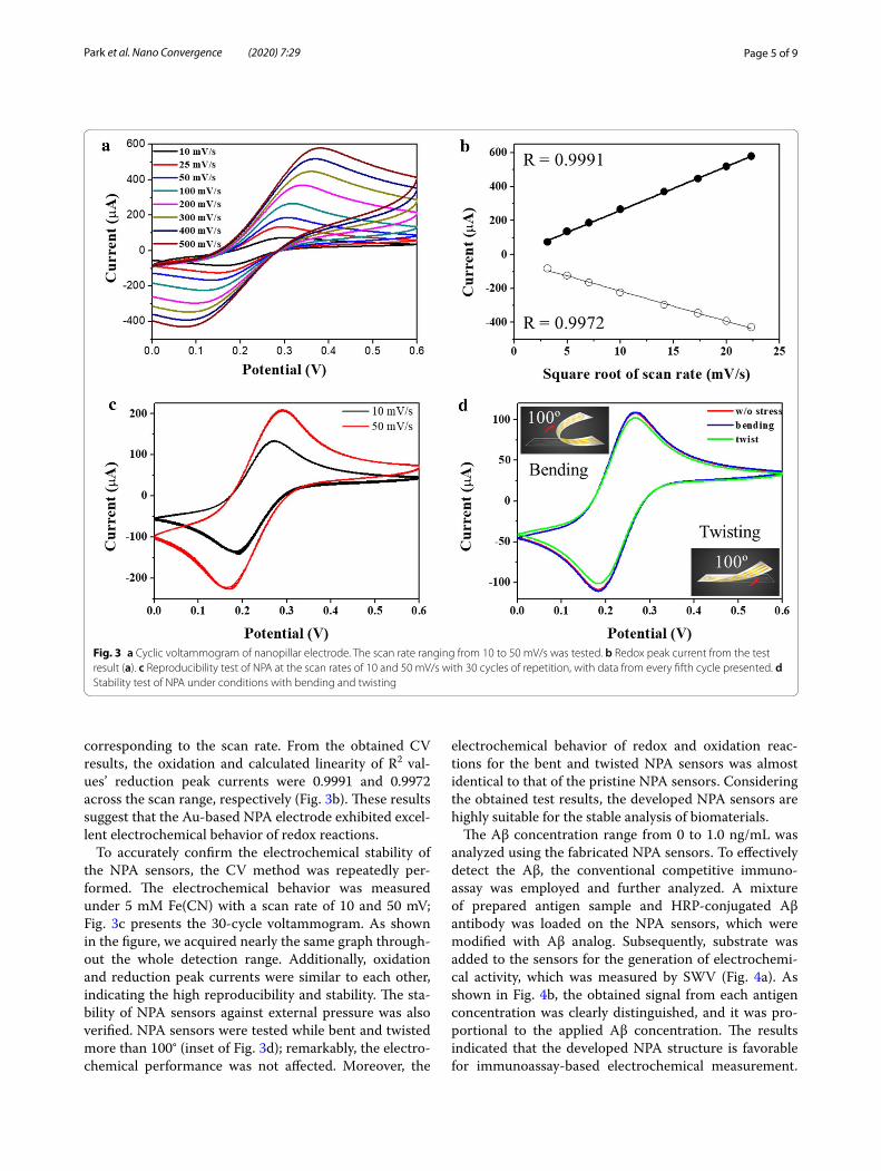
The electrochemical characteristics of the Au elec-trode on NPA sensors were intensively investigated. H2SO−4 at 50 mM was used as a supporting electrolyte. Subsequently, the redox peak current changed upon increasing the scan rate from 10 to 500 mV/s without any irregular responses, as shown in Fig. 3a. The cyclic vol-tammetry (CV) curves of the Au-based NPA sensor dis-played sharp and symmetric redox peaks. Additionally, each redox peak current’s signal was gradually increased
Fig. 2 a Photographic images of USB‑connectable NPA sensors and scanning electron microscope (SEM) images of top and side views of the indicated area. b contact angle (CA) of Au electrode and pristine NPA (scale bars are 2 μm)
Page 5 of 9Park et al. Nano Convergence (2020) 7:29
corresponding to the scan rate. From the obtained CV results, the oxidation and calculated linearity of R2 val-ues’ reduction peak currents were 0.9991 and 0.9972 across the scan range, respectively (Fig. 3b). These results suggest that the Au-based NPA electrode exhibited excel-lent electrochemical behavior of redox reactions.
To accurately confirm the electrochemical stability of the NPA sensors, the CV method was repeatedly per-formed. The electrochemical behavior was measured under 5 mM Fe(CN) with a scan rate of 10 and 50 mV; Fig. 3c presents the 30-cycle voltammogram. As shown in the figure, we acquired nearly the same graph through-out the whole detection range. Additionally, oxidation and reduction peak currents were similar to each other, indicating the high reproducibility and stability. The sta-bility of NPA sensors against external pressure was also verified. NPA sensors were tested while bent and twisted more than 100° (inset of Fig. 3d); remarkably, the electro-chemical performance was not affected. Moreover, the
electrochemical behavior of redox and oxidation reac-tions for the bent and twisted NPA sensors was almost identical to that of the pristine NPA sensors. Considering the obtained test results, the developed NPA sensors are highly suitable for the stable analysis of biomaterials.
The Aβ concentration range from 0 to 1.0 ng/mL was analyzed using the fabricated NPA sensors. To effectively detect the Aβ, the conventional competitive immuno-assay was employed and further analyzed. A mixture of prepared antigen sample and HRP-conjugated Aβ antibody was loaded on the NPA sensors, which were modified with Aβ analog. Subsequently, substrate was added to the sensors for the generation of electrochemi-cal activity, which was measured by SWV (Fig. 4a). As shown in Fig. 4b, the obtained signal from each antigen concentration was clearly distinguished, and it was pro-portional to the applied Aβ concentration. The results indicated that the developed NPA structure is favorable for immunoassay-based electrochemical measurement.
Fig. 3 a Cyclic voltammogram of nanopillar electrode. The scan rate ranging from 10 to 50 mV/s was tested. b Redox peak current from the test result (a). c Reproducibility test of NPA at the scan rates of 10 and 50 mV/s with 30 cycles of repetition, with data from every fifth cycle presented. d Stability test of NPA under conditions with bending and twisting

Page 6 of 9Park et al. Nano Convergence (2020) 7:29
To clearly verify the signal change, the calibration curve was obtained by employing the peak current from each test result (Fig. 4c). The signals in the calibration curve linearly increased in accordance with the Aβ concentra-tion. The results reveal that the antibody was effectively immobilized on the three-dimensional chip surface, and the immunoassay was successfully performed based on the nanopillar electrode. To confirm the sensitivity of the developed NPA sensors for the Aβ analysis, limit of detection (LOD) was calculated by using the standard deviation from blank and slope of regression line, and
the 0.16 ng/mL was calculated. The obtained results indi-cated that the NPA sensors have high sensitivity based on structural characterization of NPA sensors related to effective electron delivery [36–38]. Additionally, the acquired sensitivity could be due to the high surface area of the nanostructure. For confirmation of the accuracy, the R2 value in the linear curve range was calculated. The recorded linearity was 0.94, meaning that the low level of Aβ could be sensitively and clearly detected with high accuracy using the NPA sensors. The coefficient of variation was also calculated to verify the reproducibility.
Fig. 4 a Schematic illustration of electrochemical immunoassay for Aβ analysis. b Square wave voltammetry (SWV) graph from each Aβ in the buffer test. c Calibration curve of electrochemical analysis of Aβ. The linear range is presented in the inset. The error bar indicates the signal variation, and the same test was repeated three times

Page 7 of 9Park et al. Nano Convergence (2020) 7:29
To accurately identify the signal variation, the test was repeated at least three times under the same conditions, such as temperature, pH, concentration, and reaction time. The calculated CV in the whole detection range was 10%, indicating the high reproducibility. The calculated CV is presented in Fig. 4b as an error bar. Based on the findings with reliable test results, we successfully demon-strated that the fabricated NPA sensors could be effective for Aβ analysis with high sensitivity and accuracy.
To verify the practical utility of the NPA sensor for Aβ measurement, an Aβ test based on actual samples was carried out by spiking various concentrations of Aβ antigen into the artificial tear sample. The Aβ concen-tration range from 0 to 1 ng/mL was analyzed under the same principle and conditions as in the previous buffer test using the prepared sensor chip. The electrochemi-cal signals from each test are presented in Fig. 5a, and the signal changed in proportion to the Aβ concentra-tion. The oxidation peak current was employed to obtain the calibration curve, as presented in Fig. 5b. The linear curve was registered in accordance with the applied Aβ concentration, which indicated that a stable electro-chemical response was produced by immunoassay on the nanostructure. In comparison with the buffer test result, a slightly low signal was detected. The reason for this sig-nal variation is that the various components in actual tear samples, such as ions, proteins, and lipids, could interfere with electron transfer on the chip surface. With regard to the use of actual original samples, the obtained signal was highly similar to the buffer test result, which proved that the developed nano-arrayed sensor shows high electro-chemical performance toward Aβ in tear samples.
To verify the sensitivity, the LOD was calculated as same methods with previous formula, and the 0.14 ng/mL was obtained. The relatively high sensitivity of NPA sensors to actual Aβ is mainly attributable to the effective transfer of electrons produced by redox enzyme activity on the nanostructure. The signal variation was deter-mined by performing repeated tests under the same con-ditions as in previous test; a CV value of 8% was recorded with R2 of 0.92, demonstrating that the developed NPA sensors could accurately and reproducibly analyze the various concentrations of Aβ. Considering the use of actual tear samples, the test results are potentially highly promising in a clinical context, in which sensitive and accurate analyses are required.
4 ConclusionIn summary, this paper presents an in-depth study of a noninvasive way of detecting the Aβ associated with AD from human tear using a flexible and reliable nanopil-lar-based immunoelectrochemical sensor. Based on the skillful manufacturing system, we reliably fabricated the stable, reproducible, and sensitive nanopillar electrode, enabling mass production of the sensors at low cost. Additionally, using the nanostructural properties, we demonstrate the highly effective electrochemical behav-ior with effective biomolecule immobilization on the chip surface. The diverse range of Aβ in artificial tear con-firmed the performance of the as-prepared NPA biosen-sor down to 0.1 ng/mL. The NPA electrodes developed in this study could provide reliable, flexible, low-cost, and disposable sensing platforms for biosensors for testing at the point of care.
Fig. 5 a The voltammogram from Aβ in the artificial tear sample. b Calibration curve for Aβ test. The inset indicates the linear range. The error bar indicates the signal variation, and the same test was repeated in triplicate

Page 8 of 9Park et al. Nano Convergence (2020) 7:29
AbbreviationsAD: Alzheimer’s disease; NPA: Nanopillar array; CV: Cyclic voltammetric; PU: Polyurethane; Aβ: β‑amyloid; POCT: Point‑of‑care testing; Au: Gold; PBS: Phos‑phate buffered saline; BSA: Bovine serum albumin; Ti: Titanium; SEM: Scanning electron microscopy; CA: Contact angle; SWV: Square wave voltammetry.
Authors’ contributionsAll authors contributed to the writing of the manuscript, design of the figure sets, and analysis. All authors read and approved the final manuscript.
FundingThis work was supported by Nano Material Technology Development Program through the National Research Foundation of Korea (NRF) funded by the Ministry of Science and ICT (No.2017M3A7B4041761) and Basic Science Research Program (2018R1C1B3001553). This research was also supported by BioNano Health‑Guard Research Center funded by the Ministry of Science and ICT(MSIT) of Korea as Global Frontier Project (Grant number H‑GUARD_2014M3A6B2060302). This work was also supported by Nano Open Innovation Lab Cooperation Project of NNFC in 2020.
Availability of data and materialsAll the data are presented in the submitted manuscript and no supplementary information is available.
Ethics approval and consent to participateNot applicable.
Competing interestsThe authors declare that they have no competing interests.
Author details1 Division of Nano‑Bio Sensor/Chip Development, National NanoFab Center (NNFC), Daejeon 34141, Republic of Korea. 2 Department of Nano Manufactur‑ing Technology, Nano‑Convergence Mechanical Systems Research Division, Korea Institute of Machinery & Materials (KIMM), Daejeon 34103, Republic of Korea. 3 Department of Nanoengineering, University of California San Diego, La Jolla, CA 92093, USA. 4 Department of Chemical Engineering, Kangwon National University, Samcheok 25913, Republic of Korea.
Received: 22 March 2020 Accepted: 25 July 2020
References 1. N.C. Fox, R.S. Black, S. Gilman, M.N. Rossor, S.G. Griffith, L. Jenkins, M. Koller,
Neurology 64, 1563 (2005) 2. B.J. Lopresti, W.E. Klunk, C.A. Mathis, J.A. Hoge, S.K. Ziolko, X. Lu, C.C.
Meltzer, K. Schimmel, N.D. Tsopelas, S.T. DeKosky, J.C. Price, J. Nucl. Med. 46, 1959 (2005)
3. J.C. Price, W.E. Klunk, B.J. Lopresti, X. Lu, J.A. Hoge, S.K. Ziolko, D.P. Holt, C.C. Meltzer, S.T. DeKosky, C.A. Mathis, J. Cereb. Blood Flow Metab. 25, 1528 (2005)
4. T.R. Stoub, M. Bulgakova, S. Leurgans, D.A. Bennett, D. Fleischman, D.A. Turner, L. deToledo‑Morrell, Neurology 64, 1520 (2005)
5. G.B. Frisoni, N.C. Fox, C.R. Jack, P. Scheltens, P.M. Thompson, Nat. Rev. Neurol. 6, 67 (2010)
6. L. Zhou, S.Z. Zhao, S.K. Koh, L. Chen, C. Vaz, V. Tanavde, X.R. Li, R.W. Beuer‑man, J. Proteomics. 75, 3877 (2012)
7. S. Hagan, E. Martin, A. Enríquez‑de‑Salamanca, EPMA J. 7, 15 (2016) 8. L.A. Aqrawi, H.K. Galtung, B. Vestad, R. Øvstebø, B. Thiede, S. Rusthen,
A. Young, E.M. Guerreiro, T.P. Utheim, X. Chen, Ø.A. Utheim, Ø. Palm, J.L. Jensen, Arthritis Res. Ther. 19, 14 (2017)
9. J.C. Lee, S.J. Kim, S. Hong, Y. Kim, Exp. Mol. Med. 51, 53 (2019) 10. P.H. Frederikse, D. Garland, J.S. Jr Zigler, J. Piatigorsky, J. Biol. Chem. 271,
10169 (1996) 11. L.E. Goldstein, J.A. Muffat, R.A. Cherny, R.D. Moir, M.H. Ericsson, X. Huang,
C. Mavros, J.A. Coccia, K.Y. Faget, K.A. Fitch, C.L. Masters, R.E. Tanzi, L.T. Chylack Jr., A.I. Bush, Lancet 361, 1258 (2003)
12. R.M. Dutescu, Q.X. Li, J. Crowston, C.L. Masters, P.N. Baird, J.G. Culvenor, Clin Exp. Ophthalmol. 247, 1213 (2009)
13. J.A. Moncaster, R. Pineda, R.D. Moir, S. Lu, M.A. Burton, J.G. Ghosh, M. Ericsson, S.J. Soscia, A. Mocofanescu, R.D. Folkerth, R.M. Robb, J.R. Kuszak, J.I. Clark, R.E. Tanzi, D.G. Hunter, L.E. Goldstein, PLoS ONE 5, e10659 (2010)
14. M. Koronyo‑Hamaoui, Y. Koronyo, A.V. Ljubimov, C.A. Miller, M.K. Ko, K.L. Black, M. Schwartz, D.L. Farkas, NeuroImage 54, S204 (2011)
15. Y. Koronyo, B.C. Salumbides, K.L. Black, M. Koronyo‑Hanaoui, Alzhei‑mer’s disease in the retina: imaging retinal aβ plaques for early diagno‑sis and therapy assessment. Neurodegener. Dis. 10, 285–293 (2012)
16. C. La Morgia, F.N. Ross‑Cisneros, Y. Koronyo, J. Hannibal, R. Gallassi, G. Cantalupo, L. Sambati, B.X. Pan, K.R. Tozer, P. Barboni, F. Provini, P. Avan‑zini, M. Carbonelli, A. Pelosi, H. Chui, R. Liguori, A. Baruzzi, M. Koronyo‑Hamaoui, A.A. Sadun, A. Carelli, Melanopsin retinal ganglion cell loss in Alzheimer disease. Ann. Neurol 79(1), 90–109 (2016)
17. I. Koychev, B. Galna, H. Zetterberg, J. Lawson, G. Zamboni, B.H. Ridha, J.B. Rowe, A. Thomas, R. Howard, P. Malhotra, C. Ritchie, S. Lovestone, L. Rochester, Aβ42/Aβ40 and Aβ42/Aβ38 ratios are associated with meas‑ures of gait variability and activities of daily living in mild Alzheimer’s disease: a pilot study. J. Alzheimers Dis. 65, 1377–1383 (2018)
18. M. Ramakrishnan, K.K. Kandimalla, T.M. Wengenack, K.G. Howell, J.F. Poduslo, Surface plasmon resonance binding kinetics of Alzheimer’s disease amyloid β peptide‑capturing and plaque‑binding monoclonal antibodies. Biochemistry 48, 10405–10415 (2009)
19. P. Pesini, V. Pérez‑Grijalba, I. Monleón, M. Boada, L. Tárraga, P. Martínez‑Lage, I. San‑José, M. Sarasa, Reliable measurements of the β‑amyloid pool in blood could help in the early diagnosis of AD. Int. J. Alzheimer Dis. 2012, 604141 (2012)
20. S.G. Park, M.S. Ahn, Y.J. Oh, M. Kang, Y. Jeong, K.H. Jeong, Nanoplas‑monic biopatch for in vivo surface enhanced raman spectroscopy. BioChip J. 8, 289–294 (2014)
21. S. Galozzi, K. Marcus, K. Barkovits, Amyloid‑β as a biomarker for Alzhei‑mer’s disease: quantification methods in body fluids. Int. J. Alzheimer Dis. 12, 343–354 (2015)
22. P. Palladino, A.M. Aura, G. Spoto, Surface plasmon resonance for the label‑free detection of Alzheimer’s β‑amyloid peptide aggregation. Anal. Bioanal. Chem. 408, 849–854 (2016)
23. S. Shah, The nanomaterial toolkit for neuroengineering. Nano Converg. 3, 25 (2016)
24. P. Gagni, L. Sola, M. Cretich, M. Chiari, Development of a high‑sensitivity immunoassay for amyloid‑beta 1–42 using a silicon microarray plat‑form. Biosens. Bioelectron. 47, 490–495 (2013)
25. M. Gheorghiu, S. David, C. Polonschii, A. Olaru, S. Gaspar, O. Bajenaru, B.O. Popescu, E. Gheorghiu, Label free sensing platform for amyloid fibrils effect on living cells. Biosens. Bioelectron. 52, 89–97 (2014)
26. J. Oh, G. Yoo, Y.W. Chang, H.J. Kim, J. Jose, E. Kim, J.C. Pyun, K.H. Yoo, A carbon nanotube metal semiconductor field effect transistor‑based biosensor for detection of amyloid‑beta in human serum. Biosens. Bioelectron. 50, 345–350 (2013)
27. F. Liu, K.S. Choi, T.J. Park, S.Y. Lee, T.S. Seo, Graphene‑based electro‑chemical biosensor for pathogenic virus detection. BioChip J. 5, 123–128 (2011)
28. E.B. Setterington, E.C. Alocilja, Electrochemical biosensor for rapid and sensitive detection of magnetically extracted bacterial pathogens. Bio‑sensors 2, 15–31 (2012)
29. Y.M. Park, S.Y. Lim, S.J. Shin, C.H. Kim, S.W. Jeong, S.Y. Shin, N.H. Bae, S.J. Lee, J. Na, G.Y. Jung, T.J. Lee, A film‑based integrated chip for gene amplifica‑tion and electrochemical detection of pathogen causing foodborne illnesses. Anal. Chim. Acta 1027, 57–66 (2018)
30. Y.M. Park, S.Y. Lim, S.W. Jeong, Y.S. Song, N.H. Bae, S.B. Hong, B.G. Choi, S.J. Lee, K.G. Lee, Flexible nanopillar‑based electrochemical sensors for genetic detection of foodborne pathogens. Nano Convergence 5, 15 (2018)
31. Y.T. Seo, S. Jeong, J.K. Lee, H.S. Choi, J. Kim, H.Y. Lee, Innovations in bio‑medical nanoengineering: nanowell array biosensor. Nano Convergence 5, 9 (2018)
32. J.H. Yoon, S.B. Hong, S.‑O. Yun, S.J. Lee, T.J. Lee, K.G. Lee, B.G. Choi, High performance flexible pH sensor based on polyaniline nanopillar array electrode. J. Colloid. Interf. Sci. 490, 53–58 (2017)
33. K. Hashimoto, Y. Ishimori, Preliminary evaluation of electrochemical PNA array for detection of single base mismatch mutations. Lab Chip 1, 61–63 (2001)

Page 9 of 9Park et al. Nano Convergence (2020) 7:29
34. Y.S. Kim, J.H. Niazi, M.B. Gu, Specific detection of oxytetracycline using DNA aptamer‑immobilized interdigitated array electrode chip. Anal. Chim. Acta 634, 250–254 (2009)
35. K.G. Lee, B.G. Choi, B.I. Kim, T. Shyu, M.S. Oh, S.G. Im, S.‑J. Chang, T.J. Lee, N.A. Kotov, S.J. Lee, Scalable nanopillar arrays with layer‑by‑layer pat‑terned overt and covert images. Adv. Mater. 26, 6119–6124 (2014)
36. V. Anandan, Y.L. Rao, G. Zhang, Nanopillar array structures for enhancing biosensing performance. Int. J. Nanomedicine 1, 73–79 (2006)
37. C. Shin, W. Shin, H.G. Hong, Electrochemical fabrication and electrocata‑lytic characteristics studies of gold nanopillar array electrode (AuNPE) for
development of a novel electrochemical sensor. Electrochim. Acta 53, 720–728 (2007)
38. C. Lotwala, H.F. Ji, Electrochemistry on nanopillared electrodes. AIMS mater. Sci. 4, 292–301 (2017)
Publisher’s NoteSpringer Nature remains neutral with regard to jurisdictional claims in pub‑lished maps and institutional affiliations.